I N D E X
ABSTRACT pag.5
1 INTRODUCTION pag.9
1.1 Mitochondria – structure and function pag.9
1.2 Mitochondrial transport systems pag.12
1.3 Disorders related to mitochondrial carrier deficiency pag.21
2 MATERIALS AND METHODS pag.24
2.1 Construction of Expression Plasmids pag.24
2.1.1 Expression Plasmids for Glycine Carrier pag.24
2.1.2 Expression Plasmids for dPCoA Carriers pag.25
2.1.3 Expression Plasmids for OGC Carrier and site direct mutagenesis
pag.25
2.2 Bacterial expression and purification of recombinant proteins pag.26 2.3 Reconstitution of recombinant proteins into liposomes and transport
measurements
pag.26
2.4 Overexpression in Saccharomyces cerevisiae of recombinant proteins and mitochondrial isolation
pag.30
2.5 Subcellular localization of recombinant proteins pag.31 2.6 Sample preparation for yeast metabolite analysis pag.32 2.7 Glycine and ALA analysis by gas chromatography coupled to tandem
mass spectrometry (GC-MS/MS)
pag.33
2.8 Determination of mitochondrial heme and cytochrome content pag.34
2.9 Mitochondrial respiration efficiency pag.35
2.10 Comparative modelling and docking investigations pag.35 2.10.1 Comparative modelling and docking investigations of dPCoAC pag.36 2.10.2 Comparative modelling and docking investigations of BtOGC
mutants
pag.36
3 CHARACTERIZATION OF MITOCHONDRIAL GLYCINE CARRIER
pag.38
3.1 INTRODUCTION
Glycine carrier and associated diseases
pag.38
3.2 RESULTS pag.41
3.2.1 Bacterial expression of Hem25p pag.41
3.2.2 Functional characterization of recombinant Hem25p pag.42 3.2.3 Impaired glycine uptake in mitochondria lacking Hem25p pag.45
3.2.4 Subcellular localization of Hem25p pag.45
3.2.5 Growth characteristics of wild-type and hem25∆ pag.47 3.2.6 Hem25p is required for the entry of glycine into mitochondria pag.50 3.2.7 Hem25p and GlyC deficiency cause defects in respiratory chain
components
pag.51
3.2.8 Respiratory analysis of isolated mitochondria pag.55
3.3 DISCUSSION pag.57
4 CHARACTERIZATION OF MITOCHONDRIAL
DEPHOSPHOCOENZYME-A CARRIER
pag.61
4.1 INTRODUCTION
CoA carrier and associated diseases
pag.61
4.2 RESULTS pag.65
4.2.1 Evolutionary analysis of human SLC25A42 homologs in eukaryotes
pag.65
4.2.2 Bacterial expression and functional characterization of dPCoAC-A and dPCoAC-B
pag.67 4.2.3. Kinetic characteristics of the recombinant dPCoAC-A pag.71 4.2.4. Phenotype complementation of yeast LEU5 null strain by
dPCoACs
pag.74 4.2.5. Dissection of the transport features of dPCoAC through
structural analysis
pag.75
5 STRUCTURAL REARRANGEMENTS FOR SUBSTRATE
TRANSLOCATION IN THE MITOCHONDRIAL
OXOGLUTARATE CARRIER pag.83 5.1 INTRODUCTION Oxoglutarate carrier pag.83 5.2 RESULTS pag.87
5.2.1 Expression, transport activity and kinetic analysis of the reconstituted K122R mutant.
pag.87 5.2.2 Influence of sulfhydryl reagents on the WT OGC and K122R
transport activities
pag.89 5.2.3 Expression, transport activity and influence of sulfhydryl
reagents on reconstituted C184, C221, C224, K122R/C184, K122R/C221 and K122R/C224 OGC mutants
pag.91
5.2.4 Influence of substrate on the inhibition of C224, K122R/C224, C221 and K122R/C221 mutants by MTSEA
pag.92 5.2.5 Molecular modeling studies of SLC25A11_OGC pag.94
5.3 DISCUSSION pag.96
ABBREVIATIONS pag.100
5
A B S T R A C T
The mitochondrial carriers (MCs) are transmembrane proteins found in the mitochondrial inner membrane, which catalyze the translocation of solutes through the membrane. These belong to a family of carrier proteins, the SLC25 or Mitochondrial Carrier Family (MCF). Their function is to create a connection between mitochondria and cytosol, facilitating the flow of a large variety of solutes across the permeability barrier of the inner mitochondrial membrane, which is necessary for many physiological processes.
The functional information obtained from the study of mitochondrial carrier was fundamental in correlating MCs physiological and pathological roles in cellular metabolism. It was possible to identify genes, and their possible defects, responsible for the onset of certain diseases such as the Stanley syndrome, Amish microcephaly, HHH syndrome (hyperornithinemia, hyperammonemia and homocitrullinuria) and type II citrullinemia, their molecular basis and their symptoms. Further studies on the functional characterization of the gene family SLC25 will clarify other diseases caused by a mitochondrial carrier deficiency.
This work was focused in particular on the study of some carriers belonging to the MCF:
− the mitochondrial glycine carrier, important in heme synthesis and congenital sideroblastic anemia;
− the mitochondrial dephosphocoenzyme A carrier, important in regulating the compartmentalization of the CoA, the study of which is crucial for a better understanding of some neurodegenerative diseases that depend on the biosynthesis of CoA;
− the mitochondrial oxoglutarate carrier, of which the functional and structural rearrangements required for substrate transport were analyzed.
6 Mitochondrial glycine carrier
The studies were focused on the biochemical and molecular characterization of human glycine carrier protein (GlyC) and its yeast homolog (Hem25p) providing evidence that they are mitochondrial carriers for glycine. Glycine carrier is required for the uptake of glycine in the mitochondrial matrix, where this amino acid is condensed with succinyl coenzyme A to yield δ-aminolevulinic acid, necessary for heme biosynthesis. A detailed knowledge of this transporter could be helpful to clearly understand congenital sideroblastic anemia (CSA), caused by defects of heme biosynthesis in developing erythroblasts.
In particular, Hem25p was cloned into a bacterial expression system (Escherichia
coli BL21), overexpressed at high levels as inclusion bodies, and purified by Ni2+ -NTA-agarose affinity chromatography. The protein was then reconstituted in liposomes and its transport activity of glycine was observed. The kinetic constants, Km and Vmax, were calculated. Subsequently, other evidences of glycine uptake were obtained carrying out experiments on mitochondrial proteins from the yeast wild-type strain, the
hem25∆ strain and the hem25∆ HEM25-pYES2. The protein subcellular localization
was found to be mitochondrial. Furthermore, the hem25∆ mutant manifested a defect in the biosynthesis of δ-aminolevulinic acid and displayed reduced levels of downstream heme and mitochondrial cytochromes. The observed defects were rescued by complementation with yeast HEM25 or human SLC25A38 genes.
This work may suggest new therapeutic approaches for the treatment of congenital sideroblastic anemia.
Mitochondrial dephosphocoenzyme A carrier
In human, the transport of CoA across the inner mitochondrial membrane has been attributed to two different genes, SLC25A16 and SLC25A42. Presumed orthologs of both genes are present in many eukaryotic genomes, but not in that of D. melanogaster, which contains only one gene, CG4241, phylogenetically close to SLC25A42. CG4241 encodes a long and a short isoform of the dPCoA carrier, respectively dPCoAC1 and dPCoAC2, which arise from an alternative translational start site. dPCoAC1 and dPCoAC2 were expressed as inclusion bodies in E. coli C0214, and reconstituted in proteoliposomes to observe the transport activity in order to characterize them functionally.
7 The functional characterization of the D. melanogaster dPCoA carrier is of particular interest as it is the first mitochondrial carrier showing a particular substrate specificity for dPCoA and ADP.
The expression of both isoforms in a S. cerevisiae strain lacking the endogenous putative mitochondrial CoA carrier restored the growth on respiratory carbon sources and the mitochondrial levels of CoA. The results reported here and the proposed subcellular localization of some of the enzymes of the fruit fly CoA biosynthetic pathway, suggest that dPCoA may be synthesized and phosphorylated to CoA in the matrix, but it can also be transported by dPCoAC to the cytosol, where it may be phosphorylated to CoA by the monofunctional dPCoA kinase. Thus, dPCoAC may connect the cytosolic and mitochondrial reactions of the CoA biosynthetic pathway without allowing the two CoA pools to get in contact.
This work will be useful in the near future to better understand the deficiency of enzymes involved in the CoA biosynthesis associated with a neurodegenerative disorder known as neurodegeneration with brain iron accumulation (NBIA).
Mitochondrial oxoglutarate carrier
The oxoglutarate carrier (OGC) plays a key role in important metabolic pathways. Its transport activity has been extensively studied, and, to investigate new structural rearrangements required for substrate translocation,site-directed mutagenesis was used to conservatively replace lysine 122 by arginine. K122R mutant was kinetically characterized, exhibiting a significant Vmax reduction with respect to the wild-type (WT) OGC, whereas Km value was unaffected, implying that this substitution does not interfere with 2-oxoglutarate binding site.
Moreover, K122R mutant was more inhibited by several sulfhydryl reagents with respect to the WT OGC, suggesting that the reactivity of some cysteine residues towards these Cys-specific reagents is increased in this mutant. Different sulfhydryl reagents were employed in transport assays to test the effect of the cysteine modifications on single-cysteine OGC mutants named C184, C221, C224 (constructed in the WT background) and K122R/C184, K122R/C221, K122R/C224 (constructed in the K122R background). Cysteines 221 and 224 were more deeply influenced by some sulfhydryl reagents in the K122R background. Furthermore, the presence of 2-oxoglutarate significantly enhanced the degree of inhibition of K122R/C221,
8 K122R/C224 and C224 activity by the sulfhydryl reagent 2-Aminoethyl methanethiosulfonate hydrobromide (MTSEA), suggesting that cysteines 221 and 224, together with K122, take part to structural rearrangements required for the transition from the c- to the m-state during substrate translocation.
9
1 . I N T R O D U C T I O N
1.1 Mitochondria – structure and function
Mitochondria (Fig. 1.1), frequently referred as the “powerhouses of the cell”, are essential mammalian organelles typical of animal, plant, algae and protozoa, with aerobic metabolism, surrounded by two lipid bilayers. These cytoplasmic organelles are responsible for many fundamental processes, including the production of ATP (adenosine-5'-triphosphate) that can be used in energy requiring reactions. This is possible due to different mitochondrial metabolic reactions, such as the fatty acid oxidation (FAO), the citric acid cycle and the oxidative phosphorylation (OXPHOS). Shape, size, number and distribution of mitochondria are closely associated with the cell type and function. Based on the energy demands, in the cell there can be a different amount of mitochondria, from a few to some hundreds. These organelles can also be distributed evenly or be grouped in the region with the most intense metabolic activity
[1]
.
Generally rod shaped, if observed under an optical phase contrast microscope, mitochondria may appear granular or filamentary. The dimensions may vary from 1µm to10µm in length and from 0.2µm to 1µm of diameter [2].
10 The electron microscopy allows to ultrastructural analysis of these organelles, highlighting two membranes of about 6 nm of thicknesses. The outer mitochondrial membrane (OMM) is separated from the inner mitochondrial membrane (IMM) by a space of 6-8 nm, called intermembrane space (Fig. 1.2).
The IMM forms ridges within an aqueous solution, rich in protein and various metabolic intermediate molecules, called mitochondrial matrix. The number of mitochondrial ridges reflects the cellular metabolic activities: it is higher in kidney cells, striatum muscle and cardiac muscle, and is low in plant cells, depending on their different respiratory activity.
While the OMM is completely permeable to different substances and represents a barrier with low or no effectiveness for the regulation of the access of different substrates, the permeability of the IMM is rather limited. The "porins", channel proteins of the OMM, make it easily penetrable by ions and small molecules (with molecular weight of up to 10,000 Dalton), so that the composition of ions and substrates of the intermembrane space appears to be similar to the one of the cytosol.
The actual selection filter, between the cytosol and mitochondrial matrix, is represented, therefore, by the IMM. It is highly impermeable to ions and molecules with molecular weight greater than 100-150 Daltons. Such selectivity is mainly due to the presence of a high concentration of cardiolipin or diphosphatidylglycerol. Because of the different permeability of the two mitochondrial membranes, the matrix and the cytosol assume and maintain very different metabolic capacity and have different tasks.
11 Mitochondria are the only structure of of eucariotic non-plant cells, in addition to the nucleus, which contains genetic material. The mitochondrion has its own replication system; it contains the enzyme γ-DNA polymerase, which ensures a semiconservative replication.
The mitochondrial DNA (mtDNA) is relatively low, representing 1.5% of the cell total DNA, it is extremely small, contains, in fact, only 37 genes, in humans. Of these genes, 13 encode for some of the protein subunits of the respiratory chain complexes and 24 encode for molecules essential to the synthesis of these subunits (2 ribosomal RNAs, or rRNAs, and 22 transfer RNAs, or tRNAs). These proteins represent a small part of the proteins present in the mitochondria; in fact, in these organelles there are soluble proteins of the mitochondrial matrix, proteins of the outer membrane, and protein of the inner membrane, including mitochondrial transporters or carriers. All these proteins are encoded by nuclear genes, synthesized by cytoplasmic ribosomes and, subsequently, transferred into the mitochondria [3].
Unlike other cellular organelles that are produced ex-novo during cell duplication, mitochondria are duplicated by binary fission, following their DNA duplication. The complete organelles are then inherited by daughter cells, according to what is called non-mendelian or cytoplasmic inheritance.
Another important characteristic of animal mitochondria is that, at the time of sexual reproduction, they are transmitted to the children only from the mother. During fertilization, the mitochondria present in the new individual (zygote) come only from the egg cell (oocyte). Therefore a mother carrying a mutation of the mtDNA transmit this mutation to all of her children, but only daughters transmit, in turn, to their progeny (matrilineal inheritance). Differently from nuclear genes, that are present in humans only in two copies (the maternal and paternal allele), there are hundreds of mtDNA molecules within each cell.
Mitochondria are also the center where cell life and death is regulated. They are the main target of aging processes; they tend to accumulate deletions and point mutations, which lead to a change in mitochondrial morphology and functionality. Aging, together
12 with loss of efficiency of mitochondria, compromise energy production and, often, lead to tissue death.
During aging processes or as a result of specific physiological signals, a series of processes, which involve structural modifications of mitochondrial membranes, are activated.
The apoptotic signals determine transition of mitochondrial permeability; the formation of pores in the IMM causes a reduction of the membrane potential and mitochondrial swelling. The signals can also cause increased permeability of the OMM, releasing an apoptosis trigger factor, the Cytochrome C, which passes from the mitochondria to the cytosol, and sets in motion proteolytic events of cell death [2].
Since the mitochondria are present in all tissues, mitochondrial diseases can affect any organ of the body, but they mainly affect muscle and brain, because a bigger number of mitochondria are usually found in those tissues, due to their increased demand of energy.
1.2 Mitochondrial transport systems
The high degree of impermeability, that characterizes the inner mitochondrial membrane, only allows some uncharged molecules, such as O2 and CO2, to diffuse passively through it. However, numerous metabolic processes take place in the mitochondria and in the cytosol, because of the different enzyme compartmentalization. Thus, the internalization of metabolites produced outside of the mitochondria and the export in the cytosol of other metabolic intermediates that are formed in the mitochondrial matrix is necessary. Among the metabolites that migrate from the cytosol to the mitochondrial matrix we find: ADP and phosphate, involved in oxidative phosphorylation; substrates involved in the citric acid cycle, in the β-oxidation, in the biosynthesis of RNA and mitochondrial protein; coproporphyrinogen and iron, essential for the synthesis of heme, donors of functional groups (S-adenosylmethionine and folate) and co-enzymes (NAD+, FAD, TPP and CoA-SH). On the other hand, ATP, citrate, malate, PLP, δ-aminolevulinate and citrulline need to be exported out of the mitochondria.
Since all these metabolites need to migrate across the inner mitochondrial membrane, the presence of a variety of transmembrane proteins, called transporters or carriers, is
13 required, and those proteins constitute a superfamily known as Mitochondrial Carrier
Family (MCF) or SLC25 (Solute Carrier 25) family [4-6].
Numerous studies have been conducted on intact mitochondria, which allowed proving the existence of these carrier proteins. The importance of these proteins was clearly evident from the observation that the IMM possesses a protein fraction of about 76%, by far higher than the protein fraction of other biological membranes [7]. The majority of these proteins are mitochondrial carriers.
In humans, the 53 members of the MCF, known as mitochondrial carriers (MCs), are encoded by nuclear genes equally distributed in most chromosomes [5]. Different isoforms of many mitochondrial carriers exist, some of which are encoded by separate genes, while others are variants derived by alternative splicing the same gene; one example is the Phosphate carrier (PiC), responsible for the transport of phosphate, which is present in 2 isoforms derived by the SLC25A3 gene [8].
The MCs are found in all eukaryotic organisms, both unicellular and multicellular. Their expression levels vary significantly; some are synthesized in virtually all tissues, while others are tissue-specific. The limited distribution of the latter reflects the implication in particular functions [9, 10]. Often, different isoforms of the same carrier have specific tissue distribution. Since all MCs are nucleus-encoded proteins, their cytosolic biosynthesis is followed by their transport to the inner mitochondrial membrane.
The common function of carriers is to provide a link between mitochondria and cytosol, to respond to the need of cellular metabolism. This link is essential, as some physiological processes require simultaneous participation of intra- and extra- mitochondrial enzymatic reactions.
In addition to this basic function, some mitochondrial carriers play an important role in regulating and maintaining a proper balance between phosphorylation and redox potentials in cytosol and mitochondrial matrix. Moreover, some flows control numerous metabolic pathways.
14 The transported substrates vary significantly in structure and size, from smaller H+ ion
[11, 12]
, to larger charged ones, such as the ATP4- [13, 14]. The majority of the transported substrates are anions; some are cations, other zwitterion.
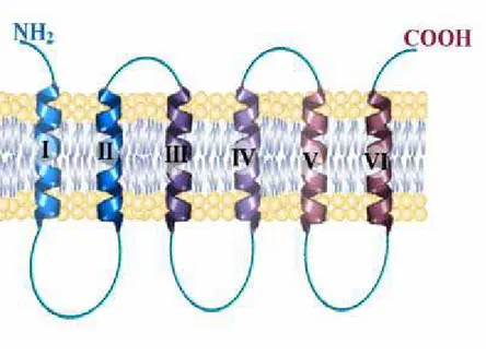
All mitochondrial carriers with known functions have a primary structure with a tripartite sequence, which contains three homologous domains, repeated in tandem, of about 100 amino acids and a similar molecular mass of about 30-35 kDa [15-17]. Each domain, consisting of two hydrophobic regions, which cross the membrane with an α-helix structure and that are connected to each other by long loops, shows a characteristic amino acid sequence, highly conserved [6]:
P-h-D / E-X-h-K / R-XK / R- (20-30 aa) -D / E-G- (4 AA) -a-K / R-G, h = hydrophobic aa; a = aromatic aa; X = any aa.
In the various mitochondrial carriers this sequence is partially modified, in one, two, or even in all three domains.
The typical tripartite structure, the presence of the two hydrophobic regions to α-helix in each domain and of the conserved sequence, emphasizes the membership of those proteins to the same family.
Most mitochondrial carriers catalyze a reaction of a specific solutes exchange. Kinetic studies, obtained by varying the concentrations of both internal and external substrates, showed, with the exception of the carnitine carrier, that all mitochondrial carriers analyzed so far agree in the function of a sequential mechanism, which implies that the substrates, one internal and one external, form a ternary complex with the carrier, so that the translocation occurs. The carnitine carrier, on the other hand, follows a ping-pong mechanism which supposes the formation of a binary complex carrier substrate: the binding site of the carrier is alternately exposed to the sides of the membrane.
On the basis of the hydropathic profile and immunochemical and enzymatic data, it has been proposed a secondary structure model according to which this carrier should be organized in six hydrophobic transmembrane segments, probably with an α-helix structure (Fig. 1.3).
15 Studies carried out using specific proteases and immunological studies showed that both the N-terminal and C-terminal end protrude in the cytoplasmic side of the membrane. The three hydrophilic segments, which connect the transmembrane domains in the side of the matrix, are called "loops" A, B, C. The two hydrophilic, shorter segments, that combine the three repeated elements, called a 'and b' protrude toward the cytoplasmic side of the mitochondrial membrane. It is possible that during the conformational change that occurs with the transport, one or more loops (A, B, C) can be inserted in the double layer lipid with a hairpin structure [6].
The tertiary structure of these proteins is not well known. This depends on the fact that these are membrane proteins, consequently hardly crystallizable due to their high hydrophobicity, their tendency to aggregate and their metastable nature.
All this does not allow a structural analysis. In fact, until now, the only crystallographic structure obtained with X-ray diffraction is the one of the carrier that catalyzes the transport of ADP/ATP (AAC1) in complex with the carboxyatractyloside [18].
Most of the isolated mitochondrial carriers and probably all the members of the MCF form homodimers, even though this issue is controversial.
The role of homodimers in the transport mechanism has not been clarified yet; there could be a formation of a channel between the two protomers or the presence of two
Fig 1.3 Schematic representation of a carrier protein, with its typical tripartite structure
16 identical channels, one for each protomer. If the mitochondrial carriers worked as dimers, these structures consist of twelve transmembrane segments, such as most of the carrier proteins [19].
As said before, 53 members of mitochondrial carriers in human have been identified and their primary structures have been studied and connected to a specific transport, by functional reconstitution of proteins in artificial membrane systems (liposomes). Such
carriers and their related isoforms are shown in Table 1 [5]. For many of them, the kinetical properties and the variation of their activities according to pH, membrane potential, phospholipids and other parameters, were clarified in detail.
Human gene name
Protein name Predominant
substrates
Tissue distribution Link to disease Human
gene locus SLC25A1 CIC (citrate carrier) Citrate,
isocitrate, malate, PEP
Liver, kidney, pancreas (also in brain, lung, heart)
22q11.21
SLC25A2 ORC2 (ornithine
carrier 2)
Ornithine, citrulline, lysine,
arginine, histidine
Liver, testis, spleen, lung, pancreas, small intestine, brain, kidney 5q31 SLC25A3 PHC (phosphate carrier)
Phosphate Isoform A: heart, skeletal muscle and diaphragm; isoform
B: liver, kidney, brain, thymus, lung,
heart, skeletal muscle, diaphragm Mitochondrial phosphate carrier deficiency 12q23
SLC25A4 ANT1 (adenine
nucleotide translocase-1)
ADP, ATP Heart, skeletal
muscle, much less in brain, pancreas, prostate, kidney, lung, thymus AAC1 deficiency, autosomal dominant progressive external ophthalmoplegia (adPEO) 4q35
17
SLC25A5 ANT2 (adenine
nucleotide translocase-2)
ADP, ATP Brain, lung, kidney, pancreas, heart,
skeletal muscle, spleen
Xq24-
SLC25A6 ANT3 (adenine
nucleotide translocase-3)
ADP, ATP Brain, lung, kidney, liver, pancreas, heart, skeletal muscle, spleen, thymus Xp22.32, Yp11.3
SLC25A7 UCP1 (uncoupling protein 1)
H+ Brown adipose
tissue
(obesity) 4q28-
SLC25A8 UCP2 (uncoupling protein 2) H+ Lung, kidney, spleen, heart obesity, type 2 diabetes, congenital hyperinsulinism 11q13
SLC25A9 UCP3 (uncoupling protein 3) H+ Skeletal muscle, lung (obesity, type II diabetes) 11q13
SLC25A10 DIC (dicarboxylate carrier) Malate, phosphate, succinate, sulphate, thiosulphate
Liver, kidney, heart, brain, lung,
pancreas
17q25.3
SLC25A11 OGC (oxoglutarate carrier) 2-oxoglutarate, malate Heart, skeletal muscle, liver, kidney, brain, pancreas 17p13.3 SLC25A12 AGC1 (aspartate/glutamat e carrier 1) Aspartate, glutamate Brain, heart, skeletal muscle, lung pancreas, kidney, but not in
liver
AGC1 deficiency, (autism)
18 SLC25A13 AGC2 (aspartate/glutamat e carrier 2) Aspartate, glutamate Liver, kidney, pancreas, heart, skeletal muscle, brain Citrullinemia type II (CTLN2), neonatal intrahepatic cholestasis (NICCD) 7q21.3
SLC25A14 UCP5 (uncoupling protein 5)
? Widely expressed,
with highest levels in brain and testis
Xq24
SLC25A15 ORC1 (ornithine carrier 1)
Ornithine, citrulline, lysine,
arginine
Liver, pancreas, lung, testis, small intestine, spleen, kidney, brain, heart
Hyperornithinemi a-hyperammonemi a-homocitrullinuria (HHH) syndrome 13q14.11 SLC25A16 GDC (Graves’disease carrier) ? Liver, kidney,
thyroid, lung, heart, skeletal muscle,
brain
10q21.3
SLC25A18 GC2 (glutamate
carrier 2)
Glutamate Brain, testis, heart, pancreas, kidney, lung 22q11.2 SLC25A19 DNC (deoxynucleotide carrier) Thiamine pyrophosphate, thiamine monophosphate, (deoxy)nucleotid es
Brain, testis, lung, kidney, liver, spleen, skeletal muscle, heart Congenital Amish microcephaly (MCPHA), neuropathy with bilateral striatal necrosis 17q25.3 SLC25A20 CAC (carnitine/acylcarnit ine carrier) Carnitine, acylcarnitine Heart, skeletal muscle, liver (also
in lung, kidney, brain, pancreas, lung,placenta) Carnitine acylcarnitine carrier deficiency 3p21.31
19 SLC25A21 ODC (oxoadipate
carrier) Oxoadipate, oxoglutarate Kidney, gall bladder, colon, liver, placenta, testis, lung, spleen,
skeletal muscle, brain, heart
14q11.2
SLC25A22 GC1 (glutamate
carrier 1)
Glutamate Pancreas, brain, liver, testis, spleen, kidney, heart, lung, small intestine, pancreatic b cells
Early epileptic encephalopathy
11p15.5
SLC25A23 APC2 ATP-Mg2+, ATP,
ADP, AMP and Pi
Kidney, lung, small intestine, pancreas, brain, liver, skeletal
muscle, heart
19p13.3
SLC25A24 APC1 ATP-Mg2+, ATP,
ADP, AMP and Pi
Testis 1p13.3
SLC25A25 APC3 ? Brain, heart,
skeletal muscle, liver, small intestine, lung, pancreas, testis
9q34.11
SLC25A26 SAMC
adenosyl-methionine, S- adenosyl-homocysteine
Ubiquitous (testis) 3p14.1
SLC25A27 UCP4 (uncoupling protein 4)
? Brain
6p11.2-q12
SLC25A28 Mitoferrin 2 (Mfrn2) Fe2+ Ubiquitous (heart,
liver, kidney)
10q23-q24
SLC25A29 ORNT3 Ornithine,
acylcarnitine
Heart, skeletal muscle, liver, brain
14q32.2
20
SLC25A31 AAC4, ANT4
(adenine nucleotide carrier 4)
ADP, ATP Testis (spermatogenesis
) 4q28.1 SLC25A32 MFT Folate 8q22.3 SLC25A33 PNC1 (pyrimidine nucleotide carrier 1) UTP Ubiquitous 1p36.22 SLC25A34 ? 1p36.21 SLC25A35 ? 17p13.1 SLC25A36 PNC2 (pyrimidine nucleotide carrier 2) Pyrimidine nucleotides 3q23
SLC25A37 Mitoferrin 1 (Mfrn1) Fe2+ Fetal liver, bone
marrow, spleen, placenta, liver,
brain
8p21.2
SLC25A38 Glycine Erythroid cells Sideroblastic
anemia
3p22.1
SLC25A39 ? Ubiquitous 17q12
SLC25A40 ? Ubiquitous at low
levels
7q21.12
SLC25A41 APC4 ATP-Mg/Pi Brain, testis, liver 19p13.3
SLC25A42 CoA, ADP, ATP,
dPCoA Ubiquitous (adipose tissue) Mitochondrial myopathy 19p13.11
SLC25A43 ? Brain, adrenal
gland, skeletal muscle
Xq24
SLC25A44 ? Ubiquitous 1q22
SLC25A45 ? Skeletal muscle,
intestine, brain, testis
11q13.1
21 SLC25A47 ? 14q32.2 SLC25A48 ? 5q31.1 SLC25A49 MTCH1 ? 6pter-p24.1 SLC25A50 MTCH2 ? 11p11.2 SLC25A51 MCART1 ? 9p13.3-p12 SLC25A52 MCART2 ? 18q12.1 SLC25A53 MCART6 ? Xq22.2
The mitochondrial carriers currently known can be divided into electrogenic and electroneutral transporters; in particular, the functional characterization of the known
carriers allows us to further divide them into three categories, according to the type of
substrate transported:
• Carriers that catalyze an electrogenic transport such as the ADP4- / ATP3- transporter; • Carriers that catalyze an electroneutral transport, in which the charges transported inward are balanced by equal charges outward (symport: anion/H+) or opposite charges inward (antiporter: anion/OH-), as the phosphate carrier;
• Carriers that catalyze a neutral transport in which the transported molecule does not have an electric charge, as in the case of the carnitine carrier.
1.3 Disorders related to mitochondrial carrier deficiency
Since almost all of the known substrates of the MCF members are involved in reaction sequences that ultimately produce ATP for the cell, efficient transport is required for mitochondrial function and cellular survival. Furthermore, amino acid mutations caused
Table 1.1 Schematic representation of the genes codifying for transport proteins, their substrates, tissue distribution, the human gene locus and the possible link to diseases [5]. The mitochondrial carriers analyzed in this work are marked with
22 by single nucleotide polymorphisms in MCF proteins have been documented in humans and are linked to clinical diseases [5].
The diseases caused by deficiencies of mitochondrial transport proteins are metabolic disorders caused by mutations in genes that encode for them, and they are transmitted as autosomal recessive, with the exception of Progressive External Ophthalmology (adPEO), inherited as autosomal dominant.
Diseases related to mitochondrial carriers can be divided into two groups [20]:
• Diseases with a genetic defect of mitochondrial carriers, whose function is linked to the oxidative phosphorylation;
• Diseases due to the alteration of mitochondrial function of those carriers that are not involved in oxidative phosphorylation but determine an alteration of physiological concentrations of important intermediates of some metabolic pathways. The symptoms in this case depend on the metabolic pathway and the type of affected tissue (tissue distribution of the carrier).
The dysfunction of oxidative phosphorylation is due to the ADP/ATP carrier and to the Pi carrier, which transport ADP and Pi, used by the complex ATP synthase for the production of ATP, in the mitochondrial matrix.
In the first group we find: • Sengers’ syndrome, • AAC1 deficiency, • PiC deficiency
• Progressive External Ophthalmology (adPEO).
The symptoms of the diseases related to these genes defects are caused by insufficient production of ATP, especially in those tissues where these carriers are strongly expressed and where is present a high activity of the oxidative phosphorylation.
In this group of diseases is also included PEO, even though the heterozygous mutation of isoform 1 of the carrier of' ADP/ATP does not lead to a direct action on the energy dysfunction, but it contributes to the mitochondrial DNA instability [20].
23 The known diseases of the second group are:
• CAC deficiency, • HHH syndrome, • ACG2 deficiency,
• Microcephaly of the Amish population • Neonatal myoclonic epilepsy [20].
Other carriers may be jointly responsible for diseases, but the mechanisms have not been clarified yet. For example, the carrier of citrate (CIC), encoded by the SLC25A1 gene seems to be involved in DiGeorge Syndrome (DGS) and the veil-craniofacial syndrome (VCFS) [20].
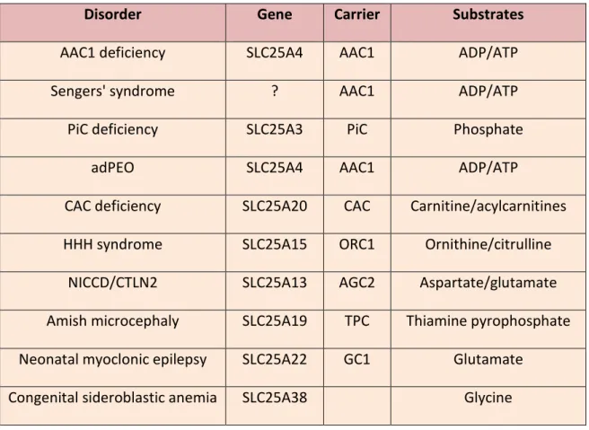
Disorder Gene Carrier Substrates
AAC1 deficiency SLC25A4 AAC1 ADP/ATP
Sengers' syndrome ? AAC1 ADP/ATP
PiC deficiency SLC25A3 PiC Phosphate
adPEO SLC25A4 AAC1 ADP/ATP
CAC deficiency SLC25A20 CAC Carnitine/acylcarnitines HHH syndrome SLC25A15 ORC1 Ornithine/citrulline
NICCD/CTLN2 SLC25A13 AGC2 Aspartate/glutamate Amish microcephaly SLC25A19 TPC Thiamine pyrophosphate Neonatal myoclonic epilepsy SLC25A22 GC1 Glutamate
Congenital sideroblastic anemia SLC25A38 Glycine
24
2 . M A T E R I A L S A N D M E T H O D S
2.1 Construction of Expression Plasmids
The coding sequence of the selected proteins (yeast HEM25 and human SLC25A38 for the glycine carrier; dPCoAC-A and dPCoAC-B, the two isoforms of the D.
melanogaster gene GC4241; the B. taurus OGC) were cloned into expression vectors,
respectively into pET21b, pRUN and pMW7 vectors for expression in E. coli, and into the yeast pYES2 expression vector (HEM25 and SLC25A38 - A and
dPCoAC-B).
Transformants selected on LB plates containing ampicillin (100 µg/ml) were screened by direct colony PCR and by restriction digestion of the purified plasmid DNA [21] and verified by DNA sequencing.
2.1.1 Expression Plasmids for Glycine Carrier
The HEM25 open reading frame was amplified from S. cerevisiae genomic DNA by PCR, using primers corresponding to the extremities of the coding sequences with additional NdeI and HindIII sites.
The coding sequence of the human SLC25A38 gene was obtained by RT-PCR reaction, using total RNA extracted from HepG2 cells as template [22, 23], and the primers with additional NdeI and HindIII sites. The amplified products were cloned into the NdeI-HindIII sites of the expression vector pET21b that had been previously modified by cloning into HindIII-XhoI sites a cDNA sequence coding for a V5 epitope followed by six histidines [24].
The HEM25-pYES2 and SLC25A38-pYES2 plasmids were constructed by cloning the coding sequences of HEM25 and SLC25A38, respectively, into the yeast pYES2 expression vector. In order to create the HEM25-pYES2, the
HEM25 cDNA was amplified by PCR using the HEM25-pET21b construct as
template. The forward and reverse primers carried BamHI and XhoI restriction sites, respectively, as linkers. The reverse primer also carried a cDNA sequence coding for a V5 epitope. Similarly, to generate the SLC25A38-pYES2 construct,
25 the coding sequence of SLC25A38 was obtained by PCR using SLC25A38-pET21b as template and the primers with additional BamHI and XhoI sites. The pET21b and pYES2 vectors, prepared as above, were transformed into E. coli DH5α cells.
2.1.2 Expression Plasmids for dPCoA Carriers
The first isoform, dPCoAC-A, corresponded to a 1098 bp open reading frame (RefSeq accession no.NM_206522) and encoded a protein of 365 residues (RefSeq accession no. NP_650891). The second one, dPCoAC-B, corresponded to an 873 bp open reading frame (RefSeq accession no. NM_169901) and encoded a protein of 290 residues (RefSeq accession no. NP_732519). Both splicing isoforms were amplified by PCR, using an Oregon-R adult flies cDNA as template and two primers (sense and antisense) carrying suitable restriction sites at their 5′ ends for the further cloning of the amplified inserts in the E. coli and S. cerevisiae expression vectors, pRUN [25] and pYES2 (Invitrogen™), respectively. We introduced the R103S mutation into the wild-type dPCoAC-A cDNA by overlap-extension PCR [26].
2.1.3 Expression Plasmids for OGC Carrier and site direct mutagenesis
The coding region for BtOGC was amplified by polymerase chain reaction (PCR) method from bovine heart cDNA [27]. The forward and reverse oligonucleotide primers corresponded to the extremities of the coding sequence for OGC with NdeI and HindIII sites. The WT OGC cDNA was employed as a template to replace lysine 122 by arginine. K122R cDNA was employed as a template to construct triple mutants having a single cysteine residue: K122R/C184, K122R/C221 and K122R/C224. As the WT OGC contains three native cysteines (in position 184, 221 and 224), in K122R/C184 cDNA cysteines located in position 221 and 224, in K122R/C221 cDNA cysteines 184 and 224 and in K122R/C224 cDNA cysteines 184 and 221 were replaced by serines, respectively. Further mutants, named C184, C221 and C224, each containing a single cysteine residue (184, 221 and 224, respectively), were constructed in the
26 WT OGC background, as previously described [28, 29]. All of the mutations were introduced in the WT or in the K122R OGC cDNA by the overlap extension PCR method [26], using oligonucleotides with appropriate mutations in their sequences. The PCR products were cloned into the expression vector pMW7 and transformed into E. coli TG1 cells.
2.2 Bacterial expression and purification of recombinant proteins
All the cloned expression vectors were overexpressed as inclusion bodies in the cytosol of E. coli cells, BL21(DE3) for Hem25p and GlyC, C0214(DE3) for dPCoAC-A, R103S dPCoAC-A mutant, dPCoAC-B and the OGC mutants.
A colony was inoculated into growth medium containing ampicillin (100 µg/ml) and the culture was grown at 37 °C for 4-5 h, or until the optical density of the culture at 600 nm was 0.7-1.0. Then isopropyl-β-D-thiogalactopyranoside was added to a final concentration of 0.4 mM to induce the expression of the carrier proteins, and incubation was continued for a further 4 to 5 hours [30]. Control cultures with the empty vector were processed in parallel. The cells were harvested by centrifugation, and used for the preparation of inclusion bodies. Cells were resuspended in TE buffer disrupted using either a French Press or a Sonicator, and then centrifuged at 4°C at 27000 g for 15 min. The pellet was resuspended in a smaller volume of the same buffer and fractionated by centrifugation at 131000 g for 4.5 h at 4°C, through a step gradient made of 40 %, 53 % and 70 % (w/v) solutions of sucrose (sucrose density gradient) [31]. Inclusion bodies were collected, washed at 4ºC with TE buffer (10 mM Tris⁄HCl, 1 mM EDTA, at the appropriate pH), and finally resuspended in the same buffer [32].
Hem25p and GlyC proteins were solubilized in 2% (w/v) sarkosyl and purified by centrifugation and Ni2+-NTAagarose affinity chromatography, as described previously [33]
.
2.3 Reconstitution of recombinant proteins into liposomes and transport measurements
The recombinant proteins were solubilized in Sarkosyl® (N-dodecanoyl-N-methylglycine sodium salt) and then reconstituted into liposomes by cyclic removal of
27 the detergent with a hydrophobic column of Amberlite beads (Bio-Rad) [34] in the presence or absence of substrates [35].
After vortexing, this mixture was recycled 13 times through the Amberlite column (4.0 × 0.5 cm). All operations were performed at 4 °C, except the passages through Amberlite, which were carried out at room temperature.
External substrate was removed from proteoliposomes on Sephadex G-75 columns, pre-equilibrated with 50 mM NaCl/10 mM HEPES at pH 7.5 (for Hem25p and GlyC) or PIPES at pH 7 (for dPCoACs and OGC) [36].
The first 600 µl of turbid eluate from the Sephadex G- 75 column were collected, and 100 µl were transferred to reaction vessels and used for transport measurements by the “inhibitor stop method” [24, 34, 36].
Transport at 25°C was started by adding, at the indicated concentrations, respectively, L- [14C] glycine, [14C] ADP, 2-oxo [1-14C] glutarate or other indicated labeled compounds, to substrate-loaded proteoliposomes (exchange) or to empty proteoliposomes (uniport).
Transport was terminated by adding PLP (pyridoxal-5′-phosphate) and BAT (bathophenanthroline), which, in combination and at high concentrations, completely inhibit the activity of several mitochondrial carriers [35]. Each sample was run in duplicate. Finally, the external substrate was removed by a Sephadex G-75 column, the proteoliposomes were eluted with 50 mM NaCl, collected in a scintillation mixture and the accumulation of radioactive substrate in the liposomes was measured by liquid scintillation (Fig. 2.1) [24, 36]. The experimental values were corrected by subtracting control values.
28 Fig. 2.1 Formation of proteoliposomes and determination of transport.
29 The reconstitution mixture for each group of proteins contained:
• Hem25p and GlyC
100 µl of purified proteins (0.5-1 µg of protein) 90 µl of 10% (w/v) Triton X-114
90 µl of 10% (w/v) L-α-phosphatidylcholine from egg yolk (Sigma-Aldrich), as sonicated liposomes
10 mM glycine (except where otherwise indicated)
20 mM HEPES at pH 7.5 (except where otherwise indicated) water to a final volume of 700 µl.
• dPCoAC-A, R103S dPCoAC-A mutant, dPCoAC-B 55 µl of purified proteins (10 µg of protein)
70 µl of 10% (w/v) Triton X-114
90 µl of 10% (w/v) L-α-phosphatidylcholine from egg yolk (Sigma-Aldrich), as sonicated liposomes
10 mM ADP (except where otherwise indicated)
20 mM PIPES at pH 7 (except where otherwise indicated) 0.4 mg of cardiolipin (Sigma-Aldrich)
water to a final volume of 700 µl. • OGC and mutants
100 µl of purified proteins 98 µl of 10% (w/v) Triton X-114
90 µl of 10% (w/v) L-α-phosphatidylcholine from egg yolk (Sigma-Aldrich), as sonicated liposomes
20 mM oxoglutarate (except where otherwise indicated) 20 mM PIPES at pH 7.0 (except where otherwise indicated) 2.3 mg/ml asolectin from soybean
water to a final volume of 700 µl.
The initial transport rate was calculated from the radioactivity taken up by proteoliposomes after 1or 2 min (in the initial linear range of substrate uptake).
For efflux measurements, proteoliposomes containing 2 mM substrate were loaded with 5 µM of labeled substrate, by carrier-mediated exchange equilibration [34]. After 30 min, the external radioactivity was removed by passing the proteoliposomes through
30 Sephadex G-75. Efflux was started by adding unlabeled external substrate or NaCl to aliquots of proteoliposomes and terminated by adding the inhibitors indicated above.
In the OGC experiments, the influence of several sulfhydryl reagents such as mersalyl, pCMBS, MTSEA, MTSES and MTSET, was investigated on single-cysteine replacement mutants through transport assays. Proteoliposomes were pre-incubated at 25 °C in the presence or absence of each sulfhydryl reagent at the desired concentration for 2 min (mersalyl or pCMBS) [31] or for 10min (MTSEA, MTSES or MTSET) [29, 37, 38]
. After removal of unbound reagent by Sephadex G-75 chromatography, transport was started by adding 0.3 mM 2-oxo[1-14C]glutarate and stopped after 30 s [39]. The influence of substrate on the inhibition of some OGC mutants by MTSEA was tested by pre-incubating proteoliposomes, preloaded with 20 mM 2-oxoglutarate, with this sulfhydryl reagent and its concentration was chosen in order to obtain only partial (approximately 50%) transport inhibition. After removal of unbound reagent and external substrate by Sephadex G-75 chromatography, transport was initiated by the addition of 1 mM 2-oxo[1-14C]glutarate and terminated after 30 s.
2.4 Overexpression in Saccharomyces cerevisiae of recombinant proteins and mitochondrial isolation
The resulting yeast expression plasmids were introduced in the deleted strain, and trasformants were selected on SC agar plates without uracil, supplemented with 2% (w/v) glucose. A colony was precultured on SC medium without uracil supplemented with 2% (w/v) glucose for 14-16 h, diluted to a final OD600 = 0.05 in YP supplemented with 3% (w/v) glycerol and 0.1% (w/v) glucose and grown to early-exponential phase. Galactose (0.4% w/v) was added 4 h before harvesting to induce recombinant protein overexpression.
Cells were pelleted by centrifugation at 3000g for 5 min at room temperature and washed with distilled water. Subsequently, they were resuspended in 2 ml/g of cells (v/w) DTE buffer (100 mM Tris–H2SO4, pH 9.4, 10 mM 1,4-Dithioerythritol) and shaken slowly for 10 min at 30°C. After incubation with DTE buffer, the cells were centrifuged again at 3000g for 5 min and then washed with sorbitol 1.2M.
31 phosphate, pH 7.4) and then incubated with the addition of 4 mg/g (w/w) Zymolyase-20T (Seikagaku Kogyo Co.) for 30-60 min at 30°C to obtain spheroplasts. Cells were then washed again with sorbitol 1.2M.
Cells were homogenized by 50 strokes in a glass–teflon potter in 14 ml/g (v/w) ice-cold homogenization buffer (0.6 M sorbitol, 10 mM Tris–HCl, pH 7.4, 1 mM PMSF, 0.2% BSA).
Cell debris and nuclei in the homogenate were removed by centrifugation at 3000g for 5 min at 4°C. The supernatant was collected and centrifuged twice at 12,000g for 15 min at 4°C in order to recover the mitochondrial fraction which was resuspended in ST buffer (0.25 M Sucrose, 10 mM Tris–HCl, pH 7) [40, 41].
After protein quantification, mitochondria were stored at −80 °C until use.
Mitochondria were solubilized in a buffer containing 3% (w/v) Triton X-100, 20 mM NaCl, 10 mM HEPES pH 7.5 or PIPES p7 and supplemented with cardiolipin. After incubation for 20 min at 4ºC, the mixture was centrifuged at 8000 x g for 20 min thereby obtaining a supernatant, referred to as mitochondrial extract. The mitochondrial extract was reconstituted by cyclic removal of detergent [33].
2.5 Subcellular localization of recombinant proteins
Indirect immunofluorescence experiments were carried out according to Pringle’s procedure with some modifications [42, 43]. Cells were grown in glycerol supplemented YP medium until early logarithmic phase, and the expression of recombinant proteins was induced adding 0.4% (w/v) galactose for 4 h at 30ºC. Then, formaldehyde solution was added directly to the cells in growth medium to a final concentration of 3.7% (w/v). After 1 h at 30ºC, the cells were pelleted by centrifugation at 1301 x g for 5 min at room temperature and resuspended in phosphate-buffered paraformaldehyde containing 100 mM potassium phosphate, pH 6.5, 1 mM MgCl2 and 3.7% (w/v) paraformaldehyde. After 16 h at 30ºC, the cells were washed with distilled water and then incubated for 10 min at 30ºC in 1 ml solution containing 200 mM Tris/HCl, pH 8.0, 1 mM EDTA and 1% (v/v) β-mercaptoethanol. The pellet was resuspended in 1 ml zymolyase buffer (1.2 M sorbitol, 20 mM potassium phosphate, 1 mM MgCl2, pH 7.4). Zymolyase-20T (Seikagaku Kogyo Co., Japan) (5 mg/g of cell, wet weight) was added and the suspension was incubated for 30-60 min at 30°C with gentle shaking for conversion into
32 spheroplasts. After digestion, cells were spinned down at 1301 x g and washed once with 1.2 M sorbitol, then resuspended gently and incubated for 2 min in 1.2 M sorbitol and 2% (w/v) SDS. Cells were washed twice with 1.2 M sorbitol.
Spheroplasts (20 µl) were incubated for 1 h at 30°C in the presence of 100 nM MitoTracker Red, 32 nM FM 4-64 Dye (N-(3-Triethylammoniumpropyl)-4-(6- (4(Diethylamino) Phenyl) Hexatrienyl) Pyridinium Dibromide) or 1 µg/ml DAPI (4',6- Diamidino-2 -Phenylindole, Dihydrochloride) (Molecular Probes, The Netherlands), washed twice with PBS containing 5 mg/ml BSA (PBS-BSA) before adding a primary antibody against V5 (1 µg/ml in PBS-BSA) and incubated for 1 h at 30ºC. After washing twice for 5 min with PBS-BSA, the secondary antibody anti-mouse FITC conjugate (Santa Cruz Biotechnology, Inc., CA, USA) was added at 1:100 dilution in PBS-BSA for 1 h at 30ºC. In the final step, the coverslips were washed three times in PBS-BSA and the cells were observed by a confocal laser scanning microscope (LSM 700, Zeiss, http://www.zeiss.com/). Observations were performed as described in [44].
2.6 Sample preparation for yeast metabolite analysis
A previous procedure for preparation of yeast cell lysates described by Villas-Bôas was used [45]. Yeast cultures were grown overnight shaking in YP media supplemented with 2% (w/v) glucose at 30ºC to OD600 = 3.5 and then were quenched by quickly adding 10 ml of overnight cultures to 40 ml of chilled methanol-water solution (60% v/v). Cells were harvested for 5 min at 1540 x g (0ºC) and the pellets were resuspended in 3 ml chilled 100% (v/v) methanol. A 1ml aliquot of this suspension was snap frozen in liquid nitrogen, thawed in an ice-bath and centrifuged at 770 x g for 20 min (0ºC). The supernatant was collected and an additional 0.5 ml of chilled 100% (v/v) methanol was added to the pellet and vortexed for 30 seconds. Suspensions were centrifuged at 770 x
g for 20 min (0ºC) and both supernatants were pooled. Metabolite extractions were
carried out on yeast mitochondria isolated from wild-type, hem25∆ and HEM25-pYES2 cells. Yeast mitochondria were obtained as described above.
33 2.7 Glycine and ALA analysis by gas chromatography coupled to tandem mass spectrometry (GC-MS/MS)
Yeast lysates and mitochondria obtained from wild-type, hem25∆ and HEM25-pYES2 cells were centrifuged, and the supernatant was transferred into a glass vial; norleucine (Sigma-Aldrich, Italy) was added as internal standard (10 µl of an 80 ng/µl norleucine solution) and freeze dried [46, 47]. The residue was reconstituted with 200 µl of DMF (N,N-dimethylformamide) (VWR International PBI S.r.l., Italy) and 50 µl of MTBSTFA (N-tertbutyldimethylsilyl- N-methyltrifluoroacetamide) (Sigma-Aldrich, Italy) [48]. Derivatization with MTBSTFA was performed at 60°C for 30 min. If not otherwise indicated, samples were placed on the GC-MS/MS autosampler tray immediately after preparation. After cooling the solution at room temperature for 5 min, 1 µl of the solution was injected into the GC-MS/MS. Samples were run on a GC-QqQ-MS (Bruker 456 gas chromatograph coupled to a triple quadrupole mass spectrometer Bruker Scion TQ) equipped with an autosampler (GC PAL, CTC Analytics AG). 1 µl of the sample was injected in split mode (split ratio 5:1). The GC was operated at a constant flow of 1.2 mL/min and analytes were separated on a Restek Rxi 5Sil MS capillary column (30 m with a 10 m “built-in” guard column, inner diameter 250 µm and film thickness 0.25 µm). The oven was kept at 130°C for 2 min after injection, then a temperature gradient of 5°C/min was employed until 220°C was reached and of 10°C/min up to 300°C. The oven was then held at 300°C for 10 min. The total run time was 38 min. The mass detector was operated at 70 eV in the electron impact (EI) ionization mode. The ion source and transfer line temperature were 200°C and 280°C, respectively. The collision gas was argon. For both ALA and Gly quantification, an MS/MS procedure was employed in Multiple Reaction Monitoring (MRM) mode using argon 99.999% (w/v) (Sapio BIC) as collision gas at a pressure of 1.5147 mbar in the collision cell. To ensure a reliable identification of analytes, two MS/MS transitions were selected within ± 0.2 min of the compounds’ retention time. Product ion abundance was maximized by optimal collision energy voltage. The dwell time per transition was 0.1 s. For identity confirmation both the match in retention time and the confirmation ratio (Q/qi), i.e. the ratio between the intensity of the quantification (Q) and confirmation (qi) transitions recorded for each compound, were required to confirm positive identification in a sample. Bruker MS Workstation 8 application manager was used to process the data. Method validation included determination of linear range, limit
34 of detection (LOD), limit of quantification (LOQ), recovery, and repeatability (Table 1 gives an overview of the relevant data). Linearity was evaluated by least-squares regression and expressed as r2. The calibration curve was linear in the investigated range of six concentration levels (0.04, 0.1, 0.2, 0.4, 0.7, and 1.0 ng/µl). LOQ and LOD were estimated, respectively, from the calibration curve as the concentration for which there is a 5% chance of having a false-positive and the concentration for which there is a coefficient of variation on the expected value less than 10%. Experiments to evaluate surrogate or marginal recovery were performed with samples spiked with 100 ng Gly and ALA: the average recoveries were calculated on five different experiments. The repeatability of the present method was estimated during the entire analytical procedure (sample pretreatment, derivatization, and GC/MSMS separation) using standard solutions (0.4 ng/µl ALA; 0.1 ng/µl Gly). The intraday and interday precision values, expressed in terms of relative standard deviation (RSD), were assessed by performing an analysis three times on the same day and by conducting the analysis on five different days in one month, respectively.
2.8 Determination of mitochondrial heme and cytochrome content
Mitochondria were isolated from wild-type, hem25∆, HEM25- pYES2 and SLC25A38-pYES2 cells and solubilized in 1% (w/v) Triton-X100. Heme was quantified by spectrophotometry according to the method of Drabkin with some modifications [49]. To determine fluorescence spectra of heme, equal amounts of mitochondrial protein were mixed with 2 mol/L oxalic acid, heated to 95°C for 30 minutes to release iron from heme and generate protoporphyrin IX. Samples were then centrifuged for 10 minutes at 1000 g at 4°C to remove debris. The fluorescence intensity of the supernatant was measured with a JASCO FP 750 fluorimeter (Jasco Corporation, Japan). The excitation wavelength was 405 nm and emission was measured at 600 nm. The 662 nm emission peak has lower fluorescence than the peak at 600 nm, but also has other interference [50]. Determination of the contents of cytochromes aa3, b-type (bII + bH + bL) and ctype (c + c1) in isolated mitochondria was carried out in 1 ml of respiratory buffer (300 mM sucrose, 1 mM EDTA, 5 mM Mops, 10 mM KH2PO4, 2 mM MgCl2, 0.1% (w/v) BSA and 5 mM succinate, pH 7.4) [50]. Individual fully reduced (with excess sodium
35 dithionite) or fully oxidized (with excess ferrycianide) absorbance spectra were recorded between 500 and 650 nm, and the concentration of each type of cytochrome was determined from the difference (reducedoxidized) spectrum at the maximum absorption value for each one, normalized by the absorbance of the respective isosbestic point. Values were calculated by the Beer-Lambert law, as described previously [51], and expressed relative to protein concentration.
2.9 Mitochondrial respiration efficiency
Mitochondrial respiration (0.2 mg of mitochondrial protein/ml) was measured in a medium consisting of 300 mM sucrose, 1 mM EDTA, 5 mM Mops, 10 mM KH2PO4, 2 mM MgCl2, 0.1% (w/v) BSA and 5 mM succinate, pH 7.4, by means of a Clark oxygen electrode at 30ºC. After 2 min, state 3 respiration was induced by the addition of 0.2 mM ADP. The rate of oxygen uptake by yeast mitochondria (V) was expressed as nmol O2 x ml-1 x min-1/mg protein. The RCR was calculated by dividing V3 (rate of oxygen uptake measured in the presence of respiratory substrates + ADP, i.e., state 3 of respiration or active state of respiration) by V4 (rate of oxygen uptake measured with respiratory substrates alone, i.e., state 4 of respiration or resting state of respiration) [52]. The enzymatic bc1 complex activity was determined by measuring the reduction of oxidized cytochrome c at 550 nm as described previously [53].
2.10 Comparative modelling and docking investigations
Computational approaches for protein function investigations [54] have been employed to investigate the function of the analyzed proteins.
Modeller [55] was used to calculate a 3D comparative model of dPCoAC-A and BtOGC by using as template the structure of the bovine AAC1 (protein data bank accession code: 1okc), crystallized in complex with its powerful inhibitor carboxyatractyloside (CATR) [18]. The structural properties of the dPCoAC-A and SLC25A11_OGC comparative models with the best energy function were evaluated using the biochemical/computational tools ofthe WHAT IF Web server [56]. Q-site Finder [57] was used to predict the potential binding sites of dPCoAC-A best model. For docking
36 analysis, CoA and dPCoA ligands were docked into the proposed binding sites of dPCoAC-A by using Autodock 1.5.2. [58].
2.10.1 Comparative modelling and docking investigations of dPCoAC
A multiple sequence alignment (MSA) between dPCoAC-A and dPCoAC-B proteins and their putative orthologs from H. sapiens and S. cerevisiae was obtained by using ClustalW [59]. The sequence of crystallized bovine ADP/ATP carrier (AAC1) [18]was introduced in the alignmentin order to use the secondary structure of the bovine AAC1 to weigh gap insertions [55]. The characterizing triplet set of dPCoACs was obtained by aligning the three repeats of the analyzed mitochondrial carriers, building an inter-repeat multiple sequence alignment (MSA) [60].
2.10.2 Comparative modelling and docking investigations of BtOGC mutants
By using the obtained 3D model of BtOGC as a protein template, mutagenesis in silico analysis was also performed to build the single OGC mutant K122R, and the triple mutants (K122R/C184, K122R/C221 and K122R/C224), in order to investigate the role played by K122R in 2-oxoglutarate translocation and, more in general, the influence of cysteine residues in 2-oxoglutarate uptake. Due to the availability of in vitro transport assays in the presence or absence of cysteine specific reagents MTSEA was added to cysteine residues of 3D models (WT OGC and triple mutants) in order to explain results from transport assays in the context of a 3D protein model. More in detail, MTSEA was alternatively added to the cysteine residues within the triple mutants K122R/ C184, K122R/C221 and K122R/C224. For docking analysis the 2-oxoglutarate ligand was docked into the proposed binding site of our 3D models using Autodock 1.5.2. [58]. A gridbox involving the residues protruding towards the WT OGC and the triple mutants carrier cavity from the c-gate to the m-gate, surrounding residues that form the MCs common substrate binding site [61, 62], and overlapping with the carboxyatractyloside binding region described in the crystallized AAC1 [18], was built to investigate the binding of 2-oxoglutarate substrate to OGC. Docking
37 simulations were performed according to validated protocols [54], in order to screen the putative binding modes of 2 oxoglutarate at this region in the WT OGC 3D model and in the analysed triple mutants in the presence or absence of MTSEA.
2.11 Molecular evolution analysis
14 eukaryotic model organisms for which whole genome sequences were available were selected, i.e. two yeast species (Saccharomyces cerevisiae and Schizosaccharomyces
pombe), two plant species (Arabidopsis thaliana and Zea mais), two nematode species
(Caenorhabditis elegans and Caenorhabditis briggsae), two insect species (Drosophila
melanogaster and Anopheles gambiae), two fish species (Danio rerio and Tetraodon nigroviridis), two amphibian species (Xenopus laevis and Xenopus tropicalis), and two
mammalian species (Mus musculus and Homo sapiens). The human protein sequence encoded by the SLC25A42 gene was queried against the non-redundant reference RNA sequence database using the tblastn version implemented in the NCBI website (https://blast.ncbi.nlm.nih.gov/). The obtained transcripts were filtered for e-value ≤10−40 and coverage ≥80%, and only one protein isoform per gene, i.e. the longest one, was retained for subsequent analyses. The 65 homologous proteins collected were multi-aligned with ClustalW [59]. N- and C-termini sequences were removed from multiple sequence alignment (MSA) and 6 MSA blocks (255 alignment columns) were retained for the following evolutionary analyses, since they correspond to the most informative mitochondrial carrier amino acids [5, 61]. After having used MEGA6 [63] to find the best amino acid substitution model for the maximum likelihood (ML) analysis, a phylogenetic tree was built in PhyML [64], using the LG substitution model with four substitution rate categories. The tree branch support was tested in PhyML using two methods, i.e. aBayes [65]and 100 bootstrap samplings.
38
3 . C H A R A C T E R I Z A T I O N O F T H E
M I T O C H O N D R I A L G L Y C I N E
C A R R I E R
3.1 INTRODUCTION
Glycine carrier and associated diseases
The human glycine carrier (human SLC25A38) is a nuclear encoded protein, localized in the inner mitochondrial membrane. This carrier belongs to the mitochondrial carrier family, and, similarly to all SLC25A family members, mediates the exchange of metabolites across the inner mitochondrial membrane and possesses a tripartite structure, typical of the mitochondrial inner membrane transporter family, consisting of three tandemly repeated sequences of approximately 100 amino acids length [5]. The gene, mapped on chromosome 3.p22.1, has a length of 14kb, presents 7 exons coding for a protein of 303 amino acids and about 33kDa [66].
SLC25A38 is highly and preferentially expressed in transferrin receptor (CD71) positive erythroid cells. Mutations in SLC25A38 cause congenital sideroblastic anemia [67]
. As its name indicates, this disease is characterized by severe anemia with hypochromia, microcytosis and ringed sideroblasts in the bone marrow, formed by iron-loaded mitochondria clustered around the erythroblast nucleus [68]. Patients are nonsyndromic and present no developmental anomalies.
Unlike the acquired sideroblastic anemia, which occurs after exposure to certain drugs or alcohol and with copper deficiency [69], congenital sideroblastic anemia (CSA) is a rare and heterogeneous disease caused by mutations of genes involved in heme biosynthesis, iron-sulfur (Fe-S) cluster biogenesis or Fe-S cluster transport and mitochondrial metabolism. The definition of this disease at molecular level has provided insight into cellular pathways associated with dysfunctional mitochondrial iron metabolism [68].
X-linked sideroblastic anemia (XLSA) is a form of non-syndromic CSA caused by a defect of the δ-aminolevulinate synthase 2 (ALAS2) gene, which encodes the first
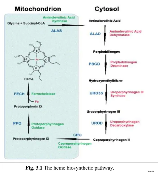
39 mitochondrial enzyme of heme biosynthesis in erythroid cells [70]. The ALAS2 catalyzes the condensation into mitochondria of glycine with succinyl-coenzyme A to yield δ-aminolevulinic acid (ALA), and requires pyridoxal 5'-phosphate (PLP; vitamin B6) as cofactor [71]. Following its synthesis, ALA is exported to the cytosol where it is converted to coproporphyrinogen III (CPgenIII). All the remaining steps of heme biosynthesis take place inside mitochondria. CPgenIII is imported into the mitochondrial intermembrane space where it is converted to protoporphyrinogen IX by coproporphyrinogen oxidase. Then, protoporphyrinogen IX is oxidized to protoporphyrin IX (PPIX) by protoporphyrinogen oxidase. Lastly, ferrous iron is incorporated into PPIX to form heme in the mitochondrial matrix, a reaction catalyzed by ferrochelatase (Fig 3.1) [72]. While all the enzymatic steps leading to the production of heme are well characterized, it is still not completely understood how ALA, CPgenIII and heme and glycine are transported across the two mitochondrial membranes.
Fig. 3.1 The heme biosynthetic pathway.
40 Missense mutations in SLC25A38 gene have been found recently, by Guernsey et al, in patients with severe non-syndromic CSA resembling XLSA but lacking ALAS2 mutations. Knocked down zebrafish orthologues of SCL25A38 (slc25a38a and
slc25a38b) caused an anemic phenotype, showing the importance of this gene for red
blood cells production and function [67].
Furthermore, the genetic deletion of the Saccharomyces cerevisiae SLC25A38 orthologue YDL119c (also named HEM25) produces a respiratory phenotype (unable to grow on glycerol), indicative of mitochondrial involvement, and shows a significant reduction of ALA levels, the first product in the heme biosynthetic pathway [67].
Based on these findings and on their structure (particularly because of a conserved arginine-asparate (RD) dipeptide sequence present in transmembrane helix 4, typical of amino acid carriers), SLC25A38 and its yeast orthologue Hem25p were hypothesized to facilitate ALA production by importing glycine into mitochondria or by exchanging glycine for ALA across the inner mitochondrial membrane. Thus, it would transport one or two substrates required in the first steps of heme biosynthesis.

![Table 1.1 Schematic representation of the genes codifying for transport proteins, their substrates, tissue distribution, the human gene locus and the possible link to diseases [5]](https://thumb-eu.123doks.com/thumbv2/123dokorg/2868158.9171/25.892.115.821.104.426/schematic-representation-codifying-transport-substrates-distribution-possible-diseases.webp)
![Fig. 4.1 Biosynthetic pathway of Co-A [92]](https://thumb-eu.123doks.com/thumbv2/123dokorg/2868158.9171/66.892.184.763.110.757/fig-biosynthetic-pathway-of-co-a.webp)

![Fig. 4.4 Inhibition of [ 14 C]ADP uptake by inhibitors and substrates externally added to proteoliposomes reconstituted with dPCoAC-A](https://thumb-eu.123doks.com/thumbv2/123dokorg/2868158.9171/75.892.150.796.236.488/inhibition-uptake-inhibitors-substrates-externally-proteoliposomes-reconstituted-dpcoac.webp)
![Fig. 4.5 Kinetics of [14C]ADP transport.](https://thumb-eu.123doks.com/thumbv2/123dokorg/2868158.9171/77.892.311.623.106.684/fig-kinetics-of-c-adp-transport.webp)
