UNIVERSITY of SASSARI
PhD school in
Biomolecular and Biotechnological Sciences
PhD Programme: Molecular and Clinical MicrobiologyXXVI cycle
PhD School Director: Prof. Claudia Crosio
Development of new technologies
to study gut microbiomes
Supervisor:
PhD candidate:
Prof. Sergio Uzzau
M. Sc. Antonio Palomba
________________
___________________
PhD School Director:
Prof. Claudia Crosio
____________________Antonio Palomba
“Development of new technologies to study gut microbiomes”
Tesi di dottorato in Scienze Biomolecolari e Biotecnologiche; Università degli Studi di Sassari
i
Table of contents
ABSTRACT ... 1 LIST OF ABBREVIATIONS... 2 LIST OF TABLES ... 6 LIST OF FIGURES... 8 CHAPTER 1 INTRODUCTION ... 11 1.1 MICROBIOME ... 12 1.1.1 What is it? ... 121.1.2 The Human Microbiome Project ... 13
1.1.3 Microbiome establishment and dynamics ... 15
1.2 TECHNIQUES TO STUDY MICROBIOMES ... 21
1.2.1 Metaculturomics ... 21
1.2.2 Metagenomic ... 23
1.2.3 Other “omic” approaches: Metatranscriptomics ... 29
1.2.4 Other “omic” approaches: Metametabolomics ... 30
1.3 METAPROTEOMICS ... 31
1.3.1 Preliminary remarks... 31
1.3.2 Protein extraction ... 32
1.3.3 Protein digestion ... 34
1.3.4 Mass spectrometry analysis ... 35
1.3.5 Peptide and protein identification... 37
1.3.6 Quantification ... 37
1.3.7 Issues in metaproteomic analysis ... 38
1.3.8 Database impact ... 39
1.3.9 Taxonomic Attribution ... 44
Antonio Palomba
“Development of new technologies to study gut microbiomes”
Tesi di dottorato in Scienze Biomolecolari e Biotecnologiche; Università degli Studi di Sassari
ii
1.5 HOW TO EXPLOIT INFORMATION ON GUT-MICROBIOME? ... 50
1.6 NON-HUMAN MICROBIOMES ... 52
CHAPTER 2 AIM OF THE PROJECT ... 59
CHAPTER 3 MATERIALS AND METHODS ... 61
3.1 SAMPLES ... 62
3.1.1 Microbial samples ... 62
2.1.2 Fecal samples ... 63
3.2 MICROBIAL MIXTURE ASSEMBLING... 64
3.3 DIFFERENTIAL CENTRIFUGATION ... 65
3.4 PROTEIN EXTRACTION ... 67
3.5 PROTEINS AND PEPTIDES QUANTIFICATION ... 67
3.6 FILTER-AIDED SAMPLE PREPARATION (FASP) ... 68
3.7 DNA EXTRACTION ... 69
3.8 DNA SEQUENCING ... 70
3.9 GENOME/METAGENOME GENE FINDING, ANNOTATION AND SIX-FRAME TRANSLATION ... 71
3.10 LC-MS/MS ANALYSIS ... 72
3.11 PROTEIN DATABASE CONSTRUCTION ... 73
3.12 PROTEIN IDENTIFICATION ... 77
3.13 PROTEOMIC DATA ANALYSIS ... 79
CHAPTER 4 RESULTS AND DISCUSSION: “DEVELOPMENT OF A RAPID AND EFFICIENT PROTOCOL BASED ON BEAD-BEATING, FASP AND SINGLE RUN LC-MS/MS FOR IN-DEPTH METAPROTEOME CHARACTERIZATION” ... 81
4.1 PRELIMINARY OPTIMIZATION OF A PROTEIN EXTRACTION METHOD FOR STRUCTURALLY DIFFERENT MICROBIAL SPECIES ... 82
4.2 OVERVIEW OF THE STUDY DESIGN FOR METAPROTEOME ANALYSIS ... 84
Antonio Palomba
“Development of new technologies to study gut microbiomes”
Tesi di dottorato in Scienze Biomolecolari e Biotecnologiche; Università degli Studi di Sassari
iii
4.3.1 Nine-organism microbial mixture (9MM) ... 85
4.3.2 Four-organism unbalanced microbial mixture (4MM) ... 87
4.4 PROTOCOL VALIDATION ON MURINE FECAL SAMPLES ... 88
CHAPTER 5 RESULTS AND DISCUSSION: “EVALUATING THE IMPACT OF DIFFERENT SEQUENCE DATABASES ON METAPROTEOME ANALYSIS: INSIGHTS FROM A LAB-ASSEMBLED MICROBIAL MIXTURE” ... 95
5.1 GLOBAL EXPERIMENTAL DESIGN ... 96
5.2 COMPARISON OF METAPROTEOMIC DATA OBTAINED USING DIFFERENT PROTEIN DATABASES ... 99
5.3 EVALUATION OF FDR BEHAVIOR AND PEPTIDE DEGENERACY ACROSS DIFFERENT DATABASES ... 103
5.4 RELIABILITY OF TAXONOMIC ATTRIBUTION BY UNIPEPT AND MEGAN ANALYSIS OF METAPROTEOMIC DATA ... 107
CHAPTER 6 RESULTS AND DISCUSSION: “OVINE GUT MICROBIOME CHARACTERIZATION” ... 118
6.1 PROTEOMICS ANALYSIS OF OVINE FECAL SAMPLES ... 119
6.2 TAXONOMIC DISTRIBUTION ... 121
6.3 INVESTIGATION OF THE OVINE “CORE” MICROBIOME ... 126
6.4 CONCLUSION... 130
CHAPTER 7 ... 145
CONCLUSIONS AND ... 145
FUTURE PERSPECTIVES ... 145
Antonio Palomba
“Development of new technologies to study gut microbiomes”
Tesi di dottorato in Scienze Biomolecolari e Biotecnologiche; Università degli Studi di Sassari
1
Abstract
Metaproteomics allows the qualitative and quantitative evaluation of the protein complement of an environment at a given time. Given the youth of this research field, significant efforts are needed to optimize sample preparation and data analysis workflows for metaproteome analysis.
A major task is aimed at developing novel, rapid and efficient workflows for shotgun metaproteomic analysis.
In the present PhD Thesis the investigation of a number of experimental methods have been developed to optimize sample preparation and its MS analysis. Methods were assessed on mock and real gut microbiome samples, combining bead-beating/freeze-thawing for protein extraction, FASP for clean-up and digestion, and single-run LC-MS/MS for peptide separation and identification. The impact of different sequence databases on data analysis was evaluated using mock microbial mixtures. Upon comparison of experimental metagenomic-derived and publicly deposited databases, complementary results suggested the use of iterative searches and suitable taxonomy filters to improve metaproteomic analysis. According to data obtained, the workflow enables protein identification also from fungi, showing high reproducibility (>99%), sensitivity (<104 bacterial CFUs) and dynamic range (>104).
Finally, this workflow was successfully applied to investigate the sheep fecal metaproteome, obtaining the identification of more than 35,000 proteins belonging to more than 700 microbial species (10 % of which fungi).
Antonio Palomba
“Development of new technologies to study gut microbiomes”
Tesi di dottorato in Scienze Biomolecolari e Biotecnologiche; Università degli Studi di Sassari
2
List of abbreviations
4MM: 4-organisms microbial mixture; 9MM: 9-organisms microbial mixture;
9MM-H: 9-organisms microbial mixture treated with method harsh; 9MM-M: 9-organisms microbial mixture treated with method mild; aa: Amino acid;
ABC: Ammonium bicarbonate; ABF: Archaea, Bacteria, Fungi;
ABFV: Archaea, Bacteria, Fungi, and Viruses; ACN: Acetonitrile;
BFV: Bacteria, Fungi, and Viruses;
BLAST: Basic local alignment search tool;
Blat: Brevibacillus laterosporus;
bp: basepair;
CDS: Coding DNA sequence; CFU: Colony-forming unit;
CID: Collision induced dissociation; DB: Database;
Antonio Palomba
“Development of new technologies to study gut microbiomes”
Tesi di dottorato in Scienze Biomolecolari e Biotecnologiche; Università degli Studi di Sassari
3
DTT: Dithiothreitol;
Ecol: Escherichia coli; Efae: Enterococcus faecalis;
ESI: Electronspray ionization;
FASP: Filter-aided sample preparation; FDR: False discovery rate;
GC: Gas chromatography; GO: Gene ontology;
HCD: High-energy collision-induced dissociation; HMP: Human microbiome project;
IAM: Iodoacetamide;
Laci: Lactobacillus acidophilus;
LC: Liquid chromatography; LCA: Lowest common ancestor;
Lcas: Lactobacillus casei (group);
MEGAN: Metagenome analyzer;
Meta-6FT: Metagenome 6-frame translation; Meta-PA: Metagenome predicted and annotated; MFM: Murine faecal microbiome;
MMM: Mock microbial mixture; MS/MS: Tandem mass spectrometry;
Antonio Palomba
“Development of new technologies to study gut microbiomes”
Tesi di dottorato in Scienze Biomolecolari e Biotecnologiche; Università degli Studi di Sassari
4
MS: Mass spectrometry;
NCBI: National center for biotechnology information; NGS: Next generation sequencing;
NIH: National institutes of health;
NSAF: Normalized spectral abundance factor; PBS: Phosphate-buffered saline;
PCR: Polymerase chain reaction;
Pmul: Pasteurella multocida; Ppen: Pediococcus pentosaceus;
ppm: Parts per million;
PSM: Peptide-spectrum match; RDP: Ribosomal database project;
Rglu: Rhodotorula glutinis;
rpm: Revolutions per minute; rRNA: Ribosomal ribonucleic acid;
Scer: Saccharomyces cerevisiae;
SDS: Sodium dodecyl sulphate;
SDS-PAGE: Sodium dodecyl sulphate - polyacrylamide gel electrophoresis; SGA-6FT: Single genomes assembly 6-frame translation;
SGA-PA: Single genomes assembly predicted and annotated; SwissProt: UniProtKB SwissProt database;
Antonio Palomba
“Development of new technologies to study gut microbiomes”
Tesi di dottorato in Scienze Biomolecolari e Biotecnologiche; Università degli Studi di Sassari
5
TBP: Tributylphosphine; TFA: Trifluoroacetic acid
TrEMBL: UniProtKB TrEMBL database; WGS: Whole genome sequencing.
Antonio Palomba
“Development of new technologies to study gut microbiomes”
Tesi di dottorato in Scienze Biomolecolari e Biotecnologiche; Università degli Studi di Sassari
6
List of Tables
Chapter 1
Table 1 - 1. Distribution of the dairy and slaughter areas among Italian regions. ... 54
Chapter 3
Table 3 - 1. Features of microorganism used in this project to assemble
microbial mixtures. ... 63 Table 3 - 2. Characteristics of the drafts genomes upon individual
sequencing of the nine microorganisms and metagenome sequencing of the 9MM. ... 71 Table 3 - 3. Public databases used for peptide identification from MS spectra.. ... 74 Table 3 - 4. Custom databases used for peptide identification from MS spectra.. ... 75 Table 3 - 5. Features of the in-house databases.. ... 76 Table 3 - 6. Restricted databases used to analyze murine and ovine fecal samples... 77
Antonio Palomba
“Development of new technologies to study gut microbiomes”
Tesi di dottorato in Scienze Biomolecolari e Biotecnologiche; Università degli Studi di Sassari
7
Chapter 4
Table 4 - 1. Features of microorganism used in this project to assemble microbial mixtures. ... 82 Table 4 - 2. Number of proteins, peptides and PSMs identified in each sample, replicate and run. ... 89 Table 4 - 3. Top 25 microbial protein families detected in the mouse stool sample. ... 92
Chapter 6
Table 6 - 1. Number of proteins, peptides and PSMs identified in each sample, replicate and run. ... 119 Table 6 - 2. Microbial protein families detected in the mouse stool
sample above 0.5% threshold. ... 120 Table 6 - 3. Host protein families. ... 124 Table 6 - 4. Archaeal protein gene ontology. ... 129 Table 6 - 5. Complete list of microbial taxa reaching the cut off value of 0.5% in at least on sheep. ... 132
Antonio Palomba
“Development of new technologies to study gut microbiomes”
Tesi di dottorato in Scienze Biomolecolari e Biotecnologiche; Università degli Studi di Sassari
8
List of Figures
Chapter 1
Figure 1 - 1. Overall distribution of the organism sequenced as part of the Human Microbiome Project around the
tree of life. (Nelson et al., 2010) ... 14
Figure 1 - 2. Ecology of human microbiome. (Gonzalez et al., 2011) ... 16
Figure 1 - 3. Genus- and phylum-level classification of Bacteria colonizing a human host (Grice and Segre 2012). ... 17
Figure 1 - 4. The concept of a core human microbiome. (Turnbaugh et al., 2007) ... 19
Figure 1 - 5. Possible syntrophic relationships in the human gut between fungal, archaeal, and bacterial microorganisms. (Hoffmann et al., 2013) ... 21
Figure 1 - 6. How to study a microbiome. ... 22
Figure 1 - 7. Trend of sequencing cost during the last years. ... 26
Figure 1 - 8. Whole genome sequencing workflow. ... 27
Figure 1 - 9. Timeline of microbial community studies using high-throughput sequencing (Gevers et al., 2012). ... 28
Figure 1 - 10. Typical proteomic workflow. (Duncan et al., 2010) ... 32
Figure 1 - 11. Diagram illustrating tandem mass spectrometry analyis worfklow (MS/MS). ... 36
Figure 1 - 12. Quantitative proteomic approaches.(Zhu et al., 2011) ... 39
Figure 1 - 13. Number of protein sequences in UniProtKB/TrEMBL and UniProtKB/SwissProt databases. ... 40
Antonio Palomba
“Development of new technologies to study gut microbiomes”
Tesi di dottorato in Scienze Biomolecolari e Biotecnologiche; Università degli Studi di Sassari
9
Figure 1 - 15. A non exhaustive overview of human gut
microorganisms among bacterial, Archaea, viral,and
Eukaryota domains. (Lagier et al., 2012b) ... 47
Figure 1 - 16. Human microbiota: onset and shaping through life stages and perturbations.(Ottman et al., 2012) ... 48 Figure 1 - 17. Groups detected in the sampled digestive tract
microbiome sites based on similarities in microbial composition.
(Segata et al., 2012) ... 49 Figure 1 – 18. Comparison of ewe diffusion and milk production
between Sardinia and the rest of Italy. ... 53
Chapter 4
Figure 4 - 1. Protein extraction method optimization. ... 83 Figure 4 - 2. Schematic representation of the protocol workflow. ... 84 Figure 4 - 3. Results obtained with lab-assembled microbial mixtures.. ... 86 Figure 4 - 4. Evaluation of reproducibility in the analysis of the MFM sample. . ... 90 Figure 4 - 5. Distribution of the identified proteins belonging to the main microbial taxa into metabolic pathways. ... 93
Chapter 5
Figure 5 - 1. Schematic illustration of the experimental design. ... 97 Figure 5 - 2. Schematic illustration of the database classes examined. ... 99 Figure 5 - 3. Comparison of metaproteomic data obtained with
different databases. ... 100 Figure 5 - 4. Comparison of metaproteomic data obtained with
different databases.. ... 102 Figure 5 - 5. Evaluation of FDR behavior using different databases. ... 104
Antonio Palomba
“Development of new technologies to study gut microbiomes”
Tesi di dottorato in Scienze Biomolecolari e Biotecnologiche; Università degli Studi di Sassari
10
Figure 5 - 6. Evaluation of peptide degeneracy using different databases... 105 Figure 5 - 7. Evaluation of shared peptide and PSMs using
different databases. ... 106 Figure 5 - 8. Reliability of taxonomic attribution using Unipept. ... 110 Figure 5 - 9. Reliability of taxonomic attribution using MEGAN. ... 111 Figure 5 - 10. Improvement of the reliability of taxonomic
attribution upon data filtering. ... 114
Chapter 6
Figure 6 - 1. Distribution of phyla identified in the ovine gut
reaching the 0.5% threshold. ... 122 Figure 6 - 2. Archaea, Bacteria, Fungi, and Viruses distribution among each analyzed animals. ... 127 Figure 6 - 3. Overall representation of the huge microbial
biodiversity identified in sheep. ... 128
Antonio Palomba
“Development of new technologies to study gut microbiomes”
Tesi di dottorato in Scienze Biomolecolari e Biotecnologiche; Università degli Studi di Sassari
11
Chapter 1
Antonio Palomba
“Development of new technologies to study gut microbiomes”
Tesi di dottorato in Scienze Biomolecolari e Biotecnologiche; Università degli Studi di Sassari
12
1.1 Microbiome
1.1.1 What is it?
The word “microbiome” was coined by Joshua Lederberg and Alexa McCray in 2001 to indicate “the ecological community of commensal, symbiotic, and pathogenic microorganisms that literally share our body space and have been all but ignored as determinants of health and disease” (Lederberg and Mccray, 2001). Numerous studies have estimated that microbial cells in an animal body could exceed tenfold the number of host cells (approximately up to 100 trillion microbial cells against 10 trillion host cells), and that the total number of genes associated with the microbial organisms could be more than hundredfold superior than the total number of human genes (Bäckhed et al., 2005; Ley et al., 2006a). Some of these microorganisms can cause illnesses, and are thus to be considered as pathogens, but many other are not only harmless, but often absolutely necessary for host healthy.
For this reason, immediately after the conclusion of human genome sequencing, Relman and Falkow have highlighted the importance of the microbial component in the host life, asserting that “it is time to embark on a comprehensive genomic inventory of the large portion of cellular life within the human body that has been ignored so far, the endogenous microflora” (Relman and Falkow, 2001). In 2007, almost in response to this appeal, the National Institute of Health (NIH) of the United States of America launched the Human Microbiome Project (HMP), whose focal point was the description of the microbial diversity associated with health and disease (Peterson et al., 2009; Turnbaugh et al., 2007).
This project, beyond the shadow of a doubt, marked the beginning of the era of microbiome studies.
Antonio Palomba
“Development of new technologies to study gut microbiomes”
Tesi di dottorato in Scienze Biomolecolari e Biotecnologiche; Università degli Studi di Sassari
13
1.1.2 The Human Microbiome Project
The HMP has been immediately and universally considered as an extraordinarily ambitious project with a great deal of skepticism about the chances of success, but good results have immediately arrived. To give an example, one of the principal objectives of HMP was the production of reference genome sequences for at least 900 bacteria from several human body sites, and, just a couple of years later, 356 genomes were produced by the NIH HMP Jumpstart Consortium (formed by: the Human Genome Sequencing Center, Baylor College of Medicine, Houston; the Broad Sequencing Platform, Broad Institute of the Massachusetts Institute of Technology/Harvard, Cambridge, Massachusetts; The J. Craig Venter Institute, Rockville, Maryland; the Washington University Genome Sequencing Center, Washington University School of Medicine, St. Louis), including 178 genomes that have been completely annotated. These sequences, representing two kingdoms (Bacteria and Archaea), nine phyla, 18 classes, and 24 orders, were distributed among the gastrointestinal tract, the urogenital/vaginal tract, the skin, the oral cavity, and the respiratory tract (Nelson
et al., 2010).
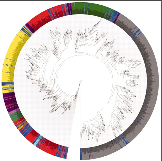
One interesting finding described in this initial report was the distribution of the new species obtained by HMP researchers around the tree of life, as depicted in Figure 1-1. This picture shows the phylogenetic tree of 16S rDNA sequences with every specific phylum marked with a different color, and the organisms sequenced as part of the HMP project in blue. Despite a lack of detail in the branching structure (and several minor artifacts as acknowledged by the authors), it can be clearly seen an overall distribution of the HMP organisms around the whole tree of life, suggesting that there are microbial species still unknown in every phylum (Nelson et al., 2010).
Antonio Palomba
“Development of new technologies to study gut microbiomes”
Tesi di dottorato in Scienze Biomolecolari e Biotecnologiche; Università degli Studi di Sassari
14
Figure 1 - 1. Overall distribution of the organism sequenced as part of the Human Microbiome Project (HMP) around the tree of life. The tree was created using 16S rDNAs representing single
species. Organisms sequenced as part of the HMP are highlighted in blue. This image shows the phylogenetic tree of 16S rDNA sequences with any specific phylum marked with a different color: Actinobacteria in yellow, Bacteroidetes in dark green, Cyanobacteria in light green, Firmicutes in red, Fusobacteria in cyan, Planctomycetes in dark red, Proteobacteria in gray, Spirochaetes in magenta, TM7 in light pink, Tenericutes in tan.
In addition, the researchers compared 16.8 million microbial sequences found in public databases (DBs) to the genome sequences in the HMP reference collection, discovering that 62 genomes in the reference collection showed similarity with 11.3 million microbial sequences in public DBs, and 6.9 million
Antonio Palomba
“Development of new technologies to study gut microbiomes”
Tesi di dottorato in Scienze Biomolecolari e Biotecnologiche; Università degli Studi di Sassari
15
of these (about 41%) corresponded with genome sequences in the reference collection. This analysis demonstrates that genomes sequenced as part of the reference collection add directly to an understanding of the human microbiome. Researchers also evaluated the microbial diversity present in the HMP reference collection, and found 29,693 previously undiscovered proteins, a number of protein superior to the estimated genes in the human genome (https://commonfund.nih.gov/hmp/) This excellent result has been followed by several others, allowing to achieve the objective initially fixed more rapidly than foreseen. For these reasons the original aim of 900 genomes (established in 2007) was changed, and the current (since 2012) objective of HMP is to sequence, or collect from publicly available sources, a total of at least 3,000 reference genomes isolated from human body sites (http://www.hmpdacc.org/).
1.1.3 Microbiome establishment and dynamics
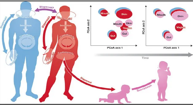
The colonization of a specific biological niche might be more controlled by abiotic and biotic factors than microbial dispersal ability (Gonzalez et al., 2011). Due to the generally unlimited dispersal capacity of microbes, the theory “everything is everywhere, but the environment selects” could be appropriate to explain the achievement dynamics of microbiome balance (Quispel, 1998; de Wit and Bouvier, 2006). As depicted in Figure 1-2, the host body can be seen as an ecosystem exposed to ecological processes including, for example, dispersal (horizontal transfer between two different individuals or between two different sites of the same individual), invasion (sudden appearance of a new “exotic” microorganism in a specific site), and succession (change in the species structure of an ecological community over time) (Gonzalez et al., 2011).
Antonio Palomba
“Development of new technologies to study gut microbiomes”
Tesi di dottorato in Scienze Biomolecolari e Biotecnologiche; Università degli Studi di Sassari
16
Figure 1 - 2.Ecology of human microbiome. The human body can be visualized as an ecosystem that is
subject to the ecological processes that structure communities, including dispersal, invasion, succession, and meta-community dynamics.
Usually, in a first phase, the chemical and physical properties of the specific body site, namely pH, aerobic or anaerobic conditions, nourishment availability, etc., select against the microorganisms impaired to survive. Progressively such microorganisms contribute to modify the native environment in order to facilitate their self-survival, making the specific site always more selective. In this way, each site and its microbial composition is distinct, as it has been clearly shown by several studies describing the vagina, penis, intestinal tract, skin, and oral microbiome, as reported in Figure 1-3 (Jenkinson, 2011; Kong, 2011; Lamont et
al., 2011; Price et al., 2010; Qin et al., 2010; Ravel et al., 2010; Turnbaugh et al.,
2009).
The microbial balance can be altered by drug application, especially antibiotic treatments that could eliminate, in addition to pathogens, also the commensal ones, resulting in a global remodeling of microbial hierarchy (Pérez-Cobas et al., 2012). Furthermore, differences in life style that can condition food assumption,
Antonio Palomba
“Development of new technologies to study gut microbiomes”
Tesi di dottorato in Scienze Biomolecolari e Biotecnologiche; Università degli Studi di Sassari
17
exposure to pets and livestock, and many other factors could influence how and where a gut microbiome is acquired (Yatsunenko et al., 2012).
Figure 1 - 3. Genus- and phylum-level classification of Bacteria colonizing a human host. Each body
site is characterized by specific bacterial taxonomy distribution. Districts with similar chemical and physical features share a greater similarity than others.
Antonio Palomba
“Development of new technologies to study gut microbiomes”
Tesi di dottorato in Scienze Biomolecolari e Biotecnologiche; Università degli Studi di Sassari
18
For this reason, the microbial composition among healthy individuals can be extremely different. In addition, the components of the human microbiome change over time, affected, for instance, by the patient disease state and medication. However, the microbiome eventually returns to a state of equilibrium, even if the composition of bacterial types has changed. Several studies have also shown that the microbiota of a specific site within the same individual is dynamic, varying naturally in correlation with age (Yatsunenko et
al., 2012).
Despite all these sources of variability, the presence of a “core microbiome”, defined as “those species-level phylotypes in a given body habitat that were observed across all sampling events”, has been demonstrated (Caporaso et al., 2011). For example, as far as gut microbiome is concerned, in 2011 Arumugam
et al. pointed out that human gastrointestinal microbiome can be clustered in
three distinct groups, identifiable by the levels of one of the three bacterial genera Bacteroides, Prevotella, and Ruminococcus, and named enterotypes 1, 2, and 3, respectively. These enterotypes, each of which rich in genes involved in specific and alternative pathways exploited to generate energy from complex carbohydrates, seem to have correlation with none of the host properties evaluated, namely gender, age, or nationality (Arumugam et al., 2011).
Nevertheless, just a year later another group begun to query whether this classification could simplify to an extreme degree the situation, suggesting, in turn, a continuum of species rather than discontinuous variation with segregated types (Jeffery et al., 2012). Nonetheless, understanding which microbial taxa constitutes a “core microbiome” is of pivotal importance to enhance knowledge concerning microbial ecology, to determine their influence upon metabolic functions, as well as to use taxonomic profiles as possible diagnostic markers (Figure 1-4) (Li et al., 2013).
Antonio Palomba
“Development of new technologies to study gut microbiomes”
Tesi di dottorato in Scienze Biomolecolari e Biotecnologiche; Università degli Studi di Sassari
19
Figure 1 - 4. The concept of a core human microbiome. The core human microbiome (red) is the set of
genes present in a given habitat in all or the vast majority of humans. The variable human microbiome (blue) is the set of genes present in a given habitat in a smaller subset of humans. This variation could result from a combination of factors.
Moreover, it is also important to note that Bacteria are the most abundant inhabitants inside microbiome, but not the only residents. Other organisms, as Fungi (forming “mycobiome”), Virus (forming “virome”), and Archaea (forming what can someone begins to call “archaeome”), although less abundant than Bacteria (also for this reason indicated as “rare biosphere”), are more variable between different individuals, and are deemed to play an increasingly pivotal role (Huffnagle and Noverr, 2013; Minton, 2012; Sogin et al., 2006; Williams, 2013). In a complex microbial communities a relative small number of microbial species dominate, but hundreds to thousands of low abundance microorganisms also exist. It is also important to note that the word “rare” is used in correlation to
Antonio Palomba
“Development of new technologies to study gut microbiomes”
Tesi di dottorato in Scienze Biomolecolari e Biotecnologiche; Università degli Studi di Sassari
20
specific environment analyzed. At mucosal sites, for instance, the most abundant bacterial specie can reach 1010 microbes per gram. Consequently, microorganisms present at 104 cells per gram can be considered “rare” since they count up for only the 0.0001% of the cellular content of the community. This “rare biosphere” may also harbor species that have an unbalanced effect (positive or negative) on the dominant members of the microbiome, a potential way by which they may support physiological or pathological effects (Huffnagle and Noverr, 2013). In support of this, accumulating evidence has delineated a correlation between species that are poorly represented within the microbiome and the host physiology, as depicted in Figure 1-5. For example, it has been shown that Methanobrevibacter (the most widespread Archaea genus in human gut) and Candida (the second most prevalent Fungi genus, after Saccharomyces, in the same human site) were positively associated with diets rich in carbohydrates, but negatively with diets high in amino acids, proteins, and fatty acids (Hoffmann et al., 2013). Moreover, even though the Prevotella/Bacteroides ratio was not significantly correlated with the fungal types, the same study demonstrated that it was significantly correlated with relative proportions of Fungi present. As far as Archaea are concerned, there was a significant correlation between archaeal genera Methanobrevibacter and Nitrososphaera with the bacterial genus Bacteroides, specifically negative with the former and positive with the latter (Hoffmann et al., 2013).
Several other studies concerning gut virome have suggested that bacteriophages (Virus infecting Bacteria) are the biggest regulators of bacterial abundance (Hofer, 2013; Williams, 2013). All together, these findings highlight that the microorganisms counterbalance in a microbiome is very complex, and numerous factors, both internal and external to the host, could have a crucial importance.
Antonio Palomba
“Development of new technologies to study gut microbiomes”
Tesi di dottorato in Scienze Biomolecolari e Biotecnologiche; Università degli Studi di Sassari
21
Figure 1 - 5. Possible syntrophic relationships in the human gut between fungal, archaeal, and bacterial microorganisms. Fungi are marked in green, Bacteria in blue, and Archaea in orange.
1.2 Techniques to study microbiomes
1.2.1 Metaculturomics
A microbiome can be studied for different purposes. On the one hand, some analyses, that could be roughed in as “descriptive”, have the main objective to thrash out, both at qualitative and quantitative level, the microbial composition of a specific environment (for instance, an anatomic site of a particular animal). On the other hand, in studies that could be defined “associative”, the chief aim is to identify a correlation between a specific physiologic or pathologic host status and some alteration in the microbial composition.
Antonio Palomba
“Development of new technologies to study gut microbiomes”
Tesi di dottorato in Scienze Biomolecolari e Biotecnologiche; Università degli Studi di Sassari
22
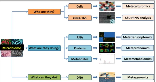
As represented in Figure 1-6, a microbiome can be characterized using different techniques. Traditionally, the best approach to study a microorganism has been culture-dependent. According to this method, two steps are crucial: plating the sample on a selective growth media, and recognizing the specific microorganism on the basis of several particular features, such as the morphological characteristics of colonies, the specific metabolic production, or the specific nutrient consumption. However, this kind of approach has huge limitations owing to the fact that, as it has been amply demonstrated by numerous evidences, more than 80% of the bacterial species present in the human body have not yet been cultured, or are even considered as unculturable (Bik et al., 2006; Gevers et
al., 2012; Grice and Segre, 2012; Turnbaugh et al., 2007). Another important
limit is that an isolated microorganism in a pure culture obtained in laboratory is less representative of community interactions, due to the loss of precious information concerning the original ecological and molecular relationships between different microorganisms.
Figure 1 - 6. How to study a microbiome. Various approaches to answer important questions
concerning a microbiome.
Microbiome
What can they do? What are they doing?
Who are they?
Cells Metaculturomics
RNA Metatranscriptomics
Proteins Metaproteomics
Metabolites Metametabolomics
DNA Metagenomics
Antonio Palomba
“Development of new technologies to study gut microbiomes”
Tesi di dottorato in Scienze Biomolecolari e Biotecnologiche; Università degli Studi di Sassari
23
To overcome some of these limitations, in 2012 Lagier et al., in a singular study, used more than 200 different culture conditions to identify microorganisms belonging to the human gut microbiome. With this analysis, researchers obtained 32,500 colonies belonging to 340 species (174 of which never described previously in the human gut) of Bacteria from seven phyla and 117 genera, including two species from rare phyla (Deinococcus-Thermus and Synergistetes), five Fungi, and a giant Virus (Senegalvirus, the largest Virus reported in the human gut). These results achieved with striking efforts were comparable, for the first (and, to date, unique) time, to those achievable with more sophisticated technologies. However, the extremely long time of this approach (also called “metaculturomics”) and the extreme complexity of the experimental design indubitably reduce its routinely application (Lagier et al., 2012).
1.2.2 Metagenomic
In the last years, along with advancements in molecular technologies, especially in sequencing and mass spectrometry (MS) instruments, alternative methods of microbial communities analysis have become available. This continuous improvement has led to the emergence of new branches of research, namely metagenomics, metaproteomics, metatranscriptomics, and metametabolomics, opening the door to a high-throughput analysis of microbial communities using culture-independent methods (Grice and Segre, 2012).
Metagenomics is based on extraction of DNA directly from a clinical or environmental sample. So far, two techniques have been the most adopted. The first one is the 16S rDNA tag sequencing, used to typify bacterial taxonomy according to information concerning 16S ribosomal RNA gene. The strong point of this method is the use of the 16S rDNA gene, which contains both highly conserved sequences, that allow polymerase chain reaction (PCR) amplification
Antonio Palomba
“Development of new technologies to study gut microbiomes”
Tesi di dottorato in Scienze Biomolecolari e Biotecnologiche; Università degli Studi di Sassari
24
using broad-range primers, and specie-specific hypervariable sequences, available for phylogenetic characterization (Hugenholtz and Pace, 1996). The
16S rDNA gene gathers up all characteristics to be a perfect marker to identify
the genome that contains it, without sequencing the entire genome. It is simple enough to be analyzed both for its reduced dimension, approximately 1500 base pairs (bps) in length, and for its high number of copies in some microbial genomes. Moreover, it is contained in every member of a population, differing only between distinct individuals with specific genomes and, in addition, it varies proportionally to the evolutionary distance between specific microbes, facilitating consequently taxonomic attribution (Morgan and Huttenhower, 2012). Actually, for all these reasons, it has reached an extremely high level of reliability, becoming the most popular technique to perform taxonomic classification (Almeida and Araujo, 2013; Carroll et al., 2012; Han et al., 2013; Hu et al., 2013; Maughan et al., 2012; Nava and Stappenbeck, 2011; Newton et
al., 2011; Santamaria et al., 2012; Shahinas et al., 2012; Tringe and Hugenholtz,
2008; Woo et al., 2008). As reported by Grice and Segre in February 2012, the
16S rDNA sequences deposited in Ribosomal Database Project (RDP) were more
than 2 millions shared in 35 different phyla (Cole et al., 2007; Grice and Segre, 2012).
The second genomic-based approach is founded on the Whole-Genome Sequencing (WGS) technology, that allows the identification of all genetic material from the different organisms making up a community in a specific ecosystem, by extracting and analyzing their DNA globally. The first studies have been focused on environmental and ecological communities, for example acid mine drainage (AMD), because of their lower complexity. The results of such analyses have been useful to pinpoint the presence of uncultivable microorganisms (Bacteria, Archaea, Fungi and Viruses), some of which, as mentioned above, can have pivotal importance to environment safeguard or host health, regardless of their abundance (Denef et al., 2010). The complexity of a
Antonio Palomba
“Development of new technologies to study gut microbiomes”
Tesi di dottorato in Scienze Biomolecolari e Biotecnologiche; Università degli Studi di Sassari
25
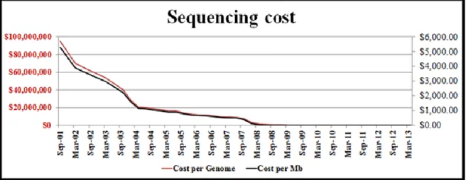
microbial community can range from very simple (extreme environments inhabited by few specialists, such as the above mentioned AMD) to very complex (environments inhabited by a variety of species, such as the gut microbiome). Obviously, the depth of knowledge which can be reached by the WGS approach can be dramatically different depending on the community under study. Simple communities, with only 76 Mb sequencing effort, can result in the assembly and analysis of almost complete genomes of the dominant species, including accurate metabolic reconstruction and detection of strain-specific genomic variants. More complex communities, with a much larger sequencing effort (almost 2 Gb), can result in very fragmented assemblies even for the most abundant species, with most of the dataset being represented by singleton sequencing reads (Chistoserdova, 2010). A huge technological advancement in sequencing instruments has made it possible to achieve these kind of results, allowing the investigation of several different genomes simultaneously. Taking into consideration, as described in Figure 1-7, that in 2002 millions of dollars were needed to obtain a complete genome sequence, it is truly amazing that, currently, the same information can be obtained with a few thousand dollars. In addition, it is also important to note that what was accessible in months or years of work just until a few years ago, now it can be obtained in few days, if not hours, with a higher reliability of the results.
A typical workflow of a WGS analysis (Figure 1-8) consists of a first fragmentation of genomic DNA, with the creation of a library of small segments, that in the next step are accurately sequenced in millions of parallel reactions.
Antonio Palomba
“Development of new technologies to study gut microbiomes”
Tesi di dottorato in Scienze Biomolecolari e Biotecnologiche; Università degli Studi di Sassari
26
Figure 1 - 7. Trend of sequencing cost during the last years. Cost per genome (red line, values on the
left) and per Mb (black line, values on the right) from 2001 to 2013 expressed in U.S. dollars. Data derived from the Genome Sequencing Program of the National Human Genome Research Institute (http://www.genome.gov/sequencingcosts/).
Then, these little nucleotide sequences, called reads, are reassembled either using a resequencing approach, that aligns the reads to a known reference genome employed as a scaffold, or using a de novo approach, where the alignment is achieved without reference information. This latter approach has the advantage to be useful to obtain genomic information about all microbial organisms (or, more in general, all organisms) independently from the preliminary level of information. Unfortunately, this strategy has the disadvantage to require very high quality data to achieve good results, i.e. an extremely high whole genome coverage, that is not always achievable, especially when several hundred different genomes are contained in the same sample. The resenquencing approach has reciprocal advantages and disadvantages compared to de novo sequencing; in fact, it is also possible to make use of data with poor coverage to identify a microbial organism, taking advantage of information concerning its reference genome. Obviously, this approach is not applicable to unknown, or not yet sequenced, microorganisms; in this regard, the lack of reference genome sequences represents the most important limitation to achieve trustworthy results with this technique. Therefore, further efforts to address these issues by
Antonio Palomba
“Development of new technologies to study gut microbiomes”
Tesi di dottorato in Scienze Biomolecolari e Biotecnologiche; Università degli Studi di Sassari
27
generating new reference genomes, such as the Human Microbiome Project has done, are highly sought after (Grice and Segre, 2012).
Figure 1 - 8. Whole genome sequencing (WGS) workflow.
To summarize, instruments with high performance in terms of both reliability and speed, combined with extremely reduced costs, have permitted to obtain in short time the sequencing of various genomes belonging to prokaryotic and
Antonio Palomba
“Development of new technologies to study gut microbiomes”
Tesi di dottorato in Scienze Biomolecolari e Biotecnologiche; Università degli Studi di Sassari
28
eukaryotic organisms, and even of whole microbial communities with very high microbial complexity, such as human gut (Figure 1-9).
Another important limitation of this kind of techniques is that it is not possible to know if the extracted DNA originated from intact, viable cells, or not. For this reason, DNA is not the ideal system to evaluate functions carried out by the community at a specific point in time (Morgan and Huttenhower, 2012).
Figure 1 - 9. Timeline of microbial community studies using high-throughput sequencing. Each
circle represents a high-throughput sequence-based 16S or shotgun metagenomic bioproject in NCBI (May 2012), indicating the amount of sequence data produced for each project (circle area and y-coordinate). Projects are grouped by human-associated (red), other animal (black), or environmental (green) communities, and shotgun metagenomic projects are marked with a grey band.
Antonio Palomba
“Development of new technologies to study gut microbiomes”
Tesi di dottorato in Scienze Biomolecolari e Biotecnologiche; Università degli Studi di Sassari
29
1.2.3 Other “omic” approaches: Metatranscriptomics
Through 16S rDNA tag sequencing and WGS it is possible to answer to two important questions, “Who is there?” and “What can they do?”, respectively, but different strategies are required to address another important question, namely “What are they doing?”. In order to obtain this kind of information, it is thus necessary to look at the expression profile (metatranscriptomics), and at metabolite (metametabolomics) or protein (metaproteomics) production. These kinds of analysis are still technically demanding and have only recently begun to be applied for studying microbial communities.
Metaproteomic techniques, probably the most interesting to assess the functions accomplished by microbes, are becoming increasingly popular. Since the main topic of this thesis is metaproteomics, this approach will be discussed in a chapter apart.
Metatranscriptomics, that is the qualitative and quantitative description of genes expressed at a given time by all organisms attending an ecological niche, can be seen as an interesting strategy to describe functionally a microbiome. Unfortunately this approach is very challenging due to various features concerning prokaryotic mRNA. For example, bacterial mRNA completely lacks the 3’-end poly(A) tail that instead marks mature molecules in eukaryoric mRNA, making their enrichment and analysis easier. In addition, this technology must deal with the intrinsic biases associated with the need for subtraction of ribosomal RNA (rRNA) that is normally the dominant RNA species extracted, usually comprising over 90% of the total RNA. As Figure 1-9 suggests, this problem could be overcome by deep sequencing of total RNA, including rRNA, since current depth of coverage would still be sufficient to obtain considerable mRNA transcripts (Lamendella et al., 2012). In other words, whether we can generate about 50 Gb of sequences, 90% of which are rRNA (and so not usable
Antonio Palomba
“Development of new technologies to study gut microbiomes”
Tesi di dottorato in Scienze Biomolecolari e Biotecnologiche; Università degli Studi di Sassari
30
to our purposes), the remaining 5 Gb of other RNA, including millions of employable transcript reads, can be sufficient to complete a transcriptomic analysis. Furthermore, RNA is more difficult to prepare and preserve compared to DNA, owing to its chemical nature. For these reasons, initially, this type of studies has been mainly applied to samples from water and soil environments; human samples, as those originated from the human gastrointestinal tract, have been successfully analyzed only in the last four years (Gosalbes et al., 2011).
1.2.4 Other “omic” approaches: Metametabolomics
A further alternative is represented by metametabolomics (also called less awkwardly “community metabolomics”), that provides information concerning the complete spectrum of small-molecules, and their changes as a consequence of a particular stimulus. This approach includes various analytical technologies such as high-resolution nuclear magnetic resolution (NMR), GC-MS and LC-MS, in combination with chemometrics and bioinformatics tools (Turnbaugh and Gordon, 2008; Wikoff et al., 2009; Xie et al., 2013). Metabolites of microbial origin can be characterized by analyzing low molecular weight compounds in biofluids (blood and urine), intestinal contents, and tissues (especially feces), achieving a metabolic fingerprint profile, associable with individual phenotypes, in correlation with physiological and pathological statuses. Metabolomics can be applied to explain the molecular mechanisms of host-microorganism interactions during a disease, taking advantage of quantitative information about specific metabolite levels, such as bile acids and short-chain fatty acids (SCFAs) that are modulated during the pathologic process (Xie et al., 2013).
Antonio Palomba
“Development of new technologies to study gut microbiomes”
Tesi di dottorato in Scienze Biomolecolari e Biotecnologiche; Università degli Studi di Sassari
31
1.3 Metaproteomics
1.3.1 Preliminary remarks
As de Hoog and Mann pointed out in their review on proteomics in 2004, “biological function is not carried out by the static genome but mainly by the dynamic population of proteins determined by an interplay of gene and protein regulation with extracellular influences”(de Hoog and Mann, 2004). In other words, the information obtainable by the global analysis of the proteins expressed in a given sample is key to carry out its full characterization. Proteomics offers the opportunity to identify the protein repertoire collectively expressed by an organism, making it possible to estimate protein abundance, either relatively or absolutely, and thus providing important insights into physiology, metabolism and cellular functionality, and/or confirming the real expression of proteins only inferred “in silico” from genome information (Hettich et al., 2012; Siggins et al., 2012; VerBerkmoes et al., 2009a; Wilmes and Bond, 2006). This approach is able to provide details on the pathways that are actively functioning in a community, and on how the expression of specific proteins can change according to time, location, or environmental stimuli (Ottman et al., 2012). In particular, when analyzing a particular microbiome, it can be more important to know which functions are carried out by the microbial components present in a biological district, than which specific microbial species are present within. Different microorganisms can in fact perform the same function, thus a divergence in microbial composition between two samples is not always correlated to an equivalent altered microbial functionality.
Unfortunately, several factors can seriously hamper a correct protein identification, and consequently a realistic proteome characterization. To give an example, one important issue in proteomics is the difficulty to access minor or under-represented proteins in a complex sample. The high dynamic range of
Antonio Palomba
“Development of new technologies to study gut microbiomes”
Tesi di dottorato in Scienze Biomolecolari e Biotecnologiche; Università degli Studi di Sassari
32
proteins, that in some samples like blood can reach 12 orders of magnitude, still remains a challenging task despite the huge improvement in MS sensitivity (Zubarev, 2013).
In general, as illustrated in Figure 1-10, a typical shotgun proteomics workflow consists of few pivotal steps: protein extraction; protein digestion; MS analysis; computational analysis.
Figure 1 - 10. Typical proteomic workflow. General approach used by peptide-centric MS technologies
for the identification of proteins in complex mixtures. After proteolysis of a protein or complex mixture of proteins, the spectra associated with protease fragments are matched with spectra generated “in silico” using information obtained from protein databases.
1.3.2 Protein extraction
The objective of the protein extraction step is to maximize the recovering of all proteins included in the sample and to minimize the presence of other molecules that can hamper the following analysis. To achieve this result, a wide assortment
Antonio Palomba
“Development of new technologies to study gut microbiomes”
Tesi di dottorato in Scienze Biomolecolari e Biotecnologiche; Università degli Studi di Sassari
33
of methods are suitable: mechanical (French Press; bead-beating; grinding), physical (boiling, freeze-thawing; snap-freezing; sonication), chemical (using buffers containing one or more components among detergents, such as sodium dodecyl sulphate, SDS, CHAPS, and Triton X-100; chaotropic agents, such as urea and guanidine hydrochloride; reducing agents, such as dithiothreitol, DTT, and tributylphosphine, TBP; and other organic/inorganic compounds, such as phenol and sodium hydroxide) or enzymatic (deoxyribonuclease; ribonuclease) approaches can be used, depending on sample features (Abram et al., 2009; Benndorf et al., 2007; Chourey et al., 2010; Fouts et al., 2012; Kan et al., 2005; Keiblinger et al., 2012; Klaassens et al., 2007; Kolmeder et al., 2012; Leary et
al., 2012; Schneider et al., 2012; Verberkmoes et al., 2009a; Wilmes and Bond,
2004).
Since Gram-positive bacteria, Gram-negative bacteria and yeasts have important structural differences, and therefore a variable susceptibility to each protein extraction method, the choice of a specific approach may significantly bias the quality and the quantity of the proteomic results in the direction of a specific category of microorganisms. In same conditions the application of a single disruption method, among those mentioned above, can be sufficient for an efficient protein extraction, but in other circumstances, with more resistant samples, a combination of two or more of them might be necessary. In this regard, the combination of strong buffer components and harsh treatments may probably help maximize extraction yields and avoid selective depletion of species showing a higher resistance to lysis, such as yeasts and Gram positive bacteria, therefore enabling a more complete representation of the microbial community proteome.
Antonio Palomba
“Development of new technologies to study gut microbiomes”
Tesi di dottorato in Scienze Biomolecolari e Biotecnologiche; Università degli Studi di Sassari
34
1.3.3 Protein digestion
The subsequent step, protein digestion, is mainly achieved using specific enzymes that break peptide bonds in a process where all reaction conditions, such as duration, temperature, and pH, are carefully controlled. The most used enzyme is trypsin, that is a serine protease able to cleave peptide chains at the carboxyl side of the amino acids lysine or arginine, except when either ones are followed by proline. This process of proteolysis is also called trypsinization (Hustoft et al., 2010). Prior to this step, it is also important to remove compounds that can limit enzymatic digestion and/or the following process steps, namely liquid chromatography (LC) separation and MS analysis. The typical way to achieve this result is using protein precipitation, which can be accomplished by adding, for instance, trichloroacetic acid, acetone, or ammonium acetate/methanol to the protein extract; then, the protein pellet is resuspended in a buffer compatible with the subsequent steps (Benndorf et al., 2007; Chourey et al., 2010; Leary et al., 2012; Sharma et al., 2012). However, significant sample losses due to protein aggregation may occur (Fic et al., 2010; Jiang et al., 2004). Another effective opportunity is to perform 1-dimension electrophoresis (1-DE) protein separation followed by in-gel digestion of the extracted proteins, which allows both the entrapment of interfering compounds within the gel matrix and the sample fractionation into gel slices (Ferrer et al., 2012; Haange et al., 2012; Kolmeder et
al., 2012). Unfortunately, although efficient, this method is labor-intensive and
time-consuming, and reproducibility may not arrive to high values (Choksawangkarn et al., 2012). A recent alternative is represented by the filter-aided sample preparation (FASP), in which sample clean-up and enzymatic cleavage take place in a molecular weight cut-off centrifuge filter (Wiśniewski et
al., 2009). This procedure has been applied with success in recent times to
environmental microbiome samples, and was demonstrated to outperform several competing methods principally for low protein amounts (Sharma et al., 2012).
Antonio Palomba
“Development of new technologies to study gut microbiomes”
Tesi di dottorato in Scienze Biomolecolari e Biotecnologiche; Università degli Studi di Sassari
35
1.3.4 Mass spectrometry analysis
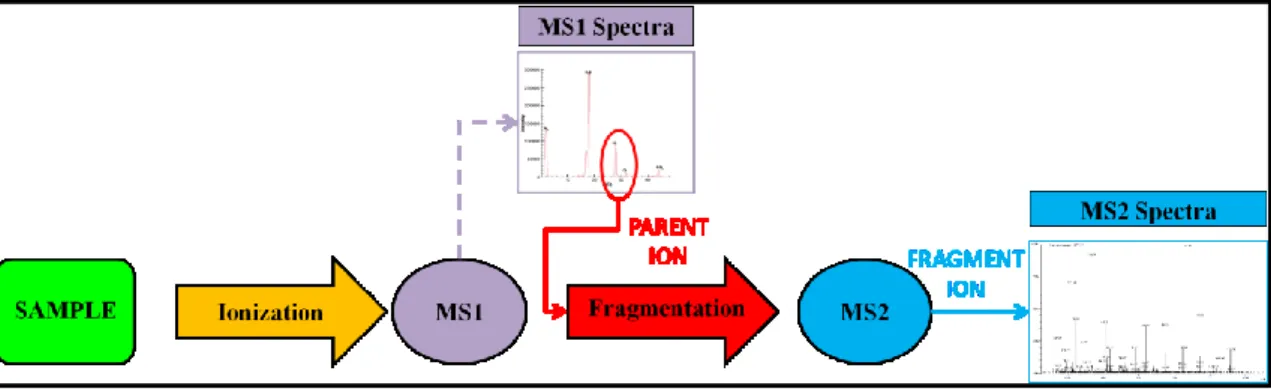
The next step, MS analysis, is key because it allows the peptide identification to be achieved. Mass spectrometers are commonly interfaced upstream with separation devices, as gas chromatographs (GC) or LC. The separated components are then introduced into the mass spectrometer. Currently, the most performing instruments are based on a tandem mass spectrometry technology (MS/MS) that combines two mass spectrometers (Figure 1-11). Briefly, these instruments work by using magnetic and electric fields to exert forces on charge particles (ions). Therefore, the peptide mixture must be charged (ionization) to be analyzed. The choice of the ionization method depends on the nature of the sample. In proteomic analysis, one of the most performing ionization sources is the electrospray ionization (ESI). This technique, that can be classified as a “soft ionization” method, tends to produce mass spectra with little or no fragment-ion content. The sample solution is sprayed across a high potential difference (a few kilovolts) from a needle into an orifice in the interface. Heat and gas flows are used to desolvate the ions existing in the sample solution. This process often produces multiply charged ions with the number of charges tending to increase as the molecular weight increases. Later, the first mass analyzer detects a spectrum from which a single mass ion, also called precursor or parent ion, with a particular mass/charge (m/z) ratio, is selected. In turn, the precursor ion produces its fragment ions due to a harder ionization obtained by colliding the selected ions with a neutral gas. This process can be named collision-induced dissociation (CID) or higher-energy collision-induced dissociation (HCD), depending on the extent of collision energy used. Finally, such fragment ions are separated into the second mass analyzer according to their m/z ratio. The resulting MS/MS spectrum consists only of product ions generated from the selected precursor ion (Guthals and Bandeira, 2012; Rotilio et al., 2012).
Antonio Palomba
“Development of new technologies to study gut microbiomes”
Tesi di dottorato in Scienze Biomolecolari e Biotecnologiche; Università degli Studi di Sassari
36
Figure 1 - 11. Diagram illustrating tandem mass spectrometry analyis worfklow (MS/MS). A sample
is injected into the mass spectrometer, ionized and accelerated, and then analyzed by the first mass analyzer (MS1). Ions from the MS1 spectra are then selectively fragmented and analyzed by the second mass analyzer (MS2) to give the spectra for the ion fragments. While the diagram indicates separate mass analyzers (MS1 and MS2), some instruments can utilize a single mass analyzer for both rounds of MS. Prior to the MS analysis step, sample complexity generally has to be reduced in order to improve the amount of information achievable by shotgun MS analysis. This has been attained in previous metaproteomic studies by carrying out a separation at the protein (mainly by 1-DE and GELFrEE approaches) and/or peptide level (most commonly by means of 2D-LC) (Kolmeder et al., 2012; Pérez-Cobas et al., 2012; Ram et al., 2005; Schneider et al., 2012; Sharma et al., 2012; Verberkmoes et al., 2009b). However, each additional fractionation step implies a corresponding increase in the quantity of starting material, laboratory effort, and/or MS measuring time required, as well as increasing challenges in analytical repeatability. In particular, 2D-LC-MS/MS, although reaching a very remarkable analysis depth, is technically challenging and, above all, requires extremely long times for a single sample to be analyzed (22 hours in a typical experimental setting) (Verberkmoes et al., 2009a). Recently, a straightforward approach based on single-run nanoLC-MS/MS has been described, enabling the identification of several thousands of proteins per run from different kinds of sample (Köcher et al., 2012; Nagaraj et al., 2012; Pirmoradian et al., 2013; Thakur et al., 2011).
Antonio Palomba
“Development of new technologies to study gut microbiomes”
Tesi di dottorato in Scienze Biomolecolari e Biotecnologiche; Università degli Studi di Sassari
37
1.3.5 Peptide and protein identification
The last step concerns peptide identification, which is carried out starting from the m/z ratio signal obtained from MS analysis. To accomplish this result, a comparison is normally performed between the experimentally obtained signal (experimental spectrum) and the theoretical spectrum, which is obtained by digesting “in silico” all protein sequences contained in a sequence database (DB) through a dedicated software called search engine (such as SEQUEST, Mascot, OMSSA, and X!TANDEM) (Craig and Beavis, 2004; Geer et al., 2004; Perkins
et al., 1999; Yates et al., 1995). The resulting collection of identified peptide
sequences is then assembled into an inventory of proteins that can account for the identified peptides. Since the likelihood of matching an MS/MS spectrum to a given peptide increases with the amount of that peptide in the sample under analysis, the number of MS/MS spectra that map to a given protein provides a quantitative estimation of the amount of that protein within the sample.
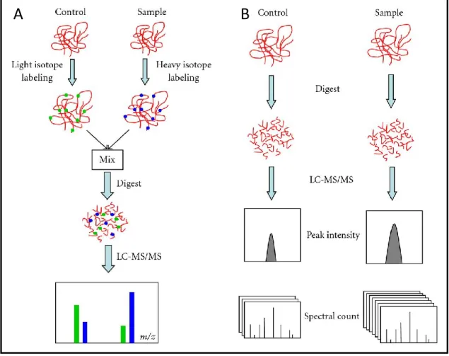
1.3.6 Quantification
A crucial issue in proteomic research concerns the accurate quantification of proteins contained in a sample (Bantscheff et al., 2012). The most accurate quantitation methods consist of using heavy or light stable isotopes incorporated into the different proteomes to bring into comparison (Hinkson and Elias, 2011). On the one side these technologies provide very accurate measurements, on the other they require additional steps in sample preparation, reducing the experimental repeatability and increasing the laboratory effort.
For this reason, alternative approaches, especially the so-called “label-free quantification”, characterized by more user-friendly procedure, are routinely
Antonio Palomba
“Development of new technologies to study gut microbiomes”
Tesi di dottorato in Scienze Biomolecolari e Biotecnologiche; Università degli Studi di Sassari
38
preferred. The label-free based techniques, as the name suggests, do not involve any kind of marking of the samples. As stated before, they are based on correlation between several mass spectrometric signals (for example, peak intensity or spectral counting), linked to a specific peptide, and the original amount of the same peptide, and therefore of the protein, in the sample. This technique reduces substantially the number of steps in the procedure, resulting in a lower labor time and a higher reproducibility (Figure 1-12). Unfortunately, however, such techniques have an important limitation concerning sensitivity. In fact, label-free quantification requires the abundance variation to be at least 2-fold to be detected, whereas with metabolic labeling approach it could be possible to describe also protein variation of a few percent (Mann et al., 2013).
1.3.7 Issues in metaproteomic analysis
Several studies have shown that the description of a protein expressed from a microbial community is very demanding, chiefly in data analysis and interpretation (Muth et al., 2013; Seifert et al., 2013). In this respect, two main issues do severely hamper the analysis of a metaproteome: first, genome sequence data might be unavailable for most of the species contained in the particular microbial community under study, thus considerably reducing the possibility of a correct matching between the experimental spectra and the theoretical spectra; second, a typical environmental sample contains thousands of proteins belonging to up to thousands of different microbial species, often having a high level of homology, making therefore both to-protein and peptide-to-taxon assignments a really tremendous task.
Antonio Palomba
“Development of new technologies to study gut microbiomes”
Tesi di dottorato in Scienze Biomolecolari e Biotecnologiche; Università degli Studi di Sassari
39
Figure 1 - 12. Quantitative proteomic approaches. (A) Shotgun isotope labeling method. After labeling
by light and heavy stable isotope, control and sample are combined and analyzed by LC-MS/MS. The quantification is calculated based on the intensity ratio of isotope-labeled peptide pairs. (B) Label-free quantitative proteomics. Control and sample are subjected to individual LC-MS/MS analysis. Quantification is based on the comparison of peak intensity of the same peptide or the spectral count of the same protein.
1.3.8 Database impact
The selection of proper protein DBs represents an extremely critical step, especially when dealing with poorly characterized microbiomes. When a novel microbial community is subjected to metaproteome analysis, without further genomic investigation, publicly available DBs have to be used for peptide/protein identification, almost for a preliminary analysis. Protein DBs can be generally distinguished into non-manually annotated with plenty of information but huge dimensions, and thus very high computing times, such as NCBI and
Antonio Palomba
“Development of new technologies to study gut microbiomes”
Tesi di dottorato in Scienze Biomolecolari e Biotecnologiche; Università degli Studi di Sassari
40
UniProtKB/TrEMBL (TrEMBL), and manually curated sequences as UniProtKB/SwissProt (SwissProt), with inverse pros and cons in comparison with the first ones (Figure 1-13)(NCBI Resource Coordinators, 2013; The UniProt Consortium, 2012).
Figure 1 - 13. Number of protein sequences in UniProtKB/TrEMBL and UniProtKB/SwissProt databases.
Unfortunately, most uncultivable species have not yet been sequenced, in spite of the great efforts made in the last few years by genome scientists, and are therefore not present within the public resources. In this case, cross-species
A