A PROTOCOL FOR THE USE OF
99MTC (V) DMSA
IN THE SCYNTIGRAPHIC DIAGNOSIS
OF CARDIAC AMYLOIDOSIS
A.F. SEDDA, G. ROSSIFusion and Technology for Nuclear Safety and Security Department Casaccia Research Centre,Rome
R. SALVATORI
Nuclear Medicine Division S.Maria GorettiHospital,Latina,Italy
C. CIPRIANI
Nuclear Medicine Division AlGa Institute,L’Aquila,Italy
RT/2017/2/ENEA
ITALIAN NATIONAL AGENCY FOR NEW TECHNOLOGIES, ENERGY AND SUSTAINABLE ECONOMIC DEVELOPMENT
A.F. SEDDA, G. ROSSI
Fusion and Technology for Nuclear Safety and Security Department Casaccia Research Centre, Rome
R. SALVATORI
Nuclear Medicine Division S. Maria Goretti Hospital, Latina, Italy
A PROTOCOL FOR THE USE OF
99m
TC (V) DMSA
IN THE SCYNTIGRAPHIC DIAGNOSIS
OF CARDIAC AMYLOIDOSIS
C. CIPRIANI
Nuclear Medicine Division AlGa Institute, L’Aquila, Italy
RT/2017/2/ENEA
ITALIAN NATIONAL AGENCY FOR NEW TECHNOLOGIES, ENERGY AND SUSTAINABLE ECONOMIC DEVELOPMENT
I rapporti tecnici sono scaricabili in formato pdf dal sito web ENEA alla pagina http://www.enea.it/it/produzione-scientifica/rapporti-tecnici
I contenuti tecnico-scientifici dei rapporti tecnici dell’ENEA rispecchiano l’opinione degli autori e non necessariamente quella dell’Agenzia
The technical and scientific contents of these reports express the opinion of the authors but not necessarily the opinion of ENEA.
A PROTOCOL FOR THE USE OF 99mTC (V) DMSA IN THE SCYNTIGRAPHIC DIAGNOSIS OF
CARDIAC AMYLOIDOSIS
A.F. Sedda, G. Rossi, R. Salvatori, C. Cipriani Abstract
99mTc is a gamma emitter radioactive isotope commonly employed in nuclear medicine diagnostic. Its utility lies in the fact that it can be conjugated to different organic molecules, for in vivo diagnostic. The 99mTc(V)-DMSA (DiMercaptoSuccinic Acid) is a radioactive tracer originally employed in the scinti-graphic diagnosis of medullary thyroid carcinoma, but has been proposed a selective tracer for the differential diagnosis of amyloidosis. We report here a case report of cardiac amyloidosis in which the prominent myocardial uptake of the tracer suggested a diagnosis of amylod disease, confirmed by histologic diagnosis. A quantitative analysis of the radioactivity distribution demonstrated a clear-cut distinction from the uptake in patients not affected by cardiac amyloidosis, so allowing quantitative criteria for a disease diagnosis employing the off-label 99mTc(V)-DMSA as a selective uptake tool.
Keywords: radioactive isotopes,99mTc, 99mTc(V)-DMSA, amyloidosis, radioactive labeling
Riassunto
Il 99mTc è un isotopo radioattivo gamma emittente comunemente impiegato in medicina nucleare
dia-gnostica. La sua utilità consiste nel fatto che può essere coniugato a differenti molecole organiche, per la diagnostica in vivo. Il 99mTc(V)-DMSA (acido dimercaptosuccinico) è un tracciante radioattivo
ori-ginariamente impiegato nella diagnosi scintigrafia del carcinoma midollare della tiroide, ma è stato anche proposto come tracciante selettivo per la diagnosi differenziale di amiloidosi. Riportiamo qui un caso di amiloidosi cardiaca in cut il preminente assorbimento del tracciante da parte del miocardio ha suggerito una diagnosi di malattia amiloide, successivamente confermata da diagnosi istologica. Un'analisi quantitativa della distribuzione della radioattività ha dimostrato una chiara distinzione, tale da permettere la definizione dei criteri quantitativi per la diagnosi della malattia, impiegando il 99mTc
(V) -DMSA off-label come strumento di captazione selettiva.
Introduction
Material and methods Results Conclusions References 7 8 9 15 16
INDEX
7
Introduction
Amyloidosis is a group of diseases caused by the deposit in various body tissues of abnormal proteins. In each type of amyloidosis, a protein produced by the body acquires the property of accumulation, in various organs and tissues, in the form of fibrils; the deposits are called amyloid fibrils. The gradual accumulation of amyloid fibrils causes important damage to involved organs. The systemic amyloidosis are rare diseases; it is estimated that in Italy about 800 new cases of amyloidosis are annually diagnosized. More than twenty histological types of amyloidosis are currently known, each caused by a different proteins that can be produced by various organs, for example liver, bone marrow, intestines. At present, the best treatment for amyloidosis is to reduce and possibly eliminate the production of the protein, and for this reason the treatment is radically different for the different types of amyloidosis. The symptoms depend on the organs that are affected by the accumulation of amyloid and from the type of amyloidosis. The most common manifestations are related to the involvement of the heart and kidney, with leg swelling (edema), difficulty in breathing - especially under stress - and general fatigue. Renal involvement may occur with changes in some blood tests (serum creatinine and serum cholesterol, above the reference limits) and urine (protein loss). The cardiac amyloidosis can generally be recognized by echocardiography; sometimes, patients with amyloidosis may also exhibit an increase in liver size, low blood pressure, loss of appetite and weight, altered sensitivity of the hands and feet, and diarrhea. As these events are not only typical of amyloidosis, but are often found in other, more common, diseases, it is essential that they be interpreted with care by the physician. There are some signs that occur rarely, but are characteristic of amyloidosis and can be a useful element r a diagnosis of the disease, such as the increase in size of the tongue, the appearance of purple spots on the skin in the and neck face, especially around the eyes.
The definitive diagnosis of amyloidosis requires the identification of amyloid deposits of a tissue sample, and is based on histological material observation, with apple green birefringence appearing under polarized light after staining tissues with Congo red, usually after a fine needle aspiration of peri-umbilical fat. The sensitivity of this test, (i.e. the probability of detecting amyloid deposits in a person suffering from the disease) is around 80% in systemic amyloidosis, if the procedure is performed by an experienced operator. If this hystology doesn't confirm the diagnosis of amyloidosis but the suspicion persists, a biopsy of a salivary gland or, in selected cases, a biopsy of the organs believed to be affected by amyloidosis can be performed.
Noninvasive affordable imaging techniques for detecting amyloidosis are therefore strongly beneficial, because early and definite diagnosis can allow for early treatment and monitoring of treatment. Instrumental non invasive tests on specific organs (heart, kidneys, liver, etc.), can be used to help in the tissue histological sampling in the affected organs, and could also be potentially useful in the follow up of the patient. Due to this reason, many clinical research to test non-invasive methods in the diagnosis of this group of diseases have been tested; among the most promising techniques, scintigraphy radiodiagnostic is considered highly
8
selective. Over the years 67Ga-citrate, 111In-benzilguanidina, 99mTc-MDP, 99mTc-pyrophosphate, 131I- 2-macroglobulin, 123I-Serum Amyloid P-component (123I- SAP), 99mTc-aprotinin, 99mTc (V) -Dimercaptosuccinic acid (99mTc (V) -DMSA) were used as clinical tracers. These last two molecules appear to possess greater specificity in setting on deposits of amyloid. Aprotinin, which shows a good specificity due to its distribution in vivo, appears mainly useful in the visualization of deposits on the liver, kidneys and spleen, but of lesser utility on the heart 1.
The 99mTc (V)-DMSA appears to have the characteristics that render it more versatile in the various districts. Also, it is not an expensive tracer, is easy to prepare, and well tested from the clinical point of view, due to its codified use in the diagnosis of medullary thyroid carcinoma. The literature reports the case of two patients 2 in which a diagnosis of amyloidosis associated with plasmacytoma was performed, with the use of 10 mCi of 99mTc (V) -DMSA; a SPECT scan showed a satisfactory spatial resolution, and the tracer was crucial in guiding biopsy confirmation. A second scientific work, described a case of cardiac amyloidosis revealed with a scintigraphy with 99mTc (V) -DMSA3; in another case report 4 a patient with amyloidosis polyneuropathy underwent scintigraphy with 201Tl-chloride, with 123 I-beta-methyl-p-iodophenylpentadecanoic acid ( 123IBMIPP), and 131I- metaiodobenzylguanidine, but only scintigraphy with
99m
Tc (V) -DMSA allowed a reliable diagnosis. A case report work 5 showed a case of amyloidosis with minimal involvement of internal organs, but with extensive skin lesions; scintigraphy with 15 mCi of 99mTc (V) -DMSA allowed to locate the sites of amyloid deposition, and to follow the patient follow-up seven months after the start of therapy. Some variants of amyloidosis are so severe as to require transplantation of liver and kidney; two transplant patients were examined for the follow-up scintigraphy with 99mTc-DMSA, up to 5 years after surgery 6. In one case a patient was examined with scintigraphy with 10 mCi of 99m Tc (V) -DMSA for a suspected amyloidosis 7; the examination showed an accumulation in some areas, but the subsequent histology revealed absence of amyloidosis, but instead the presence of myeloma. Therefore in some cases the same method seems able to highlight also this type of pathology. In another case, a patient underwent scintigraphy with 10 mCi of 99mTc(V)-DMSA for a suspected amyloidosis 8; examination showed an accumulation in kidney area, and a subsequent histology of the area revealed the diagnosis of multiple myeloma with amyloidosis.
Materials and methods.
The 99mTc(V)-DMSA has been prepared by a galenic formulation, starting from high purity analytical reagents. Briefly, 1.3 mg of DMSA, 7.8 mg of NaHCO3, 1.97 mg of Na2CO3, 10.17 of glucose and 0.166 of
SnCl2 .
2H2O (Sigma Aldrich), have been mixed under sterile hood with 1 ml of saline containing 5 mCi of
freshly eluted 99mTc. After complete dissolution of the product (1 minute), a needle equipped with a sterile 0.22 micron filter was inserted in the vial, and a flow (20 ml/min) of high purity oxygen has been bubbled
9 into the solution for 15 minutes. After this period, the solution has been filtered with a 0.22 micron sterile filter. The radiochemical purity was determined by the method described by Westera et al.9 using TLC (Merck, silica gel 60F) with a solvent system containing n-butanol/ acetic acid/water (3 : 2 : 3 v/v). The radiochemical purity was calculated as the percentage of 99mTc(V)-DMSA relative to the total activity. The radioactivity was estimated from autoradiography of TLC sheets by using a Cyclone Perkin Elmer image phosphor screen apparatus.
Results
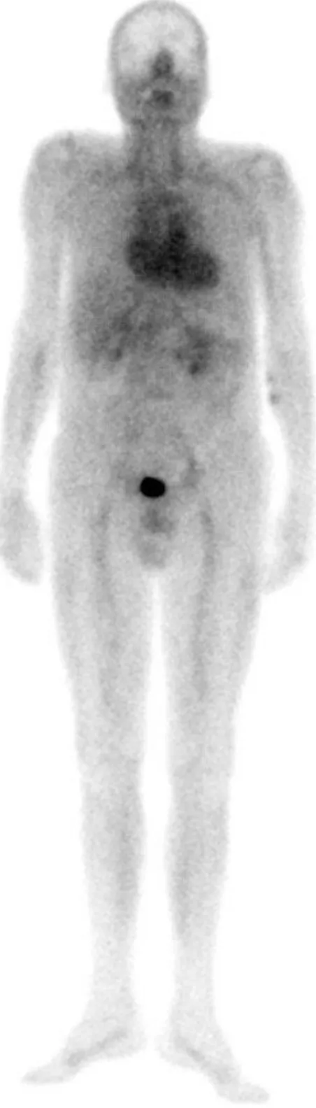
A 76 y.o. patient showed alterations in ECG and a slight left atrium dilation; the troponine and B natriuretic peptide were well beyond the upper limits, while all other blood parameters were within physiological limits. The patient was injected intravenously with 99mTc(V)-DMSA, and after 1h and 3h whole body images (both anterior and posterior views) were obtained with a GE Infinity dual-head gamma camera equipped with low energy high-resolution collimators; images were acquired using a 1024x256 matrix size. Figure 1 shows the anterior whole body view of the patient after 1 h p.i., and Figure 2 the cardiac region 3h p.i.
10
11 The normalized ratio (radioactivity on heart) / (radioactivity on lung) was found to be of 2.0, and a normalized value for the ratio (radioactivity on kidney) / (radioactivity on lung) of 1.75, for both 1h and 3h imaging. Our research group has re-examined a large cohort of patients, on which 99mTc(V)-DMSA was used in our Department for the diagnosis of bone metastases; on a group of 12 patients healthy, or affected with pathologies different from amyloidosis (mainly bone metastases) it was found a normalized value for the ratio (radioactivity on heart) / (radioactivity on lung) of 1.25 (+0.09), and a normalized value for the ratio (radioactivity on kidney) / (radioactivity on lung) of 1.66 (+0.1). Furthermore, in the past, another patient affected with amyloidosis has been examined in our work group 3, and a normalized value for the ratio (radioactivity on heart) / (radioactivity on lung) of 2.0, and a normalized value for the ratio (radioactivity on kidney) / (radioactivity on lung) of 1.70 were found. For both patients, after the 99mTc(V)-DMSA scintigraphy, a histologic analysis on peri-umbilical fat confirmed the scintigraphic diagnosis of amyloidosis.
In spite of the reduced numbers of examined patients, these data suggest that an accumulation of amyloid in the heart is clearly detectable by abnormal fixation of 99mTc-DMSA (V). In patients not affected by amyloidosis, the activity of 99mTc-DMSA (V) shows a typical distribution along the blood pool, followed by a washout; in healthy patients, after three hours from injection, only the kidneys generally shows an increased accumulation of the tracer. An example of distribution of 99mTc(V)-DMSA in a normal patient is showed in Figure 3.
12
13 In Figure 4 and 5 are reported two patient affected with bone metastases from prostatic cancer, examined in our Nuclear Medicine Department.
14
Figure 5
In both the patients affected by amyloidosis examined by our work-group, the activity on the heart after one hour and three hours, decay corrected, was substantially the same. Moreover, the tracer activity is clearly indicative of an uptake on the myocardial muscle, and not a mere blood pool distribution (see Figure 1 and 2). The results also show that in both patients renal cortical uptake is negligible , which is consistent with the low level of 99mTc(III)-DMSA in the preparation.
General criteria that can be extracted for a positive diagnosis of cardiac amyloidosis are here proposed:
1) normalized ratio (radioactivity on heart) / (radioactivity on lung) > 1.3 (tipically 2)
15 3) uptake of the radiotracer on the myocardial muscle, and not as a mere blood pool.
Conclusions
After the diagnosis of amyloid disease, it is necessary to identify the type of amyloidosis involved, in function of the protein found in deposits. This step is essential for a possible therapy, as the treatment, when possible, is radically different for each type of amyloidosis. At the present state of knowledge the goal of therapy of systemic amyloidosis is to slow or stop the production of the protein that gives rise to deposits. Of course, methods for obtaining this result are different in different types of amyloidosis and are different for the different proteins. By reducing the production of the protein that cause the amyloid, the amount of material available to form new deposits rapidly decreases, the process that led to the disease stops, amyloid deposits can be reabsorbed and the function of organs damaged, if the damage is not already irreversible, may also be retrieved completely. The ability, in the early stages of the disease, to restore the normal function of the affected organs emphasizes the importance of an early diagnosis. It is also important to support the therapy with a possible follow-up by non-invasive diagnostic, which typically consist in scintigraphic diagnosis. The execution of a diagnostic method as scintigraphy with 99mTc(V)-DMSA on a regular basis, can be proposed as a clinical diagnostic protocol. It is non-invasive, simple to perform and sufficiently reliable to be considered a tool of valuable help in routinary clinical diagnostic.
16
References
1. Minamimoto R, Kubota K, Ishii K, Morooka M, Okasaki M, Miyata Y, Nakajima K, Sato T, Igari T, Hirai R, Okazaki O, Re-evaluating the potentials and limitations of 99mTc-aprotinin scintigraphy for amyloid imaging, Am J Nucl Med Mol Imaging 2013;3(3):261-271
2. Ohta H, Endo K, Kanoh T, Konishi J, Kotoura H. Technetium-99m (V) DMSA uptake in amyloidosis. J Nucl Med. 1989;30:2049–2052.
3. Manni C, Sangiorgi G, Boemi S, De Nardo D, Cipriani C, Cannata D, Cardiac amyloidosis detected by 99mTc(V) DMSA myocardyal uptake, Clin Nucl Med 19; 1109-1111, 1994
4. Ali Syed Arbab A S, Koizumi K, Toyama K,Arai T, Yoshitomi T, Araki T, Scan findings of various myocardial SPECT agents in a case of amyloid polyneuropathy with suspected myocardial involvement,Annal Nuc Med, 11(2), 139-141, 1997
5. Ambrosone L,. Mansi L, Salvatore T, Marino F, Orabona P, Rambaldi A, Rambaldi P F, Rambaldi M, An unusual case of primary systemic amyloidosis, Jour Eur Acad Dermatol Vener, 10(1998) 53-57.
6. Stangou AJ, Banner NR, Hendry BM, Rela M, Portmann B, Wendon J, Monaghan M, Maccarthy P, Buxton-Thomas M, Mathias CJ, Liepnieks JJ, O'Grady J, Heaton ND, Benson MD, Hereditary fibrinogen A alpha-chain amyloidosis: phenotypic characterization of a systemic disease and the role of liver transplantation.
7. Ohnishi T, Noguchi S, Murakami N, et al. Pentavalent technetium-(V)-99m DMSA uptake in a patient having multiple myeloma without amyloidosis. J Nucl Med 1991; 32:1785-1787.
8. Barai S, Bandopadhayaya GP, Rathi M, Singh NG,Accumulation of Tc99m-DMSA-3 in the spleen in a case of multiple myeloma with associated amyloidosis, J Postgrad Med 51(2), 119-121, 2005.
9. Westera G, Gadze A, Horst W, A convenient method for the preparation of 99mTc(V)dimercaptosuccinic acid (99mTc(V)-DMSA), Int J Appl Radiat Isot. 1985 Apr;36(4):311-2.
ENEA
Servizio Promozione e Comunicazione www.enea.it
Stampa: Laboratorio Tecnografico ENEA - C.R. Frascati febbraio 2017