Shedding light on the polyphyletic behavior of
the genus Sterkiella: The importance of
ontogenetic and molecular phylogenetic
approaches
Daizy Bharti1,2, Santosh Kumar2*, Govindhasamay R. Varatharajan1, Komal Kamra3, Antonietta La TerzaID1*
1 School of Biosciences and Veterinary Medicine, Laboratory of Animal and Molecular Ecology, University of Camerino, Camerino, MC, Italy, 2 Zoological Survey of India, Prani Vigyan Bhawan, New Alipore, Kolkata, India, 3 Ciliate Biology Laboratory, SGTB Khalsa College, University of Delhi, Delhi, India
*[email protected](SK);[email protected](ALT)
Abstract
Present study, investigates a poorly known species of the genus Sterkiella, i.e., S. tricirrata, based on two populations isolated from soil samples collected from the Colfiorito Regional Park, Umbria Region, Italy and from the Silent Valley National Park, India. Both populations showed a highly similar morphology, however different ontogenetic pattern in between. The study confirms the validity of the species S. tricirrata which was considered to be a species within the Sterkiella histriomuscorum complex. The main ontogenetic difference between S. tricirrata and other species of the genus Sterkiella is the different mode of formation of anla-gen V and VI of the proter in the former. In the phyloanla-genetic analyses, Sterkiella tricirrata clusters with Sterkiella sinica within the stylonychine oxytrichids, in a clade away from the type species (Sterkiella cavicola) of the genus Sterkiella. The study highlights the impor-tance of ontogenetic as well as molecular data in shedding light on the polyphyletic behavior of the genus Sterkiella. A detailed description of S. tricirrata based on morphology, ontogen-esis and molecular phylogenetic methods is presented. Further, the improved diagnosis has been provided for the genus Sterkiella and the poorly known species S. tricirrata.
Introduction
Recent studies, among the hypotrich and spathidiid ciliates, have shown that detailed observa-tions of characters often resolve the discrepancy between the morphological and molecular analyses [1–4]. This reiterates the need for an integrated approach to investigate in-depth cili-ate diversity [5]. The identification of cryptic characters among hypotrich ciliates (e.g. cyst structures, morphology, details on the mode of division) has justified the separation of mor-phologically similar species reflecting distant relationships in molecular phylogeny [3,6–9]. The structure of resting cyst has provided the support for separation of morphologically simi-lar ciliate species, i.e., cyst species [3,4,7–9]. The hypotrich genusFragmospina Foissner, 2016 a1111111111 a1111111111 a1111111111 a1111111111 a1111111111 OPEN ACCESS
Citation: Bharti D, Kumar S, Varatharajan GR,
Kamra K, La Terza A (2018) Shedding light on the polyphyletic behavior of the genus Sterkiella: The importance of ontogenetic and molecular phylogenetic approaches. PLoS ONE 13(11): e0207688.https://doi.org/10.1371/journal. pone.0207688
Editor: Michael Schubert, Laboratoire de Biologie
du De´veloppement de Villefranche-sur-Mer, FRANCE
Received: February 26, 2018 Accepted: November 5, 2018 Published: November 20, 2018
Copyright:© 2018 Bharti et al. This is an open access article distributed under the terms of the Creative Commons Attribution License, which permits unrestricted use, distribution, and reproduction in any medium, provided the original author and source are credited.
Data Availability Statement: SSU rRNA gene
sequence is deposited in GenBankhttps://www. ncbi.nlm.nih.gov/(accession number: MG805314).
Funding: Financial support and facilities were
provided by the Director, Zoological Survey of India. The data from the Indian species is a part of the research conducted under the Project SR/SO/ AS-04/2004 by the Department of Science and Technology (DST), Government of India. The data
is a recent example where morphology of cyst has been incorporated as a generic character. Morphologically,Fragmospina depressa Foissner, 2016, is rather similar in the arrangement of
ciliature withSterkiella histriomuscorum (Foissner et al., 1991) Foissner, Blatterer, Berger &
Kohmann, 1991; however, the undulating membranes are arranged inAustralocirrus pattern
[3,10]. Similarly, when ontogenesis within a genus is compared, most congeners have a rather similar pattern with some minor variations [6]. Kumar et al. [2] erected a new genus, i.e.,
Metasterkiella Kumar et al., 2017, for a species isolated from the petroleum contaminated soil,
which was morphologically similar to species of the genusSterkiella Foissner, Blatterer, Berger
& Kohmann, 1991. However, this species possesses a variant character in ontogenetic pattern, i.e., it is the only known stylonychid ciliate, thus far, where cirrus V/3 was incorporated during the anlagen formation.
The relatedness between genera within the subfamilies Oxytrichinae and Stylonychinae is rather difficult to understand, despite being strongly reflected as monophyletic groups in the phy-logenetic analyses [11–15]. A probable explanation could be the insufficient availability and inter-pretation of morphological and ontogenetic data for the known species. Previous studies have shown that the stylonychid genusSterkiella is a non-monophyletic assemblage [1,2,16,17]. Ster-kiella histriomuscorum, a sibling species complex according to Berger [11], include populations which have not been studied in detail and thus considered to be synonyms under the complex.
In this study, we describe a poorly known speciesSterkiella tricirrata (Buitkamp, 1977)
Ber-ger, 1999, which is probably a synonym of one of the species within theSterkiella histriomus-corum complex together with S. terricola, according to the remarks of Berger [11]. The Indian and Italian populations ofS. tricirrata were studied and found to be highly similar in
morphol-ogy. Ontogenetic stages of both populations showed difference in the anlagen formation dur-ing the early divisional stages with respect to that ofSterkiella species [11,18,19], and thus suggesting its possible separation at the genus level. Detailed data on the morphometry and ontogenesis for both populations and molecular analyses based on SSU rRNA gene of Italian population is presented. Furthermore, this study highlights the relevance of combining onto-genetic and molecular phyloonto-genetic approaches in identifying species in polyphyletic assem-blages such as that represented by the stylonychid genusSterkiella.
Materials and methods
Description of the sampling site and sample processing
Soil samples were collected from the core zone of the Silent Valley National Park, India (11˚08’ 40.72"N; 129˚20’ 38"E) in January, 2008 and from the plains of the Colfiorito and Plestini uplands, Umbria region, Central Italy (43˚01’ 40.72"N; 12˚52’ 39.46"E) in July, 2009. Vegeta-tive cells were excysted from resting cysts from two-weeks-dried soil samples (approximately 200 g) by employing the non-flooded Petri dish method [20]. A clonal culture ofSterkiella tri-cirrata was established for both the populations as described in Kumar et al. [1], i.e., using Pringsheim’s medium for culturing and the green algaChlorogonium elongatum as food
source. Observations on the live specimens were made using a microscope with bright-field and differential interference contrast illuminations at a magnification of 100–1000×. The pro-targol staining method described by Kamra and Sapra [21] was used with some modification to reveal the ciliature. Measurements of impregnated specimens were performed at a magnifi-cation of 1000× using an ocular micrometer for Italian population and Leica software IM50 image manager for the Indian population. An Optika microscope camera was employed for photomicrography for the Italian population and Leica camera DFC320 for the Indian. The illustration of the live specimen was prepared using free-hand sketches, while those of from the Italian species is a part of the research
grant (FAR- Fondo Ateneo per la Ricerca, BioPrint project) from the University of Camerino, Italy and financial support from the Young Indian Research Fellowship through the Italian Minister of University and Research (MIUR). The funders had no role in study design, data collection and analysis, decision to publish, or preparation of the manuscript.
Competing interests: The authors have declared
impregnated specimens were made with the drawing device. Terminology is according to Ber-ger [11] and Wallengren [22].
DNA extraction, PCR amplification, and sequencing
Unfortunately, we could not perform the DNA extraction for the Indian population. Thus the methods described here, including the phylogenetic analyses of the SSU rRNA gene, exclusively refer to the Italian population. Five cells were collected from a clonal culture with the help of glass micropipettes and washed three times with autoclaved distilled water (same culture was used for live observation and protargol staining to study morphology and ontogenesis). Geno-mic DNA was extracted using the Norgen DNA Kit (Elettrofor Scientific Instruments, Borsea, Italy), following the manufacturer’s instruction [23]. Extracted DNA (5μl) was dispensed into a PCR tube containing 5μl of autoclaved distilled water, and amplifications were carried out using high-fidelity Pfx50 DNA polymerase (Invitrogen, Italy) in a total volume of 50μl with the universal eukaryotic primers Euk A (FW 5’-AACCTGGTTGAT CCTGCCAGT-3’) and Euk B (RV 5’-TGATCCTTCTGCAGGTTCACCTAC- 30) [24]. Additionally, nested primer pairs Eup 18S (FW 5’-TAG AGG GAC TTT GTG TGC AAC C-3’) and Eup 18S (RV 5’-ATC TCC CTG AAA CAC ACG TTG G-3’) were used in combination with the universal primers for amplification and sequencing. The PCR program for 18S rDNA amplification included an initial denaturation at 94˚C for 3 min, followed by 35 cycles of 94˚C for 1 min, 55˚C for 45 s and 72˚C for 80 s, with a final extension step at 72˚C for 10 min. After confirmation of the appropriate size, the PCR products were purified using the Nucleospin gel extraction kit (Qia-gen) and were then directly sequenced on both strands at StarSEQ GMBH, Germany.
Phylogenetic analyses
For phylogenetic analyses, the SSU rRNA gene sequence ofSterkiella tricirrata was aligned
with 55 SSU rRNA gene sequences of hypotrich ciliates from GenBank using the MAFFT soft-ware v. 7.047 (choosing the iterative refinement methods Q-INS-I that considers the secondary structure of the SSU rRNA molecules) [25].
Ambiguously aligned regions were identified and excluded from the phylogenetic analyses with GBlocks v.0.91b [26] using parameters optimized for rRNA alignments (minimum length of A block = 5, allowed gap positions = with half), leaving 1,644 unambiguous positions. The final alignment was then used for subsequent phylogenetic analyses after converting the FASTA (.fas) file to NEXUS (.nex) format using the open web-based tool ALTER (Alignment Transformation EnviRonment) [27]. A Bayesian inference (BI) analysis was performed using MrBayes v.3.2.1 [28] and the GTR+I+G model, as selected by the jModel Test v.2.1.3 software [29] under theAkaike Information Criterion corrected (AICc). Markov chain Monte Carlo
(MCMC) simulations were run, with two sets of four chains using the default settings, for 1,000,000 generations with trees sampled every 100 generations and discarding the first 25% of the sampled trees as burn-in. The remaining trees were used to generate a consensus tree and to calculate the posterior probabilities (PP) of all branches using the majority-rule consensus approach. The previous alignment was also used to perform a Maximum Likelihood (ML) tree by means of the Molecular Evolutionary Genetic Analysis (MEGA) software, v.5.2.2 [30] using the default parameters and the GTR+I+G model. The reliability of tree topology was assessed by 1,000 bootstrap replicates and was expressed as a percentage. Phylogenetic trees were visual-ized using the free software package FigTree v1.4 by A. Rambaut athttp://tree.bio.ed.ac.uk/ software/figtree/.
Data availability
The newly obtained SSU rRNA gene sequence ofSterkiella tricirrata is available from the
Gen-Bank/EMBL databases (accession number: MG805314). Two neotype slides of the Italian pop-ulation containing the protargol stained neotype specimen and relevant morphostatic
specimens have been deposited at the Natural History Museum, London, UK, with registration numbers NHMUK 2014.3.20.1 and NHMUK 2014.3.20.2. Further, two slides of the Indian population is deposited at the Natural History Museum, London, UK, with registration num-bers NHMUK 2011.7.4.2 and NHMUK 2011.7.4.3 and one at the type collection of the Zoolog-ical Survey of India, Kolkata, India, with registration number Pt 3067. The SSU rRNA gene sequence is deposited in GenBank (accession number: MG805314).
Nomenclatural acts
The electronic edition of this article conforms to the requirements of the amended Interna-tional Code of Zoological Nomenclature, and hence the new names contained herein are avail-able under that Code from the electronic edition of this article. This published work and the nomenclatural acts it contains have been registered in ZooBank, the online registration system for the ICZN. The ZooBank LSIDs (Life Science Identifiers) can be resolved and the associated information viewed through any standard web browser by appending the LSID to the prefix “http://zoobank.org/. The LSID for this publication is: urn:lsid:zoobank.org:pub:DB29FEE1-22B6-48CC-9E8D-661AD15BBB06. The electronic edition of this work was published in a journal with an ISSN, and has been archived and is available from the following digital reposi-tories: PubMed Central, LOCKSS.
Results
Description of
Sterkiella tricirrata
Morphometric data of the Indian and Italian population ofSterkiella tricirrata highly overlap
(Table 1). Thus only a detailed description of the Italian population is provided below; minor differences with the Indian population in some characters include: (1) body size, i.e., about 85× 40 μm (vs. 75 × 40 μm) in vivo; (2) number of cirri in right marginal rows 20 (vs. 16); and (3) number of bristles in first dorsal kinety (15 vs. 20) (Figs1A–1C,2A–2C,2F,2G,3A–3C,
4A,4B,4E and 4FandTable 1).
Size in vivo 70–90× 30–50 μm, usually about 85 × 40 μm, as calculated from some in vivo (n = 6) measurements and morphometric data inTable 1, assuming 15% preparation shrink-age [9]. Body outline oval, elliptical to broadly elliptical, both ends rounded; dorso-ventrally flattened about 2:1 (Figs1A–1C,2A–2C,2F and 2GandTable 1). Nuclear apparatus in or slightly left of midline composed of two macronuclear nodules and one to four micronuclei (Figs1A,1C,2B,2F and 2GandTable 1). Macronuclear nodules ellipsoidal to broadly ellipsoi-dal, anteriormost nodule on average 11× 7 μm in protargol preparations; contain small nucle-oli, 1–3μm across. Micronuclei usually attached to macronuclear nodules, globular, on average 2.0μm across in protargol preparations (Figs1A,1C,2B,2F and 2GandTable 1). Contractile vacuole slightly anterior of body’s midline, near left cell margin (Figs1Aand2C). Cortex semirigid; cortical granules absent. Cytoplasm colorless, filled with some crystals of about 1–3μm in size and fat droplets about 2–6 μm in diameter (Figs1A,2A and 2B). Feeds on bacteria and small flagellates in non-flooded Petri dish culture (Figs1Aand2A). Movement by rapid crawling over and between soil particles.
Cirral pattern and number of cirri rather constant. Invariably, 16 fronto-ventral-transverse cirri (Figs1A,1Band2FandTable 1). Three frontal cirri, in vivo about 15μm long, right
Table 1. Morphometric data on Italian (ITA) and Indian (IND) populations ofSterkiella tricirrata.
Characteristica Population Mean M SD SE CV Min Max n
Body, length ITA 75.3 75.0 6.6 1.4 8.8 63.0 86.0 21
IND 65.7 65.2 3.9 1.0 5.9 60.8 74.2 15
Body, width ITA 34.3 35.0 4.3 0.9 12.4 26.0 40.0 21
IND 30.4 29.3 3.2 0.8 10.4 26.1 38.2 15
Body length:width, ratio ITA 2.2 2.2 0.2 0.0 9.0 1.9 2.6 21
IND 2.2 2.2 0.2 0.0 7.7 1.9 2.5 15
Anterior body end to proximal end of adoral zone, distance ITA 26.4 27.0 1.6 0.3 5.9 22.0 29.0 21
IND 25.2 25.4 1.6 0.4 6.4 22.6 28.3 15
Body length:AZM length, ratio ITA 2.9 2.9 0.2 0.0 6.7 2.5 3.2 21
IND 2.6 2.6 0.1 0.0 5.5 2.5 3.1 15
Anterior body end to proximal end of adoral zone, % of body length ITA 35.2 34.7 2.4 0.5 6.9 31.4 40.6 21
IND 38.4 38.4 1.9 0.5 5.0 32.8 40.9 15
DE-valueb ITA 0.2 0.2 0.0 0.0 19.9 0.1 0.3 15
IND 0.3 0.3 0.0 0.0 17.1 0.2 0.3 11
Adoral membranelles, number ITA 22.9 23.0 1.3 0.3 5.8 20.0 26.0 21
IND 24.0 24.0 1.4 0.4 5.7 21.0 26.0 15
Adoral membranelles, width of largest base ITA 5.4 5.0 0.5 0.1 8.9 5.0 6.0 21
IND 5.8 6.0 0.6 0.2 10.4 5.0 7.0 11
Anterior body end to paroral membrane, distance ITA 7.9 8.0 0.8 0.2 10.6 6.5 9.0 21
IND 7.5 8.0 1.1 0.3 15.1 5.0 9.0 11
Anterior body end to anterior macronuclear nodule, distance ITA 19.2 20.0 2.6 0.6 13.3 15.0 24.0 21
IND 24.4 24.0 2.6 0.8 10.6 19.0 29.0 11
Anterior macronuclear nodule, length ITA 10.9 11.0 1.6 0.4 14.8 8.0 13.0 21
IND 8.8 8.4 1.3 0.3 14.9 7.0 12.2 15
Anterior macronuclear nodule, width ITA 7.2 7.0 0.9 0.2 12.1 5.0 9.0 21
IND 5.3 5.4 0.4 0.1 8.3 4.7 6.3 15
Posterior macronuclear nodule, length ITA 11.7 11.0 2.3 0.5 19.6 8.0 17.0 21
IND 8.6 9.0 1.2 0.4 14.0 7.0 11.0 11
Posterior macronuclear nodule, width ITA 6.8 7.0 1.0 0.2 15.1 5.0 9.0 21
IND 5.7 6.0 0.8 0.2 13.7 5.0 7.0 11
Macronuclear nodules, number ITA 2.0 2.0 0.0 0.0 0.0 2.0 2.0 21
IND 2.0 2.0 0.0 0.0 0.0 2.0 2.0 21
Anterior micronucleus, diameter ITA 2.2 2.2 0.2 0.0 10.1 1.8 2.5 21
IND 1.9 1.8 0.1 0.0 7.0 1.7 2.1 15
Micronuclei, number ITA 2.0 2.0 0.7 0.1 34.3 1.0 4.0 21
IND 2.0 2.0 0.0 0.0 0.0 2.0 2.0 15
Anterior body end to right marginal row, distance ITA 14.1 14.0 1.5 0.4 10.7 10.0 16 15
IND 16.0 16.0 2.1 0.6 13.4 12.0 19.0 11
Posterior body end to right marginal row, distance ITA 5.3 5.0 1.3 0.3 24.7 4.0 8.0 15
IND 3.6 4.0 1.1 0.3 30.8 2.0 5.0 11
Right marginal row, number of cirri ITA 19.7 20.0 1.1 0.2 5.6 18.0 21.0 21
IND 15.7 16.0 0.9 0.2 5.7 14.0 18.0 15
Anterior body end to left marginal row, distance ITA 22.5 23.0 1.6 0.4 6.9 20.0 25.0 15
IND 21.3 21.0 1.4 0.4 6.7 20.0 24.0 11
Posterior body end to left marginal row, distance ITA 1.5 1.0 0.8 0.2 53.0 1.0 3.0 15
IND 2.2 2.0 0.8 0.2 34.4 1.0 3.0 11
Left marginal row, number of cirri ITA 16.4 16.0 1.2 0.3 7.3 14.0 18.0 21
IND 14.3 14.0 1.0 0.2 6.7 13.0 16.0 15
Gap between last cirri of marginal rows ITA 9.9 10.0 1.4 0.4 14.5 7.0 12.0 15
Frontal cirri, number ITA 3.0 3.0 0.0 0.0 0.0 3.0 3.0 21
IND 3.0 3.0 0.0 0.0 0.0 3.0 3.0 21
Anterior body end to buccal cirrus, distance ITA 9.7 10.0 0.9 0.2 9.4 8.0 11.0 21
cirrus posterior of distal end of adoral zone, middle cirrus anterior of buccal cirrus, left cirrus anterior of distal end of undulating membranes. One buccal cirrus about 10μm distant from anterior body end in protargol preparation. Four frontoventral cirri, arranged in opposed J-shaped pattern (Figs1A,1Band2FandTable 1). Three postoral cirri behind buccal vertex (distance between cirrus V/3 and V/4 is double to that of cirrus IV/2 and V/4) and two slightly obliquely arranged pretransverse cirri. Invariably, three transverse cirri, in vivo about 14μm long, base of rearmost cirrus about 3μm distant from posterior body end (Figs1A,1B,2B and 2FandTable 1). Marginal rows non-confluent posteriorly, cirri about 13μm long in protargol
Table 1. (Continued)
Characteristica Population Mean M SD SE CV Min Max n
IND 10.7 11.0 1.3 0.4 11.9 8.0 12.0 11
Anterior of paroral to buccal cirrus, distance ITA 1.9 2.0 0.5 0.1 26.5 1.0 3.0 21
IND 3.5 3.0 0.5 0.2 15.1 3.0 4.0 11
Buccal cirrus, number ITA 1.0 1.0 0.0 0.0 0.0 1.0 1.0 21
IND 1.0 1.0 0.0 0.0 0.0 1.0 1.0 21
Frontoventral cirri, number ITA 4.0 4.0 0.0 0.0 0.0 4.0 4.0 21
IND 4.0 4.0 0.0 0.0 0.0 4.0 4.0 21
Distance between cirrus V/2 and V/3 ITA 19.3 19.0 4.1 1.0 21.1 10.0 27.0 15
IND 10.6 10.0 1.9 0.6 17.4 9.0 15.0 11
Postoral cirri, number ITA 3.0 3.0 0.0 0.0 0.0 3.0 3.0 21
IND 3.0 3.0 0.0 0.0 0.0 3.0 3.0 21
Pretransverse cirri, number ITA 2.0 2.0 0.0 0.0 0.0 2.0 2.0 21
IND 2.0 2.0 0.0 0.0 0.0 2.0 2.0 21
Posterior body end to rear transverse cirrus, distance ITA 3.1 3.0 0.9 0.2 29.2 1.0 4.0 15
IND 3.5 3.0 0.7 0.2 19.9 3.0 5.0 11
Transverse cirri, number ITA 3.0 3.0 0.0 0.0 0.0 3.0 3.0 21
IND 3.0 3.0 0.0 0.0 0.0 3.0 3.0 21
Dorsal kineties, number ITA 6.0 6.0 0.0 0.0 0.0 6.0 6.0 21
IND 6.0 6.0 0.0 0.0 0.0 6.0 6.0 15
Anterior body end to dorsal kinety 1, distance ITA 16.4 17.0 2.1 0.6 13.0 11.0 19.0 15
Dorsal kinety 1, number of bristles ITA 14.6 15.0 1.3 0.3 8.8 13.0 18.0 21
IND 19.9 20.0 1.7 0.4 8.7 17.0 22.0 15
Dorsal kinety 2, number of bristles ITA 16.4 16.0 1.7 0.4 10.7 14.0 21.0 21
IND 17.0 17.0 1.7 0.4 10.2 14.0 20.0 15
Dorsal kinety 3, number of bristles ITA 11.3 11.0 1.1 0.3 10.2 10.0 14.0 21
IND 11.7 12.0 2.1 0.5 17.5 7.0 14.0 15
Dorsal kinety 4, number of bristles ITA 9.9 10.0 1.0 0.2 10.0 8.0 13.0 21
IND 12.4 12.0 1.4 0.3 10.9 11.0 15.0 15
Dorsomarginal row 1, number of bristles ITA 6.9 7.0 0.9 0.2 12.4 5.0 8.0 21
IND 8.3 8.0 0.8 0.2 9.8 7.0 10.0 15
Dorsomarginal row 2, number of bristles ITA 2.9 3.0 0.8 0.2 26.5 2.0 5.0 21
IND 4.8 5.0 0.8 0.2 16.1 3.0 6.0 15
Caudal cirri, distance in between ITA 5.7 5.5 0.7 0.2 11.9 5.0 7.0 15
Caudal cirri, number ITA 3.1 3.0 0.3 0.1 9.7 3.0 4.0 21
IND 3.0 3.0 0.0 0.0 0.0 3.0 3.0 15
a Data based on mounted, protargol-impregnated, and randomly selected specimens from the clonal cultures of Italian and Indian populations fed withChlorogonium elongatum. Measurements in μm. CV–coefficient of variation in %, M–median, Max–maximum, Mean–arithmetic mean, Min–minimum, n–number of individuals investigated, SD–standard deviation, SE–standard error of arithmetic mean.
bDistal End of adoral zone [12]
preparations. Left row composed of an average of 16 cirri; right row about 5μm distant from posterior body end, composed of an average of 20 cirri (Figs1A–1Cand2FandTable 1).
Invariably six dorsal kineties with bristles about 2–3μm long in protargol preparations. Kinety 1 and 4 shortened anteriorly, kineties 2–3 bipolar, kineties 5 and 6 distinctly shortened posteriorly (Figs1Cand2GandTable 1). Three caudal cirri, one each at posterior ends of dor-sal kineties 1, 2, and 4 (Figs1Cand2GandTable 1).
Adoral zone extends about 35% of body length, on average composed of 23 membranelles with about 15μm long cilia in vivo, bases of largest membranelles about 5 μm wide in protar-gol preparations (Figs1A–1C,2A,2B and 2FandTable 1). Undulating membranes left of body’s midline, slightly curved, intersect optically near anterior third or remains parallel. Par-oral commences about 8μm from anterior body end; endoral commences at the level of buccal cirrus (Figs1A,1B,2A,2B and 2FandTable 1).
Resting cyst
Resting cysts (two-week-old) about 35μm across in vivo; cyst surface with hyaline ridges, about 3.0μm high (Fig 2D and 2E). Cyst wall 1.0–1.5μm thick. Nuclear apparatus with
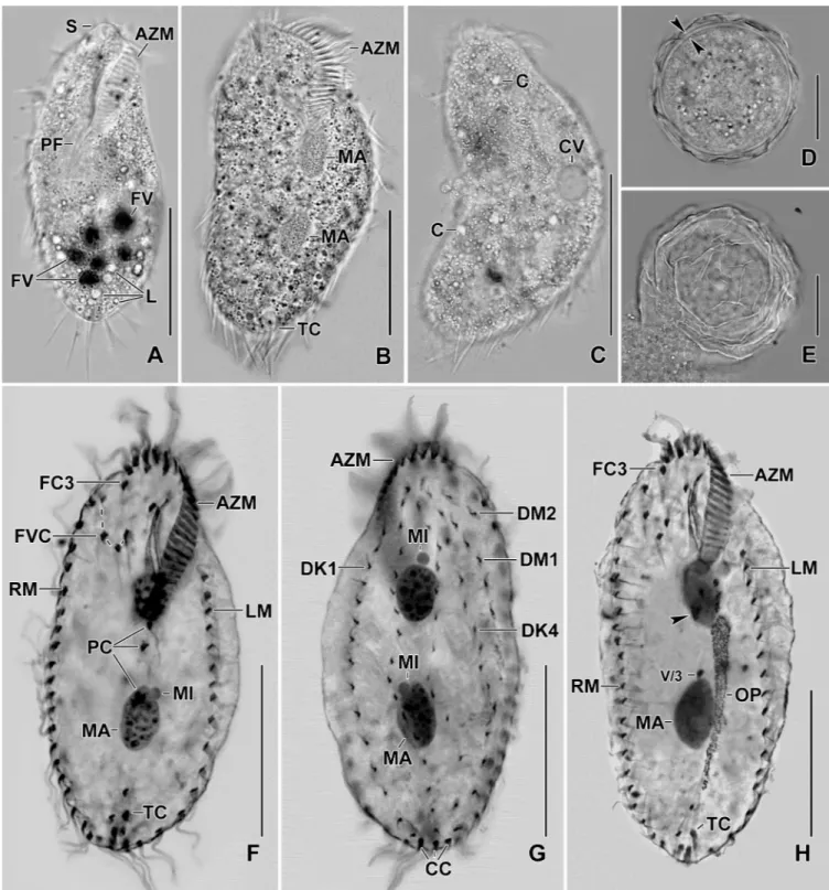
Fig 1. Line diagrams ofSterkiella tricirrata Italian population from life (A) and after protargol impregnation (B, C). (A) A representative cell with a length of 85μm. (B, C) Ventral and dorsal views of a voucher specimen, showing the ciliature and the nuclear apparatus. Note the invariably three transverse cirri typical of the species. AZM, adoral zone of membranelles; BC, buccal cirrus; CC, caudal cirri; DK1–4, dorsal kineties; DM1,2, dorsomarginal kineties; E, endoral membrane; FC3, frontal cirrus 3; FVC, frontoventral cirri; LM, left marginal row; MA, macronuclear nodules; MI, micronuclei; P, paroral
membrane; PC, postoral cirri; PTC< pretransverse cirri; RM, right marginal row. Scale bars = 40μm. https://doi.org/10.1371/journal.pone.0207688.g001
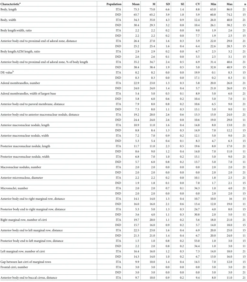
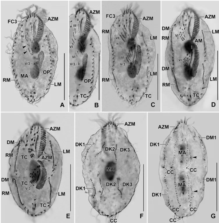
Fig 2. Photomicrographs ofSterkiella tricirrata Italian population from life (A–E) and after protargol impregnation (F–H). (A) Specimen, showing body shape, food vacuoles and lipid droplets. (B, C) Slightly compressed specimens due to cover slip pressure, showing nuclear apparatus (B), cytoplasmic crystals and contractile vacuole (C). (D, E) Resting cyst. Optical section (D), showing the cyst wall (opposed arrowheads). Squeezed cyst (E) with contents released, showing the wrinkled hyaline ridges. (F, G) Ventral view of the main voucher specimen, showing body shape, nuclear apparatus, and ciliature of the ventral (F) and dorsal surface (G). (H) An early divider, showing the formation of oral primordium close to transverse cirri. AZM, adoral zone of
membranelles; C, crystals; CC, caudal cirri; CV, contractile vacuole; DK1,4, dorsal kineties; DM1,2, dorsomarginal kineties; FC3, frontal cirrus 3; FV, food vacuoles; FVC, frontoventral cirri; L, lipid droplets; LM, left marginal row; MA, macronuclear nodules; MI, micronuclei; OP, oral primordium; PC, postoral cirri; PF, pharyngeal fibre; RM, right marginal row; S, scutum; TC, transverse cirri; V/3, postoral ventral cirrus. Scale bars = 15μm (D, E) and 30 μm (A–C, F– H).
separate macronuclear nodules (Fig 2D,4C and 4D). Cyst content includes many lipid drop-lets 1.5–3.0μm across in vivo (Fig 2D and 2E).
Notes on ontogenesis
The ontogenetic stages of Italian and Indian population show a common origin of anlagen II, III, V, and VI for the proter and the opisthe (Figs2H,5A–5E,6A–6C,7A–7C,8A–8Gand9A– 9K). Difference in the anlagen formation was observed in the Indian population, i.e., a W-shaped formation for the anlagen IV, V, and VI of the proter during the late-early stage, similar to type species of the genusSterkiella [18,19] (Fig 9E and 9F).
The oral primordium originates close to transverse cirri IV/1 and extends towards the buc-cal vertex (Figs2H,5A,7A–7C,8Aand9A–9C). The scattered basal bodies at the anterior end of the oral primordium develop into the opisthe’s anlagen I–IV (Figs5A,5B,7A–7C,8A,8B
and9A–9C). It is not clear whether disaggregating kinetosomes of cirrus IV/2 form or contrib-ute to the formation of opisthe’s anlage IV (Figs5B,5C,7A–7C,8B,8Cand9D–9F). Cirrus V/ 4 disaggregates and forms anlagen V and VI for the opisthe; anterior portions of the opisthe’s anlagen V and VI proliferate anteriorly, forming the proter’s anlagen V and VI (Figs5A–5C,
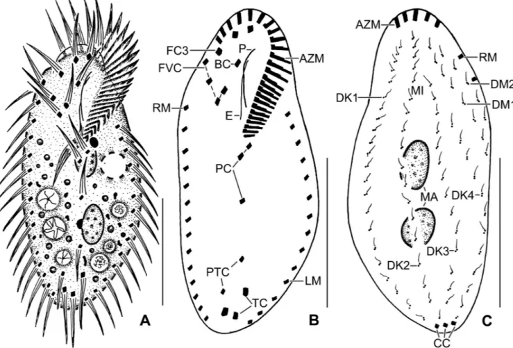
Fig 3. Line diagrams ofSterkiella tricirrata Indian population from life (A) and after protargol impregnation (B, C). (A) A representative cell with a length of 75μm. (B, C) Ventral and dorsal views of a voucher specimen, showing the ciliature and the nuclear apparatus. Note the invariably three transverse cirri. AZM, adoral zone of membranelles; BC, buccal cirrus; CC, caudal cirri; DK1–4, dorsal kineties; DM1,2, dorsomarginal kineties; FC3, frontal cirri; FVC, frontoventral cirri; LM, left marginal row; MA, macronuclear nodules; P, paroral membrane; PC, postoral cirri; PTC, pretransverse cirri; RM, right marginal row; TC, transverse cirri. Scale bars = 30μm.
7A–7C,8A,8Band9D). Cirrus V/3 does not participate in anlagen formation (Figs2H,5A– 5E,6A,7A–7C,8A–8Eand9A–9F). Anlage II of the opisthe extends anteriorly crossing the buccal vertex and joining the disaggregating buccal cirrus in early dividers (Figs5B,5C,7A– 7C,8A,8Band9D). Anlage I of the proter, i.e., the partially reorganized paroral and endoral, generates first frontal cirrus I/1 as well as the paroral and the endoral for the proter (Figs5D,
5E,6A,8C–8Eand9F–9I). Cirri III/2 and IV/3 disaggregate and give rise to the anlagen III and IV of the proter (Figs5B,7A–7C,8A,8B,9D and 9E). In the opisthe, anlage I separates from the posterior ends of anlagen II to IV and forms the paroral, endoral and cirrus I/1 (Figs
5D,5E,8C–8Eand9G–9I). Overall, five parental cirri and parental undulating membranes are involved in anlagen formation. The 16 frontal–ventral–transverse cirri arise from these anla-gen, splitting in a 1, 2, 2, 3, 4, 4 pattern (Figs5E,6A,8D,8Eand9G–9J). No transverse cirri are formed from anlagen II and III. A new adoral zone of membranelles for the opisthe develops from the oral primordium, while the parental adoral zone of membranelles is retained unchanged for the proter.
The marginal anlagen arise at each of two levels by “within-row” anlagen formation utiliz-ing one or two of the parental cirri at each level. The marginal anlagen elongate deployutiliz-ing four or five parental cirri and differentiate into new marginal rows. The remaining parental mar-ginal cirri are resorbed (Figs5D,5E,6A,8C–8E,8G,8H and 8J).
On the dorsal surface, three anlagen are formed within row from dorsal kineties 1, 2 and 3 at two levels, (one set for the proter and one for the opisthe) (Figs6B,6C,8F,8Gand9K). The
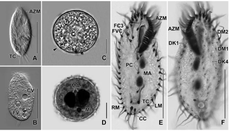
Fig 4. (A) Slightly starved specimen, showing body shape and three transverse cirri protruding beyond posterior end of the cell. (B) Slightly compressed
specimen due to cover slip pressure, showing food and contractile vacuole. (C, D) Resting cyst. Optical section (D), opposed arrowheads mark the cyst wall (C). Macronuclear nodules are separate in mature cyst (D). (E, F) Ventral view of the specimen, showing body shape, nuclear apparatus, and ciliature of the ventral (E) and dorsal surface (F). AZM, adoral zone of membranelles; CC, caudal cirri; CV, contractile vacuole; DK1,4, dorsal kineties; DM1,2, dorsomarginal kineties; FC3, frontal cirri; FV, food vacuole; FVC, frontoventral cirri; LM, left marginal row; MA, macronuclear nodules; MI, micronuclei; PC, postoral cirri; RM, right marginal row; TC, transverse cirri. Scale bars = 10μm (C, D), 30 μm (E, F) and 40 μm (A, B).
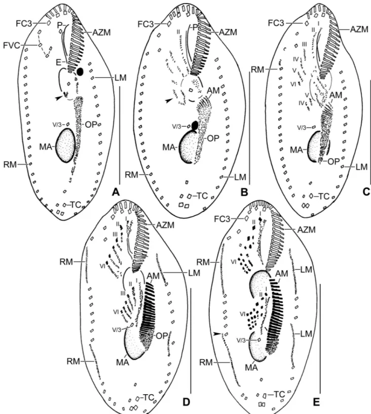
Fig 5. Line diagrams of protargol stained early dividers ofSterkiella tricirrata Italian population. (A) Arrowheads in (A, B) mark the disaggregating cirrus V/4 forming two anlagen, anterior portion of which proliferates anteriorly forming anlagen V and VI of the proter. (B) Two anlagen arise from the anterior end of the oral primordium. Anlage II of the opisthe moves anteriorly, part of this anlage merges with the parental buccal cirrus in early divider. Anlage III moves anteriorly and merges with the cirrus III/2. (C) Cirrus IV/3 disaggregates and forms anlage IV of the proter. Cirrus IV/2 forms the anlage IV of the opisthe. (D) Six anlagen are formed both for the proter and the opisthe. Posterior ends of the anlagen II to IV of the opisthe separates and form anlage I. Four anlagen for marginal cirri develop incorporating four to five parental marginal cirri in the proter and the opisthe. (E) Anlage I of the proter develops by partial reorganization of the paroral and endoral. Overall, five parental cirri (II/2, III/2, IV/3, IV/2, and V/4) disaggregate to give rise to five fronto-ventral–transverse anlagen for the proter and the opisthe. Arrowhead points to the anlagen of the dorsomarginal kineties. AM, adoral
third dorsal primordium fragments at the middle giving rise to the third and fourth kineties. The two dorso-marginal rows arise close to the anterior of right marginal row anlagen (Figs
6B,6C,8F,8Gand9K). Caudal cirri originate at the posterior end of the newly formed dorsal kineties 1, 2, and 4 (Figs6C,8Gand9K).
Nuclear division proceeds in the usual manner, i.e., in mid-dividers the macronuclear nod-ules fuse to form a single mass which divides twice to produce the typical four nodnod-ules in late dividers (Figs6B,6Cand8C–8G). The micronuclei undergo mitotic division.
SSU rRNA gene sequence and phylogeny
The SSU rRNA gene sequence ofSterkiella tricirrata Italian population is 1,628 bp in
length and has a GC content of 45.15%. It has been deposited in the NCBI database under the accession number MG805314. Phylogenetic trees inferred from the SSU rRNA gene sequences using ML and BI present similar topologies; thus, only the BI tree is shown here membranelles; AZM, adoral zone of membranelles; E, endoral membrane; FC3, frontal cirrus 3; FVC, frontoventral cirri; LM, left marginal row; MA, macronuclear nodules; OP, oral primordium; P, paroral membrane; RM, right marginal row; TC, transverse cirri; V/3, postoral ventral cirrus. Numerals denote cirral anlagen. Scale bars = 40μm.
https://doi.org/10.1371/journal.pone.0207688.g005
Fig 6. Line diagrams of protargol stained middle (A, B) and late (C) dividers ofSterkiella tricirrata Italian population. (A) Ventral surface (A), showing the formation of 16 fronto-ventral-transverse cirri from six anlagen (1:2:2:3:4:4). The newly formed fronto-ventral-transverse cirri migrate to their specific sites and dorsomarginal kineties develop close to the newly formed right marginal row. (B) Dorsal surface, showing the within row formation of dorsal kineties 1–3 at two levels. (C) Dorsal kinety 4 for the proter and the opisthe is formed by the simple fragmentation of dorsal kinety 3. AZM, adoral zone of membranelles; CC, caudal cirri; DK1–4, dorsal kineties; DM, dorsomarginal kinety; LM, left marginal row; RM, right marginal row; TC, transverse cirri; V/3, postoral ventral cirrus. Scale bars = 30μm (A, B) and 40 μm (C).
(Fig 10).Sterkiella tricirrata clusters with Sterkiella sinica (1.00 BI and 99% ML) within the
stylonychine oxytrichids group.
Discussion
Comparison of
Sterkiella tricirrata with related species and populations
Sterkiella tricirrata can be compared with species of the genus Sterkiella having two
macronu-clear nodules, i.e.,Sterkiella histriomuscorum (Foissner et al., 1991) Foissner, Blatterer, Berger
& Kohmann, 1991;S. nova Foissner & Berger, 1999; S. subtropica Chen et al., 2015; S. sinica
Chen et al., 2016, andS. ecuadoriana Foissner & Heber in Foissner, 2016. Sterkiella tricirrata
mainly differs from all the above mentioned species in having invariably three (vs. four or five) transverse cirri. Apart from transverse cirri, the Italian and Indian populations ofS. tricirrata
possess lower number of adoral membranelles (20–26 and 21–26 vs. 26–44 average from popu-lations described) in comparison withS. histriomuscorum [11]. There is one population of
Sterkiella histriomuscorum with four transverse cirri [18]; however, a reinvestigation on the
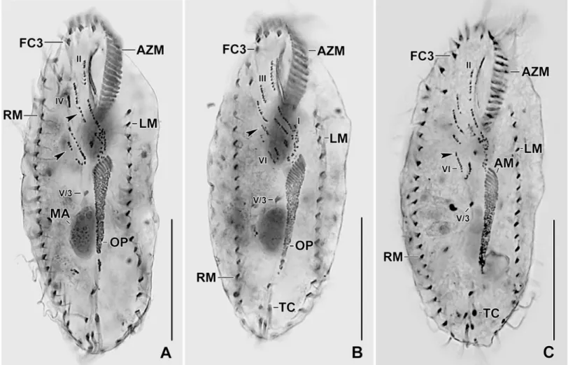
Fig 7. Photomicrographs of Protargol stained early dividers ofSterkiella tricirrata Italian population. (A–C) Anlage II of the opisthe moves anteriorly and merges with the anlage II of the proter. Arrowhead in (A) points to the part of the opisthe anlage III (note the orientation of the basal bodies and cilia) which probably joins the anlage III of the proter. Anlagen V and VI of the proter (arrowheads in B, C) originate from the anterior portion of the anlagen V and VI of the opisthe. AM, adoral membranelles; AZM, adoral zone of membranelles; FC3, frontal cirrus 3; LM, left marginal row; MA, macronuclear nodules; OP, oral primordium; RM, right marginal row; TC, transverse cirri; V/3, postoral ventral cirrus. Numerals denote cirral anlagen. Scale bars = 30μm (E, F) and 40 μm (A–D, G).
Fig 8. Photomicrographs of protargol stained early (A–D), middle (E, F), and late (G) dividers ofSterkiella tricirrata Italian population. For explanation refer to the legend ofFig 5B–5E. (A) Cirrus V/4 disaggregates and forms two anlagen for the opisthe, i.e., Anlagen V and VI; anterior portion of these anlagen proliferates anteriorly forming anlagen V and VI of the proter (arrowheads). (B) Cirrus IV/3 disaggregates and forms anlage IV of the proter. Cirrus IV/2 forms the anlage IV of the opisthe. (C) Six anlagen are formed both for the proter and the opisthe. Four anlagen for marginal cirri develop incorporating four to five parental marginal cirri in the proter and the opisthe. (D, E) The newly formed fronto-ventral-transverse cirri migrate to their specific sites and dorsomarginal kineties develop close to the newly formed right marginal row. (F, G) Within row formation of the anlagen for dorsal kineties 1–3 (F) takes place on the dorsal surface. Dorsal kinety 3 undergoes simple fragmentation forming kineties 3 and 4 (arrowheads in G). Caudal cirri are formed at the posterior end of dorsal kineties 1, 2, and 4, and the newly formed dorsomarginal kineties shift to the dorsal surface. AM, adoral membranelles; AZM, adoral zone of membranelles; CC, caudal cirri; DK1–3, dorsal kineties; DM1, dorsomarginal kineties; FC3, frontal cirrus 3; LM, left marginal row; MA, macronuclear nodules; OP, oral primordium; RM, right marginal row; TC, transverse cirri; V/3, postoral ventral cirrus. Numerals denote cirral anlagen. Scale bars = 30μm (E, F) and 40μm (A–D, G).
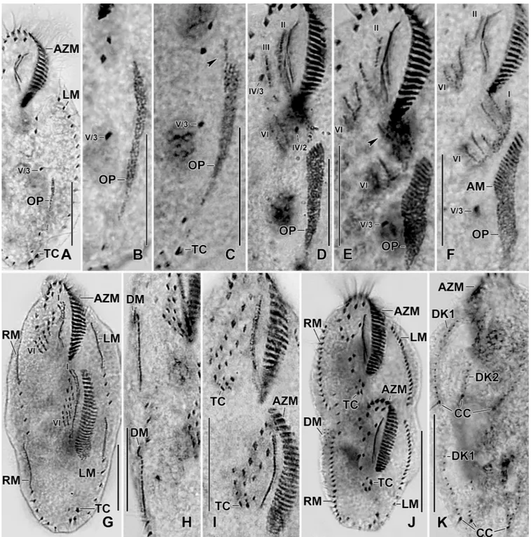
number of transverse cirri and the ontogenetic pattern is needed for a better comparison. The invariably three transverse cirri (over 250 specimens analyzed from Italian population and 50
Fig 9. Photomicrographs of protargol stained early (A–D), middle (E, F), and late (G) dividers ofSterkiella tricirrata Indian population. The ontogenesis of Indian population is rather similar to Italian population except that a W-shaped pattern is formed for anlagen IV, V, and VI for the proter in the former (E, F). For explanation refer to the legend of Figs5and8. Arrowhead in (E) points to anlage III of the opisthe. Six anlagen are formed both for the proter and the opisthe which splits transversely in 1:2:2:3:4:4 pattern. Dorsal kinety 3 undergoes simple fragmentation forming kineties 3 and 4; caudal cirri are formed at the posterior end of dorsal kineties 1, 2, and 4. AM, adoral membranelles; AZM, adoral zone of membranelles; CC, caudal cirri; DK1,2, dorsal kineties; LM, left marginal row; OP, oral primordium; RM, right marginal row; TC, transverse cirri; IV/3, fronto-ventral cirrus; V/3, IV/2, postoral ventral cirri. Numerals denote cirral anlagen. Scale bars = 20μm (A–I) and 30 μm (J, K).
specimens from Indian population) is a constant character ofSterkiella tricirrata. This,
sup-ports the validity ofSterkiella tricirrata at species rank and out of the S. histriomuscorum
com-plex; this separation is in agreement with Kumar et al. [1], who described a novelSterkiella
Fig 10. Bayesian tree inferred from the SSU rRNA gene sequences, showing the phylogenetic position ofSterkiella tricirrata (bold). Codes following the names are GenBank accession numbers. Numbers at the nodes represent the Bayesian inference (BI) posterior probabilities and the maximum likelihood bootstrap values out of 1000 replicates. A hyphen (-) represents differences between the BI and ML tree topologies. Asterisks represents values lower than 40%. The scale bar corresponds to nine substitutions per 100 nucleotide positions.
species with four macronuclear nodules mainly on the basis of four transverse cirri.Sterkiella histriomuscorum and S. nova are indistinguishable based on the morphology and gene
sequence data are required for their identification [18].Sterkiella subtropica can be separated
fromS. tricirrata Italian and Indian populations by the marine vs. terrestrial habitat, large
body size in vivo 100–200× 35–70 μm (vs. 70–90 × 30–50 μm), slightly higher number of adoral membranelles 25–39 (vs. 20–26 and 21–26), and number of right 19–27 (vs. 18–21 and 14–18) and left 18–26 (vs. 14–18 and 13–16) marginal cirri [16]. The Italian and Indian popu-lations ofSterkiella tricirrata can be separated from S. ecuadoriana by the slightly larger body
length (63–86μm and 61–74 vs. 91–150 μm), adoral membranelles (20–26 and 21–26 vs. 32– 49) and bristles in DK1 (13–18 and 17–22 vs. 27–45) [3].Sterkiella sinica can be distinguished
from Italian and Indian populations ofS. tricirrata in having an extra cirrus between the
sec-ond and third frontal cirri (vs. no such cirrus), distance between cirri V/2 and V/3 24.5μm (vs. 19.3 and 10.6μm) and cirri V/3 and V/4 4.7% (vs. 9.4% and 16.1%) of the body length, number of cirri in left 19–23 (vs. 14–18 and 13–16) marginal row, and by the lower number of bristles in DK1 (22–25 vs. 13–18 and 17–22), DK2 (20–24 vs. 14–21 and 14–20), DK3 (14–19 vs. 10– 14 and 7–14), DK4 (13–19 vs. 8–13 and 11–15), DM1 (9–15 vs. 5–8 and 7–10), and DM2 (5– 10 vs. 2–5 and 3–6) [31]. Minor differences betweenSterkiella sinica and Italian and Indian
populations ofS. tricirrata were observed in the body size 85–110 × 35–45 μm (vs. 70–
90× 30–50 μm) and number of cirri in right 18–22 (vs. 18–21 and 14–18) marginal row [31]. Other than species of the genusSterkiella, Sterkiella tricirrata can be compared with Para-sterkiella thompsoni (Foissner, 1996) Ku¨ppers et al., 2011; MetaPara-sterkiella koreana Kumar et al.,
2017; andFragmospina depressa Foissner, 2016. Sterkiella tricirrata mainly differs from Para-sterkiella thompsoni in having two (vs. three) macronuclear nodules, numbers of dorsal
kin-eties 6 (vs. 5), and presence (vs. absence) of fragmentation of dorsal kinety 3 during dorsal morphogenesis [17]. It differs fromMetasterkiella koreana mainly in the number of transverse
cirri (invariably 3 vs. 5) and in the ontogenesis, i.e., cirrus V/3 intact (vs. involved during anla-gen formation) [2].Fragmospina depressa can be separated from S. tricirrata by having a
par-oral membrane close (vs. distant) to the adpar-oral membranelles, number of transverse cirri 5 (vs. invariably 3) and the structure of resting cyst, i.e., spinous (vs. wrinkled) surface [3].
The Indian population ofSterkiella tricirrata shows minor differences in size and ciliature
with the Italian population as mentioned in the description section. The resting cyst of Indian population appears to be smooth (vs. wrinkled in Italian population); however, additional data on the resting cyst of the Indian population is required to confirm this feature. The original population described by Buitkamp [32] could not be meaningfully compared since most of the morphometric data are lacking. Main differences observed (data from the single image of a protargol stained specimen provided in Buitkamp [32] rely in the number of cirri in left (10 vs. 16 and 20 in Indian and Italian populations, respectively) and right (12 vs. 14 and 16 in Indian and Italian populations, respectively) marginal rows. Further, the original description ofS. tri-cirrata mentioned the presence of five (instead of six recorded in the present study) dorsal
kin-eties. We agree with Berger [11] since the dorsal kinety 6 is rather short it could have been easily missed by Buitkamp [32]. A reinvestigation of the Ivory Coast population will further clarify if it requires separation at the species/subspecies level.
Notes on the ontogenesis of the genus
Sterkiella
Berger and Foissner [15] reported that the anlagen V and VI of the opisthe originate de novo in species of the genusSterkiella. Later, Foissner et al. [19] provided a detailed ontogenetic data onSterkiella cavicola (Kahl, 1935) Foissner, Blatterer, Berger & Kohmann, 1991,
cirrus V/4. The same pattern is observed also forSterkiella tricirrata where cirrus V/4
gener-ates anlagen V and VI of the opisthe; however the anterior patches of both the anlagen move anteriorly and later form the proter anlagen V and VI (Table 2). On the contrary, anlagen V and VI of the proter originate from a disaggregation of cirrus IV/3 inS. cavicola [19]. Further, the ontogenetic data ofS. tricirrata shows that the anterior portions of anlagen II and III of the
opisthe proliferate anterior and merge with the disaggregating cirri II/2 and III/2 respectively to form anlagen II and III of the proter. Recently, Kumar et al. [2] erected a novel genus,
Metasterkiella, for a species having similar morphological features as that of Sterkiella histrio-muscorum; however, the former not only showed difference in the anlagen formation but also
the involvement of cirrus V/3 in anlagen formation, a feature never reported for any stylony-chid ciliate. Possibly the involvement of cirrus V/3 during anlagen formation and the semi-rigid body indicate that theM. koreana might have recently evolved from an Oxytricha-like
ancestor. As mentioned above,Sterkiella tricirrata also shows differences with Sterkiella cavi-cola in the formation of anlagen II, V and VI, i.e., confluent anlagen II and anlagen V and VI
of the opisthe give rise to anlagen V and VI of the proter by enlargement and then splitting, though cirrus V/3 remains intact during ontogenesis. The ontogenetic difference between the Indian and Italian populations, i.e., formation of a W-shaped pattern (vs. separate) by the anla-gen IV, V, and VI of the proter in late-early divider, indicates that the Indian population may represent a separate subspecies/species if the pattern mentioned is found to be stable in other populations with consensus of molecular data. As of now, we do not perform its separation from theSterkiella histriomuscorum complex and wait for further data to resolve the
phyloge-netic status of the species within the complex. However, the different morphogephyloge-netic patterns, within the genusSterkiella, as seen in the present study and Foissner et al. [19] needs to be reflected in the generic characteristics, thus we have provided an improved diagnosis of the genusSterkiella. The Austrian population of Sterkiella histriomuscorum shows some similarity
in anlagen formation withS. tricirrata [18,33]; however, a detailed investigation of its mor-phogenesis is required for a reliable comparison.
Phylogenetic position of
Sterkiella tricirrata
Sterkiella tricirrata clusters with S. sinica (1.00 BI and 99% ML;Fig 9) within the stylonychine oxytrichids, in a clade away from the type species (Sterkiella cavicola) of the genus Sterkiella;
we assume that the molecular relatedness ofS. tricirrata and S. sinica could be because of
simi-larity in the formation of anlagen. However, a detailed investigation of the ontogenesis ofS. sinica is needed to properly compare these genetically similar species. Our phylogenetic
Table 2. Comparison between species of the genusSterkiella.
Characteristic Sterkiella tricirrata Sterkiella histriomuscorum Sterkiella histriomuscorum
Sterkiella nova Sterkiella cavicola
Italy Austria Antarctica USA Austria
Early disaggregation of cirrus II/2 during ontogenesis Present Present Absent Absent Absent
W-shaped anlagen formation Absent Absent Present Present Present
Formation of confluent anlagen II for the proter and opisthe
Present Present Absent Absent Absent
Formation of anlagen V and VI of the proter from anlagen V and VI of the opisthe
Present Present Absent Absent Absent
Transverse cirri, number 3 4 5 5 5
Data source Present study Berger et al. [34] Petz & Foissner [33] Foissner & Berger
[18]
Foissner et al. [19] https://doi.org/10.1371/journal.pone.0207688.t002
analyses also shows thatS. histriomuscorum and S. cavicola behave as sisters of a larger clade
containing, in addition to the aforementionedS. tricirrata + S. sinica sub-clade, a further high
supported sub-clade includingS. tetracirrata + Gastrostyla steinii (1.00 BI, 99% ML). The
otherSterkiella species are distributed across two more clades: i) S. nova with Tetmemena bifaria (1.00 BI, 79% ML); and ii) S. subtropica with Metasterkiella koreana (1.00 BI, 100%
ML). The monophyly of the genusSterkiella is not supported in our phylogenetic analyses as
also evident in other recent studies [1,2,15–17]. Certainly, more sequences from populations of theS. histriomuscorum complex as well as from other Sterkiella species are required to
obtain better resolution. Although in recent years, the situation has been slightly improved with the establishment of genera, namely,Parasterkiella Ku¨ppers et al., 2011 and Metasterkiella
Kumar et al., 2017.Sterkiella subtropica and S. nova cluster away from the type species (S. cavi-cola); for the former species, Kumar et al. [2] suggested that it probably belongs to the genus
Metasterkiella due to highly similar morphology and gene sequence; this interpretation is also
supported by our phylogenetic analyses. The classification ofSterkiella nova has been widely
debated among classical taxonomists and molecular biologists who established this species as model organism for analyzing various biological phenomena such as epigenetic inheritance, genome rearrangement, somatic differentiation and many others [11,18]. In this regards, Foissner and Berger [18] describedS. histriomuscorum and S. nova in great detail from viable
genetic systems (via frozen resting cysts) established by molecular biologists. They mentioned
that both species are inseparable based on the morphological characters; though based on the differences in molecular sequences of actin I and DNA polα genes, they proposed them as dif-ferent species. Considering the complexity of identification it is unclear whether the gene sequences provided by Hewitt et al. [35], which is used in the present study and remains the only sequence available forS. nova, is of same species described by Foissner and Berger [18].
As of now, only differences which seem most suitable to solve the polyphyletic behavior of the genusSterkiella is the data on the ontogenetic pattern on the ventral and dorsal surface. In Sterkiella cavicola anlagen V and VI of the proter originate from cirrus IV/3 forming W-shaped
anlagen [19], whereas it forms from opisthe’s anlagen V and VI during the early ontogenetic stages inSterkiella tricirrata and the genus Metasterkiella. In our phylogenetic tree, Metaste-rkiella forms a distant clade away from that of SteMetaste-rkiella tricirrata the involvement of cirrus V/3
(vs. intact) during anlagen formation possibly justifies this distant relationships. Nonetheless, several examples exists like,Parasterkiella thompsoni, Fragmospina depressa, which would have
been easily identified asSterkiella species but separated based on detailed investigations on
mor-phology and cyst structure.Parasterkiella thompsoni shows a different ontogenetic pattern on
the dorsal surface and acquires a place distant fromSterkiella species [17], for the species of the genusFragmospina no gene sequence is available thus far. We believe that addition of related
molecular sequences, e.g.,Fragmospina, S. histriomuscorum populations, and gene sequences
from other loci will further support the monophyly of the genusSterkiella.
Soil ciliate diversity and species identification: A contribution
Ciliated protists are a highly diverse group of microbial eukaryotes that play a key role in soil microbial food webs by mediating the fluxes of nutrients and energy between different trophic levels [36]. Nevertheless, ciliate diversity in the soil is a still largely neglected research topic and this taxon is significantly less studied than other soil microbial taxa such as bacteria and fungi [37,38]. Since 2009, our group has made a significant contribution to in-depth knowledge about the diversity of soil ciliates across two continents, i.e., Europe (Italy) and Asia (India and South Korea) [1,9,39–45]. Numerous faunistic surveys performed in the framework of several projects, allowed us to isolate and describe several novel species and genera, as well as
re-describe poorly known or even misidentified species [9,39–45]. According to Foissner [46], more than 70–80% of the soil ciliate diversity is still unexplored and a single soil sample can host new species/genera such as in the case of the soil sample collected from the regional Park of Colfiorito described in this study, in which one new and one poorly known species were identified, i.e.,Pseudouroleptus plestiensis [45] andSterkiella tricirrata (present study).
In the end, our sampling effort has allowed us to contribute to strengthen the knowledge about soil ciliate diversity, providing hints about their biogeographic distributions and new distinguishing characters (i.e., cyst morphology, molecular data, ontogenetic processes, arrangement and number of cirri, etc.) among hypotrich ciliates that can be helpful in species identification within problematic (cryptic) species "complexes".
Phylum Ciliophora Doflein, 1901 Class Spirotrichea Bu¨tschli, 1889
Order Sporadotrichida Faure´-Fremiet, 1961 Family Oxytrichidae Ehrenberg, 1838
Genus Sterkiella
Improved diagnosis
Body semi-rigid. Eighteen or less frontal-ventral-transverse cirri arranged in typical oxytrichid pattern. One right and one left row of marginal cirri. Six dorsal kineties including dorsomargi-nal rows, kinety 3 with simple fragmentation; caudal cirri present. Undulating membranes in
Oxytricha pattern. Opisthe’s anlage II may contribute to proter’s anlage II. Anlagen V and VI
of the proter originate from cirrus IV/3 forming W-shaped anlagen or from anlagen V and VI of the opisthe.
Sterkiella tricirrata
Improved diagnosis (averages are from the populations of India, Italy, and
Ivory Coast)
Size about 80× 40 μm in vivo; body elongate to broadly ellipsoidal. Nuclear apparatus com-posed of two macronuclear nodules and two micronuclei on average. Invariably, 16 frontal-ventral-transverse cirri, including three transverse cirri. Right and left marginal rows posed of an average of 15 and 14 cirri, respectively. Adoral zone 37% of body length and com-posed of an average of 23 membranelles. Three narrowly spaced, inconspicuous caudal cirri. Resting cyst with wrinkled surface. Soil habitat.
Neotype material
Since the original description is incomplete and no type material is available thus according to the Article 75.3 of the ICZN (1999) we propose neotypification of theSterkiella tricirrata with
sampling site of the Italian population being the type locality. Two neotype slides of Italian population containing the protargol stained neotype specimen (Figs2Fand3A) and relevant morphostatic specimens have been deposited at the Natural History Museum, London, UK, with registration numbers NHMUK 2014.3.20.1 and NHMUK 2014.3.20.2. Further, two slides of the Indian population are deposited at the Natural History Museum, London, UK, with reg-istration numbers NHMUK 2011.7.4.2 and NHMUK 2011.7.4.3 and one at the type collection
of the Zoological Survey of India, Kolkata, India, with registration number Pt 3067. The SSU rRNA gene sequence is deposited in GenBank (accession number: MG805314).
Occurrence and ecology
Buitkamp [32] isolatedSterkiella tricirrata from the soil collected from the burnt savannah in
the Ivory Coast. The Italian population was identified from the ‘Molinaccio’ site during the summer (dry season), where it was moderately abundant in non-flooded Petri dish culture. For details on the soil physico-chemical parameters and other ciliate species identified in the same soil sample, refer to Bharti et al. [45]. The Indian population was identified from the soil sample collected from the tracts of the tropical rain forest of the Silent Valley National Park, India. For details on other ciliates species identified from the soil samples collected, refer to Kumar et al. [47]. Feeds on bacteria, small amoeba, and flagellates; clonal cultures can be raised as mentioned in materials and methods section.
Acknowledgments
We thank Director, Zoological Survey of India, for providing the necessary facilities and encouragement. The authors would also like to thank Dr. Mauro Tiberi, Dr. Giovanni Cia-bocco and Dr. Cristina Bernacconi from Osservatorio Regionale Suoli (http://suoli.regione. marche.it/) and Dr. Emilio Insom and Dr. Silvia Marinsalti from University of Camerino for their help in sampling. Special thanks to Shri Ghanshyam Prasad Singh, University of Delhi, for the help provided during sampling in the Silent Valley National Park, India.
Author Contributions
Formal analysis: Daizy Bharti, Santosh Kumar.
Funding acquisition: Santosh Kumar, Komal Kamra, Antonietta La Terza. Investigation: Daizy Bharti, Santosh Kumar, Govindhasamay R. Varatharajan.
Project administration: Daizy Bharti, Santosh Kumar, Komal Kamra, Antonietta La Terza. Supervision: Santosh Kumar, Antonietta La Terza.
Writing – original draft: Daizy Bharti, Santosh Kumar.
Writing – review & editing: Daizy Bharti, Santosh Kumar, Govindhasamay R. Varatharajan,
Komal Kamra, Antonietta La Terza.
References
1. Kumar S, Kamra K, Bharti D, La Terza A, Sehgal N, Warren A, Sapra GR. Morphology, morphogenesis, and molecular phylogeny of Sterkiella tetracirrata n. sp. (Ciliophora, Oxytrichidae), from the Silent Valley National Park, India. Eur J Protistol. 2015; 51: 86–97.https://doi.org/10.1016/j.ejop.2014.12.002PMID: 25625942
2. Kumar S, Bharti D, Shazib SUA, Shin MK. Discovery of a new hypotrich ciliate from petroleum contami-nated soil. PLoS ONE. 2017; 12(6): e0178657.https://doi.org/10.1371/journal.pone.0178657PMID: 28570607
3. Foissner W. Terrestrial and semiterrestrial ciliates (Protozoa,Ciliophora) from Venezuela and Gala´pa-gos. Denisia. 2016; 35: 1–912.
4. Kumar S, Foissner W. High cryptic soil ciliate (Ciliophora, Hypotrichida) diversity in Australia. Eur J Pro-tistol. 2016; 53: 61–95.https://doi.org/10.1016/j.ejop.2015.10.001PMID:26844781
5. Warren A, Patterson DJ, Dunthorn M, Clamp JC, Achilles-Day UEM, Aescht E, et al. Beyond the “Code”: A guide to the description and documentation of biodiversity in ciliated protists (Alveolata, Cilio-phora). J Eukaryot Microbiol. 2017; 64: 539–554.https://doi.org/10.1111/jeu.12391PMID:28061024
6. Blatterer H, Foissner W. Morphological and ontogenetic comparison of two populations of Parentocirrus hortualis Voss 1997 (Ciliophora, Hypotrichida). Linzer Biol Beitr. 2003; 35: 831–854.
7. Foissner W, Xu K. Monograph of the Spathidiida (Ciliophora, Haptoria). Vol. I: Protospathidiidae, Arcuospathidiidae, Apertospathulidae. Monogr Biol. 2007; 81: 1–485.
8. Foissner W, Wolf KW, Kumar S, Xu K, Quintela-Alonso P. Five new spathidiids (Ciliophora: Haptoria) from Caribbean tank bromeliads. Acta Protozool. 2014; 53: 159–194.
9. Kumar S, Bharti D, Quintela-Alonso P, Shin MK, La Terza A. Fine-tune investigations on three stylony-chid (Ciliophora, Hypotricha) ciliates. Eur J Protistol. 2016; 56: 200–218.https://doi.org/10.1016/j.ejop. 2016.09.006
10. Kumar S, Foissner W. Biogeographic specializations of two large hypotrich ciliates: Australocirrus shii and A. australis and proposed synonymy of Australocirrus and Cyrtohymenides. Eur J Protistol. 2015; 51: 210–228.https://doi.org/10.1016/j.ejop.2015.02.002PMID:26004119
11. Berger H. Monograph of the Oxytrichidae (Ciliophora, Hypotrichia). Monogr Biol. 1999; 78: 1–1080. 12. Berger H. Monograph of the Urostyloidea (Ciliophora, Hypotricha). Monogr Biol. 2006; 85: 1–1304. 13. Berger H. Monograph of the Amphisiellidae and Trachelostylidae (Ciliophora, Hypotricha). Monogr Biol.
2008; 88: 1–737.
14. Berger H. Monograph of the Gonostomatidae and Kahliellidae (Ciliophora, Hypotricha). Monogr Biol. 2011; 90: 1–741.
15. Berger H, Foissner W. Cladistic relationships and generic characterization of oxytrichid hypotrichs (Pro-tozoa, Ciliophora). Arch Protistenkd. 1997; 148: 125–155.
16. Chen X, Gao F, Al-Farraj S, Al-Rasheid K, Xu K, Song W. Morphology and morphogenesis of a novel mangrove ciliate, Sterkiella subtropica sp. nov. (Protozoa, Ciliophora, Hypotrichia), with phylogenetic analyses based on small-subunit rDNA sequence data. Int J Syst Evol Microbiol. 2015; 65: 2292–2303. https://doi.org/10.1099/ijs.0.000253PMID:25872955
17. Ku¨ppers GC, Paiva TS, Borges BN, Harada ML, Garraza GG. Mataloni G. An Antarctic hypotrichous cil-iate, Parasterkiella thompsoni (Foissner) nov. gen., nov. comb., recorded in Argentinean peat-bogs: morphology, morphogenesis, and molecular phylogeny. Eur J Protistol. 2011; 47: 103–123.https://doi. org/10.1016/j.ejop.2011.01.002PMID:21459562
18. Foissner W, Berger H. Identification and ontogenesis of the nomen nudum hypotrichs (Protozoa: Cilio-phora) Oxytricha nova (= Sterkiella nova sp. n.) and O. trifallax (= S. histriomuscorum). Acta Protozool. 1999; 38: 215–248.
19. Foissner W, Agatha S, Berger H. Soil ciliates (Protozoa, Ciliophora) from Namibia (Southwest Africa), with emphasis on two contrasting environments, the Etosha region and the Namib Desert. Part I: text and line drawings. Part II: photographs. Denisia. 2002; 5: 1–1459.
20. Foissner W. Soil protozoa: fundamental problems, ecological significance, adaptations in ciliates and testaceans, bioindicators, and guide to the literature. Progr Protistol. 1987; 2: 69–212.
21. Kamra K, Sapra GR. Partial retention of parental ciliature during morphogenesis of the ciliate Coniculos-tomum monilata (Dragesco and Njine´, 1971) Njine´, 1978 (Oxytrichidae, Hypotrichida). Eur J Protistol. 1990; 25: 264–278.https://doi.org/10.1016/S0932-4739(11)80179-3PMID:23195974
22. Wallengren H. Zur Kenntnis der vergleichenden Morphologie der hypotrichen Infusorien. Bih K svensk VetenskAkad Handl. 1900; 26: 1–31.
23. Gong J, Kim S-J, Kim S-Y, Min G-S, Roberts DMcL, Warren A, Choi J-K. Taxonomic redescriptions of two ciliates, Protogastrostyla pulchra n. g., n. comb. and Hemigastrostyla enigmatica (Ciliophora: Spiro-trichea: Stichotrichia), with phylogenetic analyses based on 18S and 28S rRNA gene sequences. J Eukaryot Microbiol. 2007; 54: 468–478.https://doi.org/10.1111/j.1550-7408.2007.00288.x 24. Medlin L, Elwood HJ, Stickel S, Sogin ML. The characterization of enzymatically amplified eukaryotic
16S-like rRNA coding regions. Gene. 1988; 71: 491–499. PMID:3224833
25. Katoh K, Standley DM. MAFFT multiple sequence alignment software version 7: improvements in per-formance and usability. Mol Biol Evol. 2013; 30: 772–780.https://doi.org/10.1093/molbev/mst010 PMID:23329690
26. Castresana J. Selection of conserved blocks from multiple alignments for their use in phylogenetic anal-ysis. Mol Biol Evol. 2000; 17: 540–552.https://doi.org/10.1093/oxfordjournals.molbev.a026334PMID: 10742046
27. Glez-Peña D, Go´mez-Blanco D, Reboiro-Jato M, Fdez-Riverola F, Posada D. ALTER: program-oriented format conversion of DNA and protein alignments. Nucleic Acids Res. 2010; 38(Suppl. 2): W14–18. 28. Ronquist F, Teslenko M, van der Mark P, Ayres DL, Darling A, Hohna S, Larget B, Liu L, Suchard MA,
Huelsenbeck JP. MrBayes 3.2: efficient Bayesian phylogenetic inference and model choice across a large model space. Syst Biol. 2012; 61: 539–542.https://doi.org/10.1093/sysbio/sys029
29. Posada D. jModelTest: Phylogenetic Model Averaging. Mol Biol Evol. 2008; 25: 1253–1256.https://doi. org/10.1093/molbev/msn083PMID:18397919
30. Tamura K, Peterson D, Peterson N, Stecher G, Nei M, Kumar S. MEGA5: molecular evolutionary genet-ics analysis using maximum-likelihood, evolutionary distance, and maximum parsimony methods. Mol Biol Evol. 2011; 28: 2731–2739.https://doi.org/10.1093/molbev/msr121PMID:21546353
31. Chen L, Zhao X, Shao C, Miao M, Clamp JC. Morphology and phylogeny of two new ciliates, Sterkiella sinica sp. nov. and Rubrioxytricha tsinlingensis sp. nov. (Protozoa, Ciliophora, Hypotrichia) from north-west China. Syst Biodivers. 2017; 131–142.
32. Buitkamp U. Die Ciliatenfauna der Savanne von Lamto (Elfenbeinku¨ste). Acta Protozool. 1977; 16: 249–276.
33. Petz W, Foissner W. Morphology and infraciliature of some soil ciliates (Protozoa, Ciliophora) from con-tinental Antarctica, with notes on the morphogenesis of Sterkiella histriomuscorum. Polar Rec. 1997; 33: 307–326.
34. Berger H, Foissner W, Adam H. Morphological variation and comparative analysis of morphogenesis in Parakahliella macrostoma (Foissner, 1982) nov. gen. and Histriculus muscorum (Kahl, 1932), (Cilio-phora, Hypotrichida). Protistologica. 1985; 21: 295–311.
35. Hewitt EA, Mu¨ller KM, Cannone J, Hogan DJ, Gutell R, Prescott DM. Phylogenetic relationships among 28 spirotrichous ciliates documented by rDNA. Mol Phylogenet Evol. 2003; 29(2): 258–67. PMID: 13678681
36. Geisen S. The bacterial-fungal energy channel concept challenged by enormous functional versatility of soil protists. Soil Biol Biochem. 2016; 102: 22–25.https://doi.org/10.1016/j.soilbio.2016.06.013 37. Geisen S, Mitchell EAD, Wilkinson DM, Adl S, Bonkowski M, Brown MW, et al. Soil protistology
rebooted: 30 fundamental questions to start with. Soil Biol Biochem. 2017; 111: 94–103.https://doi.org/ 10.1016/j.soilbio.2017.04.001
38. Murase J. Quest of soil protists in a new era. Microbes Environ. 2017; 32(2):99–102.https://doi.org/10. 1264/jsme2.ME3202rhPMID:28652550
39. Kumar S, Bharti D, Marinsalti S, Insom E, La Terza A. Morphology, morphogenesis, and molecular phy-logeny of Paraparentocirrus sibillinensis n. gen., n. sp., a “Stylonychine Oxytrichidae” (Ciliophora, Hypo-trichida) without transverse cirri. J Eukaryot Microbiol. 2014; 61:247–259.https://doi.org/10.1111/jeu. 12103
40. Kumar S, Bharti D, Marinsalti S, Insom E, La Terza A. Corrigendum to “Morphology, morphogenesis, and molecular phylogeny of Paraparentocirrus sibillinensis n. gen., n. sp., a “Stylonychine Oxytrichidae” (Ciliophora, Hypotrichida) without transverse cirri by Kumar et al”. J Eukaryot Microbiol. 2017; 64: 906– 907.https://doi.org/10.1111/jeu.12451PMID:29139234
41. Bharti D, Kumar S, La Terza A. 2016. Rigidosticha italiensis n. gen., n. sp. (Ciliophora, Spirotricha), a novel large hypotrich ciliate from the soil of Lombardia, Italy. Eur J Protistol. 2016; 56:112–118.https:// doi.org/10.1016/j.ejop.2016.08.004
42. Bharti D, Kumar S, La Terza A. Two gonostomatid ciliates from the soil of Lombardia, Italy; including note on the soil mapping project. J Eukaryot Microbiol. 2015; 62:762–772.https://doi.org/10.1111/jeu. 12234PMID:25976551
43. Bharti D, Kumar S, La Terza A. Corrigendum to “Two gonostomatid ciliates from the soil of Lombardia, Italy; including note on the soil mapping project by Bharti et al”. J Eukaryot Microbiol. 2017; 64: 907. https://doi.org/10.1111/jeu.12470PMID:29139231
44. Bharti D, Kumar S, La Terza A. Description and molecular phylogeny of a novel hypotrich ciliate from the soil of Marche Region, Italy; including notes on the MOSYSS Project. J Eukaryot Microbiol. 2017; 64: 678–690.https://doi.org/10.1111/jeu.12404PMID:28211199
45. Bharti D, Kumar S, La Terza A. Morphology, morphogenesis and molecular phylogeny of a novel soil cil-iate, Pseudouroleptus plestiensis n. sp. (Ciliophora, Oxytrichidae), from the uplands of Colfiorito, Italy. Int J Syst Evol Microbiol. 2014; 64: 2625–2636.https://doi.org/10.1099/ijs.0.062885-0PMID: 24824635
46. Foissner W, Chao A, Katz LA. Diversity and geographic distribution of ciliates (Protista: Ciliophora). Bio-divers Conserv. 2008; 17: 345–363.
47. Kumar S, Kamra K, Sapra GR. Ciliates of the Silent Valley National Park, India: urostyloid hypotrichs of the region with a note on the habitat. Acta Protozool. 2010; 49: 339–364.