Interaction of rotavirus nonstructural protein NSP5
with the viral replication complex
Thesis submitted for the degree of Doctor Philosophiae
(Perfezionamento in Genetica Molecolare e Biotecnologie)
Candidate:
CONTENTS
ABSTRACT... 3
LIST OF ABBREVIATIONS... 5
INTRODUCTION... 6
1. VIRUSCLASSIFICATION... 7
2. DESCRIPTIONOFTHEVIRION ... 8
2.1. OVERVIEW ... 8
2.2. SPATIAL ORGANIZATION OF THE TRIPLE-LAYERED PARTICLE ... 9
2.3. THE VIRAL GENOME ... 12
2.4. GENE-PROTEIN ASSIGNMENT ... 14 3. ROTAVIRUSPROTEINS ... 16 VP1 ... 16 VP2 ... 17 VP3 ... 19 VP4 ... 19 VP6 ... 23 VP7 ... 25 NSP1 ... 27 NSP2 ... 28 NSP3 ... 32 NSP4 ... 33 NSP5 ... 37 NSP6 ... 42 4. VIRUSREPLICATION... 43 4.1. OVERVIEW ... 43
4.2. CELL ATTACHMENT AND ENTRY ... 45
4.3. TRANSCRIPTION ... 48
4.4. TRANSLATION ... 50
4.5. GENOME REPLICATION AND PACKAGING... 52
4.6. VIRUS MORPHOGENESIS ... 59
4.7. VIRUS RELEASE ... 63
5. PATHOGENESIS,ILLNESS,IMMUNERESPONSEANDVACCINES... 64
6. INVESTIGATINGINTERACTIONSANDFUNCTIONSOFROTAVIRUSPROTEINS:THE CHALLENGEOFREVERSEGENETICS ... 67
MATERIALS AND METHODS... 69
CELL CULTURE... 69
VIRUS PROPAGATION... 69
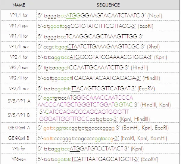
CONSTRUCTION OF PLASMIDS... 69
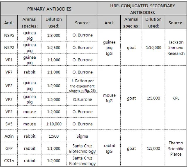
PRODUCTION OF ANTIBODIES... 73
TRANSIENT TRANSFECTION AND LABELLING WITH [35S]-METHIONINE OF MA104 CELLS ... 74
CELLULAR LYSIS ... 75
CHEMICAL DSP CROSS-LINKING AND UV TREATMENT OF CELLS ... 75
IMMUNOPRECIPITATION, PAGE AND WESTERN IMMUNOBLOT ANALYSIS ... 76
RNase TREATMENT OF PROTEIN COMPLEXES ... 77
λ-PHOSPHATASE TREATMENT OF IMMUNOPRECIPITATES ... 78
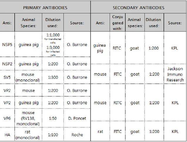
INDIRECT IMMUNOFLUORESCENCE MICROSCOPY... 78
PURIFICATION OF VIRAL PARTICLES OBTAINED FROM CELLS EXPRESSING SV5-TAGGED VP1 .... 79
EDTA TREATMENT OF PURIFIED TLPs ... 80
RESULTS... 81
RESULTS (1)... 82
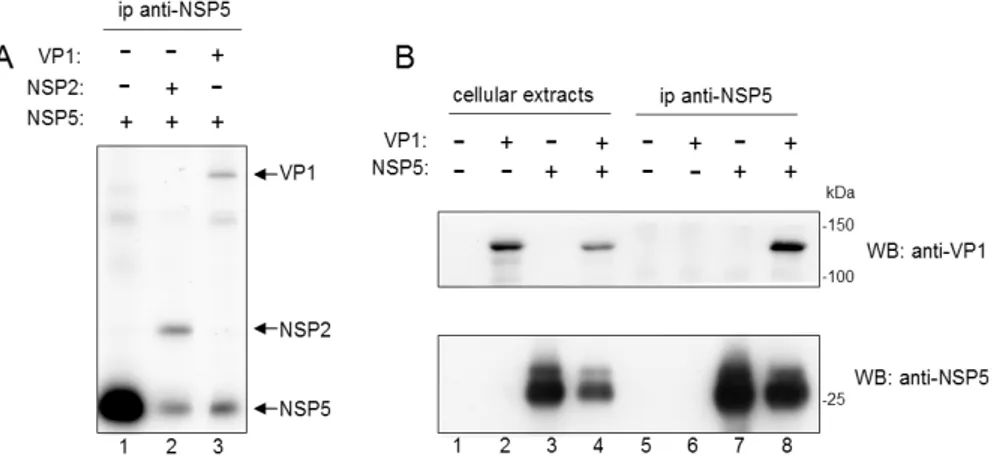
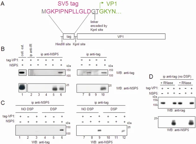
VP1 coimmunoprecipitates with NSP5... 82
Tag-VP1 can act as a structural replacement of VP1 ... 86
Tag-VP1 colocalizes with NSP5 in viroplasms and in VLS ... 89
VP1 interacts more strongly with NSP5 than with NSP2 ... 91
The C-terminal 48 amino acids of NSP5 are essential for interaction with VP1... 93
The VP1 C-terminal 15 amino acids seem to be involved in interaction with NSP5 ... 100
RESULTS (2)... 102
VP2 induces NSP5 to form VLS ... 102
VP2 increases NSP5 hyperphosphorylation ... 108
VLS formation and VP2-induced NSP5 hyperphosphorylation ... 111
From VLS to viroplasms ... 116
DISCUSSION... 122
ABSTRACT
Rotavirus morphogenesis starts in intracellular inclusion bodies called viroplasms, where synthesis of the11 dsRNA genome segments and their packaging in new viral particles take place. RNA replication is mediated by several viral proteins, of which VP1, the RNA-dependent RNA polymerase, and VP2, the core scaffolding protein, were shown to be sufficient to provide replicase activity in vitro. In vivo, however, viral replication complexes also contain the nonstructural proteins NSP2 and NSP5, which were shown to be essential for replication, to interact with each other and to form viroplasm-like structures (VLS) when coexpressed in uninfected cells.
In order to gain a better understanding of the intermediates formed during viral replication, this work focused on the interactions of NSP5 with VP1, VP2 and NSP2. We constructed a tagged form of VP1 and by coimmunoprecipitation experiments we demonstrated that VP1 and NSP5 interact in virus-infected cells as well as in the absence of other viral proteins or viral RNA in cotransfected cells. Using deletion mutants of NSP5 or different fragments of NSP5 fused to EGFP, we identified the 48 C-terminal amino acids as the region essential for interaction with VP1. On the other hand, removal of the C-terminal 15 amino acids from tagged VP1 resulted in a less efficient coimmunoprecipitation with NSP5, suggesting an involvement of the C-terminus of VP1. Interaction of NSP5 with VP2 was investigated by coexpression of the two proteins in uninfected cells, which resulted in a strong hyperphosphorylation of NSP5 and in the formation of VLS, that we named VLS(VP2i) to distinguish them from those induced by NSP2, here designated as VLS(NSP2i). VLS(VP2i) were shown to assemble independently of the phosphorylation degree of NSP5 and to recruit the viroplasm-resident proteins NSP2, VP1, VP2 and VP6 (the protein forming the middle
layer of the virion). Attempts to coimmunoprecipitate NSP5 and VP2 failed both from infected and cotransfected cells.
Tagged VP1 was found to localize in VLS (both VP2i and NSP2i) and in viroplasms, and to be able to replace wild-type VP1 structurally by being incorporated into progeny viral particles. Coexpression of different combinations of tagged VP1, NSP5, NSP2 and VP2 showed that the interaction of VP1 with NSP5 is not affected by the other viral proteins and is stronger than the interaction with NSP2. In addition, an inhibitory effect of VP1 on the levels of NSP5 hyperphosphorylation induced by both NSP2 and VP2 was observed.
Altogether, these data confirmed an important role for NSP5 in replication, related with the interactions with the two structural proteins essentially involved in viral genome synthesis, and suggested that NSP5 plays a key role in architectural assembly of viroplasms and in recruitment of the other viroplasmic proteins.
LIST OF ABBREVIATIONS
3D
aa three-dimensional amino acids ATP adenosine triphosphate bp
CPE base pair cytopathic effect C-terminal carboxy-terminal DLP double-layered particle
DMEM Dulbecco’s modified Eagle’s medium DMSO dimethylsulfoxide
DSP Dithiobis(succinimidylpropionate) dsRNA double-strand RNA
EDTA ethylenediamine tetraacetic acid EGFP
EM enhanced green fluorescent protein electron microscopy ER
ERGIC endoplasmic reticulum ER-Golgi Intermediate Compartment FCS
FITC foetal calf serum fluorescein isothiocyanate GST
HA glutathione-S-transferase hemagglutinin HIT histidine triad
HRP horseradish peroxidase IPTG
λ-PPase isopropyl-β-D-thiogalactopyranoside lambda-phosphatase MOI multiplicity of infection
NSP
nt nonstructural protein nucleotides N-terminal amino-terminal ORF open reading frame
PAGE polyacrylamide gel electrophoresis PBS Phosphate buffered saline p.i. p.t. RdRp RITC RNase siRNA sn ssRNA TBS post infection post transfection
RNA-dependent RNA polymerase rhodamine isothiocianate Ribonuclease
small interfering RNA supernatant
single-strand RNA Tris buffered saline TLP triple-layered particle UTR
UV untranslated region ultraviolet VLP virus-like particle VLS viroplasm-like structures
INTRODUCTION
Rotavirus was identified as a cause of human acute gastroenteritis (AGE) in 1973, when it was found in duodenal biopsies from children with acute non-bacterial gastroenteritis (25, 99), and then it has been recognized as the major etiologic agent of gastroenteritis in infants and young children worldwide. According to data collected until 2006, rotavirus is responsible for 500,000-600,000 deaths every year, 80% of which occur in developing countries, and in addition represents a significant cause of morbidity in developed countries (203). Two oral, live-attenuated vaccines have been shown to be effective and safe in several clinical trials and were licensed by many countries worldwide (73, 91). Several post-marketing surveillance studies are now under way to monitor the impact of these vaccines. Moreover, since human rotaviruses exhibit a huge genomic and antigenic diversity and since the pressure of anti-rotavirus vaccine-specific antibodies might select novel strains, many studies are aimed at evaluating the present geographical distribution of the different strains and its variations over time (73).
Studies on the molecular biology of rotavirus have so far led to a large but not exhaustive knowledge of the mechanisms, by which the virus replicates inside the host cell. One of the main limits is the lack of a universal reverse genetics system that enables to manipulate the virus genome and to identify roles and functions of the different viral proteins. In fact, for several of them the essential involvement in viral replication has been clearly demonstrated, but the exact function remains unknown, representing the goal of many research works.
1. VIRUS CLASSIFICATION
Rotaviruses are classified as a genus within the family Reoviridae, which includes non-enveloped viruses with segmented, double-stranded RNA (dsRNA) genomes. The name is derived from Latin “rota”, meaning “wheel”, and is due to the wheel-like appearance of the virion observed by electron microscopy. A triple-layered icosahedral protein capsid encloses 11 genome segments (91).
Rotaviruses are classified serologically into groups: viruses sharing cross-reacting antigens detectable by serologic tests with different monoclonal and polyclonal antibodies against VP6 (the protein forming the middle layer) belong to the same group. Five groups have been firmly established (A to E) and two more groups (F, G) are likely to exist; group A rotaviruses are those of major medical interest. Viruses within group A are classified further either into serotypes by cross-neutralization studies or into genotypes by sequence comparison. Serotypes are defined in a binary classification system by VP4 and VP7, which are the components of the external layer and are targets of neutralizing antibodies: serotypes determined by VP7 are termed G (which stands for glycoprotein) and those defined by VP4 are named P (which stands for protease-sensitive protein). Genotypes are based on identities between sequences of cognate genome segments. So far, 16 different G genotypes and 27 P genotypes have been detected. Whilst for G serotypes and G genotypes the correlation is practically complete, there is no concordance between P serotypes and P genotypes and they are still designated separately (the serotype in open Arabic numbers and letters and the genotype in Arabic numbers in squared brackets: for example, the human Wa strain is classified as G1P1A[8]) (91). A new classification system has recently been proposed based on the sequences of all 11 genome
segments and phylogenetic analyses, which allow to identify in a comprehensive fashion distinct genotypes and reassortment events (175, 176).
2. DESCRIPTION OF THE VIRION
2.1. OVERVIEW
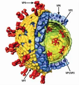
Structural studies using cryoelectron microscopy and computer image reconstruction have provided a description of the virion as an icosahedral particle of 75nm diameter and consisting of three concentric capsid protein layers (235, 300). The complete viral particle (= the infectious virion) is called triple-layered particle (TLP), a particle where the outer layer is missing is named double-layered particle (DLP) and a subviral particle containing only the innermost layer is designated as a single-layered particle or “core”. The genome consisting of 11 segments of double-strand RNA (dsRNA) is packaged within the inner layer, together with two proteins involved in transcription and genome replication: the RNA-dependent RNA polymerase (RdRp), VP1, and the capping enzyme, VP3. The inner layer is formed by 120 molecules of VP2, the middle layer by 260 trimers of VP6 and the outer layer by 780 molecules (260 trimers) of VP7 and 60 trimers of VP4 (Fig. 1) (91). The entire structure has a left handed T=13 icosahedral symmetry and is perforated by 132 aqueous channels of three types (I-III). The type I channels play an important role in viral transcription as through them the newly formed messenger RNA (mRNA) exits into the cytoplasm. A detailed description of the virion 3D structure and of the genome organization is provided in the following paragraphs (91).
2.2. SPATIAL ORGANIZATION OF THE TRIPLE-LAYERED PARTICLE
The outer layer of the TLP is formed by the major protein VP7, which is uniformly distributed giving rise to a smooth outer surface. The 780 molecules of VP7, organized as trimers, form a T=13l (l for levo) icosahedral lattice defining the different types of aqueous channels (235). Sixty VP4 projections are anchored near the type II channels surrounding each 5-fold vertex (232, 299). The VP4 spikes protrude for about 100-120Å and present a bi-lobed head at the distal end (232, 300). Recent structural studies suggest that the each spike is a VP4 trimer, in which two VP4 molecules are associated and form the visible spike and the third VP4 molecule is flopping and not visible by cryo-EM (see Fig. 5 in chapter 3) (81). VP4 interacts with both VP7 and the middle layer protein VP6 and through these interactions plays an important role in maintaining the precise geometric arrangements between the outer and the middle layer (299).
FIG. 1: cut-away view of the rotavirus TLP showing the outer layer (VP7 in yellow and VP4 in red), the middle layer (VP6 in blue) and the inner layer (VP2 in green) surrounding the enzymes VP1 and VP3 (in red), which are anchored to the inside of the VP2 layer at the five-fold axes (From Jayaram et al, 2004).
The intermediate layer is composed of 780 VP6 polypeptides organized in 260 trimers on a T=13 lattice, which produce the typical bristle-like structure of DLPs (235, 300) (Fig. 2). VP6 trimers contain conserved peptides for interactions with the inner layer protein VP2 on the inside and with VP7 and VP4 at the outside. While the lateral interactions between VP6 trimers are not sufficient to make a closed icosahedral structure, the interactions with VP2 drive the assembly of correctly sized rotavirus particles (48, 174). The arrangements of the VP6 trimers and their contacts with the VP7 trimers are such that the aqueous channels in the outer and in the middle layers lye in register; however, VP7 induces a slight displacement of the VP6 trimers flanking the 5-fold axis, which is responsible for the different diameter of the type I channels in the two layers (156, 162).
The inner layer is a thin, relatively smooth spherical structure formed by 120 molecules of VP2 assembling to 60 asimmetric dimers, which are arranged in a T=1 icosahedral simmetry (160, 234). Since VP2 is the only protein capable to form stable virus-like particles when expressed alone in insect cells or also with various combinations of VP6, VP4 and VP7 (62), it has been considered the rotavirus scaffolding protein (62, 153). Although VP2 forms a relatively smooth shell, a small portion extends further inward at the 5-fold axes to form a pentagonal structure (234) (Fig. 2). Small pores surrounding the 5-fold axes pass through the VP2 layer connecting the core environment with the outside (233, 234).
The aqueous channels are classified in three types based on their position on the icosahedral structure (235). All channels are about 55Å wide at the outer surface, with the exception of type I channels that have a narrower opening (about 40Å), constrict then further and widen again to reach the maximum width in proximity of the inner shell (235). Their depth through the two outer shells is about 140Å (235). Nascent mRNA transcripts exit the DLPs through type I channels, which run down the
icosahedral 5-fold axes, suggesting an important role for these channels in viral transcription (159).
Inside the core, positioned at each of the 12 pentameric edges of the VP2 lattice, there are complexes formed by one molecule of VP1 and one of VP3, which means that 12 copies of VP1 and VP3 are contained in each viral particle (234). Even if the exact position of the genome segments has not been determined yet, the dsRNA genome was visualized as an ordered dodecahedral structure, in which the dsRNA helices are packed around the VP1-VP3 complex (234) (Fig. 2).
FIG. 2: Structural organization of RNA inside rotavirus. a) cut-away view of the rotavirus DLP showing internal organization: VP6 is represented in shades of blue, VP2 in shades of green; the VP1-VP3 complex at the 5-fold axes is shown in red and portions of VP2 at the 5-fold in green. The dodecahedral shell of the ordered RNA, shown also separately in b), is represented in yellow. (From Prasad et al, 1996).
2.3. THE VIRAL GENOME
Rotavirus genome consists of eleven segments of dsRNA, each encoding one protein, with the exception of segment 11 of some strains that encodes two (NSP5 and NSP6). The RNA segments can be extracted from purified or semipurified virus and resolved by polyacrylamide gel electrophoresis (PAGE), showing a migration pattern of eleven different bands (Fig. 4). The nucleotide sequence of all segments is known for several rotavirus strains. All are between 660 and 3300 bp in size and share the following general features (91):
- lack of a poly(A) tail;
- 5’ cap structure m7GpppG(m)GPy on the positive-sense strand; - uncapped minus-strand;
- high content of A+U (58-67%);
- complete complementarity of both RNA strands;
- one single ORF (except for segment 11 of some strains).
On one side the different genome segments share sequence signals in order to be transcribed and replicated by the same RdRp, on the other they contain signals to be distinguished from one another during packaging. While assembly and encapsidation signals remain still unknown, some of the signals for transcription and replication have been identified in both untranslated regions (UTRs) (76). In particular, for the medically important group A rotaviruses the plus-strand RNAs start with the consensus sequence 5’-GGC(A/U)6-8-3’ and end with the consensus sequence
5’-UGUGACC-3’ (76) (Fig. 3). The conserved sequence at the 3’UTR has been identified as a signal promoting minus-strand RNA synthesis (215, 294). In particular, the 3’-terminal CC have been shown to be crucial for the formation of the initiation complex of RNA replication (51, 53). However, they are dispensable for specific
binding of the RdRp, which instead requires the four nucleotides 5’-UGUG-3’ of the 3’-terminal consensus sequence and also other signals positioned in non-conserved regions positioned upstream (280). Within the same conserved sequence, the last four nucleotides 5’-GACC-3’ function as translation enhancers (56). The conserved sequence within the 5’-UTR, in particular the second G, has also been shown to play a role in the attachment of the RdRp and cofactors into a stable initiation complex for minus-strand RNA synthesis (281) (Fig. 3). Based on computer modelling, the 5’ and 3’ UTRs are predicted to stably anneal to form a panhandle structure, from which the 3’-terminal conserved sequence extends as un-base-paired tail (54, 206) and this structure has been proposed to be stabilized by the RdRp (281).
Rotavirus genomes (better investigated for group A rotaviruses) show an extensive diversity, which is essentially due to the following mechanisms (135):
FIG. 3: Schematic representation of a group A rotavirus plus-strand RNA. The conserved sequences at the 5’ and 3‘ ends are indicated. Both sequences were shown to be essential for the formation of the minus-strand initiation complex. They are predicted to stably base-pair forming a panhandle structure. The dinucleotide GG indicated in purple is conserved within all groups of rotaviruses and the second G was shown to be essential for specific recognition by the polymerase VP1. Another recognition signal for VP1 is at the 3’UTR. Both signals are underlined. The sequence indicated in green is a translation enhancer.
- accumulation, sometimes fixation, of point mutations (genomic drift);
- genome segment reassortment (genomic shift): a dual infection with two co-circulating human strains or a human and an animal strain leads to a viral progeny containing novel assortments of genome segments;
- gene rearrangements (75): considerable tracts of sequence within a single genome segment may be altered by deletions or duplications. Most gene rearrangements involve segment 11 and consist in a partial head-to-tail duplication of the dsRNA sequence (113).
2.4. GENE-PROTEIN ASSIGNMENT
The genome segments code for six structural proteins found in virus particles (VP1, VP2, VP3, VP4, VP6, VP7) and five or six nonstructural proteins (NSPs) found in infected cells but not in mature virus particles (NSP1-NSP6; as mentioned above, some strains have only a single ORF in segment 11 and lack NSP6) (91). Some NSPs are found in subcellular locations, some others in cytoplasmic viral inclusion bodies called viroplasms and considered sites of viral genome replication and packaging (91).
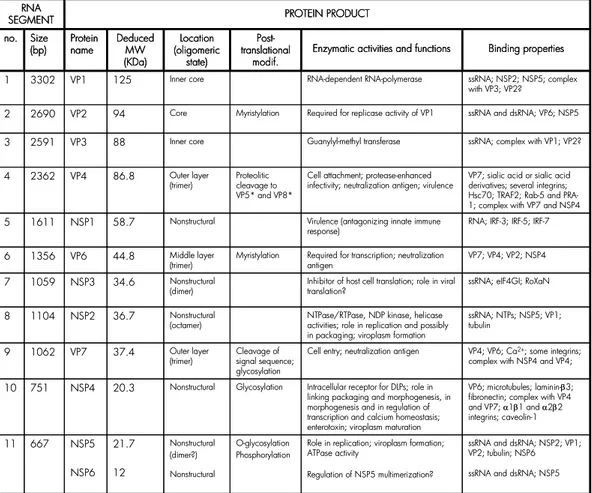
Comparative studies of the electrophoretic pattern of genome segments deriving from different strains have shown that the relative migration order of the eleven genes can differ (91). Therefore, the gene-protein assignment is specific for each strain (Fig. 4). Table 1 lists the RNA segments and their protein products for the simian rotavirus SA11 strain. All functions reported in literature for rotavirus proteins are also summarized. A more detailed description of their properties and functions is provided in the following chapter.
PROTEIN PRODUCT RNA
SEGMENT
Binding properties Enzymatic activities and functions
Post-translational modif. Location (oligomeric state) Deduced MW (KDa) Protein name Size (bp) no.
ssRNA and dsRNA; NSP2; VP1; VP2; tubulin; NSP6 ssRNA and dsRNA; NSP5 Role in replication; viroplasm formation;
ATPase activity Regulation of NSP5 multimerization? O-glycosylation Phosphorylation Nonstructural (dimer?) Nonstructural 21.7 12 NSP5 NSP6 667 11 VP6; microtubules; laminin-β3; fibronectin; complex with VP4 and VP7; α1β1 and α2β2 integrins; caveolin-1 Intracellular receptor for DLPs; role in
linking packaging and morphogenesis, in morphogenesis and in regulation of transcription and calcium homeostasis; enterotoxin; viroplasm maturation Glycosylation Nonstructural 20.3 NSP4 751 10 VP4; VP6; Ca2+; some integrins; complex with NSP4 and VP4; Cell entry; neutralization antigen
Cleavage of signal sequence; glycosylation Outer layer (trimer) 37.4 VP7 1062 9 ssRNA; NTPs; NSP5; VP1; tubulin
NTPase/RTPase, NDP kinase, helicase activities; role in replication and possibly in packaging; viroplasm formation Nonstructural (octamer) 36.7 NSP2 1104 8
ssRNA; eIF4GI; RoXaN Inhibitor of host cell translation; role in viral
translation? Nonstructural (dimer) 34.6 NSP3 1059 7 VP7; VP4; VP2; NSP4 Required for transcription; neutralization
antigen Myristylation Middle layer (trimer) 44.8 VP6 1356 6
RNA; IRF-3; IRF-5; IRF-7 Virulence (antagonizing innate immune
response) Nonstructural 58.7 NSP1 1611 5
VP7; sialic acid or sialic acid derivatives; several integrins; Hsc70; TRAF2; Rab-5 and PRA-1; complex with VP7 and NSP4 Cell attachment; protease-enhanced
infectivity; neutralization antigen; virulence Proteolitic cleavage to VP5* and VP8* Outer layer (trimer) 86.8 VP4 2362 4
ssRNA; complex with VP1; VP2? Guanylyl-methyl transferase Inner core 88 VP3 2591 3
ssRNA and dsRNA; VP6; NSP5 Required for replicase activity of VP1
Myristylation Core 94 VP2 2690 2 ssRNA; NSP2; NSP5; complex with VP3; VP2? RNA-dependent RNA-polymerase Inner core 125 VP1 3302 1 PROTEIN PRODUCT RNA SEGMENT Binding properties Enzymatic activities and functions
Post-translational modif. Location (oligomeric state) Deduced MW (KDa) Protein name Size (bp) no.
ssRNA and dsRNA; NSP2; VP1; VP2; tubulin; NSP6 ssRNA and dsRNA; NSP5 Role in replication; viroplasm formation;
ATPase activity Regulation of NSP5 multimerization? O-glycosylation Phosphorylation Nonstructural (dimer?) Nonstructural 21.7 12 NSP5 NSP6 667 11 VP6; microtubules; laminin-β3; fibronectin; complex with VP4 and VP7; α1β1 and α2β2 integrins; caveolin-1 Intracellular receptor for DLPs; role in
linking packaging and morphogenesis, in morphogenesis and in regulation of transcription and calcium homeostasis; enterotoxin; viroplasm maturation Glycosylation Nonstructural 20.3 NSP4 751 10 VP4; VP6; Ca2+; some integrins; complex with NSP4 and VP4; Cell entry; neutralization antigen
Cleavage of signal sequence; glycosylation Outer layer (trimer) 37.4 VP7 1062 9 ssRNA; NTPs; NSP5; VP1; tubulin
NTPase/RTPase, NDP kinase, helicase activities; role in replication and possibly in packaging; viroplasm formation Nonstructural (octamer) 36.7 NSP2 1104 8
ssRNA; eIF4GI; RoXaN Inhibitor of host cell translation; role in viral
translation? Nonstructural (dimer) 34.6 NSP3 1059 7 VP7; VP4; VP2; NSP4 Required for transcription; neutralization
antigen Myristylation Middle layer (trimer) 44.8 VP6 1356 6
RNA; IRF-3; IRF-5; IRF-7 Virulence (antagonizing innate immune
response) Nonstructural 58.7 NSP1 1611 5
VP7; sialic acid or sialic acid derivatives; several integrins; Hsc70; TRAF2; Rab-5 and PRA-1; complex with VP7 and NSP4 Cell attachment; protease-enhanced
infectivity; neutralization antigen; virulence Proteolitic cleavage to VP5* and VP8* Outer layer (trimer) 86.8 VP4 2362 4
ssRNA; complex with VP1; VP2? Guanylyl-methyl transferase Inner core 88 VP3 2591 3
ssRNA and dsRNA; VP6; NSP5 Required for replicase activity of VP1
Myristylation Core 94 VP2 2690 2 ssRNA; NSP2; NSP5; complex with VP3; VP2? RNA-dependent RNA-polymerase Inner core 125 VP1 3302 1
FIG. 4: PAGE gel showing the 11 dsRNA genome segments of the rotavirus SA11 strain. The genome segments are numbered on the left and the encoded proteins are indicated on the right. Segment 11 of this strain has also an alternative ORF encoding the nonstructural protein NSP6.
3. ROTAVIRUS PROTEINS
VP1
VP1 is the viral RNA-dependent RNA polymerase (RdRp) acting as both, the transcriptase (for mRNA synthesis) and the replicase (for minus-strand RNA synthesis). Several lines of evidence indicate that VP1 is the viral RdRp: (i) VP1 contains sequence motifs that are shared by RdRps of other RNA viruses (188); (ii) VP1 has NTP-binding activity and, when cross-linked with the nucleotide analog 8-azido-ATP, inhibits RNA transcription (284); (iii) VP1 specifically recognizes multiple functionally distinct elements at the 3’ end of viral plus-strand RNAs (53, 207, 280); (iv) recombinant VP1 can direct template-dependent minus-strand synthesis in vitro in the presence of VP2 (212, 308). To date, regions of VP1 responsible for specific recognition of the templates and the enzymatic activity have not been identified and this is in part due to the lack of a solved 3D structure. Based on a bioinformatic programme comparing shared motifs of different viral RdRps, an overall structural conservation has been identified in the middle of the sequence, whereas the N- and C-termini have been predicted as unique regions (288), which could be related to unique functions of VP1 or to interactions with specific rotavirus proteins. Interactions of VP1 with NSP2 (145) and NSP5 (2, 8) have been described by coimmunoprecipitation experiments from extracts of both infected and transfected cells. Moreover, VP1 may interact with VP2 because: (i) they form virus-like particles (VLPs) when coexpressed in insect cells (307, 308); (ii) purified recombinant VP1 and VP2 can drive synthesis of dsRNA in template-dependent assays only when both are present (212, 280); (iii) far-Western blot analysis showed a binding between VP1 and VP2 involving the N-terminus of VP2 (aa 1 to 25) (307).
VP2
VP2 is the major scaffolding component of rotavirus core: 120 molecules of VP2 form the innermost capsid layer of rotavirus enclosing the genomic dsRNA (153). Since VP2 is the only protein capable to form stable VLPs when expressed alone in insect cells or also in various combinations with VP6, VP4 and VP7, it has been considered the rotavirus scaffolding protein (62, 153). However, VP2 shell has both a structural and a functional role: interactions with trimers of VP6 (forming the intermediate particle layer) are responsible both for the stability of DLPs and for mRNA synthesis and transcript extrusion (48, 162); VP2 binds to genomic dsRNA, as demonstrated by UV cross-linking experiments (152), and its higher affinity for ssRNA than for ds nucleic acids (35) suggests a possible role in the encapsidation of ssRNAs used as templates for dsRNA synthesis; in fact, VP2 is required by VP1 in in vitro replication assays to achieve the replicase activity directing minus-strand synthesis (212, 280). The N-terminus of VP2 is responsible for both its nonspecific RNA binding activity (35, 152) and the proper assembly of VP1 and VP3 into the core (307) forming a complex located on the inner surface of the VP2 layer at the icosahedral fivefold axes (234). More in detail, deletion of the first 26 amino acids completely abolished the RNA binding activity, indicating that either the first 26 amino acids contain the binding site(s) or they allow VP2 to fold in such a way that RNA binding can occur through several other RNA binding sequences identified in the first 132 amino acids (152). Baculovirus recombinant-mediated co-expression of N-terminally truncated forms of VP2 and VP1 and/or VP3 showed that the N-terminal 92 aminoacids are essential for incorporation of VP1 and VP3 in virus-like particles (VLPs), although they are not for VP2-VLP formation (307). Moreover, far-Western blot analyses using a series of truncated VP2 forms showed that full-length VP2 binds to VP1, any
N-terminal truncation lacking amino acids 1 to 25 fails to bind VP1 and a C-N-terminal 296-aa truncation maintains the ability to bind VP1 (307).
By cryoelectron microscopy and image reconstruction of VLPs formed either by full-length VP2 or by the truncation lacking the N-terminal 92 amino acids, it has been proposed that the 120 molecules of VP2 are arranged as dimers, each extending between neighbouring fivefold axes, and that the N-termini are located near the icosahedral vertices (159). Interestingly, previous cryoelectron microscopy experiments on native DLPs had shown significant interactions between the inner surface of VP2 layer and the genomic dsRNA near the icosahedral fivefold axes and minor interactions along the icosahedral twofold axes (234), supporting the idea of VP2 having an important role in organizing the genomic dsRNA within the core. While the N-terminal region of VP2 seems to be essential for the spatial architecture of the core, it does not seem to be involved in interactions with VP6 and with the nonstructural protein NSP5. It has been reported that both full-length VP2 and a VP2 deletion mutant lacking the N-terminal 92 amino acids can form VLPs incorporating VP6 (49) and both coimmunoprecipitate with NSP5 and form aggregate structures in insect cells with NSP5 and VP6, and with either of them (23). Moreover, it has been shown that an insect cell lysate containing recombinant baculovirus-derived NSP5 dislodges VP6 from purified VLPs formed by VP2 and VP6, but not from purified DLPs (23). Since the differences between the two types of purified particles are the presence of genome segments and the stronger interaction between VP2 and VP6 in DLPs, it has been proposed that the interaction between the VP2 core and NSP5 occurs at early times to prevent the VP6 binding and thus the formation of defective particles until the set of genomic RNA segments inside core is complete (23).
VP3
VP3 is a basic protein responsible for capping rotavirus mRNAs through its guanylyl-transferase and methyl-guanylyl-transferase activities (52, 164, 225). It is one of the minor components of the core (163), a component of early replication intermediates and, together with VP1, seems to be the first protein to associate with viral (+)ssRNAs during packaging and RNA replication (104), possibly through its unspecific affinity for ssRNA (209). Together with VP1 and ssRNA, VP3 interacts with the N-terminus of VP2 (307) and this association might have a role in RNA replication. In fact, it has been shown that the presence of a functional VP3 is important for RNA replication (286) and that in in vitro replication assays VLPs containing VP1, VP2 and VP3 synthesize minus-strand RNAs more efficiently than VLPs formed only by VP1 and VP2 (308). The exact role of VP3 in this context is unclear. It has been proposed that VP3 increases the rate at which the components of the replicase complex assemble into functional structures (212).
VP4
VP4 is one of the two proteins of the outer layer of rotavirus. It is a non-glycosylated protein with essential roles in virus attachment and penetration. Antibodies against VP4 can neutralize by blocking cell entry and protect against rotavirus gastroenteritis (201, 244). In virions uncleaved with trypsin, molecules of VP4 are distributed on the surface of the VP7 shell as flexible stalks occupying each of the 60 symmetry-equivalent positions. In the gut lumen trypsin cleavage of VP4 maximises rotavirus infectivity (94, 98): this cleavage generates two fragments, VP5* (60KDa) and VP8* (28KDa), which remain associated with virions and confer the spike appearance of VP4 observed by cryoelectron microscopy in trypsin-cleaved virions (260, 299). VP8* forms the “heads” of the spikes and binds cellular receptors in the virus attachment phase (82); VP5*, together with the N-terminus of VP8*, forms the spike
“body”, which is linked by an asymmetric “stalk” to a “foot” buried beneath the VP7 shell (81) and interacting with VP6 (299). The VP5* residues in the spike body are involved in membrane penetration and integrin binding (83, 120, 171). It appears that each spike is indeed a VP4 trimer (81) and that the rearrangement induced by trypsin cleavage allows a switch from a flexible structure into rigid spikes (64, 81), where two primed VP4 molecules are associated and present the VP8* core for receptor binding and the third primed VP4 molecule is flopping and not visible by cryo-EM (81) (Fig. 5A). It has been proposed that a second rearrangement event occurs during cell entry, in which two VP5* subunits fold back on themselves and join a third VP5* subunit to form a tightly associated trimer, shaped like a folded umbrella, from which VP8* dissociates (81) (Fig. 5B). This second rearrangement is thought to occur to unmask the membrane interaction region of VP5* allowing cell penetration (81). To support this VP4 rearrangement model, there is the evidence that a globular domain of VP5* containing the potential membrane interaction region has the intrinsic molecular property of alternatively forming dimers and trimers (301). As a further confirmation, it has been observed that at elevated pH the VP4 spike undergoes an irreversible conformational change from a bilobed structure to a distinctly stunted trilobed structure (217).
VP8* contains a sialoside-binding region within a shallow groove on its surface, which is involved in sialic acid binding and hemagglutination in sialidase-sensitive rotavirus strains (82, 98). However, the capacity of VP8* of binding sialic acid derivatives has also been recognized for some sialidase-insensitive strains (28), and new carbohydrate binding regions in VP8* are being discovered in both types of strains (28, 82, 127). Interestingly, the VP8* sialic acid binding domain has a galectin fold, which might be responsible for features shared by VP4 and galectins, like the translocation to the plasma membrane via a Golgi-independent pathway or the capacity of activating the TRAF2-NF-kB-inducing kinase signalling pathway (155). The binding of VP8* to TRAF2, a member of a family of adapter proteins involved in transducing signals generated by ligands of Tumor Necrosis factor (TNF), and the consequent activation of NF-kB, which in turn causes the secretion of selected chemokines, suggests a role for VP4 in determining cell-specific responses to rotavirus infection (155).
FIG. 5: Models of two VP4 conformations. (A) The primed state. Two rigid subunits form the spike visible in electron cryomicroscopy image reconstructions of trypsin-primed virions. A third subunit is flexible. VP8* is represented in gray, VP5* in green bean-shape, with a red membrane interaction region. (B) The putative post-membrane penetration state. VP8* has dissociated and the VP5* antigen domain has folded back. From Yoder et al, 2006.
VP5* is involved in cell attachment (304, 305) and is considered the main part of the molecule responsible for cell entry through a hydrophobic fusion domain that was shown to be able to permeabilize membranes in the absence of other rotavirus proteins (72). Since it was found that VP5* cannot permeabilize lyposomes or bacteria, it has been proposed that it has a selective permeabilizing activity, possibly by forming transient pores that could allow the decrease of calcium levels in the proximity of the plasma membrane or in endosomes (83, 107). VP5*, together with VP7, is also responsible for integrin binding, which is important not only for cell attachment and entry (involving mainly α2 integrins), but also for facilitating virus spread or host immune response modulation (involving mainly α4 integrins) (119, 129, 304): through its peptide sequence DGE, VP5* interacts with α2β1 integrin (120, 121, 128) and it is likely to be involved in binding to α4β1 and α4β7 integrins through its peptide sequence YGL (119). Furthermore, VP5* has been shown to bind Hsc70 (303) in a post-attachment step important for cell entry (124). The role of VP4 in the morphogenesis of rotavirus has been studied by experiments of RNA interference silencing VP4 expression (65, 67). The formation of spike-less TLPs in rotavirus-infected cells transfected with an siRNA against VP4 suggested that VP4 is not required for virus assembly (67). Since these spike-less TLPs have larger diameter than wild-type TLPs, it has been proposed that VP4 tightens the structure of the viral particle (67). Interestingly, VP4 silencing reduces the association of rotavirus particles with rafts and causes an accumulation of non-enveloped particles in the endoplasmic reticulum (ER) (65), suggesting a role for VP4 in trafficking newly synthesized particles. Many reports indicate that VP4 is indeed associated with rafts (68, 252), which are dynamic microdomains in cellular membranes enriched in cholesterol and sphingolipids and exert critical roles in membrane trafficking. However, the role of VP4 in virion assembly and release remains still unclear. During rotavirus infection the
newly synthesized VP4 seems to be localized both in the cytosol and in the plasma membrane, starting early after infection. More precisely, it has been detected in an area between viroplasms and ER (220), colocalizing in the ER proximity with NSP4 (108), with which it might interact (10), also because of the recognized ability to form heterotrimers with NSP4 and VP7 (170). It has also been identified in the ER-Golgi Intermediate Compartment (ERGIC) (65), in transiently enveloped particles (229) and in the plasma membrane in lipid rafts, associated with microtubules and exposed on the external face of the apical plasma membrane (69, 196, 252). The data from RNA interference experiments, the VP4 cellular distribution, the described interactions [with NSP4/VP7 and with the ER chaperone grp78 (296)], and the fact that VP4 needs to be added before VP7 to reconstitute in vitro infectious TLPs from non-infectious DLPs (283), support the idea that VP4 assembly into particles occurs inside or in the vicinity of the ER and not at a later time point when VP7-coated particles are already formed. However, since rafts are considered to be absent in the ER and the block of enveloped particles in the ER induced with chemical reagents does not correlate with a reduced cell surface targeting of VP4, it has been proposed that VP4 assembly in viral particles occurs as a post-ER event (69). Thus, two pools of VP4 might exist, one associating with viral particles in the ER and the other independently targeted to the plasma membrane (65). Further experiments are needed to prove this assumption. Finally, an involvement in localization and trafficking of the free cytosolic VP4 in infected cells has been suggested for two cellular proteins, Rab-5 and PRA1, both participating in vesicular trafficking and able to form complexes with free VP4 at late steps of infection (88).
VP6
VP6 is the major capsid protein of rotavirus. It forms 260 trimers that constitute the intermediate layer (91, 114, 250). The structure of the VP6 trimer consists of an
elongated, tower-like molecule, where the three subunits wrap around a central 3-fold axis with a right-handed twist. The VP6 monomer consists of two domains (H and B) positioned along the long dimension of the tower-like molecule and both involved in intra-trimer interactions. Each trimer contains conserved surfaces for interactions with other structural proteins, at the bottom for binding to VP2, at the top for contacting VP7, at the sides of domain H for binding to VP4 and at the sides of domain B for lateral interactions with other VP6 trimers. These inter-trimer interactions do not contain all the information to form a closed icosahedral shell, since interactions with the VP2 inner layer are also required for a correct assembly. A model for assembly has been proposed, in which the center of each of the twenty faces of the VP2 layer acts as a nucleation point for the growth of the middle layer and the driving force for assembly propagation appears to be the inter-trimer interactions rather than interactions with the VP2 layer (1, 174). A Zn2+ ion is located at the center of the VP6 trimer coordinated
by three histidines, one for each VP6 monomer (174). Mutation of the Zn2+
coordination site renders the trimer more sensitive to proteases, but has no effect on the interaction with VP2 or on the DLP transcription activity (89). When coexpressed in insect or mammalian cells, VP2 and VP6 can in fact form double-layered virus-like particles (Dl-VLPs) (62, 110). However, while recombinant VP2 is able to form core-like particles in insect cells by itself (153), VP6 forms only tubular or spherical structures in the absence of other viral proteins (161, 240). The spherical structures are larger than Dl-VLPs, highlighting the importance of VP2 in the assembly of viral particles (161). The main parameter affecting interactions between VP6 molecules, and consequently the formation of spheres or tubes, is pH: at pH 3.5-5.5 these interactions generate spherical particles, at pH 5.5-7.0 large tubes and at pH above 7.0 small tubes (161). These observations suggested that the protonation state of VP6 is critical for assembly, probably affecting lateral inter-trimer contacts (161).
By connecting the outer layer and the inner layer, VP6 has a fundamental role in the structure of the virion as a physical adapter between the biological functions exerted by the two linked layers: cell entry and genome packaging (233). Moreover, VP6 seems to play an active role in transcription, because in its absence the process does not take place (24). In particular, it has been shown that: (i) hydrophobic interactions at the interface with VP2 are responsible for the DLP stability and subtle electrostatic interactions in the same area influence transcription and transcript extrusion (48); (ii) structural changes induced by anti-VP6 antibodies (97, 156) or the geometrical orientation of the trimers induced by interaction with VP7 (162) inhibit transcriptional activity. VP6 might also have a role in the budding of newly formed DLPs in the ER: in fact, during viral infection VP6 is localized at the periphery of viroplasms and in close proximity of the ER (220) and is thought to interact with the cytoplasmic tail of NSP4 (180, 275, 277), which is the viral ER-transmembrane protein mediating the budding of viral particles into the ER (9, 10, 19). Interestingly, NSP4 silencing experiments showed a drastic change of the VP6 distribution in infected cells: instead of being localized in viroplasms, in the absence of NSP4 VP6 forms filaments not colocalizing with tubulin, actin or vimentin (165). Furthermore, the same phenotype was observed when the expression of another nonstructural protein was silenced: NSP5 (166). However, at present there is no evidence of an interaction between VP6 and NSP5. On the contrary, it has been shown that NSP5 does not bind DLPs and that VP6 hinders the interaction between NSP5 and VP2 (23).
VP7
VP7 is a protein organized in 260 trimers forming the outer layer of rotavirus (300), in which VP4 molecules are inserted. In most rotavirus strains, but not in all of them, VP7 is N-glycosylated. It has a high content of conserved cysteines that form disulfide bonds important for the correct folding of the protein and the maturation of viral
particles (185). During the replicative cycle the newly synthesised VP7 is inserted in the ER membrane with a luminal orientation (143). The VP7 targeting to the ER is due to two hydrophobic regions located at the N-terminus, which are cleaved off after insertion of VP7 in the ER membrane, but are still necessary for the retention of VP7 in the ER (169, 231, 266, 295). The C-terminus is thought to be protruding into the cytoplasm and have important roles for the assembly of mature particles (60).
The conformation and stability of VP7 are calcium-dependent (79); in particular, the trimerization of VP7 requires calcium (80). In fact, many lines of evidence indicate that both the loss of the outer layer during cell entry and the formation of the same layer during virus assembly are mediated by calcium-dependent conformational changes of VP7: in vitro calcium chelation triggers TLPs uncoating (61); the outer layer contains calcium (258); low concentrations of calcium induce a VP7 solubilization (103, 247) shown to be necessary for the permeabilizing activity of the protein (47); in the absence of calcium, VP7 cannot interact with VP4 and NSP4 in the heterotrimeric complex formed during virus assembly (170, 230).
VP7 has an important role during cell entry, not only because of its membrane permeabilizing activity (47), but also because of its binding to αvβ3 (306) and αxβ2 integrins (120). VP7 also has a regulatory role in transcription, because it appears to induce a structural change of VP6 (156) and a reorganization of the VP6 trimer around the particle 5-fold axes (162) that inhibit the DLP transcriptional activity; moreover, it causes a steric hindrance that blocks the exit of nascent mRNAs through the type I channels (159). VP7 silencing experiments also revealed an important role of VP7 in removing the transient envelope during virus morphogenesis (165) and a contribution of VP7 to the NSP4 function of regulating calcium homeostasis during infection (302).
NSP1
NSP1 is an RNA-binding nonstructural protein that accumulates in the cytoplasm of infected cells in association with the cytoskeleton (132). It is the least conserved rotavirus protein; only the N-terminal zinc binding motif is completely conserved (189). This cystein-rich region has been shown to be essential for binding viral RNAs at the 5’UTR (37, 131) and it is important, but not sufficient, for allowing NSP1 binding to the cellular transcription factor IRF3 (Interferon Regulatory Factor 3) (117). This cellular protein is activated following virus infection and promotes the expression of IFNβ, which is secreted and induces the neighbouring uninfected cells to express another transcription factor, IRF7 (253). IRF7 is the main molecule responsible for the production of type I IFN (IFNα and β) in the anti-viral host response. NSP1 has been shown to target IRF3 to the proteasome (14), and it has been proposed to act as a E3 ubiquitin protein ligase based on the fact that the arrangement of the cysteine residues of the zinc binding motif resembles the RING fingers of the E3 ligase (116). However, NSP1 of some rotavirus strains does not have IRF3 as a target, despite having an intact zinc-binding domain (116). Furthermore, it has recently been shown that NSP1 induces the proteasome degradation of IRF7 (15). This is probably related with the ability of the virus to replicate in specialized trafficking cells (macrophages and dendritic cells) constitutively expressing IRF7 (11) and IRF5, which is another factor that upregulates IFNI expression and is involved in triggering apoptosis during viral infection (15). Therefore, NSP1 may be considered as a broad-spectrum antagonist of the innate immune response that limits the virus spread (15). Since truncated forms of NSP1 lacking portions at the C-terminus lose their ability of interacting with these IRF factors, the C-terminus seems to play an important role in this context and its sequence variability in strains infecting different animal species suggests that NSP1 may confer species specificity to different rotaviruses (15). NSP1
has thus an important role in virus spread, confirmed also by reassortment experiments that showed a strong correlation between the genome segment encoding NSP1 and virus virulence and spread (36) and by the demonstration that truncations of NSP1 are responsible for the formation of smaller plaques following rotavirus infection (214). However, NSP1 does not have a role in virus replication, since virus mutants encoding C-truncated forms of NSP1 replicate efficiently (214) and NSP1 silencing by RNA interference does not affect virus replication (261).
Interestingly, it has been shown that the stability of NSP1 is maintained by the other viral proteins, alone or in combination with viral RNA, since in their absence the proteasome-mediated degradation of NSP1 is significantly increased (222).
NSP2
NSP2 is a basic, conserved protein with an important role in virus replication. This role has been demonstrated both by analysis of cells infected with a temperature-sensitive (ts) mutant of SA11 rotavirus strain, tsE(1400), with a lesion in the genome segment encoding NSP2 (239) and by RNA interference experiments in which NSP2 expression was silenced (261). Both the growth of cells infected with tsE(1400) at a non-permissive temperature and the NSP2 silencing revealed a critical function of NSP2 in viroplasm formation, in genome replication and packaging and in the production of infectious viral progeny (239, 261). The requirement of NSP2 in viroplasm formation is also confirmed by the fact that NSP2 forms viroplasm-like structures (VLS) when transiently coexpressed with NSP5 in the absence of other viral proteins (96) (see Fig. 9 in chapter 4). Moreover, NSP2 has been found in early replication complexes (104, 211) and the immunoprecipitation with a monoclonal antibody against NSP2 of an extract of infected cells allowed to recover a viral complex with replicase activity (5).
The functional form of NSP2 is a doughnut-shaped octamer derived from self-assembly of NSP2 monomers and from tail-to-tail interactions of two tetramers (138, 139, 255, 273) (Fig. 6).
Each monomer has two distinct domains separated by an electropositive deep cleft that contains a histidine triad (HIT)-like motif (138) responsible for binding and hydrolysis of NTPs (46). The catalytic residue of this motif is a His in position 225, which becomes phosphorylated through the covalent attachment of the γP released after a Mg2+-dependent hydrolysis reaction of the linkage between the γ and β
phosphates (46, 151, 287). To catalyze this reaction, NSP2 can use both NTPs (270) and pppRNA (287) as substrates, thus exerting both NTPase and RTPase activities. Moreover, the NTPase activity has been shown to be associated with a phosphoryl-transfer function to NDPs similar to that of cellular NDP kinases (151). While the enzymatic activities of NSP2 can be attributed to the monomeric subunits,
FIG. 6: NSP2 Structure. Structure of the NSP2 monomer (left), showing a deep cleft (arrow), which may be the site for NTP binding. Structure of the functional NSP2 octamer showing a view down the four-fold (middle) and a view down one of the two-fold axis in octamer (right). The deep grooves shown (right, arrows) are lined by basic residues and may be the sites for binding viral mRNA during genome replication and packaging. From Jayaram et al, 2004.
its binding properties require the formation of the octamer. More in detail, it has been shown that NSP2 has a sequence-independent ssRNA binding (270) and a nucleic acid helix destabilizing activities (272) and it is able to bind NSP5 (2, 85, 139) and VP1 (8, 145); the 35Å central hole of the octamer, lined by neutral residues, was shown to be not involved in RNA and NSP5 binding, for which instead the four deep positively-charged grooves extending diagonally across the octamer are required (139). Interestingly, the binding sites of NSP2 with RNA and with a NSP5 mutant lacking the first 65 amino acids and the last 10 amino acids correspond, suggesting a competition between NSP5 and RNA for interaction with NSP2 (139). Furthermore, coimmunoprecipitation of NSP2 with NSP5 using anti-NSP5 antibodies was shown to require UV treatment, and to occur even after RNase digestion of the cellular extracts (2). Based on these findings, it has been suggested that NSP2-NSP5 interaction is not directly mediated by RNA and may be the consequence of a conformational change in NSP2 bound to RNA and/or nucleotides stabilised by the UV treatment (2).
The ssRNA binding and helix-destabilizing activities of NSP2 have been suggested to relax positive-strand RNA templates to facilitate genome replication (272) and the NSP5 binding seems to be important for viroplasm formation (96) and might regulate the NSP2-RNA interaction during genome replication (139). On the other hand, the roles of the enzymatic activities remain completely obscure. In vivo complementation studies have shown that the HIT-dependent enzymatic activities are necessary for dsRNA synthesis, but not for viroplasm formation, and it has been hypothesized that these activities are used only after establishment of cellular sites of replication and assembly (271). This could have something to do with the fact that the NTP binding causes a conformational change from a more relaxed to a more compact form of NSP2 (255) and also the RNA binding induces noticeable conformational changes,
whilst NSP5 binding does not (139). Moreover, it has been shown that a ssRNA molecule with a 5’ γ-phosphate is preferred to NTPs as a substrate (287), suggesting that the hydrolytic activity of NSP2 may regulate the ssRNA binding activity or vice
versa. Interestingly, the NSP5 binding activity does not interfere with the enzymatic
activity of the HIT-like motif (287) and the catalytically inactive NSP2 mutant H225A supports VLS formation (46). Since NSP2 was found in early replication complexes, it has been proposed that the RTPase activity removes the γ-P from nascent negative-strand RNAs, but not from positive-negative-strand RNAs because of the capping modification interfering with the RTPase function (287). However, dsRNAs are not substrates for the RTPase activity of NSP2 either and the role of this activity remains still obscure. For the NDP kinase activity, a role in the homeostasis of nucleotide pools has been suggested: NSP2 would guarantee sufficient levels of NTPs for transcription and replication and of ATP for processes requiring energy like transcription, NSP5 phosphorylation, RNA packaging and translocation (151), and the recently reported ATP-ase activity of NSP5 (13). Besides NDPs, also NSP5 had been proposed as a substrate for the phosphotransfer activity of NSP2 (290), however it has then been shown that the catalytic activity of NSP2 is not involved in the phosphorylation of NSP5, since NSP2 mutants that are NTP-ase defective still promote NSP5 hyperphosphorylation (46). Interestingly, and surprisingly, in vitro replication assays with recombinant VP1 and VP2 showed that NSP2 inhibits the initiation phase, but not the elongation phase, of the synthesis of dsRNA through its nonspecific RNA binding activity (291). More precisely, NSP2 does not block the recognition of the template by the polymerase, instead it interferes with the function of VP2 through an unknown mechanism, possibly by competing for binding to ssRNA (291).
NSP3
NSP3 is a slightly acidic protein, whose role in the viral replicative cycle is still not completely clarified. During virus infection, NSP3 is distributed diffusely throughout the cytoplasm, possibly associated with the cytoskeleton (177). NSP3 is composed of two functional domains separated by a dimerization domain (223). The N-terminal domain is required for sequence-specific RNA binding and the dimerization is necessary for the strong binding of a single molecule of RNA (74, 223). The sequence GACC within the consensus sequence at the 3’UTR of all viral mRNAs has been demonstrated as the shortest target recognized by NSP3 (227), and the binding of NSP3 to this sequence protects the 3’ end from RNase digestion (226). Interestingly, NSP3 can recognize only mRNAs derived from the same serogroup, which could be one of the reasons why genomes of rotaviruses of different serogroups cannot reassort (227). The central domain of NSP3 is responsible for dimerization, forming a coil-coiled structure that confers to NSP3 dimer a heart shape (74, 223). By yeast two-hybrid assays, this domain has also been shown to be involved in interaction with RoXaN (Rotavirus X protein associated with NSP3), which is represented by two related proteins in mammalian cells (RoXaNI and RoXaNII) that might be important for translation regulation (292). In fact, RoXaNI can form a ternary complex with NSP3 and the other cellular ligand of NSP3: eIF4GI (eukaryotic Initiation Factor 4GI) (292). The NSP3 C-terminal domain is indeed involved in interaction with this cellular factor (223, 224), which is a scaffolding protein that binds the cap binding protein eIF4E, the RNA helicase eIF4A and the poly(A)-binding protein (PABP), in order to promote an efficient initiation of translation (134, 237, 274). NSP3 binds to eIF4GI in the same region recognized by PABP (224): it has been proposed that by bridging the cap-binding protein eIF4E via its binding to eIF4GI and the 3’ end of viral mRNAs via its direct binding to the consensus
sequence, NSP3 promotes the circularization of viral mRNAs and their translation, while evicting PABP from eIF4GI impairs the translation of cellular mRNAs (224, 289). While there is a general agreement on the inhibitory activity of NSP3 on the synthesis of cellular proteins (191, 202, 224, 289), supported also by the recent observation that NSP3 causes a translocation of PABP to the nucleus (192), the role of promoting synthesis of viral proteins is now brought into question: in fact, RNA interference experiments that silenced NSP3 expression showed that NSP3 is not required for viral protein synthesis and, even more, its silencing correlates with an increased synthesis of viral RNA and with a concomitant increase in the yield of viral progeny (191). Furthermore, silencing the eIF4GI expression confirmed that its interaction with NSP3 is not required for viral protein synthesis (191). It has then been proposed that the interactions with eIF4GI and with the 3’ end of viral RNAs have two distinct functions: the first would help sequestering eukaryotic translation initiation complexes, thus inhibiting cellular protein synthesis, the second would prevent viral plus-strand RNAs from being selected for replication, thus ensuring a pool of transcripts available for translation, or would protect viral mRNAs from degradation (191).
NSP4
NSP4 is a multifunctional nonstructural glycoprotein (143) with key roles in virus transcription, morphogenesis and pathogenesis. It is the only nonstructural protein that does not bind to RNA. It is a 175 amino acid long, transmembrane protein targeted to the ER membrane by a signal sequence, which remains uncleaved. After insertion in the ER, most of the protein is exposed in the cytosol. NSP4 contains three predicted hydrophobic domains, H1, H2 and H3. H1 is a small domain, which is N-glycosylated in two sites and located in the ER lumen; H2 is a transmembrane domain and was initially thought to correspond to the signal sequence (19), until a
region in the cytosolic C-terminus downstream the H3 domain was shown to be involved in the ER retention (183); H3 is embedded in lipid bilayers on the cytoplasmic side of the ER (19) and is followed by a predicted amphipathic α-helix (AAH), which overlaps a folded coiled-coil region and is thought to mediate NSP4 oligomerization into dimers and tetramers stabilized by Ca2+ (32, 170, 277); finally,
a hydrophilic C-terminus forms an extended cytosolic domain involved in binding of immature viral particles (DLPs) (31, 32, 143, 200, 277). Adjacent to the AAH region there is a microtubule-binding domain (297). Corresponding to the coiled-coil oligomerization domain there is also a region that binds two proteins of the extracellular matrix, laminin-β3 and fibronectin, with implications for the role of NSP4 in pathogenesis (30). In addition, the final part of the same domain contains two distinct integrin interaction motifs, shown to be responsible for binding to α1β1 and α2β1 integrins and for triggering signalling cascades important for the enterotoxin function of NSP4 (257). Furthermore, the same region is involved in a hydrophobic interaction with caveolin-1, a component of a subset of lipid raft microdomains called
caveolae, which is thought to be involved in the intracellular transport of NSP4 (182,
204, 267). As already mentioned, the C-terminus of NSP4 acts as an intracellular receptor that binds newly made DLPs and mediates their budding into the ER lumen (9). More in detail, it has been shown that the extreme twenty residues of the C-terminus bind to VP6 (200), and that a region comprising part of the coiled-coil domain and part of the C-terminus contains sites important for VP4 binding (10). Upon budding of DLPs into the ER, NSP4 has been shown to hetero-oligomerize with VP4 and VP7 (170). The C-terminus of NSP4 has also enterotoxigenic properties: in fact, the region spanning amino acids 112–175 can be cleaved through a still unknown mechanism and secreted from rotavirus-infected cells to function as an enterotoxic peptide (309).
The two N-linked high-mannose oligosaccharide residues of NSP4 appear to be critical for the assembly function of NSP4, as treatment of infected cells with tunicamycin, a drug that blocks N-linked glycosylation, leads to the accumulation of enveloped particles in the ER (69, 218). The fact that the only other glycoprotein of rotavirus, VP7, is unglycosylated in some strains suggests that the oligosaccharides of NSP4 are those important for virus assembly. With this regard, glycosylation of NSP4 was reported to be not essential for NSP4 binding to DLPs or for oligomerization, but required for binding with the ER-associated molecular chaperon calnexin in an interaction shown to be not critical for virus assembly (184). The exact role of NSP4 glycosylation remains still unclear.
Three pools of NSP4 with different functions have been identified in infected cells: - a first pool is represented by the NSP4 molecules localized in the ER
membrane with the role of mediating the budding of immature viral particles into the ER, as described in paragraph 4.6 (91);
- a second pool comprises the NSP4 molecules localized in the ERGIC
compartment and in the plasma membrane through their association to microtubules, lipid rafts or caveolae: these molecules might have a role in directing trafficking of vesicles carrying newly formed viral particles to the plasma membrane, as described in paragraph 4.7, or be simply detected there during their transit as “free NSP4 molecules” towards the plasma membrane and the extracellular environment. With this regard, there are reports in favor of a Golgi-independent pathway for trafficking NSP4 through the cell (142, 204, 267, 297, 309) and there is also a report showing that full-length NSP4 can be actively secreted through a Golgi-dependent pathway (40). This discrepancy has been attributed to the fact that the NSP4 molecules bypassing the Golgi network are involved in virus morphogenesis, while those
secreted through the Golgi-dependent pathway are involved in the enterotoxin function (40);
- the third pool includes NSP4 molecules distributed in cytoplasmic vesicular structures associated with the autophagosomal marker LC3 and with viroplasms; they have been proposed to promote the formation of large viroplasms by recruiting early viroplasms into putative “scaffold vesicles” (21). Silencing NSP4 expression with siRNAs revealed the involvement of NSP4 in many aspects of the replicative cycle: regulation of calcium homeostasis (302), translocation of viroplasmic proteins (165, 262), viroplasm maturation (262), assembly of packaged particles (165, 262), association of rotavirus particles with rafts (65) and, finally, regulation of viral transcription by limiting plus-strand RNA accumulation (262). This latter function is thought to be exerted by NSP4 promoting conversion of transcriptionally active DLPs into quiescent TLPs (262).
NSP4 has also been identified as an enterotoxin (12, 137, 309). In fact, the delivery of purified recombinant NSP4 or synthetic NSP4 peptides into animal models causes age-dependent diarrhoea (12, 130). NSP4 is thought to exert its enterotoxigenic activity both after secretion as a truncated peptide (309) or as a full-length protein (40) and directly as soon as it is synthesized in infected cells (20, 38, 269). In both cases, an increase in calcium levels is observed, which has also been correlated with an age-dependent Cl- secretion (193). However, in the first case, this
calcium increase is mediated by the activation of signalling pathways involving phospholipase C (PLC) (78) or phosphoinositide-3-kinase (PI3K) (257) after interaction with receptors located in neighbouring cells. Among these, α1β1 and α2β1 integrins have recently been identified (257). In the second case, NSP4 modifies the plasma membrane permeability (197), increaseas the intracellular calcium levels through a PLC-independent mechanism (22) and alters the actin network organization in a
calcium-dependent manner by altering the phospohrylation status of the remodelling protein cofilin (20). Changes in subcortical actin dynamics can affect cellular processes like endo- and exocytosis, tight-junction biogenesis and ion transport, thus directly contributing to rotavirus pathogenesis (20, 39, 141, 199, 269).
NSP5
NSP5 is a highly conserved O-glycosylated phosphorylated nonstructural protein (112, 293) with affinity for ssRNA and dsRNA (290) and capable of forming homomultimers (109, 228). It plays a key role in the rotavirus replication cycle, as demonstrated by its silencing through siRNAs (45, 166). In fact, the lack of NSP5 in infected cells results in the significant reduction of viral proteins, viroplasms, viral transcripts, dsRNA genomes, and viral progeny (45, 166). Also the distribution of the viral proteins is affected, most of them appearing dispersed instead of concentrated in viroplasms, with the peculiarity of VP6 shifting from a viroplasm location to a fibrous array (166). The involvement of NSP5 in rotavirus replication is also suggested by its detection in early replication complexes (104), by its interaction with the two structural proteins essentially involved in genome replication, VP1 and VP2 (2, 8, 23), by its localization in viroplasms (220) and by the fact that it forms VLS when coexpressed with NSP2 in uninfected cells (96) (see Fig. 9 in chapter 4). However, the exact roles in rotavirus replication of NSP5, of its post-translational modifications and also of its interactions remain still unclear. NSP5 has been extensively studied both in the full-length format and in many truncated variants, which provided insight into the involvement of different NSP5 regions in processes like viroplasm formation, phosphorylation, capability of multimerizing and interactions with other viral proteins.
The NSP5 polypeptide consists of 196-198 amino acids (depending on the virus strain), has a high serine (21%) and threonine (4.5%) content and, after separation by
SDS-PAGE, displays several phosphorylated isoforms, of which the most abundant is the band at 28KDa, followed by the band at 26KDa; there is then a heterogeneous pattern of fainter bands of molecular weight ranging from 32 to 34KDa (3, 26, 228). All these bands were shown to correspond to phophorylated isoforms by labelling experiments (3, 26, 228), even the band at 26KDa that is resistant to phosphatase treatment (Fig. 7).
As already mentioned, NSP5 is post-translationally modified by O-linked monomeric residues of N-acetylglucosamine (3, 112). Since lambda-phosphatase (λ-PPase) treatment of NSP5 labelled with [1,6-3H]glucosamine showed that all bands can be
converted into the 26KDa form with no loss of carbohydrate moieties, the addition of phosphates rather than O-glycosylation has been suggested to generate the different mobilities of the 26, 28 and 32-34KDa forms (3).
While the role of O-glycosylation is completely unknown, a lot of data, sometimes controversial, have been collected on multimerization and phosphorylation processes. NSP5 forms homomultimers through a region mapped to the 20 C-terminal amino acids, which have a predicted α-helical structure (256, 279). Consistent with this, mutants lacking the 10 C-terminal or the 18 C-terminal amino acids are unable to
FIG. 7: SDS-PAGE analysis of NSP5. Lane 1: immunoblot analysis of extracts of SA11-infected MA104 cells (4h post-infection) reacted with anti-NSP5 serum. Lanes: 2-4: immunoprecipitation of NSP5 from virus-infected cells labelled in vivo with [35S]-methionine and treated with phosphatases, as
multimerize (228, 279); in addition, the conserved cysteines in positions 170 and 173 have been shown to be not required for multimerization (279).
The mechanism leading to the NSP5 phosphorylation in infected cells is not yet completely elucidated. There are reports indicating that NSP5 expressed in bacteria, and thus free of contaminating kinases of mammalian cells, purified and incubated with [γ-32P]ATP has a low level of auto-kinase activity in vitro, associated with a
recently reported ATPase activity (13, 26, 290). However, the auto-kinase property is unlikely to be sufficient to generate a fully phophorylated state of NSP5 and the conversion from hypo- to hyperphosphorylated isoforms of NSP5 seems to be mediated by cellular casein kinase-like enzymes (43, 84, 87). Furthermore, a viral cofactor appears to be required to generate all the phosphorylated isoforms in the relative amounts observed in infected cells (3, 228). For instance, by coexpression experiments in uninfected cells NSP2 has been shown to be able to up-regulate the NSP5 hyperphosphorylation, most likely as a consequence of the interaction between the two proteins (2) and certainly not because of the NTPase activity of NSP2 (46). Thus, it is clear that many factors are involved in the hyperphosphorylation of NSP5, which appears to be a multi-step process (84). By the use of deletion and point mutants and siRNAs against the cellular kinase CK1α, it has been shown that the phosphorylation of serine 67 by CK1α and an intact C-terminus (84, 256, 279) are both required for initiating the cascade of NSP5 hyperphosphorylation (43, 84). Moreover, in the absence of other viral proteins, certain NSP5 deletion mutants lacking either the first 33 amino acids (Δ1) or the amino acids 81-130 (Δ3) can undergo hyperphosphorylation (87), probably because of an intact C-terminus able to multimerize and a conformation where serine 67 is available for phosphorylation; in addition, in the absence of other viral proteins, a mutant in which serine 67 is mutated to aspartic acid to mimic a phosphorylated residue is fully