IOS Press
Reactivity of Cortical Alpha Rhythms to Eye
Opening in Mild Cognitive Impairment and
Alzheimer’s Disease: an EEG Study
Claudio Babiloni
a,b,∗, Roberta Lizio
c, Fabrizio Vecchio
d, Giovanni B. Frisoni
e, Michela Pievani
e,
Cristina Geroldi
e, Fracassi Claudia
e, Raffaele Ferri
f, Bartolo Lanuzza
fand Paolo M. Rossini
b,gaDepartment of Biomedical Sciences, University of Foggia, Foggia, Italy bCasa di Cura San Raffaele Cassino, Italy
cIRCCS San Raffaele Pisana, Rome, Italy
dA.Fa.R., Dip. Neurosci. Osp. FBF; Isola Tiberina, Rome, Italy eIRCCS “S. Giovanni di Dio-F.B.F.”, Brescia, Italy
fDept of Neurology, IRCCS Oasi Institute for Research on Mental Retardation and Brain Aging (IRCCS), Troina-Italy
gNeurol. University “Campus Biomedico” Rome, Italy
Accepted 9 August 2010
Abstract. Cortical sources of resting eyes-closed alpha rhythms are typically abnormal in mild cognitive impairment (MCI) and
Alzheimer’s disease (AD) subjects. Here we tested the hypothesis of a progressive impairment of cortical alpha reactivity to eye-opening across amnesic MCI and mild AD subjects, reflecting another aspect of the impairment of cortical neural synchronization. Resting electroencephalography (EEG) data were recorded in 36 normal elderly subjects (Nold), 91 amnesic MCI, and 31 mild AD subjects during eyes-closed and -open conditions. EEG sources were estimated by LORETA software. In the eye-closed condition, posterior alpha 1 (8–10.5 Hz) sources were lower in MCI and AD than Nold subjects. The opposite was true for occipital delta sources (2–4 Hz). Reactivity to the eyes-open condition showed posterior alpha 1 and alpha 2 (10.5–13 Hz) sources was high in the Nold, intermediate in the MCI, and low in the AD subjects. Furthermore, occipital alpha 1 reactivity across MCI and AD subjects was correlated to the cognitive impairment as revealed by Mini-Mental State Examination score. In conclusion, at least at group level, the continuum across amnesic MCI and mild AD status is related to an impaired reactivity of cortical neuronal synchronization to eyes opening at alpha rhythms.
Keywords: Alzheimer’s disease, amnesic mild cognitive impairment, delta, theta, and alpha rhythms, electroencephalography, eyes-closed resting state, eyes-open resting state, low resolution brain electromagnetic tomography (LORETA)
INTRODUCTION
Mild cognitive impairment (MCI) is a clinical state between elderly normal cognition and dementia,
featur-∗Correspondence to: Prof. Claudio Babiloni, Ph.D, Department
of Biomedical Sciences, University of Foggia, V.le Pinto, 1 71100 Foggia, Italy. Tel.: +39 0881 713276; Fax: +39 0881 711716; E-mail: [email protected].
ing memory complaints and cognitive impairment on neuropsychological testing not yet fulfilling the clinical picture of dementia [1–3]. Amnesic MCI is regarded as a precursor of Alzheimer’s disease (AD) [4–6] since recent studies have shown a high rate of progression to AD [3,7,8]. In cognitively intact elderly subjects, the incidence of AD ranges from 0.17 to 3.86% [3, 9], while in amnesic MCI subjects it ranges from 6 to 25% [3]. However, the “transition” hypothesis is
lenged by observations indicating that not all MCI sub-jects deteriorate over time [10,11], as AD cumulative incidence rates range from 40 to 60% [10–12]. These data motivate investigations aimed at understanding the neurobiological basis of MCI condition, in order to re-fine diagnostic/prognostic procedures and to target new pharmacological interventions [13–16].
To evaluate the general mechanisms of cortical neu-ral synchronization in MCI and AD conditions, the power of resting-state (“resting”) eyes-closed elec-troencephalographic (EEG) rhythms has been evaluat-ed. When compared to healthy elderly (Nold) subjects, AD patients have been characterized by high power of delta (0–4 Hz) and theta (4–7 Hz) rhythms, and low power of posterior alpha (8–12 Hz) and/or beta (13– 30 Hz) rhythms [17–23]. Furthermore, posterior al-pha rhythms have shown a power decrement in MCI compared to Nold subjects [19,23–27], and have been related to the risk of progression to AD [28]. Fine to-pography of these rhythms has been specified by the magnetoencephalographic (MEG) counterpart of EEG activity [28,29]. However, a certain variability of rest-ing eyes-closed brain rhythmicity in pathological agrest-ing might limit its practical utility for personalized diagno-sis and prognodiagno-sis, especially at earlier stages of MCI condition. For example, recent high-resolution mag-netoencephalographic evidence has shown no statisti-cally significant difference of brain rhythms in normal elderly and MCI subjects [30].
To obtain more informative data on cortical neural synchronization, resting alpha rhythms have been eval-uated by comparing EEG or MEG oscillations collect-ed in eyes-closcollect-ed and -open conditions. Previous EEG studies have repeatedly reported a poor suppression (re-activity) of alpha power during eye opening in AD com-pared with Nold subjects [21,31–33]. This lack of reac-tivity has been also used to predict the long term dete-rioration of higher functions in subjects with cognitive decline [34]. Furthermore, MEG studies have shown that compared with Nold subjects, AD patients were characterized both by lower occipital and temporal al-pha power in the eyes-closed condition and by lower reactivity of alpha rhythms during eye opening [35].
In only one previous EEG study, alpha rhythms to the eyes opening have been compared among Nold, MCI, and AD subjects [33]. The results showed sta-tistically significant differences between Nold and AD (p < 0.05) but not between MCI and AD subjects [33]. MEG studies have also shown that posterior sources of alpha rhythms were lower in power during eyes-open compared with-closed conditions in Nold, MCI, and
AD subjects, and that the reactivity of alpha rhythms was greater in the Nold than AD subjects; again, no difference in reactivity was observed between MCI and AD subjects [36]. This lack of difference in the men-tioned EEG and MEG studies might be due to some reasons. On one hand, previous evidence might lead support to the idea that cortical neural mechanisms at the basis of eyes opening are normal in MCI subjects. On the other hand, the lack of EEG differences might be due to the use of relatively small groups and/or high inter-subjects variability of neurodegenerative process-es in MCI subjects. Thprocess-ese reasons may also explain MEG evidence showing no statistically significant dif-ference of resting eyes-closed brain rhythms in Nold and MCI subjects [30].
To gain statistical power and sensitivity, we have re-cently proposed a methodological approach including the use of large groups of MCI or AD subjects, cheap digital EEG recordings, and regional source analysis of EEG rhythms by a popular called low resolution brain electromagnetic tomography (LORETA) that takes into account the head volume conduction effects and can be downloaded from Internet towards extensive clinical applications [37]. Thanks to this approach, we have shown progressive differences of resting cortical alpha rhythms along Nold, amnesic MCI, and mild AD sub-jects [27,28,38,39]. In the present study, we applied the methodological approach mentioned above to test the hypothesis of a progressive impairment of corti-cal alpha reactivity to eye opening across Nold, am-nesic MCI, and AD subjects. Therefore, this study ex-plores another aspect of the impairment of cortical neu-ral desynchronization with respect to the mechanisms of cortical neural synchronization associated to the con-dition of eyes closed resting state, the latter being in-vestigated by a bulk of previous EEG studies of the Consortium formed by the present Authors’ research groups [27,28,38,39]. As a novelty, here we evaluat-ed topographical reactivity of alpha rhythms to eyes opening among relatively large groups of Nold, am-nesic MCI, and mild AD subjects, in order to conclu-sively ascertain if this reactivity is normal or abnormal at group level in the grey zone between preclinical and clinical stages of AD. At this early stage of the research, we focused on the group analysis as a basis of future longitudinal investigations on the practical diagnostic or prognostic utility of the present methodological ap-proach at single subject level. Noteworthy, amnesic MCI status is not necessary the precursor of AD, and AD, with its specific neuropathological alterations, can be a qualitatively different state from the others.
METHODS
Subjects and diagnostic criteria
In this study, we recruited 91 amnesic MCI subjects and 31 AD patients. Local institutional ethics com-mittees approved the study. All experiments were per-formed with the inper-formed and overt consent of each par-ticipant or caregiver, in line with the Code of Ethics of the World Medical Association (Declaration of Helsin-ki) and the standards established by the Author’s Insti-tutional Review Board.
The present inclusion and exclusion criteria for am-nesic MCI subjects were based on previous seminal re-ports [1–3,40–45]. Summarizing, the inclusion crite-ria were as follows: (i) objective memory impairment on neuropsychological evaluation, as defined by per-formances > 1.5 standard deviation below the mean value of age- and education-matched controls for a bat-tery of neuropsychological tests, to assess cognitive performance in the domains of memory (i.e., Busckhe-Fuld and Memory Rey tests), language, executive func-tion/attention, and visuo-construction [46–56]; (ii) nor-mal activities of daily living as documented by the his-tory and evidence of independent living [57]; and (iii) clinical dementia rating score of 0.5 [58]. The exclu-sion criteria included: (i) mild AD, as diagnosed by standard protocols including NINCDS-ADRDA [59]; (ii) evidence (including magnetic resonance imaging (MRI) procedures) of concomitant dementia such as frontotemporal, vascular dementia, reversible demen-tias (including pseudo-depressive dementia), marked fluctuations in cognitive performance compatible with Lewy body dementia and/or features of mixed de-mentias; (iii) evidence of concomitant extra-pyramidal symptoms; (iv) clinical and indirect evidence of de-pression as revealed by the Geriatric Dede-pression Scale (GDS; [60]) scores lower than 14 (no depression); (v) other psychiatric diseases, epilepsy, drug addiction, al-cohol dependence (as revealed by a psychiatric inter-view), and use of psychoactive drugs including acetyl-cholinesterase inhibitors or other drugs enhancing brain cognitive functions; and (vi) current or previous un-controlled or complicated systemic diseases (including diabetes mellitus) or traumatic brain injuries.
A battery of neuropsychological tests was performed to assess cognitive performance in the domains of mem-ory, language, executive function/attention, and visuo-construction abilities in MCI and AD. The tests to as-sess memory were the immediate and delayed recall measure of the Rey Auditory Verbal Learning Test [48,
49], the delayed recall of Rey figures [50], the delayed recall of a 3-word list [51], and the delayed recall of a story [52]. The tests to assess language were the 1-minute verbal fluency for letters [53], the 1-minute verbal fluency for fruits, animals or car trades [53], and the Token test [52,54]. The tests to assess executive function and attention were the Trail Making Test part A and B [55], the attentive matrices [52], the Digit for-ward, and the Digit backward [56]. Finally, the tests to assess visuo-construction were the copy of Rey fig-ures [50], the Raven of Progressive matrices [46], and the Clock Drawing test [47].
Probable AD was diagnosed according to NINCDS-ADRDA [59] and DSM IV criteria. The recruited AD patients underwent general medical, neurological, and psychiatric assessments. Patients were also rated with a number of standardized diagnostic and severi-ty instruments that included Mini-Mental State Evalu-ation (MMSE; [61]), Clinical Dementia Rating Scale (CDR; [58]), GDS [60], Hachinski Ischemic Score (HIS, [62]), and Instrumental Activities of Daily Liv-ing scale (IADL, [57]). NeuroimagLiv-ing diagnostic pro-cedures (MRI) and complete laboratory analyses were carried out to exclude other causes of progressive or reversible dementias, in order to have a clinically ho-mogenous mild AD group. Exclusion criteria includ-ed any evidence of: (i) frontotemporal dementia, di-agnosed according to criteria of Lund and Manchester Groups (1994), (ii) vascular dementia, diagnosed ac-cording to NINDS-AIREN criteria [63], (iii) extra-pyramidal syndromes, (iv) reversible dementias (in-cluding pseudodementia of depression); and (v) Lewy body dementia, according to the criteria by McKei-th [64].
Benzodiazepines, antidepressant, and/or antihyper-tensive drugs (when present) were withdrawn for about 24 h before the EEG recordings, in order to pair the period from the last assumption of the drugs and EEG recording across the MCI and AD subjects (i.e., note that the effects of drugs discontinuation are typically observed after longer periods).
For the purposes of this study, we used resting eyes-closed EEG data of 36 Nold subjects taken from a lo-cal archive. The Nold subjects were recruited mostly among non-consanguineous patients’ relatives and un-derwent physical and neurological examinations as well as cognitive screening. Subjects affected by chronic systemic illnesses, those receiving psychoactive drugs, or with a history of neurological or psychiatric disease were excluded. All Nold subjects had a GDS score lower than 14 (no depression).
Table 1
Demographic and neuropsychological data of healthy elderly (Nold), mild cognitive impairment (MCI), and mild Alzheimer’s disease (AD) subjects
Nold MCI AD
N 36 91 31
Age (years) 63.9 (± 2.8 SE) 70.6 (± 0.4 SE) 70.5 (± 1.8 SE) Education (years) 9.7 (± 0.8 SE) 7.3 (± 0.4 SE) 6.5 (± 0.6 SE) MMSE 28.24 (± 0.25 SE) 26.05 (± 0.23 SE) 21.16 (± 0.75 SE)
IAF 9.4 (± 0.19 SE) 9.4 (± 0.13 SE) 9.0 (± 0.23 SE)
Male/Female 14/22 33/59 7/24
Table 2
Results of neuropsychological tests of interest in the MCI and AD groups. The p value of the statistical ANOVA comparison is reported
MCI AD MCI vs AD
Rey list immediate recall 32.72 (± 1.26 SE) 16.44 (± 1.71 SE) p <0.001 Rey list delayed recall 5.98 (± 0.44 SE) 0.83 (± 0.34 SE) p <0.001 Figure Rey list recall 9.24 (± 0.80 SE) 1.27 (± 0.57SE) p <0.001 Delayed recall of 3 words (object) 2.78 (± 0.05 SE) 1.33 (± 0.21 SE) p <0.001 Delayed recall of 3 words (place) 2.84 (± 0.06 SE) 1.62 (± 0.24 SE) p <0.001 Delayed recall of 3 words (coupling) 2.49 (± 0.11 SE) 0.91 (± 0.22 SE) p <0.001 Verbal fluency for letter 25.59 (± 1.15 SE) 19.30 (± 1.94 SE) p <0.005 Verbal fluency for category 27.07 (± 0.95 SE) 17.37 (± 1.62 SE) p <0.001
Token test 31.50 (± 0.45 SE) 27.45 (± 1.13 SE) p <0.001
Figure Rey copy 28.79 (± 0.88 SE) 15.25 (± 2.22 SE) p <0.001
Table 1 reports demographic and neuropsychological data of Nold, MCI, and AD subjects. Table 2 reports the score to some neuropsychological tests of interest in the MCI and AD groups as well as the relative statistically significant differences (p < 0.05). As expected, the score of the neuropsychological tests was worse in the AD group than in the MCI group.
EEG recordings
EEG recordings were performed by specialized clin-ical units in dimly light rooms in the late morning. Dur-ing EEG recordDur-ing, subjects seated on a comfortable reclined chair. EEG data were collected (0.3–70 Hz bandpass; cephalic reference) from 19 electrodes po-sitioned according to the International 10–20 System (i.e., Fp1, Fp2, F7, F3, Fz, F4, F8, T3, C3, Cz, C4, T4, T5, P3, Pz, P4, T6, O1, O2) during standard resting state eyes-closed and eyes-open conditions. To mon-itor eye movements, the horizontal and vertical elec-trooculogram (0.3–70 Hz bandpass) was also collect-ed. All data were digitized in a continuous recording mode (5 min of EEG eyes-closed and 5 min of EEG eyes-open; 128–256 Hz sampling rate). In order to keep constant the level of vigilance, the experimenters controlled on-line the subject and the EEG traces. Per-sonnel of recording units were familiar with the issue of vigilance in resting state elderly subjects. Experi-menters verbally alerted the subject any time that
be-havioral and/or EEG drowsiness appeared, especially during the eyes-closed condition.
The EEG data were analyzed and fragmented off-line in consecutive epochs of 2 s. The EEG epochs with ocular, muscular, and other types of artifact were preliminarily identified by a computerized automatic procedure. EEG epochs with sporadic blinking arti-facts (less than 10% of the total) were corrected by an autoregressive method [65]. Two independent exper-imenters, blind to the diagnosis, manually confirmed the EEG segments accepted for further analysis. Spectral analysis of the EEG data
The digital FFT-based power spectrum analysis (Welch technique, Hanning windowing function, no phase shift) was calculated in order to establish the individual alpha frequency (IAF) peak, defined as the frequency associated to the maximum power of resting EEG rhythms at the extended alpha range of 6-14 Hz. For this purpose, we strictly followed the procedure for the computation of IAF as originally proposed by Klimesch’s group, which tested such procedure in sev-eral validation studies (for a review, see [66]). Specif-ically, IAF peak was detected on the individual EEG power spectrum obtained averaging the EEG power spectral of all scalp electrodes [66]. Mean IAF peak was 9.4 Hz (±0.19 standard error, SE) in the Nold subjects, 9.4 Hz (±0.13 SE) in the MCI subjects, and
9.0 Hz (±0.23 SE) in the AD subjects (Table 1). No statistically significant ANOVA difference was found among the groups. Although the subjects’ groups were characterized by quite similar mean IAF peaks, this value was used as a covariate (together with age, edu-cation, gender and resting eyes-closed alpha rhythms) in the statistics. This allowed the removal of any possi-ble residual confounding effects on the cortical sources of alpha rhythms in the comparison among the Nold, MCI, and AD groups. Indeed, the IAF is a frequency of special importance, since it is associated with maxi-mum power of resting eyes-closed EEG rhythms [66]. The above procedure minimized the possibility that small differences in the IAF peak could confound the comparisons of cortical alpha sources among the Nold, MCI, and AD groups.
The frequency bands of interest were delta (2–4 Hz), theta (4–8 Hz), alpha 1 (8–10.5 Hz), alpha 2 (10.5– 13 Hz), beta 1 (13–20 Hz), and beta 2 (20–30 Hz), in continuity with a bulk of previous studies of this Con-sortium on the cortical sources of resting EEG rhythms in aging [22,27,37,67–69]. The choice of the fixed EEG bands did not account for the IAF peak. However, this should not affect the results, since more than 90% of the subjects had the IAF peak within the alpha 1 band (8–10.5 Hz).
Cortical source of EEG rhythms as computed by LORETA
LORETA software as provided at http://www.unizh. ch/keyinst/NewLORETA/LORETA01.htm was used for the estimation of cortical sources of EEG rhythms [70– 72]. LORETA is a functional imaging technique be-longing to a family of linear inverse solution pro-cedures [73] modeling 3D distributions of EEG sources [72], which has been successfully used in re-cent EEG studies on brain aging [18,22,67–69]. Of note, here we preferred LORETA to its well known evolution called standardized LORETA (sLORETA), to have results fully comparable to those obtained by us-ing LORETA in several previous studies of this Consor-tium on eyes-closed resting state EEG rhythms along pathological aging [27,28,38,67–69].
LORETA computes 3D linear solutions (LORETA solutions) for the EEG inverse problem within a 3-shell spherical head model including scalp, skull, and brain compartments. The brain compartment is restricted to the cortical gray matter/hippocampus of a head model co-registered to the Talairach probability brain atlas and digitized at the Brain Imaging Center of the Montreal
Table 3
Brodmann areas included in the cortical regions of inter-est (ROIs) of the present study. LORETA solutions were collapsed in frontal, central, parietal, occipital, temporal ROIs
Loreta brodmann areas into the regions of interest (ROIs) Frontal 8, 9, 10, 11, 44, 45, 46, 47
Central 1, 2, 3, 4, 6 Parietal 5, 7, 30, 39, 40, 43 Temporal 20, 21, 22, 37, 38, 41, 42 Occipital 17, 18, 19
Neurological Institute [74]. This compartment includes 2394 voxels (7 mm × 7 mm × 7 mm), each voxel containing an equivalent current dipole. Therefore, the theoretical spatial resolution of LORETA brain source volume is of 7 mm. However, the real spatial resolution is much lower for the following reasons.
LORETA solutions consisted of voxel current densi-ty values able to predict EEG spectral power densidensi-ty at scalp electrodes, independently of the electrode refer-ence used. These solutions were normalized by divid-ing the LORETA current density values at each vox-el for the power density value obtained averaging the LORETA current density values across all frequencies (0.5–45 Hz) and all 2394 voxels of the brain volume. After the normalization, the solutions lost the original physical dimension and were represented by an arbi-trary unit scale (for sake of brevity and clarity, we ref-ereed to this scale as LORETA current density). This procedure reduced inter-subjects variability and fitted the LORETA solutions in a Gaussian distribution [75, 76].
These solutions of the EEG inverse problem are under-determined and ill conditioned, since the number of spatial samples (electrodes) is lower than that of the unknown variables estimated by solving the EEG in-verse problem (current density at each voxel). In order to properly address this problem, the cortical LORETA solutions predicting scalp EEG spectral power densi-ty were regularized to estimate distributed rather than punctual EEG source patterns [70–72]; this blurring regularization reduced the theoretical spatial resolution of the source estimation to centimeters. To further take into account the low spatial resolution of the LORE-TA solutions, we used our MATLAB software to col-lapse the voxels of LORETA solutions at frontal, cen-tral, parietal, occipital, temporal, and limbic regions of the brain model coded into Talairach space. The Brodmann areas listed in Table 3 formed each of these regions of interest (ROIs). On the whole, the proce-dures and transformations of the present methodologi-cal approach produced a putative spatial resolution of
several centimeters, corresponding to the spatial differ-ence among the epicenters of the mentioned cortical macroregions of interests. Such a low spatial resolution made it marginal the fact that here the EEG electrode positions were not co-registered to brain source models based on individual magnetic resonance imaging. We could not perform this co-registration, since the offi-cial LORETA package does not support the integration of the electrode positions and individual brain sources. As a main advantage, the use of the official LORE-TA package ideally allows to all EEG research units to replicate the present results.
Statistical analysis of the LORETA solutions
Statistical analysis was performed by ANOVAs using subjects’ age, education, gender, IAF, and MMSE as covariates (p < 0.05). The Mauchly’s test evaluated the sphericity assumption. Correction of the degrees of freedom was made with the Greenhouse–Geisser procedure. The Duncan test was used for the post-hoc testing (p < 0.05).
To evaluate the control hypothesis of differences in Nold, MCI, and AD subjects as cortical sources of rest-ing eyes-closed EEG rhythms, an ANOVA used the following factors: Group (Nold, MCI, AD; indepen-dent variable), Band (delta, theta, alpha 1, alpha 2, be-ta 1, bebe-ta 2), and ROI (central, fronbe-tal, pariebe-tal, oc-cipital, temporal). The existence of LORETA source differences among the groups would be confirmed by a statistical ANOVA effect including the factor Group.
The working hypothesis stated that Nold, MCI, and AD subjects are expected to be characterized by a dif-ferent “reactivity” to eye opening of cortical sources of resting alpha rhythms. For a given frequency band, the “reactivity” was defined as the power difference of re-gional normalized LORETA solutions in the eyes-open condition minus eyes-closed condition For example, we computed the individual difference of the LORETA solution for occipital alpha 1 region between eyes-open condition and eyes-closed condition, and so on for the other regions, frequency bands of interest, and indi-viduals. Negative values of this difference indexed a decrease of alpha power during the eyes opening. The power difference was used as a dependent variable for an ANOVA having the factors Group (Nold, MCI and AD; independent variable), Band (delta, theta, alpha 1, alpha 2, beta 1, beta 2), and ROI (central, frontal, pari-etal, occipital, temporal). In this ANOVA, additional covariates were the regional cortical sources of the rest-ing eyes-closed alpha 1 and alpha 2 rhythms. The
ex-istence of LORETA alpha source differences between the groups would be confirmed by a statistical ANOVA effect including the factor Group.
Regional power differences of the normalized LORETA solutions fitting the pattern of EEG power suppression “Nold>MCI>AD” were evaluated as lin-ear correlations with MMSE score in the MCI and AD subjects as a whole group (Pearson correlation test; Bonferroni corrected, p < 0.05), namely the grey zone in the continuum between preclinical and clinical stages of the disease.
RESULTS
Comparison of cortical sources of resting state eyes-closed EEG rhythms
The Nold group presented alpha 1 sources with the maximal values of amplitude distributed in parieto-occipital regions. Delta, theta, and alpha 2 sources had moderate amplitude values when compared to alpha 1 sources. Finally, beta 1 and beta 2 sources were char-acterized by lowest amplitude values. Compared to the Nold group, the MCI group showed a decrease in ampli-tude of parietal, occipital, and temporal alpha sources. With respect to the Nold and MCI groups, the AD group showed an amplitude increase of widespread delta and theta sources, along with a strong amplitude reduction of parietal, occipital, and temporal alpha sources.
An ANOVA was used to test the control hypoth-esis that the above differences were statistically sig-nificant (p < 0.05). Indeed, there was a statistical-ly significant ANOVA interaction (F(40,3100) = 4.67;
p <0.001) among the factors Group (AD, MCI, Nold),
Band (delta, theta, alpha 1, alpha 2, beta 1, beta 2), and ROI (central, frontal, parietal, occipital, temporal), with age, education, gender, IAF, and MMSE as co-variates. In line with the control hypothesis, some EEG sources showed different amplitude across Nold, MCI, and AD subjects: namely, delta sources in frontal and temporal areas, alpha 1 sources in parietal, and occip-ital areas (p < 0.001), and alpha 2 sources in parietal, and occipital areas (p < 0.005 to 0.001). These re-sults were globally in line with previous evidence [38, 67–69].
Reactivity of resting alpha EEG rhythms
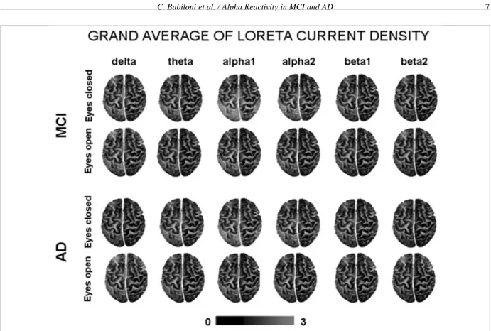
For illustrative purposes, Fig. 1 plots the grand av-erage of the LORETA solutions (i.e., normalized
pow-Fig. 1. Grand average of the LORETA solutions (i.e., relative power current density at cortical voxels) modeling the distributed EEG cortical sources at delta, theta, alpha 1, alpha 2, beta 1, and beta 2 bands in MCI and AD subjects for the two conditions, namely eyes-closed and eyes-open. The left side of the maps (top view) corresponds to the left hemisphere. Legend: LORETA, low resolution brain electromagnetic tomography. Color scale: all power density estimates were scaled based on the averaged maximum value (i.e., alpha 1 power value of occipital region in MCI eyes-closed). The maximal value of power density is reported under the figure. (Colors are visible in the electronic version of this article at http://dx.doi.org/10.3233/JAD-2010-100798)
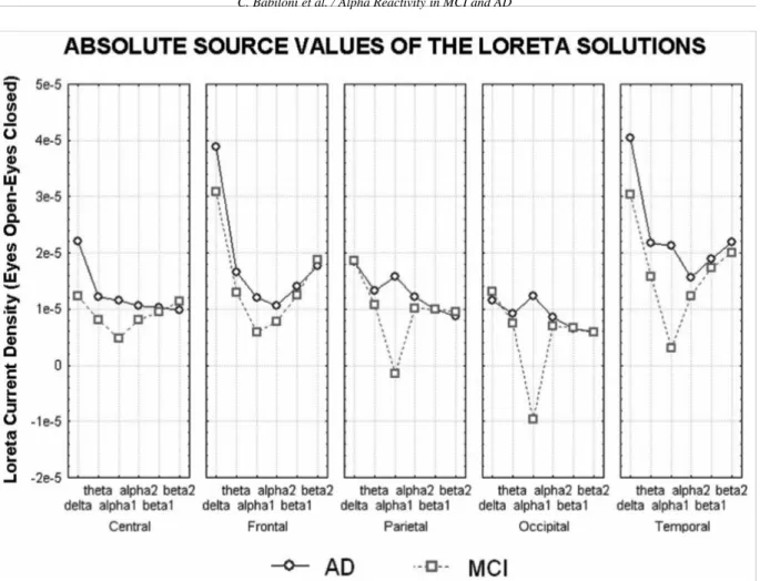
er current density at cortical voxels) modeling the dis-tributed EEG cortical sources for delta, theta, alpha 1, alpha 2, beta 1, and beta 2 bands in the MCI and AD subjects. These solutions refer to the eyes-closed and eyes-open conditions. In both MCI and AD groups, the cortical sources of alpha 1 and alpha 2 were low-er in amplitude in the eyes-open than eyes-closed con-dition, as an effect of the reactivity of cortical neural synchronization to the eyes-open condition. An oppo-site effect was observed in cortical delta sources. For control purposes, Fig. 2 plots the power difference of regional absolute LORETA solutions in the eyes-open minus eyes-closed condition modeling the distributed EEG cortical sources for delta, theta, alpha 1, alpha 2, beta 1, and beta 2 bands in the MCI and AD subjects. The figure shows a similar trend with respect to the normalized LORETA sources.
To test the statistical significance of these differ-ences of the normalized LORETA solutions, an ANO-VA was used (age, education, gender, IAF, MMSE,
and resting eyes-closed alpha rhythms as covariates;
p <0.05). Figure 3 shows the power difference of re-gional normalized LORETA solutions in the eyes-open minus eyes-closed condition (i.e., “reactivity”) rela-tive to a statistical ANOVA interaction (F(40,3100) = 3.75; p < 0.001) among the factors Group (AD, MCI, Nold), Band (delta, theta, alpha 1, alpha 2, beta 1, beta 2), and ROI (central, frontal, parietal, occipital, temporal). The planned post-hoc testing showed that the EEG source suppression pattern Nold>MCI>AD was fitted by the following normalized regional LORE-TA solutions: parietal, occipital, and temporal alpha 1 sources (p < 0.001) as well as parietal and occipital alpha 2 sources (p < 0.05 to 0.001). These results were confirmed by a control ANOVA analysis prob-ing LORETA solutions for each ROI considered sep-arately (central, frontal, parietal, occipital, temporal). Again, the source pattern Nold>MCI>AD was fitted by the following 3 normalized regional LORETA so-lutions: parietal (F(10,775) = 2.89; p < 0.005),
oc-Fig. 2. Absolute source values of the LORETA solutions (eyes-open minus eyes-closed) in MCI and AD subjects for the delta, theta, alpha 1, alpha 2, beta 1, and beta 2 bands. (Colors are visible in the electronic version of this article at http://dx.doi.org/10.3233/JAD-2010-100798)
cipital (F(10,775) = 4.55; p < 0.001), and temporal (F(10,775) = 3.28; p < 0.001) alpha 1 sources. Pari-etal and occipital alpha 2 sources just fitted the EEG source suppression pattern AD and MCI < Nold.
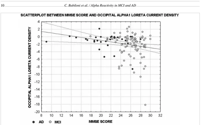
The amplitude of the above statistically significant alpha 1 and alpha 2 source power differences (i.e., re-activity to eyes-open condition; p < 0.05 to 0.001) was correlated with the MMSE score in the MCI and AD subjects as a whole group (Pearson test, p < 0.05). Bonferroni correction for the five repetitions of the test set the statistical threshold to p < 0.01 to obtain the Bonferroni corrected p < 0.05. The MMSE score neg-atively correlated (r = −0.24, p = 0.01) with occipital alpha 1 source power difference (Fig. 4); the higher the MMSE score, the higher the source power difference as expressed by negative values. No statistically sig-nificant correlation was observed (p > 0.01) between the MMSE and the remaining alpha power source dif-ferences at parietal and temporal alpha 1 as well as at parietal and occipital alpha 2.
Control analyses
We performed some control analyses to ascertain if the results of the main statistical analysis were affected by relevant confounding variables.
In a first control analysis, we performed a correla-tion between the resting state eyes closed alpha pow-er showing the pattpow-ern Nold>MCI>AD and the alpha reactivity to eyes opening (Pearson’s test; p < 0.05), in order to evaluate the relationships between the cor-tical neural synchronization/desynchronization in the two conditions. The results showed a statistically sig-nificant correlation for parietal, occipital, and temporal alpha 1 sources as well as for parietal and occipital al-pha 2 sources (r = from −0.83 to −0.95, p < 0.001). The higher the eyes closed alpha power, the higher the alpha reactivity to eyes opening. Of note, this correla-tion was quite high but unable to entirely explain the pattern Nold>MCI>AD of the alpha reactivity to eyes opening. Indeed, we obtained significant inter-groups
Fig. 3. Normalized power differences at the regional normalized LORETA solutions (eyes-open minus eyes-closed) relative to a statistical ANOVA interaction (F(40,3100) = 3.75; p < 0.001) among the factors Group (AD, MCI, Nold), Band (delta, theta, alpha 1, alpha 2, beta 1, beta 2), and ROI (central, frontal, parietal, occipital, temporal). Subjects’ age, education, gender, IAF, MMSE and resting alpha Loreta Current Density eyes closed were used as covariates. The planned post-hoc testing showed that the source pattern eyes-closed > eyes-open was fitted by the following 5 normalized regional LORETA solutions: parietal, occipital and temporal alpha 1 sources (p < 0.005 to 0.001) as well as parietal and occipital alpha 2 sources (p < 0.05 to 0.001). Legend: the rectangles indicate the cortical regions and frequency bands in which LORETA solutions presented statistically significant LORETA patterns (p < 0.05). (Colors are visible in the electronic version of this article at http://dx.doi.org/10.3233/JAD-2010-100798)
effects of alpha reactivity to eyes opening although rest-ing state eyes closed alpha power at relevant sources was used as a covariate in the statistical model.
In a second control analysis, we performed an ANO-VA using the power difference of regional absolute (as opposed to normalized) LORETA solutions in the eyes-open minus eyes-closed condition as a dependent variable. The ANOVA factors were Group (AD, MCI, Nold), Band (delta, theta, alpha 1, alpha 2, beta 1, beta 2), and ROI (central, frontal, parietal, occipital, temporal). Subjects’ age, education, gender, IAF, and MMSE served as covariates. In contrast to the ANOVA using the regional relative LORETA solutions, the con-trol analysis showed no statistically significant interac-tion among the factors (p > 0.05). These control results confirmed that with respect to relative EEG power,
ab-solute EEG power depends on (irrelevant) properties of conductibility of subjects’ skull, and may be less sensi-tive to the changes due to brain neural synchronization. In a third control analysis, we performed an ANO-VA using the LORETA solutions relative to a low-frequency alpha sub-band of 7–9 Hz in the eyes-open minus eyes-closed condition as a dependent variable. This sub-band was lower than mean IAF (i.e., around 9 Hz), to control that the results were not affected by high alpha frequencies. The ANOVA factors were Group (AD, MCI, Nold) and ROI (central, frontal, pari-etal, occipital, temporal). Subjects’ age, education, gender, IAF, and MMSE served as covariates. There was a statistically significant interaction between the factors (F(8,620) = 2.39; p < 0.01). Post-hoc testing indicated that parietal, occipital and temporal alpha
re-Fig. 4. Scatter plots relative to the results of a correlation analysis between MMSE and occipital alpha 1 sources of EEG rhythms (eyes-open minus eyes-closed) in the MCI and AD subjects as a whole group. (Colors are visible in the electronic version of this article at http://dx.doi. org/10.3233/JAD-2010-100798)
activity to the eyes opening showed differences among the AD and the Nold and MCI subjects (p < 0.01), in line with the results of the main statistical analysis.
In a fourth control analysis, we performed an ANO-VA including sub-groups of subjects that were careful-ly matched as number (31 AD, 31 MCI, and 31 Nold subjects), mean age (AD = 70.5 years; MCI = 70.2 years; Nold = 66.0 years), and mean education (AD = 6.5, MCI = 6.7, and Nold = 7.3 years). This allowed a good control of the inter-groups variability. The depen-dent variable was the normalized LORETA solution in eyes-open minus eyes-closed condition. The ANOVA design was the same of the main analysis. There was a statistical ANOVA interaction (F(40,1800) = 3.68; p < 0.0001) among the factors Group (AD, MCI, Nold), Band (delta, theta, alpha 1, alpha 2, beta 1, beta 2), and ROI (central, frontal, parietal, occipital, temporal). The planned post-hoc testing substantially confirmed the results of the main analysis, showing that the EEG source suppression pattern Nold>MCI>AD was fit-ted by parietal, occipital, and temporal alpha 1 sources (p < 0.05).
In a fifth control analysis, we performed a control data analysis to cross-validate the present LORETA re-sults with an analysis of EEG spectral power density
computed at the scalp electrodes. We strictly followed the general methodological approach used for the anal-ysis of the LORETA solutions. The EEG spectral pow-er density at each electrode was normalized to the EEG spectral power density averaged across all frequencies (0.5–45 Hz) and the electrodes, which were represen-tative of the following scalp regions: (i) Fp1, Fp2, F7, F3, Fz, F4, and F8 electrodes for the frontal region; (ii) C3, Cz and C4 electrodes for the central region; (iii) P3, Pz and P4 electrodes for the parietal region; (iv) T3, T4, T5, and T6 electrodes for the temporal region; (v) O1 and O2 electrodes for the occipital re-gion. For a given region, the regional power density for the eyes-closed (eyes-open) condition was defined as the power density averaged across all electrodes of that region. The reactivity of regional EEG power den-sity to the eyes opening and the statistical comparisons were computed according to the procedures used for the analysis of the LORETA solutions. The relative ANOVA used the power difference of regional scalp solutions in the eyes-open minus eyes-closed condition as a dependent variable. The ANOVA factors were Group (AD, MCI, Nold), Band (delta, theta, alpha 1, alpha 2, beta 1, beta 2), and Scalp ROI (central, frontal, parietal, occipital, temporal). Subjects’ age, education,
gender, IAF, and MMSE served as covariates. Results showed a statistical ANOVA interaction (F(40,3100) = 7.14; p < 0.001) among the factors Group (AD, MCI, Nold), Band (delta, theta, alpha 1, alpha 2, beta 1, be-ta 2), and Scalp ROI (central, fronbe-tal, pariebe-tal, occipibe-tal, temporal). The planned post-hoc testing showed that the scalp EEG reactivity pattern Nold>MCI>AD was fitted by the following normalized regional scalp solu-tions: parietal, occipital, and temporal alpha 1 (p < 0.05 to 0.001) as well as parietal and occipital alpha 2 (p < 0.05 to 0.001). The amplitude of the above sta-tistically significant alpha 1 and alpha 2 scalp solutions (i.e., reactivity to eyes-open condition; p < 0.05 to 0.001) was correlated with the MMSE score in the MCI and AD subjects as a whole group (Pearson test, p < 0.05). Bonferroni correction for the 5 repetitions of the test set the statistical threshold to p < 0.01 to obtain the Bonferroni corrected p < 0.05. No statistically signif-icant correlation was observed (p > 0.01) between the MMSE and the mentioned alpha scalp solutions in the MCI and AD subjects as a whole. Summarizing, the results showed that in good accordance with the LORE-TA solutions reported above, posterior scalp alpha reac-tivity to the eyes opening showed differences between the Nold and MCI subjects and between the MCI and AD subjects. However, in contrast with the LORETA solutions reported above, the occipital scalp alpha 1 reactivity was not correlated to the MMSE score.
DISCUSSION
This study tested the hypothesis of a progressive im-pairment of cortical alpha reactivity to the eyes-open compared with eyes-closed condition across Nold, am-nesic MCI and AD subjects, reflecting the functional impairment of cortical neural desynchronization in the grey zone of the continuum between preclinical and clinical stages of AD. This issue represents the step for-ward of the present study with respect to previous stud-ies of this Consortium (i.e., Authors’ research groups) focused on the mechanisms of cortical neural synchro-nization associated to the condition of eyes closed rest-ing state [27,28,38,67,68]. With respect to the EEG literature [30,33,36] the novelty of the present EEG study is to focus on topographical reactivity of alpha rhythms to eyes opening and to use relatively large groups of Nold, amnesic MCI, and mild AD subjects, to better take into account for head volume conduction effects and inter-groups variability of resting state EEG rhythms.
In the control eye-closed condition, posterior corti-cal sources of alpha rhythms were lower in power in the MCI and AD subjects than in the normal elderly subjects. Furthermore, the opposite was true for frontal and temporal cortical sources of delta rhythms. These control results were in line with previous EEG evidence of our Consortium showing progressive differences of resting eyes-closed cortical alpha rhythms along the shadow line across Nold, amnesic MCI, and mild AD subjects [27,28,38,67,68].
With respect to the closed condition, the eyes-open condition induced a suppression of posterior alpha sources that was greater in the Nold than MCI subjects and in the MCI than AD subjects. In the occipital low-frequency alpha sources, this reactivity was proportion-al to the subjects’ globproportion-al cognitive function as reveproportion-aled by MMSE score across MCI and AD individuals (i.e., the grey zone of the disease).
Notably, the eyes-closed alpha power and alpha reac-tivity to eyes opening pointed to a remarkable correla-tion and to a similar source pattern in the MCI and AD subjects. However, these variables provided at least in part independent physiological information, since the inter-groups differences in alpha reactivity were still significant when the eyes closed alpha sources were used as covariate in the statistical model. On the whole, it can be speculated that the synchronizing neural cir-cuits at the basis of eyes closed alpha rhythms largely corresponded to the neural circuits that desynchronized during eyes opening.
The present results agree with previous EEG and MEG studies showing that alpha power during eye opening is suppressed more in Nold than in AD sub-jects, as a reflection of impaired cortical neural desyn-chronization in visual systems of the patients [21,31– 33,35,36]. They also agree with previous sugges-tion that alpha reactivity to eye opening might predict the deterioration of higher functions in subjects with cognitive decline [33]. Since some of previous EEG and MEG studies have shown that differences between MCI and AD subjects at resting state are not ever dis-cernible [30,33,36], we suggest that ideally, EEG and MEG techniques are used in a combined way to better understand neurophysiology of brain rhythms in am-nesic MCI and AD subjects for basic research and clin-ical applications. High spatial resolution MEG tech-niques would be effective in the fine topographical esti-mation of tangentially-oriented cortical sources of brain rhythms, while the present low-resolution EEG ap-proach would be sensitive to the global activity of both tangentially- and radially-oriented cortical sources. Of
course, this scenario may come true when MEG tech-nology could be largely accessible, and automation of the data analysis procedures could reduce time and costs for the production of clinical reports.
An open issue of the present study is whether the present methodological approach for the evalu-ation of resting state EEG rhythms be used alone for personalized diagnosis and prognosis of individual MCI subjects, given the intrinsic variability of alpha rhythms [30,77]. We think that the present method-ological approach is promising for future clinical ap-plications. Indeed, it represents a simple, quick and cheap experimental model to probe the relationships between aging and mechanisms of cortical neural syn-chronization/desynchronization in two complementary modes of resting state (i.e., presence/absence of visual stimulus processing), without the confounding effects of anxiety, fatigue, boring, and task complexity. In-deed, these mechanisms reflect the integrity of neural populations and synapses as well as the efficiency of functional connectivity and brain reactivity. Such ex-perimental model can be used with elderly individuals ranging from full cognitive performance to severe cog-nitive impairment, and is not affected by meta-learning and effects of the repetition of the experimental record-ings over time. The present results suggest that at least at group level, AD progression is related to an impaired reactivity of the mechanisms desynchronizing posteri-or cposteri-ortical neurons at alpha rhythms during eyes open-ing. This conclusion represents a first important step for understanding the relationship between early stages of AD neurodegeneration and mechanisms of cortical neural synchronization/desynchronization. It also rep-resents an important motivation to ascertain the clini-cal relevance of the present results (i.e., early diagno-sis, prognodiagno-sis, therapy monitoring) by future longitu-dinal studies. In this regard, we are aware that the high intrinsic inter-subjects variability of EEG reactivity to eyes opening might prevent its practical clinical use for early diagnosis/prognosis of AD in single MCI indi-viduals. However, we think that there are promising perspectives for the combined used of this EEG marker together with other EEG (i.e., resting state eyes closed), neuroimaging (MRI, PET-FDG, MEG), and biological (CSF, blood) markers. Of special interest the coreg-istration of resting state EEG rhythms and functional MRI (fMRI). fMRI reflects the ratio between deoxy-hemoglobin and oxydeoxy-hemoglobin blood (BOLD), has a low temporal resolution (> 1 s) and a very high spa-tial resolution (< 1 cm), and is especially suitable to investigate spatial details of both cortical and
subcorti-cal activation [78]. The resting state fMRI represents an indirect measurement of the functional connectivity among brain regions, and perfectly complements the properties of resting state EEG rhythms [79].
A crucial question is then: “What is the function-al meaning of a reduced function-alpha reactivity to eye open-ing along MCI and AD subjects?” Duropen-ing slow-wave sleep, corticofugal slow oscillations (< 1 Hz) are effec-tive in grouping thalamic-generated delta rhythms (1– 4 Hz) and spindling activity (7–14 Hz) rhythms [80]. Delta would dominate EEG rhythms and alpha (about 8–12 Hz) would be low in amplitude. In the case of brain arousal, spindles, high and low components of the delta rhythms are blocked by the inhibition of reticulo-thalamic (7–14 Hz), thalamo-cortical (1–4 Hz), and in-tracortical (< 1 Hz) oscillators. These rhythms are re-placed by fast oscillations (beta and gamma) induced by forebrain (nucleus basalis) cholinergic inputs to hip-pocampus and cortex as well as by thalamocortical pro-jections [80,81]. In the condition of waking rest, 8– 10 Hz alpha oscillations represent the dominant resting rhythms of human brain [82–85], and have been linked to intelligence quotient, memory, and cognition [66]. Alpha rhythms are mainly generated by the coordinat-ed interplay between cortico-cortical and thalamocor-tical dynamics, as suggested by studies in higher mam-mals (lower mammam-mals show negligible or absent alpha rhythms; for a review see [86]. Given that primary visual cortex seems not to be seriously affected in the prodromal stages of AD, it can be speculated that the link between the present results (impairment of corti-cal desynchronization mechanisms underlying EEG al-pha reactivity) and neuropathological events or lesions characterizing AD may rely upon abnormal postsynap-tic potentials generated in large pyramidal neurons of occipital-parietal cortical regions and in the neurons conveying signals from parietal nodes of attention net-works to visual cortex. This may be due to synap-tic and neural loss associated to AD neurodegenerative processes occurring initially and predominantly in the medial temporal lobe structures including hippocam-pus and amygdala [13]. Hippocampal and amygdalar atrophy in AD have been documented, although thala-mic degeneration in the early stages of the disease is an open issue [13,87–90]. The hippocampus connects directly with the anterior thalamus via the fornix and mammillary bodies as well as with the pulvinar via the temporopulvinar tract. Integrity of these connections is essential for episodic memory [91], which is specifical-ly impaired in AD [92]. Keeping in mind this premises, it should be methodologically remarked that estimation
of EEG sources by LORETA directly modeled cortical but not thalamic generators of alpha rhythms. Indeed, LORETA software uses dipole source into a volume fitting cortical grey matter but not thalamus, basal gan-glia and other sub-cortical structures. Therefore, cor-tical sources of alpha rhythms can be affected by al-tered inputs from thalamus but LORETA is not able to directly model thalamic abnormalities. An abnormal interplay between human thalamic and cortical neural populations can be investigated by combined record-ings of EEG and PET-FDG or fMRI. To this regard, it has been shown that alpha power is positively correlat-ed with the cerebral blood flow, glucose metabolism, and BOLD signals in the thalamus, whereas it is neg-atively correlated with these measurements in visual areas [93–96].
In this theoretical framework, the role of choliner-gic systems is still poorly known. On one hand, there is no evidence of the participation of the basal fore-brain, via its cholinergic innervation to the cortex, in the generation of alpha rhythms in higher mammals. In cholinergic basal forebrain, lesion and stimulation of nucleus basalis of Meynert mainly affect fast cortical oscillations in the range of gamma rhythms [97]. Fur-thermore, human occipital cortex (peristriate areas, BA 19) receives a relatively scarce density of cholinergic axons from the intermediate Ch4 subdivision [98]. On the other hand, it has been emphasized the role of par-allel cholinergic tracts from basal forebrain to human hippocampus-amygdala, thalamus, and cerebral cortex in the selection/modulation of cortical excitability for attention and visual processes and consciousness [99, 100], which are typically related to alpha rhythms [66]. These cholinergic tracts would be targeted by AD neu-rodegenerative processes [101–105], especially in AD patients not responding to long term cholinergic thera-py [106]. Instead, brainstem cholinergic innervation of the thalamus would be relatively spared [103,107–110]. Furthermore it has been reported that scopolamine (a cholinergic antagonist) mimicked in healthy subjects the typical pattern of alpha and theta rhythms observed in AD patients [77], and the loss of cholinergic con-nections to cerebral cortex has been shown to reduce the power of resting posterior alpha sources in amnesic MCI subjects [111,112]. Moreover, it has been shown that posterior sources of resting delta and alpha rhythms were especially impaired in AD patients not respond-ing to cholinergic therapy at 1-year follow up [113]. Finally, recent MEG data have pointed to a complex interaction of several resting state EEG rhythms as the reflection of the balance of different neuromodulation systems [114].
Keeping in mind these data, progressive impair-ment of the alpha reactivity to eye opening in the con-tinuum along amnesic MCI and mild AD conditions might reflect the synaptic and neural loss of large cor-tical pyramidal neurons including those convey sig-nals through white-matter to visual cortex, in order to modulate arousal-attention and, hence, the transmis-sion and processing of information [115,116]. Fu-ture studies should clarify the role played by choliner-gic, monoaminergic and glutamatergic systems in the modulation of cortical excitability and alpha rhythms in amnesic MCI and AD subjects [117,118]. In this framework, an intriguing working hypothesis for these future studies is that cholinergic systems play a re-markable role in the desynchronization of posterior al-pha rhythms during eyes opening (and mental activity), whereas cortico-cortical and thalamo-cortical interac-tions rhythms play a remarkable role in the synchro-nization of posterior alpha rhythms in the condition of eyes closed resting state.
In conclusion, the novelty of present study was to test the hypothesis of a progressive impairment of cortical alpha reactivity to eye opening among relatively large groups of Nold, amnesic MCI and AD subjects, pos-sibly reflecting the functional impairment of cortical neural desynchronization in the grey zone between pre-clinical and pre-clinical stages of AD. Compared with the eyes–closed condition, the eyes-open condition showed a power reduction of posterior alpha 1 and 2 sources that was greater in the Nold than in MCI subjects and in the MCI than AD subjects. In the occipital alpha 1 sources, this reduction was proportional to the subjects’ global cognitive function as revealed by MMSE score across MCI and AD individuals. The present results suggest that at least in its early stages and at group lev-el, AD progression is related to an impaired reactivity of the mechanisms desynchronizing cortical neurons at alpha rhythms during background visual information processing. This conclusion represents a first impor-tant step for understanding the relationship between early stages of AD neurodegeneration and mechanisms of cortical neural desynchronization as a reflection of functional brain connectivity and physiological reac-tivity. However, despite their important heuristic value, the present results are valid at group level and are not conclusive about the utility of the present methodologi-cal approach for diagnostic/prognostic purposes in sin-gle amnesic MCI subjects. Future longitudinal “follow up” studies should evaluate the clinical relevance of the present results towards practical clinical applications.
ACKNOWLEDGMENTS
This research was developed thank to the financial support of Fatebenefratelli Association for Biomedical Research (AFaR), Tosinvest Sanita’ (Cassino, Pisana). Italian Ministry of Health (Strategic research project entitled “Diagnosis of incipient Alzheimer disease”), and European Committee (IMI JU 2008 project entitled “PharmaCog”). Dr. Roberta Lizio has contributed to this study in the framework of her Ph.D. fellowship in Neurophysiology at the Department of Physiology and Pharmacology, University of Rome Sapienza, Rome, Italy.
Authors’ disclosures available online (http://www.j-alz.com/disclosures/view.php?id=571).
REFERENCES
[1] Flicker CS, Ferris H, Reisberg B (1991) Mild cognitive im-pairment in the elderly: predictors of dementia. Neurology 41, 1006-1009.
[2] Petersen RC, Smith GE, Ivnik RJ, Tangalos EG, Schaid SN, Thibodeau SN, Kokmen E, Waring SC, Kurland LT (1995) Apolipoprotein E status as a predictor of the development of Alzheimer’s disease in memory-impaired individuals. JAMA 273, 1274-1278.
[3] Petersen RC, Doody R, Kurz A, Mohs RC, Morris JC, Ra-bins PV, Ritchie K, Rossor M, Thal L, Winblad B (2001) Current concepts in mild cognitive impairment. Arch Neurol 58, 1985-1992.
[4] Galluzzi S, Cimaschi L, Ferrucci L, Frisoni GB (2001) Mild cognitive impairment: clinical features and review of screen-ing instruments. Agscreen-ing (Milano) 13, 183-202.
[5] Scheltens P, Fox N, Barkhof F, De Carli C (2002) Structural magnetic resonance imaging in the practical assessment of dementia: beyond exclusion. Lancet Neurol 1, 13-21 [6] Arnaiz E, Almkvist O (2003) Neuropsychological features
of mild cognitive impairment and preclinical Alzheimer’s disease. Acta Neurol Scand Suppl 179, 34-41.
[7] Bachman DL, Wolf PA, Linn RT, Knoefel JE, Cobb JL, Be-langer AJ, White LR, D’Agostino RB (1993) Incidence of dementia and probable Alzheimer’s disease in a general pop-ulation: the Framingham Study. Neurology 43, 515-519. [8] Gao S, Hendrie HC, Hall KS, Hui S (1998) The
relation-ships between age, sex, and the incidence of dementia and Alzheimer disease: a meta-analysis. Arch Gen Psychiatry 55, 809-815.
[9] Frisoni GB, Padovani A, Wahlund LO (2004) The prede-mentia diagnosis of Alzheimer disease. Alzheimer Dis Assoc Disord 18, 51-53.
[10] Bennett DA, Wilson RS, Schneider JA, Evans DA, Beckett LA, Aggarwal NT, Barnes LL, Fox JH, Bach J (2002) Nat-ural history of mild cognitive impairment in older persons. Neurology 59, 198-205.
[11] Larrieu S, Letenneur L, Orgogozo JM, Fabrigoule C, Amieva H, Le Carnet N, Barberger-Gateau P, Dartigues JF (2002) Incidence and outcome of mild cognitive impairment in a population-based prospective cohort. Neurology 59, 1594-1549.
[12] Fisk JD, Merry HR, Rockwood K (2003) Variations in case definition affect prevalence but not outcomes of mild cogni-tive impairment. Neurology 61, 1179-1184.
[13] Braak H, Braak E (1991) Neuropathological stageing of Alzheimer-related changes. Acta Neuropathol 82, 239-259. [14] Rogers J, Webster S, Lue LF, Brachova L, Civin WH,
Emmer-ling M, Shivers B, Walker D, McGeer P (1993) Inflammation and Alzheimer’s disease pathogenesis. Neurobiol Aging 17, 681-686.
[15] Small GW, La Rue A, Komo S, Kaplan A, Mandelkern MA (1995) Predictors of cognitive change in middle-aged and older adults with memory loss. Am J Psychiatry 152, 1757-1764.
[16] Nestor PJ, Scheltens P, Hodges JR (2004) Advances in the early detection of Alzheimer’s disease. Nat Med 10, S34-S41. [17] Dierks T, Ihl R, Frolich L, Maurer K (1993) Dementia of the Alzheimer type: effects on the spontaneous EEG described by dipole sources. Psychiatry Res 50, 51-162.
[18] Dierks T, Jelic V, Pascual-Marqui RD, Wahlund LO, Julin P, Linden DEJ, Maurer K, Winblad B, Nordberg A (2000) Spatial pattern of cerebral glucose metabolism (PET) cor-relates with localization of intracerebral EEG-generators in Alzheimer’s disease. Clin Neurophysiol 111, 1817-1824. [19] Huang C, Wahlund LO, Dierks T, Julin P, Winblad B, Jelic
V (2000) Discrimination of Alzheimer’s disease and mild cognitive impairment by equivalent EEG sources: a cross-sectional and logitudinal study. Clin Neurophysiol 11, 1961-1967.
[20] Ponomareva NV, Selesneva ND, Jarikov GA (2003) EEG alterations in subjects at high familial risk for Alzheimer’s disease. Neuropsychobiology 48, 152-159.
[21] Jeong J (2004) EEG dynamics in patients with Alzheimer’s disease. Clin Neurophysiol 115, 1490-1505.
[22] Babiloni C, Binetti G, Cassetta E, Cerboneschi D, Dal Forno G, Del Percio C, Ferreri F, Ferri R, Lanuzza B, Miniassi C, Moretti DV, Nobili F, Pascual-Marqui RD, Rodriguez G, Ro-mani GL, Salinari S, Tecchio F, Vitali P, Zanetti O, Zappasodi F, Rossini PM (2004) Mapping Distributed Sources of Cor-tical Rhythms in Mild Alzheimers Disease. A Multi-Centric EEG Study. NeuroImage 22, 57-67.
[23] Koenig T, Prichep L, Dierks T, Hubl D, Wahlund LO, John ER, Jelic V (2005) Decreased EEG synchronization in Alzheimer’s disease and mild cognitive impairment. Neuro-biol Aging 26,165-171.
[24] Zappoli R, Versari A, Paganini M, Arnetoli G, Muscas GC, Gangemi PF, Arneodo MG, Poggiolini D, Zappoli F, Battaglia A (1995) Brain electrical activity (quantitative EEG and bit-mapping neurocognitive CNV components), psycho-metrics and clinical findings in presenile subjects with initial mild cognitive decline or probable Alzheimer-type dementia. Ital J Neurol Sci 16, 341-376.
[25] Elmstahl S, Rosen I (1997) Postural hypotension and EEG variables predict cognitive decline: results from a 5-year follow-up of healthy elderly women. Dement Geriatr Cogn Disord 8, 180-187.
[26] Jelic V, Johansson SE, Almkvist O, Shigeta M, Julin P, Nord-berg A, Winblad B, Wahlund LO (2000) Quantitative elec-troencephalography in mild cognitive impairment: longitu-dinal changes and possible prediction of Alzheimer’s disease. Neurobiol Aging 21, 533-540.
[27] Babiloni C, Binetti G, Cassetta E, Dal Forno G, Del Percio C, Ferreri F, Ferri R, Frisoni G, Hirata K, Lanuzza B, Miniassi C, Moretti DV, Nobili F, Rodriguez G, Romani GL, Salinari S, and Rossini PM (2006) Sources of cortical rhythms change
as a function of cognitive impairment in pathological aging: a multi-centric study. Clin Neurophysiol 117, 252-268. [28] Rossini PM, Del Percio C, Pasqualetti P, Cassetta E, Binetti
G, Dal Forno G, Ferreri F, Frisoni G, Chiovenda P, Mini-assi C, Parisi L, Tombini M, Vecchio F, Babiloni C (2006) Conversion from mild cognitive impairment to Alzheimer’s disease is predicted by sources and coherence of brain elec-troencephalography rhythms. Neuroscience 143, 793-803. [29] Fern´andez A, Hornero R, Mayo A, Poza J, Maest´u F,
Or-tiz Alonso T (2006) Quantitative magnetoencephalography of spontaneous brain activity in Alzheimer disease: an ex-haustive frequency analysis. Alzheimer Dis Assoc Disord 20, 153-159.
[30] Osipova D, Rantanen K, Ahveninen J, Ylikoski R, H¨app¨ol¨a O, Strandberg T, Pekkonen E (2006) Source estimation of spontaneous MEG oscillations in mild cognitive impairment. Neurosci Lett 405, 57-61.
[31] Stam CJ, Jelles B, Achtereekte HA, van Birgelen JH, Slaets JP (1996) Diagnostic usefulness of linear and nonlinear quan-titative EEG analysis in Alzheimer’s disease. Clin Electroen-cephalogr 27, 69-77.
[32] Stevens A, Kircher T (1998) Cognitive decline unlike normal aging is associated with alterations of EEG temporo-spatial characteristics. Eur Arch Psychiatry Clin Neurosci 248, 259-266.
[33] van der Hiele K, Vein AA, van der Welle A, van der Grond J, Westendorp RG, Bollen EL, van Buchem MA, van Dijk JG, Middelkoop HA (2007) EEG and MRI correlates of mild cog-nitive impairment and Alzheimer’s disease. Neurobiol Aging 28, 1322-1329.
[34] van der Hiele K, Bollen EL, Vein AA, Reijntjes RH, West-endorp RG, van Buchem MA, Middelkoop HA, van Dijk JG (2008) EEG markers of future cognitive performance in the elderly. J Clin Neurophysiol 25, 83-89.
[35] Berendse HW, Verbunt JP, Scheltens P, van Dijk BW, Jonkman EJ (2000) Magnetoencephalographic analysis of cortical activity in Alzheimer’s disease: a pilot study. Clin Neurophysiol 111, 604-612.
[36] Kurimoto R, Ishii R, Canuet L, Ikezawa K, Azechi M, Iwase M, Yoshida T, Kazui H, Yoshimine T, Takeda M (2008) Event-related synchronization of alpha activity in ear-ly Alzheimer’s disease and mild cognitive impairment: an MEG study combining beamformer and group comparison. Neurosci Lett 443, 86-89.
[37] Babiloni C, Binetti G, Cassarono A, Dal Forno G, Del Percio C, Ferreri F, Ferri R, Frisoni G, Galderisi S, Hirata K, Lanuzza B, Miniassi C, Mucci A, Nobili F, Rodriguez G, Romani GL, and Rossini PM (2006) Sources of cortical rhythms in adults during physiological aging: a multi-centric EEG study. Human Brain Mapping 27, 162-172.
[38] Babiloni C, Cassetta E, Binetti G, Tombini M, Del Percio C, Ferreri F, Ferri R, Frisoni G, Lanuzza B, Nobili F, Parisi L, Rodriguez G, Frigerio L, Gurz`ı M, Prestia A, Verzieri F, Eusebi F, Rossini PM (2007) Resting EEG sources corre-late with attentional span in mild cognitive impairment and Alzheimer’s disease. Eur J Neurosci 25, 3742-3757. [39] Babiloni C, Visser PJ, Frisoni G, De Deyn PP, Bresciani L,
Jelic V, Nagels G, Rodriguez G, Rossini PM, Vecchio F, Colombo D, Verhey F, Wahlund LO, Nobili F (2010) Corti-cal sources of resting EEG rhythms in mild cognitive impair-ment and subjective memory complaint. Neurobiol Aging 31, 1787-1798.
[40] Albert M, Smith LA, Scherr PA, Taylor JO, Evans DA, Funkenstein HH (1991) Use of brief cognitive tests to
iden-tify individuals in the community with clinically diagnosed Alzheimer’s disease. Int J Neurosci 57, 167-178.
[41] Devanand DP, Folz M, Gorlyn M, Moeller JR, Stem J (1997) Questionable dementia: clinical course and predictors of outcome. J Am Geriatr Soc 45, 321-328.
[42] Petersen RC, Smith GE, Waring SC, Ivnik RJ, Kokmen E, Tangelos EG (1997) Aging, memory, and mild cognitive impairment. Int Psychogeriatr 9, 65-69.
[43] Rubin EH, Morris JC, Grant EA, Vendegna T (1989) Very mild senile dementia of the Alzheimer type. I. Clinical as-sessment. Arch Neurol 46, 379-82
[44] Zaudig M (1992) A new systematic method of measurement and diagnosis of "mild cognitive impairment" and dementia according to ICD-10 and DSM-III-R criteria. Int Psychogeri-atr 4(Suppl 2), 203-219.
[45] Portet F, Ousset PJ, Visser PJ, Frisoni GB, Nobili F, Scheltens P, Vellas B, Touchon J (2006) MCI Working Group of the Eu-ropean Consortium on Alzheimer’s Disease Mild cognitive impairment (MCI) in medical practice: a critical review of the concept and new diagnostic procedure. Report of the MCI Working Group of the European Consortium on Alzheimer’s Disease. J Neurol Neurosurg Psychiatry 77, 714-718. [46] Raven JC (1965) Guide to using coloured progressive
matri-ces. H.K. Lewis, London, UK.
[47] Shulman KI, Gold DP, Cohen CA, Zucchero CA (1993) Clock drawing and dementia in thecommunity: A longitudi-nal study. Int J Geriatr Psychiat 8, 487-496.
[48] Rey A (1958) Memorisation d‘une serie de 15 mots en 5 rep-etitions. In: A. Rey (Hrsg.). L‘examen clinique en psycholo-gie. Presses Universitaires de France: Paris.
[49] Carlesimo GA, Caltagirone C, Gainotti G, the Group for the Standardization of the Mental Deterioration Battery (1996) The Mental Deterioration Battery: normative data, diagnos-tic reliability and qualitative analyses of cognitive impair-ment. Eur Neurol 36, 378-384.
[50] Rey A (1968) Reattivo Della Figura Complessa Manuale. Organizzazioni Speciali, Firenze.
[51] Chandler MJ, Lacritz LH, Cicerello AR, Chapman SB, Honig LS, Weiner MF, Cullum CM (2004) Three-word recall in normal aging. J Clin Exp Neuropsychol 26, 1128-1133. [52] Spinnler H, Rognoni G (1987) Standardizzazione e taratura
italiana di test neuropsicologici. Ital J Neurol Sci, vol. suppl.6 N◦8.
[53] Novelli G (1986) Three clinical tests for the assessment of lexical retrieval and production. Norms from 320 normal subjects. Arch Psicol Neurol Psichiatria 47, 477-506. [54] De Renzi E, Vignolo LA (1962) The token test: A sensitive
test to detect receptive disturbances in aphasics. Brain 85, 665-678.
[55] Reitan RM (1958) Validity of the Trail Making Test as an indication of organic brain damage. Percept Motor Skill 1958, 271-276.
[56] Orsini A, Grossi D, Capitani E, Laiacona M, Papagno C, Vallar G (1987) Verbal and spatial immediate memory span: normative data from 1355 adults and 1112 children. Ital J Neurol Sci 8, 539-548.
[57] Lawton MP, Brodie EM (1969) Assessment of older people: self maintaining and instrumental activity of daily living. J Gerontol 9, 179-186.
[58] Hughes CP, Berg L, Danziger WL, Coben LA, Martin RL (1982) A new clinical scale for the staging of dementia. Br J Psychiatry 140, 566-572.
[59] McKhann G, Drachman D, Folstein M, Katzman R, Price D, Stadlan EM (1984) Clinical diagnosis of Alzheimer’s
dis-ease: report of the NINCDS- ADRDA Work Group under the auspices of Department of Health and Human Services Task Force on Alzheimer’s disease. Neurology 34, 939-944. [60] Yesavage JA, Brink TL, Rose TL, Lum O, Huang V, Adey, M, Leirer VO (1982–83) Development and validation of a geriatric depression screening scale: a preliminary report. J Psychiatr Res 17, 37-49.
[61] Folstein MF, Folstein SE, McHugh PR (1975) ‘Mini Mental State’: a practical method for grading the cognitive state of patients for clinician. J Psychiat Res 12, 189-198. [62] Rosen WG, Terry RD, Fuld PA, Katzman R, Peck A (1980)
Pathological verification of ischemic score in differentiation of dementias. Ann Neurol 7, 486-488.
[63] Rom´an GC, Tatemichi TK, Erkinjuntti T, Cummings JL, Masdeu JC, Garcia JH, Amaducci L, Orgogozo JM, Brun A, Hofman A, Moody D M, O’Brien MD, Yamaguchi T, Graf-man J, Drayer B P., Bennett DA, Fisher M, Ogata J, Kok-men E, Bermejo F, Wolf PA, Gorelick PB, Bick KL, Pajeau AK, Bell MA, DeCarli C, Culebras A, Korczyn AD, Bogous-slavsky J, Hartmann A, and Scheinberg P (1993) Vascular dementia: diagnostic criteria for research studies. Report of the NINDS-AIREN International Workshop. Neurology 43, 250-260.
[64] McKeith IG, Perry EK, Perry RH (1999) Report of the second dementia with Lewy body international workshop: diagnosis and treatment. Consortiumon Dementia with Lewy Bodies. Neurology 53, 902-905.
[65] Moretti DV, Babiloni F, Carducci F, Cincotti F, Remondi-ni E, RossiRemondi-ni PM, Salinari S, BabiloRemondi-ni C (2003) Computer-ized processing of EEG-EOG-EMG artifacts for multicentirc studies in EEG oscillations and event-related potentials. Int J Psychophysiol 47, 199-216.
[66] Klimesch W (1999) EEG alpha and theta oscillations reflect cognitive and memory performance: a review and analysis. Brain Res Rev 29, 169-195.
[67] Babiloni C, Benussi L, Binetti G, Bosco P, Busonero G, Cesaretti S, Dal Forno G, Del Percio C, Ferri R, Frisoni G, Guidoni R, Rodriguez G, Squitti R, Rossini PM (2006) and Genotype (cystatin C) and EEG phenotype in Alzheimer disease and mild cognitive impairment: a multicentric study. Neuroimage 29, 948-964.
[68] Babiloni C, Benussi L, Binetti G, Cassetta E, Dal Forno G, Del Percio C, Ferreri F, Ferri R, Frisoni G, Guidoni R, Miniassi C, Rodriguez G, Romani GL, Squitti R, Ventriglia MC, and Rossini PM (2006) Apolipoprotein E and alpha brain rhythms in mild cognitive impairment: A multicentric EEG study. Ann Neurol 59, 323-334.
[69] Babiloni C, Frisoni G, Steriade M, Bresciani L, Binetti G, Del Percio C, Geroldi C, Miniassi C, Nobili, F, Rodriguez G, Zappasodi F, Carfagna T, Rossini PM (2006) Frontal white matter volume and delta EEG sources negatively cor-relate in awake subjects with mild cognitive impairment and Alzheimer’s disease. Clin Neurophysiol 117, 1113-1129. [70] Pascual-Marqui RD, Michel CM (1994) LORETA (low
res-olution brain electromagnetic tomography): new authentic 3D functional images of the brain. ISBET Newsletter ISSN 5, 4-8.
[71] Pascual-Marqui RD, Lehmann D, Koenig T, Kochi K, Mer-lo MC, Hell D, Koukkou M (1999) Low resolution brain electromagnetic tomography (LORETA) functional imag-ing in acute, neuroleptic-naive, first-episode, productive schizophrenia. Psychiatry Res 90, 169-179.
[72] Pascual-Marqui RD, Esslen M, Kochi K, Lehmann D (2002) Functional imaging with low resolution brain
electromagnet-ic tomography (LORETA): a review. Meth Findings Exp Clin Pharmacol 24, 91-95.
[73] Vald`es P, Picton TW, Trujillo N, Bosch J, Aubert E, Riera J (1998) Constraining EEG-MEG source imaging with statis-tical neuroanatomy. Neuroimage 4, 635.
[74] Talairach J, Tournoux P (1988) Co-Planar Stereotaxic Atlas of the Human Brain. Stuttgart: Thieme.
[75] Leuchter AF, Cook IA, Newton TF, Dunkin J, Walter DO, Rosenberg Tompson S, Lachenbruch PA, Weiner H (1993) Regional differences in brain electrical activity in dementia: use of spectral power and spectral ratio measures. Electroen-ceph Clin Neurophysiol 87, 385-393.
[76] Nuwer MR (1988) Quantitative EEG. I: tecniques and prob-lems of frequency analysis and topographic mapping. J Clin Neurophysiol 5, 1-43.
[77] Osipova D, Ahveninen J, Kaakkola S, Jaaskelainen IP, Hut-tunen J, Pekkonen E (2003) Effects of scopolamine on MEG spectral power and coherence in elderly subjects. Clin Neu-rophysiol 114, 1902-1907.
[78] Babiloni C, Pizzella V, Gratta CD, Ferretti A, Romani GL (2009) Chapter 5 fundamentals of electroencefalography, magnetoencefalography, and functional magnetic resonance imaging. Int Rev Neurobiol 86, 67-80.
[79] Mantini D, Perrucci MG, Del Gratta C, Romani GL, Corbet-ta M (2007) Electrophysiological signatures of resting sCorbet-tate networks in the human brain. Proc Natl Acad Sci U S A 104, 13170-13175.
[80] Steriade M (2003) Neuronal Substrates of Sleep and Epilep-sy. Cambridge Univ. Press, Cambridge (UK), p. 522. [81] Steriade M, Amzica F, Contreras D (1996) Synchronization
of fast (30-40 Hz) spontaneous cortical rhythms during brain activation. J Neurosci 16, 392-417.
[82] Steriade M, Llinas RR (1988) The functional states of the thalamus and the associated neuronal interplay. Physiol Rev 68, 649-742.
[83] Klimesch W (1996) Memory processes, brain oscillations and EEG synchronization. Int J Psychophysiol 24, 61-100. [84] Klimesch W, Doppelmayr M, Pachinger T, Russegger H
(1997) Event-related desynchronization in the alpha band and the processing of semantic information. Brain Res Cogn Brain Res 6, 83-94.
[85] Klimesch W, Doppelmayr M, Russegger H, Pachinger T, Schwaiger J (1998) Induced alpha band power changes in the human EEG and attention. Neurosci Lett 244, 73-76. [86] Pfurtscheller G, Lopez da Silva F (1999) Event-related
EEG/MEG synchronization and desynchronization: basic principles. Clin Neurophysiol 110, 1842-1857.
[87] R¨ub U, Del Tredici K, Del Turco D, Braak H (2002) The intralaminar nuclei assigned to the medial pain system and other components of this system are early and progressive-ly affected by the Alzheimer’s disease-related cytoskeletal pathology. J Chem Neuroanat 23, 279-290.
[88] Xuereb JH, Perry RH, Candy JM, Perry EK, Marshall E, Bon-ham JR (1991) Nerve cell loss in the thalamus in Alzheimer’s disease and Parkinson’s disease. Brain 114, 1363-79. [89] Paskavitz JF, Lippa CF, Hamos JE, Pulaski-Salo D,
Drach-man DA (1995) Role of the dorsomedial nucleus of the tha-lamus in Alzheimer’s disease. J Geriatr Psychiatry Neurol 8, 32-37.
[90] Zarei M, Patenaude B, Damoiseaux J, Morgese C, Smith S, Matthews PM, Barkhof F, Rombouts SA, Sanz-Arigita E, Jenkinson M (2010) Combining shape and connectivity anal-ysis: an MRI study of thalamic degeneration in Alzheimer’s disease. Neuroimage 49, 1-8