Demonstrating specificity of bioactive peptide
nucleic acids (PNAs) targeting microRNAs for
practical laboratory classes of applied
biochemistry and pharmacology
Jessica Gasparello1, Chiara Papi1, Matteo Zurlo1, Roberto Corradini2, Roberto GambariID1,3*, Alessia Finotti11 Department of Life Sciences and Biotechnology, University of Ferrara, Ferrara, Italy, 2 Department of Chemistry, Life Sciences and Environmental Sustainability, University of Parma, Parma, Italy,
3 Interuniversity Consortium for Biotechnology (CIB), Trieste, Italy
Abstract
Practical laboratory classes teaching molecular pharmacology approaches employed in the development of therapeutic strategies are of great interest for students of courses in Bio-technology, Applied Biology, Pharmaceutic and Technology Chemistry, Translational Oncology. Unfortunately, in most cases the technology to be transferred to learning students is complex and requires multi-step approaches. In this respect, simple and straightforward experimental protocols might be of great interest. This study was aimed at presenting a lab-oratory exercise focusing (a) on a very challenging therapeutic strategy, i.e. microRNA ther-apeutics, and (b) on the employment of biomolecules of great interest in applied biology and pharmacology, i.e. peptide nucleic acids (PNAs). The aims of the practical laboratory were to determine: (a) the possible PNA-mediated arrest in RT-qPCR, to be eventually used to demonstrate PNA targeting of selected miRNAs; (b) the possible lack of activity on mutated PNA sequences; (c) the effects (if any) on the amplification of other unrelated miRNA sequences. The results which can be obtained support the following conclusions: PNA-mediated arrest in RT-qPCR can be analyzed in a easy way; mutated PNA sequences are completely inactive; the effects of the employed PNAs are specific and no inhibitory effect occurs on other unrelated miRNA sequences. This activity is simple (cell culture, RNA extraction, RT-qPCR are all well-established technologies), fast (starting from isolated and characterized RNA, few hours are just necessary), highly reproducible (therefore easily employed by even untrained students). On the other hand, these laboratory lessons require some facilities, the most critical being the availability of instruments for PCR. While this might be a problem in the case these instruments are not available, we would like to under-line that determination of the presence or of a lack of amplified product can be also obtained using standard analytical approaches based on agarose gel electrophoresis.
a1111111111 a1111111111 a1111111111 a1111111111 a1111111111 OPEN ACCESS
Citation: Gasparello J, Papi C, Zurlo M, Corradini
R, Gambari R, Finotti A (2019) Demonstrating specificity of bioactive peptide nucleic acids (PNAs) targeting microRNAs for practical laboratory classes of applied biochemistry and pharmacology. PLoS ONE 14(9): e0221923.https://doi.org/ 10.1371/journal.pone.0221923
Editor: Maxim Antopolsky, Helsingin Yliopisto,
FINLAND
Received: May 24, 2019 Accepted: August 19, 2019 Published: September 11, 2019
Copyright:© 2019 Gasparello et al. This is an open access article distributed under the terms of the Creative Commons Attribution License, which permits unrestricted use, distribution, and reproduction in any medium, provided the original author and source are credited.
Data Availability Statement: All relevant data are
within the manuscript and its Supporting Information files.
Funding: This work was supported by Fondazione
Fibrosi Cistica (FFC), Project “MicroRNA Therapeutics in CF: Targeting CFTR and inflammation networks (MICRORNA-CF)”, FFC#3/ 2016 to RG. The funders did not play any role in the study design, data collection and analysis,
Introduction
Large number of students engaged in scientific disciplines are expected to very interested in authentic laboratories experiences in molecular biology classrooms [1,2]. Accordingly, practi-cal laboratory classrooms based on teaching molecular pharmacology approaches employed in the development of therapeutic strategies are of great interest for students of courses in Bio-technology, Applied Biology, Pharmaceutic and Technology Chemistry, Translational Oncol-ogy. Unfortunately, despite several experiences are important, they are challenging as, in most of the cases, the technology to be transferred to learning students is complex and requires multi-step approaches [3]. Furthermore, several technologies require complex instrumenta-tion(s) and costly reagents and supplies [3]. Finally, several techniques are difficult to be fol-lowed in in real lab (wet-lab) setting in the case of large class size resulting in student crowding [4]. Based on these considerations virtual laboratories have been proposed, which facilitate learning of technologies requiring complex instruments, costly reagents and materials, highly trained personnel [2,5–8].
However, we should underline that the student’s expectation might require also the organi-zation of wet-labs for acquiring complex skills and the ability to discuss challenging biomedical approaches [9,10]. In this respect, simple and straightforward experimental protocols might be useful and of great interest, especially in the era of personalized medicine and molecular targeting.
This study is aimed at presenting a laboratory exercise focusing (a) on a very challenging therapeutic strategy, i.e. microRNA therapeutics [11–14], and (b) on the employment of bio-molecules of great interest in applied biology and pharmacology, i.e. Peptide Nucleic Acids (PNAs) [15–17].
MicroRNAs (miRNAs) are a family of evolutionary conserved small (19 to 25 nucleotides in length) noncoding RNAs playing important roles in the post-transcriptional control of gene expression, operated at the level of mRNA translation and based on the miRNA-dependent recognition of 3’UTR, CDS and 5’UTR mRNA sequences [18–22]. Excellent reviews on miRNA biology are available and might be considered in the teaching materials available to the students [23,24]. A second point is that microRNAs are novel and very important targets for therapeutic strategies [13,14,25–28]; the anti-miRNA and miRNA replacement approaches to modify miRNA-regulated gene expression are summarized in Figure A inS1 File). In this study we focused on the teaching of methods for characterize the specificity of biomolecules to be employed in anti-miRNA strategies [29–44].
The considered biomolecule are based on Peptide Nucleic Acids, DNA analogues described for the first time by Nielsen et al. [45], in which the sugar-phosphate backbone has been replaced by N-(2-aminoethyl)glycine units [15–17,46] as depicted in Figure B inS1 File. PNAs have been demonstrated to be very efficient tools for pharmacologically-mediated alteration of gene expression, bothin vitro and in vivo [47–49], in consideration of the possibility to be used as antisense molecules targeting mRNAs, triple-helix forming molecules targeting eukaryotic gene promoters, artificial promoters, decoy molecules targeting transcription factors [15–
17,46–49]. Relevant in the context of the proposed practical laboratory exercise, PNAs have been demonstrated to be able of altering miRNA functions, bothin vitro and in vivo [50–58]. This has been recently reviewed by Manicardi et al. [57] and more information is depicted in the Figure C inS1 File.
The protocol here presented considers two PNAs, a PNA targeting miR-221-3p (R8-PNA-a221) and causing activation of apoptosis of treated cancer cells through inhibition of miR-221-3p functions, and a PNA targeting miR-145-5p (R8-PNA-a145), able to induce in increase of the expression of CFTR through inhibition of the CFTR regulator: miR-145-5p.
decision to publish, and preparation of the manuscript.
Competing interests: The authors have declared
that no competing interests exist.
Abbreviations: PNA, peptide nucleic acid; miRNA,
microRNA; RT-PCR, reverse transcription polymerase-chain reaction.
We have recently found that a PNA targeting miR-221-3p (R8-PNA-a221) [53], bearing an oligoarginine peptide (R8) enabling efficient uptake by glioma cells [52,55,59], was able to
strongly inhibit 221-3p in U251, U373 and T98G glioma cells. This inhibition of miR-221-3p activity was associated with increased expression of the miR-221 target p27Kip1, ana-lyzed by RT-qPCR and by Western blotting [52,60] (see Figure D inS1 File). As far as targeting miR-145-5p, we have described a PNA against miR-145-5p which inhibits the activity of the target miRNA and enhances the expression of the miR-145-5p regulated Cystic Fibrosis Trans-membrane Conductance Regulator (CFTR) in Calu-3 Cells [61,62] (see Figure E inS1 File).
Materials and methods
Materials
Peptide nucleic acids (PNAs). PNAs can be purchased from several companies, including
Panagene Inc. (www.panagene.com; Yuseong-gu, Daejeon, South Korea). Alternatively, PNAs against miRNAs can be synthesized following the procedures described in Manicardi et al. [57] and Fabbri et al. [61]. The data here presented are based on PNAs described in the paper by Brognara et al. [52]. The sequences of PNA-a221 and PNA-a145 are reported inTable 1; PNAs with a mutated sequence were used as negative controls.
Cell lines. 1. U251 human glioma cell line (Sigma-Aldrich, St.Louis, Missouri, USA;
cat.09063001)
2. Calu-3 human airway epithelial Calu-3 cell line (American Type Culture Collection: ATCC HTB-55)
Cell culture. 1. RPMI-1640 medium (Sigma-Aldrich, St.Louis, Missouri, USA)
2. D-MEM medium (Gibco, Thermo Fisher Scientific, Walthman, Massachusetts, USA) 3. 100 U/mL penicillin and 100μg/mL streptomycin (Sigma-Aldrich, St.Louis, Missouri, USA)
4. Fetal bovine serum (FBS, Biowest, Nauillè, France)
5. Non-Essential Amino Acids Solution 100X (NEAA, Gibco, Thermo Fisher Scientific, Walthman, Massachusetts, USA)
6. To determine cell growth a Z2 Coulter Counter (Coulter Electronics, Hialeah, Florida, USA)
RNA extraction. 1. Trypsin-EDTA (Sigma-Aldrich, St.Loius Missouri, USA)
2. FBS (FBS, Biowest, Nauillè, France)
3. DPBS (Gibco, Thermo Fisher Scientific, Walthman, Massachusetts, USA) 4. Tri-Reagent (Sigma-Aldrich, St.Loius Missouri, USA) was employed for cell lysis 5. Extracted RNA was quantified using SmartSpec Plus Spectrophotometer (Bio-Rad, Her-cules, CA, USA)
6. The quality of the RNA was determined by spectrophotometric analysis and by agarose gel electrophoresis
MicroRNA reverse transcription reaction. 1. Specific stem loop primers for miRNA
Reverse Transcription (RT) have been purchased from Applied Biosystems (Thermo Fisher
Table 1. Sequences of PNAs. Mutated bases are underlined.
PNA name Sequence
R8-PNA-a221 H-RRRRRRRR-AAACCCAGCAGACAATGT-Gly-NH2
R8-PNA-a221-MUT H-RRRRRRRR-AATCCCACCAGAGAAAGT-Gly-NH2
R8-PNA-a145 H-RRRRRRRR-AGGGATTCCTGGGAAAAC-Gly-NH2
R8-PNA-a145-MUT H-RRRRRRRR-AGAGATGCCTTGGAGAAC-Gly-NH2 https://doi.org/10.1371/journal.pone.0221923.t001
Scientific, Walthman, Massachusetts, USA). The ID of employed assays (including primers and probes for RT-qPCR and RT-ddPCR reaction) have been reported inTable 2.
2. Reverse transcriptase (RT) reactions were performed using the TaqMan MicroRNA Reverse Transcription Kit (Thermo Fisher Scientific, Walthman, Massachusetts, USA).
3. MicroRNA reverse transcription reaction was performed using GeneAmp PCR System 9700 (Thermo Fisher Scientific, Walthman, Massachusetts, USA)
Real-time quantitative PCR of microRNA. 1. Primers and probes for miRNA
amplifica-tion have been obtained from Applied Biosystems (Thermo Fisher Scientific, Walthman, Mas-sachusetts, USA). The ID of employed assays has been reported inTable 2.
2. All RT-qPCR reactions were conducted using TaqMan Universal PCR Master Mix, no AmpErase UNG (Thermo Fisher Scientific, Walthman, Massachusetts, USA)
3. Real-time PCR was performed using the CFX96 Touch Real-time PCR Detection System (Bio-Rad, Hercules, CA, USA) and data collection and analysis was performed using CFX Manager Software version 3.1 (Bio-Rad, Hercules, CA, USA).
Droplet Digital PCR (ddPCR) analysis of microRNA. 1. Primers and probes for miRNA
amplification have been obtained from Applied Biosystems (Thermo Fisher Scientific, Walth-man, Massachusetts, USA). The ID of employed assays has been reported inTable 2.
2. DNA polymerase and the necessary reagents for miRNA amplification are contained in ddPCR Supermix for Probes (No dUTP) (Bio-Rad, Hercules, CA, USA)
3. Water in oil emulsion was automated created using Automated Droplet Generator (AutoDG, Bio-Rad, Hercules, CA, USA), DG32 Automated Droplet Generator Cartridges (Bio-Rad, Hercules, CA, USA) and Automated Droplet Generation Oil for Probes (Bio
Rad, Hercules, CA, USA)
4. MicroRNA amplification was performed using GeneAmp PCR System 9700 (Thermo Fisher Scientific, Walthman, Massachusetts, USA)
5. Generated droplets were analyzed using QX200 Droplet Reader (Bio-Rad, Hercules, CA, USA) and data analysis was performed using QuantaSoft version 1.7.4 (Bio-Rad, Hercules, CA, USA)
Methods
Human cell lines and culture conditions. U251 and Calu-3 cells were cultured in
humid-ified atmosphere of 5% CO2/air. U251 glioma cell line were maintained in culture medium composed by RPMI-1640 (Sigma-Aldrich) and 10% FBS (Biowest) supplemented with 100 units/mL penicillin and 100 g/mL streptomycin, while Calu-3 were cultured in D-MEM medium (Gibco) supplemented with 10% fetal bovine serum, 100 units/mL penicillin and 100 g/mL streptomycin and 1% NEAA (100x) (Non-Essential Amino Acids Solution; Gibco).
Total RNA extraction. Cells were trypsinized and collected by centrifugation at 1500
RPM for 10 min at 4˚C, washed with DPBS and lysed with Tri-Reagent (Sigma-Aldrich),
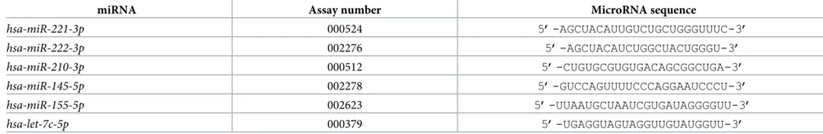
Table 2. Assays employed for miRNA quantification by RT-qPCR and RT-ddPCR.
miRNA Assay number MicroRNA sequence
hsa-miR-221-3p 000524 5’-AGCUACAUUGUCUGCUGGGUUUC-3’ hsa-miR-222-3p 002276 5’-AGCUACAUCUGGCUACUGGGU-3’ hsa-miR-210-3p 000512 5’-CUGUGCGUGUGACAGCGGCUGA-3’ hsa-miR-145-5p 002278 5’-GUCCAGUUUUCCCAGGAAUCCCU-3’ hsa-miR-155-5p 002623 5’-UUAAUGCUAAUCGUGAUAGGGGUU-3’ hsa-let-7c-5p 000379 5’-UGAGGUAGUAGGUUGUAUGGUU-3’ https://doi.org/10.1371/journal.pone.0221923.t002
according to manufacturer’s instructions. The isolated RNA was washed once, with cold 75% ethanol, dried and dissolved in nuclease free water (Sigma-Aldrich) before use. The obtained RNA was stored at -80˚C until the use. The quality of the RNA was determined by spectropho-tometric analysis and by agarose gel electrophoresis. The ratio 260/280 nm was used for deter-mining the overall quality. The electrophoresis on 0,8% agarose in TAE (Tris-acetate-EDTA) buffer was employed for quality checking.
MicroRNA reverse transcription. Obtained total RNA was quantified using SmartSpec
Plus Spectrophotometer (Bio-Rad) and 300 ng of total RNA were reverse transcribed using TaqMan MicroRNA Reverse Transcription Kit (Thermo Fisher Scientific) and specific stem loop primers (Thermo Fisher Scientific) following manufacturer instructions. Obtained miRNA-specific cDNA was stored at -80˚C until PCR analysis.
Real-time quantitative PCR of microRNA. ThreeμL of obtained cDNA were amplified in 25μL (final volume) of RT-qPCR reaction mix, containing 2X TaqMan Universal PCR Mas-ter Mix, no AmpErase UNG (Thermo Fisher Scientific) and 20X TaqMan MicroRNA Assay (Thermo Fisher Scientific) indicated inTable 2. Incremental concentrations (from 25 nM to 200 nM) of anti-miR PNAs were added to the RT-qPCR reaction mix of PNA treated mix, while no PNA was added to the control samples. Sequences of the employed PNAs are reported inTable 1. All qPCR reactions, including no-template controls (NTC) and RT-minus controls, were run in duplicate, using the CFX96 Touch Real Time PCR Detection Sys-tem (Bio-rad). Data analysis and graphic elaborations were performed using CFX Manager Software version 3.1 (Bio-Rad).
Droplet Digital PCR analysis of microRNA. The ability of PNA to arrest miRNA
ampli-fication reaction was also tested using ddPCR; at this purpose 1μL of 1:50 diluted cDNA obtained from U251 cell line was added to ddPCR reaction mix containing 2X ddPCR Super-mix for Probes (no dUTP) (Bio-Rad) and 20X TaqMan MicroRNA Assay (Thermo Fisher Sci-entific). In this case three different concentration anti-miR PNAs were employed: 25, 50 and 100 nM, while no PNA was added to control samples. 20μL of ddPCR reaction mix were mixed with Automated Droplet Generation Oil for Probes (Bio-Rad) and 40μL of droplets emulsion was automatically generated using Automated Droplet Generator (AutoDG) (Bio-Rad). The emulsion was amplified using GeneAmp PCR System 9700 (Thermo Fisher Scien-tific) using the following thermal cycler condition 95˚C for 10 min, 40 cycles of 95˚C for 15 s and 60˚C for 1 min and a final step of 98˚C for 10 min. Genereted droplets were read using the QX200 Droplet Reader, and data analysis was performed with QuantaSoft version 1.7.4 (Bio-Rad).
Statistics. Results are expressed as mean± standard error of the mean (SEM). Compari-sons between groups were made by using paired Student’st test and a one-way analysis of
vari-ance (ANOVA). Statistical significvari-ance was defined withp<0.01.
Results
Cell culture and RNA extraction
The objective of these procedures is to obtain the RNA samples to be employed by the students during the practical laboratory. Depending on the time and the program, it can be also included as a part of the lesson(s). U251 human glioma [63,64] and Calu-3 human airway epi-thelial [65,66] cell lines can be cultured in humidified atmosphere of 5% CO2/air in D-MEM
medium (Gibco) supplemented with 10% fetal bovine serum (Biowest), 100 units/mL penicil-lin and 100μg/mL streptomycin and 1% NEAA (100X) (Non-Essential Amino Acids Solution, Gibco). For RNA extraction cultured cells are trypsinized and collected by centrifugation at 1500 rpm for 10 minutes at 4˚C, washed with cold DPBS (Gibco), lysed with Tri-Reagent
(Sigma-Aldrich), according to manufacturer’s instructions. The isolated RNA is washed once with cold 75% ethanol, dried and dissolved in nuclease-free water (Sigma-Aldrich). The quality of the RNA was determined by spectrophotometric analysis and by agarose gel electrophoresis. The ratio 260/280 nm was used for determining the overall quality (representative examples are shown in Figure F inS1 File). Agarose gel electrophoresis was employed for quality check-ing (representative examples are shown in Figure F inS1 File).
Functions of miR-221-3p and miR-145-5p in U251 and Calu-3 cells
The reason for selecting miR-221-3p and miR-145-5p as miRNA PNA targets is related to pre-viously published studies demonstrating that PNA-mediated inhibition of the activity of these two miRNAs is associated with clinically relevant effects. Brognara et al. [53] reported that a PNA against miR-221-3p is able to induce apoptosis of the treated glioma cell line. More recently Brognara et al. [54] demonstrated that two PNAs, one against miR-221, the other against miR-222-3p were able to induced higher levels of apoptosis when administered to the glioma cells in combination. These results are relevant in the development of PNA-based miRNA targeting in experimental oncology. The key experiments of these studies can be pre-sented as a background to the class using the Figure D inS1 File. As far as miR-145-5p, Fabbri et al. [61] proposed the use of an anti-miR PNA for targeting miR-145-5p, a microRNA reported to suppress the expression of the Cystic Fibrosis Transmembrane conductance Regu-lator (CFTR) gene. Sequence dependent targeting of miR-145-5p was demonstrated in Calu-3 cells, allowing to enhance expression of the miR-145-5p regulated CFTR gene, analyzed at mRNA (RT-qPCR) and protein (western blotting) level. These results are relevant in the devel-opment of PNA-based miRNA targeting for cystic fibrosis [62,67,68]. The key experiments of these studies can be presented as a background to the class using the Figure E inS1 File.
Outline of the practical laboratory program
The outline of the main practical laboratory, starting from the isolated RNA described in sec-tion 3.1, will answer to the following quessec-tions:
• Are PNAs (PNA-a221 and PNA-a145) able to arrest RT-qPCR designed for the amplification of the target miRNA sequences (miR-221-3p for a221 and miR-145-5p for PNA-a145)?
• Are mutated PNAs (PNA-a221-MUT and PNA-a145-MUT) active?
• Are unrelated miRNA sequences amplified in the presence of PNA-a221 and PNA-a145, supporting selectivity of PNA-mediated effects?
Effects of PNA-a221 on RT-qPCR amplification of miR-221-3p sequences
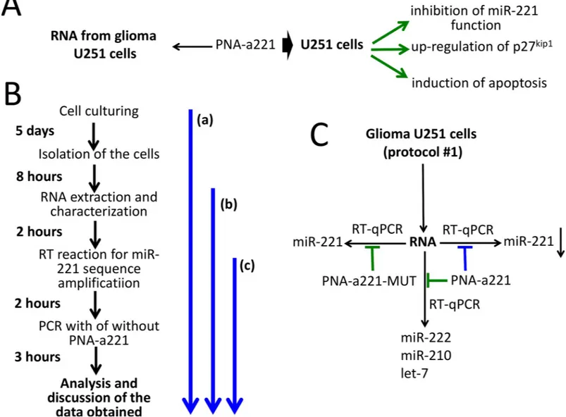
Fig 1shows the outline of the experiments based on the use of PNA-a221. InFig 1Athe biolog-ical effects of the R8-PNA-a221 on glioma cell lines are summarized (see also Figure D inS1 File).Fig 1Bshows the timing of the proposed practical laboratory activity. The key activity is shown as the segment (c) ofFig 1Band starts from the RNA preparation, is based on the per-forming of the PCR following RT in the absence or in the presence of PNA-a221 (as further depicted inFig 1C). Alternatively, U251 cell culture and RNA extraction/characterization— segment (a) ofFig 1B—or only RNA extraction—segment (b) ofFig 1B—might be considered for inclusion in the practical teaching protocol, depending on the available time.
The first set of key results that can be obtained during this practical exercise are shown in
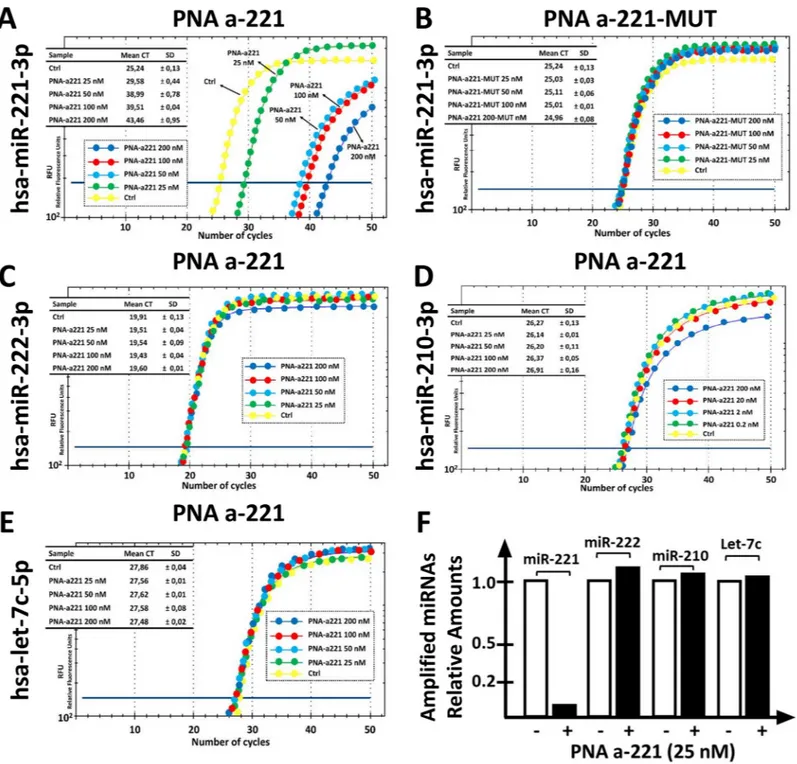
dramatic inhibitory effects on the RT-qPCR amplification of miR-221-3p sequences. It can be easily concluded that 25 nM PNA is sufficient to cause a 75% inhibition of PCR amplification (Fig 2A). On the contrary, the mutated R8-PNA-a221 (for the sequence of the R8-PNA-a221 and R8-PNA-a221-MUT seeTable 1) is completely inactive, even when added at 200 nM (Fig 2A). This first set of results was obtained with high levels of reproducibility, obtaining highly significant values when the data concerning treatments with a221 and R8-PNA-a221-MUT are compared (p <0.0000123 in five independent determinations. The second part of this laboratory exercise is considered inFig 2, panels C-E. The results obtained demonstrate that the treatment with the R8-PNA-a221 has no effects of the RT-qPCR amplification of
miR-Fig 1. Biological effects of a PNA targeting miR-221-3p and outline of the practical laboratory program. A. Scheme of the background available in the literature on
the biological effects of the R8-PNA-a221 on human glioma cell lines (Fabbri et al, 2017). More detailed information is shown in Figure A inS1 File. B. Timing of the laboratory practice, depending on the starting activity (identified by the blue arrows). The key activity is shown as the segment (c). Alternatively, U251 cell culture and RNA extraction/characterization (a) or only RNA extraction (b) might be considered. C. Scheme of the laboratory practice finalized to verify the specificity of the biological activity of the PNA-a221. The extracted U251 RNA is used for RT-qPCR in the presence of the PNA-a221 and the PNA-a221-MUT. The amplified miRNAs are indicated. Expected results (blue: inhibition; green: no inhibition) when PNA-a221 and PNA-a221-MUT are employed and miR-221, miR-222, miR-210 and let-7 sequences amplified by RT-qPCR. Specificity can be demonstrated if inhibition of the RT-qPCR product is obtained amplifying miR-221-3p in the presence of PNA-a221.
222-3p, miR-210-3p and miR let-7c-5p sequences. Altogether, these data are consistent with the hypothesis that the effects of R8-PNA-a221 are highly specific. Of particular interest are the data demonstrating that the R8-PNA-a221 has no effect on the RT-qPCR amplification of miR-222-3p, which shares with miR-221-3p extensive sequence homologies and similar
Fig 2. Effects of the a221 on the RT-PCR amplification of miRNA sequences. A,B. Effects of increasing amounts of a221 (A) and mutated
PNA-a221-MUT (B) on the amplification of miR-221-3p sequences. C-E. Effects of increasing amounts of PNA-a221 on the amplification of miR-222-3p (C), miR-210-3p (D) and let-7c-5p (E) sequences. F. Summary of the effects of 25 nM PNA-a221 on amplification of the indicated miRNA sequences. The comparison of the effects of 50, 100 and 200 nM PNA-a221 are presented in Figure E inS1 File.
biological effects. The summary of the effects of 25 nM R8-PNA-a221 is shown inFig 2F, all the quantitative data in Figure G inS1 File.
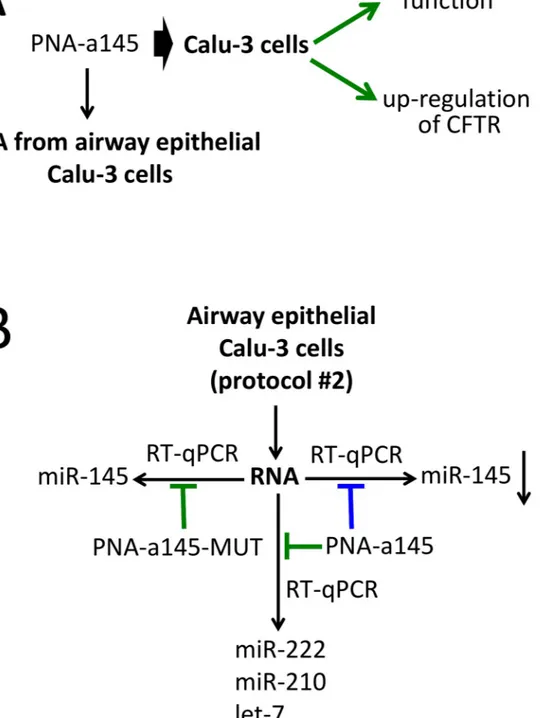
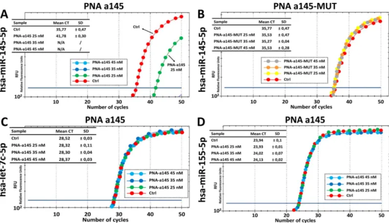
Fig 3. Biological effects of a PNA targeting miR-145-5p and outline of the practical laboratory program. A. Scheme of the
background available in the literature on the biological effects of the R8-PNA-a145 on the Calu-3 cell line (Fabbri et al., 2017). More detailed information is shown in Figure D inS1 File. B. Scheme of the laboratory practice finalized to verify the specificity of the biological activity of the PNA-a145. The extracted Calu-3 RNA is used for RT-qPCR in the presence of the PNA-a145 and the PNA-a145-MUT (green: no inhibition; blue: inhibition). The amplified miRNAs (miR-145-5p, let-7c-5p and miR-155-5p) are indicated. Specificity can be demonstrated if inhibition of the RT-qPCR product is obtained amplifying miR-145-5p in the presence of PNA-a145.
Effects of PNA-a145 on RT-qPCR amplification of miR-145-5p sequences
Fig 3shows the outline of the experiments based on the use of PNA-a145. InFig 3Athe biolog-ical effects of the R8-PNA-a145 on Calu-3 cells (see also Figure E inS1 File) are summarized. The timing of the proposed practical laboratory activity is similar to that reported inFig 1Bfor the R8-PNA-a221. The key activity is shown inFig 3B.
The first key results that can be obtained during this practical exercise are shown inFig 4, panel A and B. As clearly evident, the R8-PNA-a145 has dramatic inhibitory effects on the RT-qPCR amplification of miR-145-5p sequences. It can be easily concluded that 25 nM PNA is sufficient to cause a 90% inhibition of PCR amplification (Fig 4A). On the contrary, the mutated R8-PNA-a145 (for the sequence of the R8-PNA-a145 and R8-PNA-a145-MUT see
Table 1) is completely inactive (Fig 4B). The second part of this laboratory exercise is consid-ered inFig 4, panels C and D. The results obtained demonstrate that the treatment with the R8-PNA-a145 has no effects of the RT-qPCR amplification of miR-155-5p and let-7c-5p sequences. Altogether, these data are consistent with the hypothesis that the effects of R8-PNA-a145 are highly specific. The summary of the effects of different concentrations of R8-PNA-a145 is shown in Figure H inS1 File).
Employment of other RT-PCR systems: Droplet digital PCR
Some of the experiments reported inFig 2were repeated using droplet digital PCR (ddPCR), another RT-PCR system routinely used for miRNA quantification [69]. Thanks to sample par-tition ddPCR allows the absolute miRNA sequences quantification with more precision
Fig 4. Effects of the PNA-a145 on the RT-PCR amplification of miRNA sequences. A,B. Effects of 25 nM PNA-a145 (A) and mutated PNA-a145-MUT (B) on the
amplification of miR-145-5p sequences. C,D. Effects of 25 nM PNA-a145 on the amplification of miR-155-5p (C) and let-7c-5p (D) sequences. A summary of increasing concentrations of PNA-a145 are presented in Figure F inS1 File.
compare to traditional RT-qPCR methods. Considering the dramatic reduction of miR-221-3p amplification detected by RT-qPCR when 200 nM of R8-PNA-a221 are employed, only three PNA concentrations were considered: 25, 50 and 100 nM. Obtained key results are simi-lar to those obtained by RT-qPCR, with minor differences easily explained by the differences within miRNA quantification methods. In fact, while in RT-qPCR 25 nM of R8-PNA-a221 is sufficient to inhibit of 75% the miR-221-3p sequence amplification, when the same PNA con-centration is employed in RT-ddPCR very limited effects (reduction of 17%) were detected, while more significative effects were recorded with 50 nM of PNA (35%) and 100 nM (100%) (Fig 5). Obtained data are quite expected considering sample and PNA partitioning in thou-sands of droplets. According to those obtained by RT-qPCR no effects of R8-PNA-a221 were detected on others miRNA sequences (i.e miR-222-3p) and no activity was founded when R8-PNA-a221-MUT was employed, even at highest concentration (100 nM) (Fig 6).
Discussion
Simple experiments answering to key issues in applied pharmacology could be of great interest in the teaching, with particular focus to the possibility to set-up practical exercises in labora-tory practice delivered to student in the field of biotechnology, pharmaceutics, applied biology.
Fig 5. Effects of R8-PNA-a221 on miR-221-3p sequence detection by RT-ddPCR. A. 1D RT-ddPCR plot obtained after the addition of incremental concentration of
R8-PNA-a221 or R8-PNA-a221-MUT to the reaction mix. B. miR-221-3p content in 1:50 diluted cDNA obtained from U251 cells: 2D plots. C. miR-221-3p content detected after the addition of incremental R8-PNA-a221concentrations. D. miR-221-3p content detected after the addition of incremental R8-PNA-a221-MUT concentrations.
One of the emerging pharmaceutical approaches is the so called miRNA therapy, based on antimiRNA strategy or on miRNA replacement, depending on the role covered by the target miRNA [11–14]. Alteration of microRNA expression has been demonstrated to be associated with different human pathologies [69–75], as well as guided alterations of specific miRNAs have been suggested as novel approaches to develop innovative therapeutic protocols [39,41,
42,76–78]. Several reports conclusively demonstrated that microRNA are deeply involved in tumor onset and progression, behaving as tumor promoting miRNAs (oncomiRNA and metastamiRNAs) as well as tumor suppressor miRNAs [79–83]. In general, a miRNA able to promote cancer targets mRNA coding for tumor-suppression proteins, while microRNAs exhibiting tumor-suppression properties usually target mRNAs coding oncoproteins [14,84,85].
As far as the antimiRNA therapy, among the most interesting biomolecules to be analyzed are peptide-nucleic acids (PNAs). These are in fact reagents of great impact in antisense ther-apy, and have been proposed in a large spectrum of applications.
For instance, PNAs have been recently proposed as antisense molecules targeting mRNAs, as molecules able to target gene promoters through the formation of triple-helix structures, artificial promoters, or decoy molecules able to target transcription factors [17,47,85–89].
In this manuscript we present simple experiments that can be the basis for laboratory prac-tical teaching with the aim of determining: (a) the possible PNA-mediated arrest in RT-qPCR, to be eventually used to demonstrate PNA targeting of selected miRNAs; (b) the possible lack of activity on mutated PNA sequences; (c) the effects (if any) on the amplification of other unrelated miRNA sequences.
The results which can be obtained during this laboratory teaching activity support the fol-lowing conclusions: PNA-mediated arrest in RT-qPCR can be analyzed in a easy way; mutated PNA sequences are completely inactive; the effects of the employed PNAs are specific and no inhibitory effect occurs on other unrelated miRNA sequences.
Fig 6. Effects of R8-PNA-a221 on miR-222-3p sequence detection by RT-ddPCR. A. 1D RT-ddPCR plot obtained after the addition of incremental concentration of
R8-PNA-a221, miR-222-3p is amplified. B. miR-222-3p content in 1:50 diluted cDNA obtained from U251 cells: 2D plots. C. miR-222-3p content detected after the addition of incremental R8-PNA-a221concentrations.
This activity is simple (cell culture, RNA extraction, RT-qPCR are all well-established tech-nologies), fast (starting from isolated and characterized RNA, few hours are just necessary), highly reproducible (therefore easily employed by even untrained students).
On the other hand, these laboratory lessons require some facilities, the most critical being the availability of instruments for PCR. While, this might be a problem in the case these instru-ments are not available, we would like to underline that determination of the presence or of a lack of amplified product can be also obtained using standard analytical approaches based on agarose gel electrophoresis.
Supporting information
S1 File. These Supplementary materials include some Figures that can be used for explaining
the impact of PNAs in experimental therapeutic protocols (Figures A-C), the effects of PNAs against microRNAs miR-221-3p (Figure D) and miR-145-5p (Figure E). In addition, in the Supplementary Figure F the analysis of integrity of the RNA preparation is shown (see the pro-tocols for the laboratory practice depicted in Figs1and3of the main text). Finally, Figures G and H report the inhibitory effects of the PNAs against miR-221-3p (a221 and PNA-a221-MUT, Figure G) and of the PNAs against miR-145-5p (PNA-a221 and PNA-PNA-a221-MUT, Figure H) on RT-qPCR amplification of the target miR-221-3p and miR-145-5p, and of the control miRNA sequences (miR-222-3p, let-7c-5p and miR-210-3p for PNA-a221; let-7c-5p and miR-155-5p for PNA-a145).
(DOCX)
Acknowledgments
We thank Prof. Giulio Cabrini (Laboratory of Molecular Pathology, Laboratory of Clinical Chemistry and Haematology, University-Hospital, Verona, Italy) for useful comments to this study.
Author Contributions
Conceptualization: Jessica Gasparello, Roberto Gambari, Alessia Finotti. Data curation: Jessica Gasparello, Chiara Papi, Matteo Zurlo.
Funding acquisition: Roberto Gambari.
Methodology: Jessica Gasparello, Roberto Corradini.
Resources: Roberto Corradini, Roberto Gambari, Alessia Finotti. Supervision: Roberto Gambari, Alessia Finotti.
Writing – original draft: Roberto Gambari.
Writing – review & editing: Jessica Gasparello, Roberto Corradini, Roberto Gambari, Alessia
Finotti.
References
1. Greenhalgh T, Howick J, Maskrey N. Evidence Based Medicine Renaissance Group. Evidence based medicine: a movement in crisis? BMJ. 2014; 348: g3725.https://doi.org/10.1136/bmj.g3725PMID: 24927763
2. Polly P, Marcus N, Maguire D, Belinson Z, Velan GM. Evaluation of an adaptive virtual laboratory envi-ronment using Western Blotting for diagnosis of disease. BMC Med Educ. 2014; 14: 222.https://doi. org/10.1186/1472-6920-14-222PMID:25331335
3. Laidlaw A, Aiton J, Struthers J, Guild S. Developing research skills in medical students: AMEE Guide No. 69. Med Teach. 2012; 34: e754–71.https://doi.org/10.3109/0142159X.2012.704438PMID: 22905661
4. Amgad M, Man Kin Tsui M, Liptrott SJ, Shash E. Medical Student Research: An Integrated Mixed-Meth-ods Systematic Review and Meta-Analysis. PLoS One. 2015; 10: e0127470.https://doi.org/10.1371/ journal.pone.0127470PMID:26086391
5. Coyne L, Merritt TA, Parmentier BL, Sharpton RA, Takemoto JK. The Past, Present, and Future of Vir-tual Reality in Pharmacy Education. Am J Pharm Educ. 2019; 83:7456.https://doi.org/10.5688/ ajpe7456PMID:31065173
6. de Vries LE, May M. Virtual laboratory simulation in the education of laboratory technicians-motivation and study intensity. Biochem Mol Biol Educ. 2019; 47:257–262.https://doi.org/10.1002/bmb.21221 PMID:30748084
7. Garcia-Bonete MJ, Jensen M, Katona G. A practical guide to developing virtual and augmented reality exercises for teaching structural biology. Biochem Mol Biol Educ. 2019; 47: 16–24.https://doi.org/10. 1002/bmb.21188PMID:30475432
8. Xu X, Allen W, Miao Z, Yao J, Sha L, Chen Y. Exploration of an interactive "Virtual and Actual Com-bined" teaching mode in medical developmental biology. Biochem Mol Biol Educ. 2018; 46: 585–591. https://doi.org/10.1002/bmb.21174PMID:30311730
9. Rubio M, Sa´nchez-Ronco M, Mohedano R, Hernando A. The impact of participatory teaching methods on medical students’ perception of their abilities and knowledge of epidemiology and statistics. PLoS One. 2018; 13:e0202769.https://doi.org/10.1371/journal.pone.0202769PMID:30133528
10. Mo¨ller R, Shoshan M. Does reality meet expectations? An analysis of medical students’ expectations and perceived learning during mandatory research projects. BMC Med Educ. 2019; 19: 93.https://doi. org/10.1186/s12909-019-1526-xPMID:30925877
11. Chakraborty C, Sharma AR, Sharma G, Doss CGP, Lee SS. Therapeutic miRNA and siRNA: Moving from Bench to Clinic as Next Generation Medicine. Mol Ther Nucleic Acids. 2017; 8: 132–143.https:// doi.org/10.1016/j.omtn.2017.06.005PMID:28918016
12. Christopher AF, Kaur RP, Kaur G, Kaur A, Gupta V, Bansal P. MicroRNA therapeutics: Discovering novel targets and developing specific therapy. Perspect Clin Res. 2016; 7: 68–74.https://doi.org/10. 4103/2229-3485.179431PMID:27141472
13. Gambari R, Fabbri E, Borgatti M, Lampronti I, Finotti A, Brognara E, et al. Targeting microRNAs involved in human diseases: a novel approach for modification of gene expression and drug develop-ment. Biochem Pharmacol. 2011; 82: 1416–1429.https://doi.org/10.1016/j.bcp.2011.08.007PMID: 21864506
14. Gambari R, Brognara E, Spandidos DA, Fabbri E. Targeting oncomiRNAs and mimicking tumor sup-pressor miRNAs: New trends in the development of miRNA therapeutic strategies in oncology (Review). Int J Oncol. 2016; 49: 5–32.https://doi.org/10.3892/ijo.2016.3503PMID:27175518
15. Nielsen PE. Gene targeting and expression modulation by peptide nucleic acids (PNA). Curr Pharm Des. 2010; 16: 3118–3123. PMID:20687874
16. Gambari R. Peptide-nucleic acids (PNAs): a tool for the development of gene expression modifiers. Curr Pharm Des. 2001; 7: 1839–1862. PMID:11562312
17. Nielsen PE. Peptide nucleic acids (PNA) in chemical biology and drug discovery. Chem Biodivers. 2010; 7: 786–804.https://doi.org/10.1002/cbdv.201000005PMID:20397216
18. Filipowicz W, Jaskiewicz L, Kolb FA, Pillai RS. Post-transcriptional gene silencing by siRNAs and miR-NAs. Curr Opin Struct Biol. 2005; 15: 331–341. PMID:15925505
19. He L, Hannon GJ. MicroRNAs: small RNAs with a big role in gene regulation. Nat Rev Genet. 2004; 5: 522–531. PMID:15211354
20. Lim LP, Lau NC, Garrett-Engele P, Grimson A, Schelter JM, Castle J, et al. Microarray analysis shows that some microRNAs downregulate large numbers of target mRNAs. Nature. 2005; 433: 769–773. PMID:15685193
21. Sontheimer EJ, Carthew RW. Silence from within: endogenous siRNAs and miRNAs. Cell. 2005; 122: 9–12. PMID:16009127
22. Monga I, Kumar M. Computational Resources for Prediction and Analysis of Functional miRNA and Their Targetome. Methods Mol Biol. 2019; 1912: 215–250. https://doi.org/10.1007/978-1-4939-8982-9_9PMID:30635896
23. Alvarez-Garcia I, Miska EA. MicroRNA functions in animal development and human disease. Develop-ment. 2005; 132: 4653–4662. PMID:16224045
24. O’Brien J, Hayder H, Zayed Y, Peng C. Overview of MicroRNA Biogenesis, Mechanisms of Actions, and Circulation. Front Endocrinol (Lausanne). 2018; 9: 402.
25. Taylor MA, Schiemann WP. Therapeutic Opportunities for Targeting microRNAs in Cancer. Mol Cell Ther. 2014; 2: 1–13.
26. Nana-Sinkam SP, Croce CM. Clinical applications for microRNAs in cancer. Clin Pharmacol Ther. 2013; 93: 98–104.https://doi.org/10.1038/clpt.2012.192PMID:23212103
27. Piva R, Spandidos DA, Gambari R. From microRNA functions to microRNA therapeutics: novel targets and novel drugs in breast cancer research and treatment. Int J Oncol. 2013; 43: 985–994.https://doi. org/10.3892/ijo.2013.2059PMID:23939688
28. Mollaei H, Safaralizadeh R, Rostami Z. MicroRNA replacement therapy in cancer. J Cell Physiol. 2019; 234: 12369–12384.https://doi.org/10.1002/jcp.28058PMID:30605237
29. Cheng CJ, Bahal R, Babar IA, Pincus Z, Barrera F, Liu C, et al. MicroRNA silencing for cancer therapy targeted to the tumour microenvironment. Nature. 2015; 518: 107–110.https://doi.org/10.1038/ nature13905PMID:25409146
30. Weiler, Hunziker J, Hall J. Anti-miRNA oligonucleotides (AMOs): ammunition to target miRNAs impli-cated in human disease? Gene Ther. 2006; 13: 496–502. PMID:16195701
31. Lu Y, Xiao J, Lin H, Bai Y, Luo X, Wang Z, et al. A single antimicroRNA antisense oligodeoxyribonucleo-tide (AMO) targeting multiple microRNAs offers an improved approach for microRNA interference. Nucleic Acids Res. 2009; 37: e24.https://doi.org/10.1093/nar/gkn1053PMID:19136465
32. Obad S, dos Santos CO, Petri A, Heidenblad M, Broom O, Ruse C, et al. Silencing of microRNA families by seed-targeting tiny LNAs. Nat Genet. 2011; 43: 371–378.https://doi.org/10.1038/ng.786PMID: 21423181
33. Elme´n J, Lindow M, Schu¨tz S, Lawrence M, Petri A, Obad S, et al. LNA-mediated microRNA silencing in non-human primates. Nature. 2008; 452: 896–899.https://doi.org/10.1038/nature06783PMID: 18368051
34. Stenvang J, Silahtaroglu AN, Lindow M, Elmen J, Kauppinen S. The utility of LNA in microRNA based cancer diagnostics and therapeutics. Semin Cancer Biol. 2008; 18: 89–102.https://doi.org/10.1016/j. semcancer.2008.01.004PMID:18295505
35. Staedel C, Varon C, Nguyen PH, Vialet B, Chambonnier L, Rousseau B, et al. Inhibition of Gastric Tumor Cell Growth Using Seed-targeting LNA as Specific, Long-lasting MicroRNA Inhibitors. Mol Ther Nucleic Acids. 2015; 4: e246.https://doi.org/10.1038/mtna.2015.18PMID:26151747
36. Ebert MS, Neilson JR, Sharp PA. MicroRNA sponges: competitive inhibitors of small RNAs in mamma-lian cells. Nat Methods. 2007; 4: 721–726.https://doi.org/10.1038/nmeth1079PMID:17694064 37. Ebert MS, Sharp PA. MicroRNA sponges: progress and possibilities. RNA. 2010; 16: 2043–2050.
https://doi.org/10.1261/rna.2414110PMID:20855538
38. Kluiver J, Slezak-Prochazka I, Smigielska-Czepiel K, Halsema N, Kroesen BJ, van den Berg A. Genera-tion of miRNA sponge constructs. Methods. 2012; 58: 113–117.https://doi.org/10.1016/j.ymeth.2012. 07.019PMID:22836127
39. Kluiver J, Gibcus JH, Hettinga C, Adema A, Richter MK, Halsema N, et al. Rapid generation of micro-RNA sponges for micromicro-RNA inhibition. PLoS One. 2012; 7: e29275.https://doi.org/10.1371/journal. pone.0029275PMID:22238599
40. Tay FC, Lim JK, Zhu H, Lin LC, Wang S. Using artificial microRNA sponges to achieve microRNA loss-of-function in cancer cells. Adv Drug delivery Rev. 2015; 81: 117–127.
41. Liu Y, Han Y, Zhang H, Nie L, Jiang Z, Fa P, et al. Synthetic miRNA-mowers targeting miR-183-96-182 cluster or miR-210 inhibit growth and migration and induce apoptosis in bladder cancer cells. PLoS One. 2012; 7: e52280.https://doi.org/10.1371/journal.pone.0052280PMID:23284967
42. Bak RO, Hollensen AK, Mikkelsen JG. Managing microRNAs with vector-encoded decoy-type inhibi-tors. Mol Ther. 2013; 21: 1478–1485.https://doi.org/10.1038/mt.2013.113PMID:23752312 43. Wang H, Xu T, Jiang Y, Yan Y, Qin R, Chen J. MicroRNAs in human glioblastoma: from bench to
beside. Front Biosci (Landmark Ed). 2015; 20: 105–18. PMID:25553442
44. Lennox KA, Behlke MA. Chemical modification and design of antimiRNA oligonucleotides. Gene Ther. 2011; 18: 1111–1120.https://doi.org/10.1038/gt.2011.100PMID:21753793
45. Nielsen PE, Egholm M, Berg RH, Buchardt O. Sequence-selective recognition of DNA by strand dis-placement with a thymine-substituted polyamide. Science. 1991; 254: 1497–1500. PMID:1962210 46. Nielsen PE. Targeting double stranded DNA with peptide nucleic acid (PNA). Curr Med Chem. 2001; 8:
545–550. PMID:11281841
47. Gambari R. Biological activity and delivery of peptide nucleic acids (PNA)-DNA chimeras for transcrip-tion factor decoy (TFD) pharmacotherapy. Curr Med Chem. 2004; 11: 1253–1263. PMID:15134518 48. Gambari R, Borgatti M, Bezzerri V, Nicolis E, Lampronti I, Dechecchi MC, et al. Decoy
gene expression in cystic fibrosis cells infected with Pseudomonas aeruginosa. Biochem Pharmacol. 2010; 80: 1887–1894.https://doi.org/10.1016/j.bcp.2010.06.047PMID:20615393
49. Pandey VN, Upadhyay A, Chaubey B. Prospects for antisense peptide nucleic acid (PNA) therapies for HIV. Expert Opin Biol Ther. 2009; 9: 975–989.https://doi.org/10.1517/14712590903052877PMID: 19534584
50. Fabani MM, Gait MJ. MiR-122 targeting with LNA/2’-O-methyl oligonucleotide mixmers, peptide nucleic acids (PNA), and PNA-peptide conjugates. RNA. 2008; 14: 336–346.https://doi.org/10.1261/rna. 844108PMID:18073344
51. Fabani MM, Abreu-Goodger C, Williams D, Lyons PA, Torres AG, Smith KG, et al. Efficient inhibition of miR-155 function in vivo by peptide nucleic acids. Nucleic Acids Research. 2010; 38: 4466–4475. https://doi.org/10.1093/nar/gkq160PMID:20223773
52. Brognara E, Fabbri E, Aimi F, Manicardi A, Bianchi N, Finotti A, et al. Peptide nucleic acids targeting miR-221 modulate p27Kip1 expression in breast cancer MDA-MB-231 cells. Int J Oncol. 2012; 41: 2119–2127.https://doi.org/10.3892/ijo.2012.1632PMID:22992757
53. Brognara E, Fabbri E, Bazzoli E, Montagner G, Ghimenton C, Eccher A, et al. Uptake by human glioma cell lines and biological effects of a peptide-nucleic acids targeting miR-221. J Neurooncol. 2014; 118: 19–28.https://doi.org/10.1007/s11060-014-1405-6PMID:24595467
54. Brognara E, Fabbri E, Montagner G, Gasparello J, Manicardi A, Corradini R, et al. High levels of apopto-sis are induced in human glioma cell lines by co-administration of peptide nucleic acids targeting miR-221 and miR-222. Int J Oncol. 2015; 48: 1029–1038.https://doi.org/10.3892/ijo.2015.3308PMID: 26708164
55. Manicardi A, Fabbri E, Tedeschi T, Sforza S, Bianchi N, Brognara E, et al. Cellular Uptakes, biostabil-ities and anti-miR-210 activbiostabil-ities of chiral Arginine-PNAs in leukaemic K562 cells. Chembiochem. 2012; 13: 1327–1337.https://doi.org/10.1002/cbic.201100745PMID:22639449
56. Gupta A, Quijano E, Liu Y, Bahal R, Scanlon SE, Song E, et al. Anti-tumor Activity of miniPEG-γ -Modi-fied PNAs to Inhibit MicroRNA-210 for Cancer Therapy. Mol Ther Nucleic Acids. 2017; 9: 111–119. https://doi.org/10.1016/j.omtn.2017.09.001PMID:29246289
57. Manicardi A, Gambari R, de Cola L, Corradini R. Preparation of Anti-miR PNAs for Drug Development and Nanomedicine. Methods Mol Biol. 2018; 1811: 49–63.https://doi.org/10.1007/978-1-4939-8582-1_ 4PMID:29926445
58. Yan LX, Wu QN, Zhang Y, Li YY, Liao DZ, Hou JH, et al. Knockdown of miR-21 in human breast cancer cell lines inhibits proliferation, in vitro migration and in vivo tumor growth. Breast Cancer Res. 2011; 13: R2.https://doi.org/10.1186/bcr2803PMID:21219636
59. Fabbri E, Manicardi A, Tedeschi T, Sforza S, Bianchi N, Brognara E, et al. Modulation of the biological activity of microRNA-210 with peptide nucleic acids (PNAs). ChemMedChem. 2011; 6: 2192–2202. https://doi.org/10.1002/cmdc.201100270PMID:22012891
60. Garofalo M, Quintavalle C, Romano G, Croce CM, Condorelli G. miR221/222 in Cancer: Their Role in Tumor Progression and Response to Therapy Curr Mol Med. 2012; 12: 27–33.
61. Fabbri E, Tamanini A, Jakova T, Gasparello J, Manicardi A, Corradini R, et al. A Peptide Nucleic Acid against MicroRNA miR-145-5p Enhances the Expression of the Cystic Fibrosis Transmembrane Con-ductance Regulator (CFTR) in Calu-3 Cells. Molecules. 2017; 23: E71.https://doi.org/10.3390/ molecules23010071PMID:29286300
62. Finotti A, Gasparello J, Fabbri E, Tamanini A, Corradini R, Dechecchi MC, et al. Enhancing the Expres-sion of CFTR Using Antisense Molecules Against MicroRNA miR-145-5p. Am J Respir Crit Care Med. 2019; In Press.
63. Li H, Lei B, Xiang W, Wang H, Feng W, Liu Y et al. Differences in Protein Expression between the U251 and U87 Cell Lines. Turk Neurosurg. 2017; 27: 894–903.https://doi.org/10.5137/1019-5149.JTN. 17746-16.1PMID:27651343
64. Bigner DD, Bigner SH, Ponte´n J, Westermark B, Mahaley MS, Ruoslahti E, et al. Heterogeneity of Genotypic and Phenotypic Characteristics of Fifteen Permanent Cell Lines Derived from Human Glio-mas. Journal of Neuropathology & Experimental Neurology. 1981; 40: 201–229.
65. Shen BQ, Finkbeiner WE, Wine JJ, Mrsny RJ, Widdicombe JH. Calu-3: A human airway epithelial cell line that shows cAMP-dependent Cl-secretion. Am J Physiol. 1994; 266: L493–L501. PMID:7515578 66. Kreft ME, Jerman UD, LasičE, Hevir-Kene N, Rizˇner TL, Peternel L e al. The characterization of the
human cell line Calu-3 under different culture conditions and its use as an optimized in vitro model to investigate bronchial epithelial function. Eur J Pharm Sci. 2015; 69: 1–9.https://doi.org/10.1016/j.ejps. 2014.12.017PMID:25555374
67. Dutta RK, Chinnapaiyan S, Rasmussen L, Raju SV, Unwalla HJ. A Neutralizing Aptamer to TGFBR2 and miR-145 Antagonism Rescue Cigarette Smoke- and TGF-β-Mediated CFTR Expression. Mol Ther. 2019; 27: 442–455.https://doi.org/10.1016/j.ymthe.2018.11.017PMID:30595527
68. Lutful Kabir F, Ambalavanan N, Liu G, Li P, Solomon GM, Lal CV, et al. MicroRNA-145 Antagonism Reverses TGF-βInhibition of F508del CFTR Correction in Airway Epithelia. Am J Respir Crit Care Med. 2018; 197: 632–643.https://doi.org/10.1164/rccm.201704-0732OCPMID:29232160
69. Finotti A, Allegretti M, Gasparello J, Giacomini P, Spandidos DA, Spoto G, et al. Liquid biopsy and PCR-free ultrasensitive detection systems in oncology. Int J Oncol. 2018; 53: 1395–1434.https://doi. org/10.3892/ijo.2018.4516PMID:30085333
70. Gillen AE, Gosalia N, Leir SH Harris A. MicroRNA regulation of expression of the cystic fibrosis trans-membrane conductance regulator gene. Biochem J. 2011; 438: 25–32.https://doi.org/10.1042/ BJ20110672PMID:21689072
71. Oglesby IK, Chotirmall SH, McElvaney NG, Greene CM. Regulation of cystic fibrosis transmembrane conductance regulator by microRNA-145, -223, and -494 is altered inΔF508 cystic fibrosis airway epi-thelium. J Immunol. 2013; 190: 3354–3362.https://doi.org/10.4049/jimmunol.1202960PMID: 23436935
72. Chen L, Kang C. miRNA interventions serve as ’magic bullets’ in the reversal of glioblastoma hallmarks. Oncotarget. 2015; 6: 38628–38642.https://doi.org/10.18632/oncotarget.5926PMID:26439688 73. Costa PM, Cardoso AL, Mano M, de Lima MC. MicroRNAs in glioblastoma: role in pathogenesis and
opportunities for targeted therapies. CNS Neurol Disord Drug Targets. 2015; 14: 222–238. PMID: 25613511
74. Piwecka M, Rolle K, Belter A, Barciszewska AM,Żywicki M, Michalak M, et al. Comprehensive analysis of microRNA expression profile in malignant glioma tissues. Mol Oncol. 2015; 9: 1324–1340.https://doi. org/10.1016/j.molonc.2015.03.007PMID:25864039
75. Banelli B, Forlani A, Allemanni G, Morabito A, Pistillo MP, Romani M. MicroRNA in Glioblastoma: An Overview. Int J Genomics. 2017;7639084.https://doi.org/10.1155/2017/7639084PMID:29234674 76. Jung J, Yeom C, Choi YS, Kim S, Lee E, Park MJ, et al. Kang S.W., Kim S.B. & Chang S. Simultaneous
inhibition of multiple oncogenic miRNAs by a multi-potent microRNA sponge. Oncotarget. 2015; 6: 20370–20387.https://doi.org/10.18632/oncotarget.4827PMID:26284487
77. Bertucci A, Lu¨lf H, Septiadi D, Manicardi A, Corradini R, De Cola L. Intracellular delivery of peptide nucleic acid and organic molecules using zeolite-L nanocrystals. Adv Healthc Mater. 2014; 3: 1812– 1817.https://doi.org/10.1002/adhm.201400116PMID:24789252
78. Bertucci A, Prasetyanto EA, Septiadi D, Manicardi A, Brognara E, Gambari R, et al. Combined delivery of temozolomide and anti-mir221 PNA using mesoporous silica nanoparticles induces apoptosis in resistant glioma cells. Small. 2015; 11: 5687–5695.https://doi.org/10.1002/smll.201500540PMID: 26395266
79. Liu S, Yin F, Zhang J, Wicha MS, Chang AE, Fan W, et al. Regulatory roles of miRNA in the human neu-ral stem cell transformation to glioma stem cells. J Cell Biochem. 2014; 115: 1368–1680.https://doi.org/ 10.1002/jcb.24786PMID:24519663
80. Zhou J, Wang W, Gao Z, Peng X, Chen X, Chen W, et al. MicroRNA-155 promotes glioma cell prolifera-tion via the regulaprolifera-tion of MXI1. PLoS One. 2013; 8: e83055.https://doi.org/10.1371/journal.pone. 0083055PMID:24376632
81. D’Urso PI, D’Urso OF, Storelli C, Mallardo M, Gianfreda CD, Montinaro A, et al. miR-155 is up-regulated in primary and secondary glioblastoma and promotes tumour growth by inhibiting GABA receptors. Int J Oncol. 2012; 41: 228–234.https://doi.org/10.3892/ijo.2012.1420PMID:22470130
82. Chan XH, Nama S, Gopal F, Rizk P, Ramasamy S, Sundaram G, et al. Targeting glioma stem cells by functional inhibition of a prosurvival oncomiR-138 in malignant gliomas. Cell Rep. 2012; 2: 591–602. https://doi.org/10.1016/j.celrep.2012.07.012PMID:22921398
83. Khalil S, Fabbri E, Santangelo A, Bezzerri V, Cantu` C, Di Gennaro G, et al. miRNA array screening reveals cooperative MGMT-regulation between miR-181d-5p and miR-409-3p in glioblastoma. Onco-target. 2016; 7: 28195–28206.https://doi.org/10.18632/oncotarget.8618PMID:27057640
84. Svoronos AA, Engelman DM, Slack FJ. OncomiR or Tumor Suppressor? The Duplicity of MicroRNAs in Cancer. Cancer Res. 2016; 76: 3666–3670.https://doi.org/10.1158/0008-5472.CAN-16-0359PMID: 27325641
85. Hnedzko D, McGee DW, Karamitas YA, Rozners E. Sequence-selective recognition of double-stranded RNA and enhanced cellular uptake of cationic nucleobase and backbone-modified peptide nucleic acids. RNA. 2017; 23: 58–69.https://doi.org/10.1261/rna.058362.116PMID:27742909
86. Borgatti M, Lampronti I, Romanelli A, Pedone C, Saviano M, Bianchi N, et al. Transcription factor decoy molecules based on a peptide nucleic acid (PNA)-DNA chimera mimicking Sp1 binding sites. J Biol Chem. 2003; 278: 7500–7509. PMID:12446679
87. Pession A, Tonelli R, Fronza R, Sciamanna E, Corradini R, Sforza S, et al. Targeted inhibition of NMYC by peptide nucleic acid in N-myc amplified human neuroblastoma cells: cell-cycle inhibition with induc-tion of neuronal cell differentiainduc-tion and apoptosis. Int J Oncol. 2004; 24: 265–272. PMID:14719101 88. McNeer NA, Chin JY, Schleifman EB, Fields RJ, Glazer PM, Saltzman WM. Nanoparticles deliver
tri-plex-forming PNAs for site–specific genomic recombination in CD34+ human hematopoietic progeni-tors. Mol Ther. 2011; 19: 172–180.https://doi.org/10.1038/mt.2010.200PMID:20859257
89. Macadangdang B, Zhang N, Lund PE, Marple AH, Okabe M, Gottesman MM, et al. Inhibition of multi-drug resistance by SV40 pseudovirion delivery of an antigene peptide nucleic acid (PNA) in cultured cells. PLoS One. 2011; 6: e17981.https://doi.org/10.1371/journal.pone.0017981PMID:21445346