UNIVERSITY OF CATANIA
DEPARTMENT OF AGRI-FOOD AND ENVIRONMENTAL SYSTEMS MANAGEMENT
INTERNATIONAL PhD PROGRAMME IN
PLANT HEALTH TECHNOLOGIES AND PROTECTION OF AGRO-ECOSYSTEMS
CYCLE XXV (2009-2012)
Ben Attia Sarra
Chronobiological plasticity in Honeybee and its association
with differences in the organization of the molecular pathway
between central and peripheral clocks: the brain and flight
muscle tissues
COORDINATOR SUPERVISOR
Prof. Carmelo Rapisarda Prof. Carmelo Rapisarda
CO-SUPERVISOR Prof. Gaetana Mazzeo
DEDICATION
َنِم َو اًتوُيُب ِلاَبِجْلا َنِم يِذِخَّتا ِنَأ ِل ْحَّنلا ىَلِإ َكُّبَر ٰىَح ْوَأ َو
َنوُش ِرْعَي اَّمِم َو ِرَجَّشلا
(Verse 16:68)And thy Lord taught the bee to build its cells in hills, on trees, and in
“men’s” habitations;
ُجُر ْخَي ۚ ًلًُلُذ ِكِّب َر َلُبُس يِكُل ْساَف ِتا َرَمَّثلا ِّلُك ْنِم يِلُك َّمُث
ْنِم
يِف َّنِإ ۗ ِساَّنلِل ٌءاَفِش ِهيِف ُهُنا َوْلَأ ٌفِلَت ْخُم ٌباَرَش اَهِنوُطُب
َنوُرَّكَفَتَي ٍم ْوَقِل ًةَي َلَ َكِل
َٰذ
(Verse 16:69)Then to eat of all the produce (of the earth), and follow the ways of
thy Lord made smooth: there issues from within their bodies a drink of
varying colors, wherein is healing for men: verily in this is a Sign for
those who give thought.
Sura لحنلا - An-Nahl
(Hive Bee found in Holy Quran)
ACKNOWLEDGMENTS
Simply by words, it is not easy to acknowledge and warmly thank all who supported my work and helped my efforts, allowing me to reach my goals and put into practice my research ideas.
In a chronological order, Dr Barbara Murphy (from the School of Agriculture, Food Science & Veterinary Medicine of the University College Dublin, Ireland) has been of a basic importance in my higher education. Under her supervision, I developed my master thesis and I got my Master of Science. She introduced me in the fancy world of chronobiology and, in the course of our many pleasant conversation, “turned on my lamp” on the possibility to explore chronobiological behavior in insects, with special reference to social ones.
Together with her, I like to express my gratitude to my principal PhD supervisor, Prof. Carmelo Rapisarda (from the Department of Agri-food and Environmental Systems Management of the University of Catania, Italy), without whom I would not have been able to conclude this PhD. Since the beginning, he trusted my project proposal and was always ready to help me whatever the problem was. I am grateful for his supernatural help, unlimited availability, great advice and generosity.
As soon as I got admission to the PhD program, Prof. Guy Bloch (from the Alexander Silberman Institute of Life Sciences of the Hebrew University of Jerusalem, Israel) gave me fundamental suggestions for translating my basic research ideas into an articulated and structured protocol. Together with Dr Murphy, he facilitated also my initial approach to the literary survey on chronobiology related aspects of honeybees and bumblebees biology. That was also the time when Prof. Bloch started to plan a closer external co-supervision of my PhD work, warmly inviting me to spend a research period in his lab, in Israel. Unfortunately, this important research experience and this challenging opportunity for scientific collaboration could not materialize because of my Tunisian nationality and the barriers that still divide humanity from a real and lasting brotherhood.
Part of the research, whose results are exposed in these pages, has been realized at the Department of Biology of the University of Puerto Rico, USA. Here, the wise and patient scientific assistance by Prof. Tugrul Giray have been of great importance to me, for updating my knowledge on the basic methodologies to be applied in chronobiological studies of social insects and allowing me to get the first results of my researches, especially on field investigation. Many more persons have been of great importance in making fruitful and productive my long stay at the University of Puerto Rico. Among all, I must give a special mention to Dr José Luis Agosto Rivera, for his continuing practical suggestions on laboratory methods. I never forgot, also, the kind hospitality of Dr Riccardo Papa, in whose laboratory of molecular genetics I made part of my analysis. Moreover, I express my gratitude to Dr Adrinel Vasquez, for her great advice and support in some laboratory work aspects and especially for the cheerful and friendly moments we spent together.
Many colleagues and friends have contributed to make easy and pleasant my stay in Puerto Rico, sometimes even with only a smile, with a simple gesture or a kind and grateful help. Among the many ones, I like to remember Arian, Carlos, Charles, Emmanuel and Manuel.
At the University of Catania (Italy), the main seat of my PhD program, I developed nearly all my activities at the Department of Agri-food and Environmental Systems Management, where the kind co-tutoring of Prof. Gaetana Mazzeo is fully worth of gratitude, for her precious help and availability. Within the whole laboratory research program whose results are reported here, most of the work involving rearing and manipulation of honeybees, as well as management of their colonies and specimens, has been done under her wise supervision. A small part of the laboratory activity realized at the University of Catania has been kindly hosted by the Molecular Biology Laboratory of the Department of Agricultural and Food Science (DISPA), and the kind and warm hospitality given to me by Dr Stefano La Malfa and Dr Gaetano Di Stefano is acknowledged with many thanks.
Thanks to my close colleagues in the PhD program at the University of Catania, for their friendship and nice moments we shared together. I like to mention, especially: Ahmed Khalid Lensa, Ramzi Rosa Elena Salvo, Vincenzo, Yosra.
How to say “thanks” to Devid, my officemate, who always was ready to stop his work and move to kindly help me, every time I was desperately launching an “S.O.S.”? And now, I do not want to forget the four pillar of my life: my great dad Abdel Jelil, my lovely mum Amel, and my sweet sisters Imene and Selma. Though so physically far from me during all my PhD, they have been able to be always closer to me than any other one in the world.
ABSTRACT
Circadian rhythms govern the behavior, physiology, and metabolism of living organisms, enabling them to anticipate changes of environmental conditions. Within Insects, in honey bee’s colonies an ontogeny in circadian rhythms of behavior was suggested to be related to an age- based division of labor, to best serve their social organization: young adult tend to perform tasks in the nest, nursing brood around the clock, with no circadian rhythms for the 2–3 first weeks, and then shift to foraging outside for the remainder period of life, enabling visiting flowers at time of maximal nectar and pollen availability. In addition to light, social cues is one of the primary “zeitgeber” that entrain the locomotor activity rhythm in honeybees, as notably showed by the robustness of circadian activity of young nurses soon after their release far from social interactions with the conspecifics. Such context dependent plasticity in circadian behavior was often associated with a variation in the main core clock genes mRNA expression profile in the brain, but it has not been yet examined whether such variations extend to other body tissues of the honeybee. In this thesis, I identified for the first time oscillating transcription of core clock genes in the flight muscle of workers, which temporal cycling pattern differed significantly in LD and under DD, in a task rather than age dependent manner. Although the autonomy of such oscillator from central orchestration has yet to be determined, the consistent temporal cycling pattern of mRNA level enriched for genes highly involved in muscle metabolism, suggest exciting future opportunities to discover additional clock–controlled genes in the honeybee. The ensemble of progresses, might give answer to the kind of relationships between different clocks, involving eventually total and partial autonomy, especially in such an excellent model in chronobiology:
the Honeybee.
Key words: Honeybee, flight muscle, temporal cycling pattern, clock genes, metabolic genes, division of labor.
TABLE OF CONTENTS
DEDICATION
2ACKNOWLEDGEMENTS
3ABSTRACT
6TABLE OF CONTENTS
7LIST OF TABLES
9LIST OF FIGURES
101.
INTRODUCTION
111.1
Basic principles of circadian rhythm
121.1.1 Entrainment 13
1.1.2 Free-running 13
1.1.3 Temperature compensation 14
1.2
Organization of the circadian system in insects
151.2.1 Localization of the master clock 16
1.2.2 The core molecular clock 18
1.2.3 Multiple tissues express clock genes 22
1.3
The honey bee: an interesting model for understanding
mechanisms of life history transitions
241.3.1 Honey bee as a model system 24
1.3.2 The social clock of Honey bee 26
1.3.2.1 Social influences on the ontogeny of circadian rhythm 26 1.3.2.2 Social modulation of plasticity in the expression of circadian rhythm 29
1.4
Thesis objectives
302.
FIELD RESEARCH
322.1
Introductory notes
322.2
Materials and methods
342.2.1 Bees 34
2.2.2 Brain and flight muscle dissection 35
2.2.3 RNA isolation and cDNA synthesis 36
2.2.4 Real-time quantitative RT-PCR 36
2.2.5 Statistical analysis 37
2.3
Results
372.3.1 Flight muscle tissue expresses robust circadian oscillations for
circadian genes 37
2.3.2 Circadian gene expression patterns varies with age in brain and
2.3.3 Circadian Clock regulation of muscle’s specific gene “Nautilus” and
division of labor 39
2.4
Discussion
422.4.1 Flight muscle tissue expresses robust circadian oscillations for
circadian genes 42
2.4.2 Circadian Clock regulation of muscle’s specific gene “Nautilus” and
division of labor 45
3.
LABORATORY RESEARCH
463.1
Introduction
463.2
Materials and methods
493.2.1 Experimental design 49
3.2.1.1 Housing colonies 49
3.2.1.2 Collection of samples 50
3.2.2 Brain and flight muscle dissection 52
3.2.3 RNA isolation 52
3.2.4 cDNA conversion 52
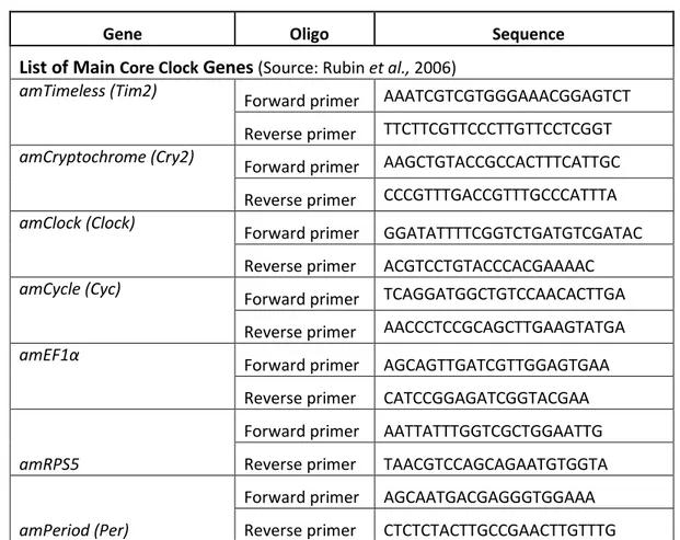
3.2.5 Selection of genes and primers design 53
3.2.6 Quantitative Real-time RT-PCR 54
3.2.7 Data analysis 55
3.3
Results
553.3.1 Daily variation in the expression of clock genes in brain and flight muscle
tissue according to age development of honeybee 56
3.3.2 Daily variation in the expression of metabolic genes in flight muscle
tissue according to age development of honeybee 73
3.4
Discussion
744.
FINAL REMARKS
79LIST OF TABLES
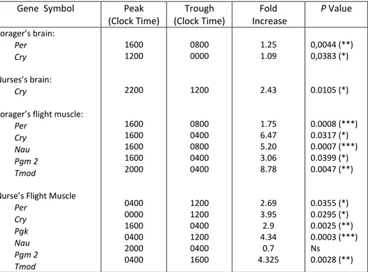
Tab.1. Peak times, trough times, and fold increases of genes found to significantly vary over time in the case of nurses and foragers bees, using
one-way-ANOVA 40
Tab.2. Core Clock and Metabolic gene’s Primers for Real-time quantitative PCR
reactions carried out in this study. 53
Tab.3. Peak times, trough times, and fold increases of genes found to significantly vary over time in honeybee tissues at five different ages old, using
LIST OF FIGURES
Fig. 1. Fundamental components of the circadian system 15
Fig. 2. Circadian oscillators in Drosophila head (A) 18
Fig.3. The core molecular clock in Drosophila melanogaster. Model of the PER/TIM
feedback loop 19
Fig. 4. The core molecular clock in Drosophila melanogaster. Model of the CLK
feedback loop 21
Fig. 5. Behavioral development in a Single Cohort Colony (SCC) 26 Fig. 6. Age-related task performance (“Temporal polytheism”) by worker honey
bees 28
Fig.7. mRNA levels of core clock genes, relative to the internal control gene
amRPS5, in the honey bees flight muscle tissue, over a 24-h period 38 Fig.8. Age related differences in mRNA levels of core clock genes and clock
controlled gene nau, relative to the internal control gene amRPS5, in either brain or peripheral flight muscle tissue of the honey bees, over a 24-h
period 41
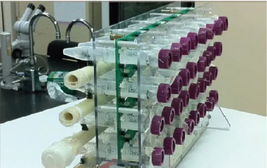
Fig. 9. “Environmental chamber” for housing the hive
Fig.10. Individual Eppendorf tubes into the frame to place individually bees under
constant conditions of darkness. 51
Fig.11. Twenty – four –hour profiles of 1-day-old honeybee brain core clock genes
expression 58
Fig.12. Twenty – four –hour profiles of 1-day-old honeybee flight muscle core
clock genes expression 59
Fig.13. Twenty – four –hour profiles of 1-day-old honeybee flight muscle
metabolic genes expression. 60
Fig.14. Twenty – four –hour profiles of 3-day-old honeybee brain core clock genes
expression. 61
Fig.15. Twenty – four –hour profiles of 3-day-old honeybee flight muscle core
clock genes expression. 62
Fig.16. Twenty – four –hour profiles of 3-day-old honeybee flight muscle
metabolic genes expression. 63
Fig.17. Twenty – four –hour profiles of 7-day-old honeybee brain core clock genes
expression. 64
Fig.18. Twenty – four –hour profiles of 7-day-old honeybee flight muscle core
clock genes expression. 65
Fig.19. Twenty – four –hour profiles of 7-day-old honeybee flight muscle
metabolic genes expression. 66
Fig.20. Twenty – four –hour profiles of 17-day-old honeybee brain core clock
genes expression. 67
Fig.21. Twenty – four –hour profiles of 17-day-old honeybee flight muscle core
clock genes expression. 68
Fig.22. Twenty – four –hour profiles of 17-day-old honeybee flight muscle
metabolic genes expression. 69
Fig.23. Twenty – four –hour profiles of 24-day-old honeybee brain core clock
genes expression. 70
Fig.24. Twenty – four –hour profiles of 24-day-old honeybee flight muscle core
clock genes expression. 71
Fig.25. Twenty – four –hour profiles of 24-day-old honeybee flight muscle
1. INTRODUCTION
Almost all organisms are exposed to the earth’s rotation around its axis and around the sun with predictable changes in the geophysical environment. More precisely, the periodic variation in the daily light and dark span and the seasonal changes in day length give rise to numerous biological rhythms in physiology and behavior. These rhythms are observed in the whole organism but also in single organs and recur at approximately regular intervals: from 20 to 28 h (=circadian); < 24 h (=ultradian); > 24 h (=infradian) and ± 1 year (=circannual) (reviewed by Halberg, 1969). They are synchronized in a meaningful way, allowing organisms to efficiently anticipate periodic events and to place themselves adaptively in the appropriate spatio-temporal conditions of the planet, by adjusting organism physiology and behavior to such changes and thereby optimize survival.
The study of biological rhythms, known as chronobiology, has been focused mainly on biological rhythms that are synchronized to the environmental 24 h-cycle, no doubt for their predominance in nature and their relevant control of a variety of biological processes. In fact, it is common to see: honey bee visiting flowers precisely at the same time of the day, even when nectar source is removed; plants that continuously open and close their leaves regularly, also if placed under constant darkness; fruit flies eclosing in the morning, regardless of constant laboratory conditions, and exhibiting same rhythm pattern in mating or egg laying activity. Similar relevant circadian coordination is conserved across a wide range of taxa including prokaryotes, birds, mammals or fishes. The most obvious explanations given for these conservation is that circadian rhythms are ubiquitous, they evolved under the influences of periodic geophysical environmental forces and have been fine-tuned under strong selective pressure.
In order to determine whether these rhythms are directly driven by a periodic environmental stimulation (exogenous rhythm) or whether they are an intrinsic characteristic of a circadian system of the organism (endogenous rhythm), applied and basic research questions have been used to uncover both the mechanisms and impact of circadian rhythms in tandem. Results suggest that most of the daily fluctuations in behavioral and physiological variables are not merely passive responses to the daily alternation of light and dark, as they persist even when no external time cues are present, such as in constant darkness. Indeed, circadian rhythms are driven by an endogenous clock (or a pacemaker) and have an endogenous period of approximately 24 h. In the
absence of a synchronizing “Zeitgeber” (time giver) (Aschoff, 1981), these rhythms “free run” with an intrinsic natural period, which shortens or lengthens closely to 24 hours and exhibit a “temperature compensation”, that allows their period length to remain over a wide range of physiological temperatures. Such fundamental characteristics account largely in providing organisms with a “Temporal organization” or “Time keeping” or “Biological clock”, allowing them to perform biological functions “at the right time of the day” by coordinating a functional relationship between various metabolic processes within the organism and the periodic factors that belong to world outside (reviewed by Sanders et al., 2002).
1.1 Basic principles of circadian rhythm
In the modern era, the endogenous nature of circadian timekeeping has been illustrated through the use of several experimental models, most of them developed on pheno - type based screen of mutagen-exposed flies (Konopka & Benzer, 1971). Such studies allowed the discovery of 3 period mutants: long (perL, ± 29 h), short (perS, ± 19 h) and arrhythmic (per0), which all mapped to a single locus, called period (per), on the X-chromosome, that could affect the persistence of either eclosion rhythm or locomotor activity of flies. Such findings facilitated further researches (Bargiello et al., 1984; Reddy et al., 1984) that confirm in Drosophila the same period as the first clock gene, indeed for the large protein of more than 1200 amino acids that it encodes. Later, such studies and others applying transgenic experiments in rodents, has characterized several other clock genes and reinforced basic understandings of the molecular mechanisms that coordinate the rhythmic regulation of circadian behavioral and physiological phenotypes (as reviewed by Hardin., 2005).
At this stage, endogenous circadian clocks became defined as internal molecular timekeeping mechanisms that reside in diverse range of cell types in a variety of organisms, from unicellular bacteria to vertebrates, and regulate the timing of behavior and physiology (Dunlap et al., 2004). Their key features are the following.
1.1.1 Entrainment
The most distinguishing characteristic of a circadian rhythm is the stability of the period.
In the presence of the 24 h environmental cycle, where the light-dark periods follow the cyclic pattern of the solar day, organisms adjust their circadian pattern to this dominant environmental entrainer or zeitgeber (Pittendrigh & Minis, 1964). The most important and universal entraining agent is the light-dark cycle generated by the earth’s rotation. Numerous studies have investigated the effect of photic entrainment upon organisms. It seems that under experimental conditions, the animal when housed within a 24 h light-dark (LD) cycle, with 12 h of light and 12 h of darkness (LD: 12:12), the entrained clock tend to follow such zeitgeber time in a very stable way, allowing a behavior period length of 24 h too (Aschoff, 1960; Mrosovsky et al., 1992). Other findings revealed that increasing the light intensity under constant light (LL) lengthens the circadian period of nocturnal animals and shortens that of diurnal ones (Aschoff, 1979a). This supports the idea that, in contrast to others factors, light intensity has the largest effect on the free-running period (Aschoff, 1979a, 1979b). In addition, light induced phase shifts also depend on the properties of the light pulse (duration and irradiance) (Daan & Pittendrigh, 1976).
The most important and universal entraining agent is the light-dark cycle generated by the earth’s rotation; non-photic cues exists too, such as temperature cycle, food availability, social interactions, which, although playing a secondary role, are able to provide stimuli and entrain the clock to the driving oscillation of the surrounding environment of the organism.
So, through entrainment, the zeitgebers do not create rhythms but do determine their placement in time.
1.1.2 Free-running
However, after elimination of external stimuli, the cycles persist with a period of close to, but not exactly 24 h. This persistence seems to indicate the existence of some kind of internal timekeeping mechanism or biological clock (Pittendrigh & Calderola, 1973).
Under such constant conditions of continuous light (LL) or darkness (DD), the period to which the internal biological clock can be entrained tends to be longer or shorter than 24 h, ranging between
20 to 28 h, depending on various factors: intrinsic factors, such as the physiological status of the individual animal (age, hormone levels), and its genetic background (diurnal or nocturnal species), and on external factors (light intensity, temperature) and the physiological state of the organism (feeding conditions, previous experiment etc.) (Aschoff, 1979a, 1979b; Pittendrigh & Daan, 1976). Thus, under such conditions, the “time” is often measured relatively to the endogenous circadian rhythm. Such time, is defined by researchers as circadian time (CT) and corresponds to a relative point during the organism’s behavior rhythm (Pittendrigh, 1966).
Interestingly, once a shift in external cues occurs due to phase shifts of the light - dark cycle or travel across time zones for example, what follows are several unstable transitional cycles between the stable periods established under synchronizing LD condition and those of the new environment until rhythms are aligned to the new cues. This re-alignment is called entrainment (Enright, 1981). Generally, the resulting plot of such phases changes, against the circadian time of such perturbations, is considered to give a phase response curve (PRC) for the oscillation (Saunders et al., 1994).
1.1.3 Temperature compensation
Another fundamental property of the circadian clocks is their ability to maintain a roughly 24-hour periodicity over a range of physiological temperatures; this means that they exhibit a sort of “temperature compensation”. Such vital characteristic was revealed as universal, occurring across all taxa, and of high functionality, as it allows organisms to keep track of time. In fact, most kinds of biological processes in cells are affected by temperature changes, and a clock that speed up and slow down would not be useful. So Thus, through temperature compensation, a circadian clock is able to efficiently resist and to adapt to environmental changes.
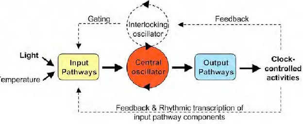
Although the understanding and definitions of circadian system have developed as discoveries have been made, a system could not be called “circadian clock” from a chronobiological point of view, if it does not have the three fundamental and linked components as following (Fig. 1):
An input pathway: represents all the corresponding ways of perception of the information coming from the environment, which are necessary for clock synchronization.
A central clock or pacemaker: that generates cell-autonomous and self-sustaining rhythms. It consists of a series of interconnected positive and negative transcriptional and translational feedback loops governing protein stability and degradation in an evolutionary conserved way across organisms (Pittendrigh, 1960).
An output pathway: output pathways that influence physiological function and behavior through gene expression [generally called clock controlled genes (ccgs)], neural and humoral channels.
Fig. 1. Fundamental components of the circadian system
“The circadian system consists of three basic elements: an input pathway (yellow), an endogenous pacemaker generating circadian rhythms (red), and an output pathway (blue). Environmental signals, called Zeitgeber (German for “time giver) are transduced to the master clock via input pathways. The input signals are received via receptors and then sent to the central pacemaker that generates oscillations. The output pathway finally translates the oscillation into rhythms such as genome transcription in cyanobacteria or sleep-wake cycles in animals. Some organisms contain more complex circadian clocks (shown as dotted line) that include multiple, interlocking oscillators and positive or negative feedback from clock controlled activities to the pacemaker and/or input components” (Jud, 2009) (figure modified from Gardner et al., 2006).
1.2 Organization of the circadian system in insects
In respect of the fact that Drosophila melanogaster Meigen is one of the most studied insects and due to the fact that its clock investigation has led for many years the way to a complexity of molecular aspects of circadian research, that greatly outnumber basic foundations in other insects, the identification and description of the circadian system organization will be mainly focused on these flies, pointing out eventual differences with other insects (especially bees), when judged of relevance and needed to be outlined.
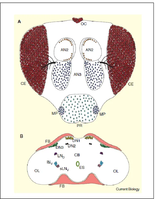
1.2.1 Localization of the master clock (Fig. 2)
In flies, circadian rhythms are regulated by a cell autonomous pacemaker, located bilaterally in the lateral protocerebrum just at the border to the optic lobes and traditionally divided into two main groups of neurons, the lateral neurons (LNs) and the dorsal neurons (DNs) (reviewed by Taghert & Shafer, 2006; Nitabach & Taghert, 2008). Such cell clusters have been determined through cytological staining for the clock gene products and their products, which are prevalently period (per) and timeless (tim) (Liu et al., 1992; Saez & Young, 1996). Although both neurons are important, the major influence on the daily locomotor activity and pupal eclosion rhythms confer to the LNs the role of key pacemaker neurons (Zerr et al., 1990; Ewer et al., 1992; Frisch et al., 1994; Helfrich-Forster, 1998). In adult flies, lateral neurons are traditionally divided into a group of four large – ventrolateral neurons (I-LNvs), five small – ventrolateral neurons (s-LNvs), which project directly to the medulla and dorsal protocerebrum, respectively (Stanewsky et al., 1997; Kaneko & Hall, 2000). Both groups have the ability to express the pigment dispersing factor (pdf) gene, usually used to reveal their projection pattern (Helfrich-Forster, 1995) and well-known as encoding the neuropeptide PDF that plays a key output signal from the circadian clock at peripheral tissues. Unlikely, pdf gene is not expressed by the third more dorsally located group, namely the dorsolateral neurons (LNds) (Kaneko & Hall, 2000), which also projects to the dorsal brain. On the other hand, the dorsal neurons (DNs) also split into three groups, which are categorized according to their localization within the dorsal brain, mainly dorsal neurons 1s (DN1s), dorsal neurons 2s (DN2s) and dorsal neurons 3s (DN3s). Kaneko & Hall (2000) and Helfrich-Forster (2002) illustrated the neuronal functioning of DN1 and DN3 projections towards sLNv cell bodies in interconnecting clocks gene expression between neurons.
Although these different groups of clock gene expressing neurons contribute to the control of behavioral activity, it appears through mutant analyses that different aspects of locomotor activity rhythms are not equally regulated by all neurons (Grima et al., 2004), which make some of them being master and others subordinate clocks for behavioral rhythmicity. Under LD cycle, locomotor activity seems to be under the control of two separate oscillators that generate consequently two activity peaks. The 4 s-LNvs appear to be responsible for the morning activity peak (M peak) whereas the LNds and possibly a subset of DN1s regulate the evening peak together (E peak) (Helfrich-Forster, 2000).
These clock neurons also regulate the anticipatory increase in activity corresponding to the M and E peaks. In constant conditions s-LNvs appear to be sufficient to provoke robust activity rhythms, indicating a more important role for these clocks in DD (Grima et al., 2004). However, the LNds and the 5th s-LNv are thought to be capable for modulating the phase of the activity pattern. In flies, projections sent from sLNvs into the dorsal brain, which contains the rhythmically released neuropeptide PDF (Park et al., 2000), drives the locomotor activity rhythm (Renn et al., 1999; Helfrich-Forster, 1995) in a circadian manner and synchronize the timing of different clocks.
This central oscillator receive light input from retinal photoreceptors in the compound eyes and extra-retinal photoreceptors within the brain, allowing synchronization with the environmental light-dark cycles, and possess multiple output pathways to control diverse endocrine, autonomic and behavioral functions, particularly locomotor activity (Fig. 2).
Fig. 2 - Circadian oscillators in Drosophila head (A)
External structures containing circadian oscillators. A frontal view of a Drosophila head is shown. OC, ocelli; CE, compound eyes; AN2, second antennal segment; AN3, third antennal segment; MP, maxillary palps; PR, proboscis. (B) Oscillator cells within and surrounding the brain. A frontal section through a Drosophila brain and surrounding tissues is shown. CB, central brain; OL, optic lobes; FB, fat body; ES, esophagus; LND, dorsal lateral neurons; ILNv, large ventral lateral neurons; sLNv, small ventral lateral neurons; DN1, dorsal neuron 1s; DN2, dorsal neuron 2s; DN3, dorsal neuron 3s (Illustrated by Hardin, 2005).
1.2.2 The core molecular clock
Since the first clock gene period has been cloned in Drosophila, thousands of studies have been published identifying several other clock genes that play an important role in the timekeeping mechanism (reviewed by Hardin, 2005). As a consequence, several molecular models have been
assembled based on in vitro observations, which have been central to our understanding of how the clock is regulated at the molecular level (Figure 3).
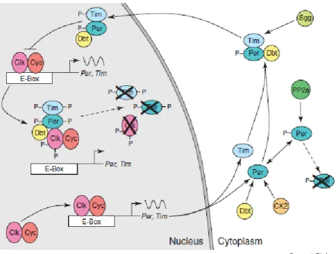
Fig. 3. The core molecular clock in Drosophila melanogaster. Model of the PER/TIM feedback loop (illustrated by Hardin, 2005)
In flies, the molecular-genetic basis of circadian time-keeping may rely on a two interconnected transcriptional – transitional feedback loops which control expression of several genes, that can generally be divided into groups according to the molecular nature of their products (as transcription activator, transcription repressor or protein degradation player) and the changes created on their own steady – state levels (reviewed by Hardin, 2005) along the day. More in details, the flies core clock is composed of a positive loop – rhythm driving factors, consisting of two transcription factors CLOCK and CYCLE proteins (Allada et al., 1998; Rutila et al., 1998) that contain a basic helix – loop – helix (bHLH) DNA binding domain and a PER –ARNT- SIM (PAS) dimerization domain, that activate transcription of many genes, including period (per) and timeless (tim), and a negative loop elements, the PERIOD (PER) and TIMELESS (TIM) proteins, that acts as transcription repressor in the molecular cycle (Dunlap, 1999). Generally, both per and tim mRNAs and proteins, exhibit rhythmic abundance, with peaks resulting respectively during the early and the later part of the night, reflecting the corresponding behavioral rhythm (reviewed by Sharma,
2003). On the other hand, clock genes show cyclic expression too, with mRNA and protein level peaking early in the morning, whereas cycle is constantly expressed. In wild type flies, to initiate the negative loop, CLK heterodimerizes with CYCLE in the middle of the day, binding an E-box regulatory elements in the regulatory regions of per and tim, thereby activating their transcription, which peaks its level early in the night (Fig. 3). PER and TIM proteins interact to form an unstable PER:TIM heterodimer and translocate into the nucleus, without accumulating till late evening, when they peak their level. After a delay, due to phosphorylation of per dependently upon two kinases, DBT and CASEIN KINASE 2 (CK2), leading to its degradation, till stabilization of phosphorylated per by binding either to tim or PROTEIN PHOSPHATASE 2a (PP2a), which is supposed to remove the phosphates added to PER (reviewed by Hardin, 2005). In the meanwhile, as TIM levels increases, the remaining complex TIM-PER-DBT translocates to the nucleus upon SHAGGY (SGG) – dependent TIM phosphorylation, which, in concert with CK-2 dependent PER phosphorylation, lead to their transport into the nucleus (reviewed by Hardin, 2005). Recently, it has been argued that PER and TIM can enter into the nucleus as dimer complex formed by protein – protein interaction without any DNA – binding domains, due to PER capacity to associate with other transcription factors to negatively regulate its own transcription (reviewed by Sharma, 2003) and to act as transcriptional repressor, specially that tim sequence lacks of DNA-binding domain and PAS domain (reviewed by Shirasu et al., 2003). Once in the nucleus, TIM-PER-DBT complex binds to the CLK-CYC heterodimer, inhibiting further per and tim expression by preventing the DNA binding to E-box of CLK-CYC dimer complex (Lee et al., 1999; reviewed by Hardin, 2005), thus repressing CLK-CYC activity. In the nucleus, DBT phosphorylation leads to PER and CLK degradation, while TIM destabilization is triggered via a light – mediated pathway, by tyrosine phosphorylation. Consequently, the accumulation of the hypophosphorylated CLK, in addition to a PER-TIM heterodimer decays, trigger heterodimerization of CLK with CYCLE and the starting of per and tim transcription again; thus the cycle repeats itself, which resets the mechanism of circadian clock (reviewed by Hardin, 2005).
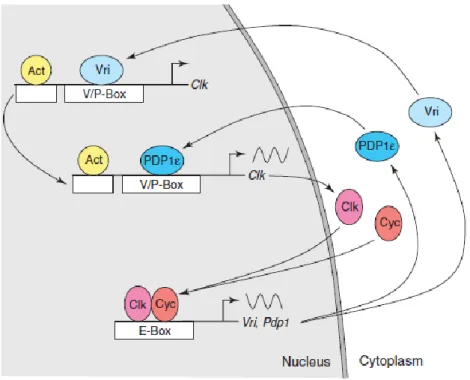
Another identified component playing an important role in the second transcriptional – translational interlocked feedback loop is the transcription factor encoded by vrille (vri) and par domain protein 1 ԑ (pdp1ԑ) genes. Both seems to regulate directly Clk transcription by specific binding to Clk promoter elements and functions as respectively repressor and activator elements of Clk transcription (Fig. 4) (Cyran et al., 2003; Glossop et al., 2003; as reviewed by Hardin, 2005).
The CLK-CYC heterodimer drive expression of vri and pdp1ԑ by binding to the E-Box sequence in the promoter region, trigging their highly transcription, respectively by late day and early night (Cyran et al., 2003; Glossop et al., 2003; as reviewed by Hardin, 2005), with vri mRNA cycling in phase with those of per and tim. Once VRI accumulates, it tends to bind the VRI/PDP1ԑ box regulatory elements and repress CLK transcription till VRI displacement form the (V/Pbox) by PDP1ԑ, and consequently de-repression of CLK transcription (Cyran et al., 2003). In addition to the effect on the central clock’s components, VRI affects the output system through the peptide PDF (reviewed by Williams & Sehgal, 2001), that is strongly suggested to function as a circadian transmitter.
Fig. 4. The core molecular clock in Drosophila melanogaster. Model of the CLK feedback loop (illustrated by Hardin, 2005).
Molecular genetic studies in Drosophila identify some additional clock components. These circadian regulatory molecules act to refine the transcriptional/translational feed-back loops. Light changes the levels of these additional clock components, resetting the clock to different time of day. In Drosophila, the entrainment of the circadian clock relies on the degradation of TIM in response to light. Cryptochrome (CRY) is the major mediator for the rapid light-induced degradation of TIM, which has been demonstrated to be mediated by a ubiquitin-proteasome
pathway into neural and peripheral clock cells (reviewed by Dubruille & Emery, 2008). Light activation of CRY, according to a light dependent rhythm (Emery et al., 1998), permits its binding to TIM (Ceriani et al., 1999), which in turn leads to TIM degradation by a tyrosine kinase phosphorylation. Thus, TIM degradation through the CRY photoreceptor, in addition to its ability to synchronize the internal clocks to the 24 h cycle of sunlight, plays a relevant role in shortening or advancing the phase of the clock, according to the time of exposition to light - pulses (during early or late night) (reviewed by Allada & Chung, 2010) and indirectly to the levels of tim mRNA (reviewed by Hardin, 2005).
1.2.3 Multiple tissues express clock genes
In flies, concerns regarding the overall organization of the circadian system trigger the characterization of several essential clock genes; and the analysis of their expression patterns gave evidences that the molecular components of the circadian clock are largely conserved outside the central master in a broad range of organs (reviewed by Giebultowicz, 1999). This, together with relevant researches investigating clock molecule cycle in various biological functions, such as metabolism, reproduction or excretion, without focusing primarily on the central clock, reinforced evidences that circadian oscillators may function by maintaining coordinated physiological processes, in a tissue – autonomous fashion (Giebultowicz, 1999; Brown & Schibler, 1999). Although many advances have been made recently in enlarging our understanding of the peripheral clocks and their role in circadian timing, many questions remain unanswered yet, regarding how many clocks oscillate in pluricellular animals, such as insects, how much precise influence have the master clock over fly peripheral oscillators, in the sense if they do require input from the nervous system, and in which manner such peripheral ensemble of clocks ensures the internal temporal organization of the organism.
In insects, the first organs that have been revealed to have an independent pacemaking function and to be directly entrained by environmental signals is the testes – vas deferens complex in the gypsy moth, Lymantria dispar L., that has shown a light entrainable sperm release rhythm when investigated in vitro (Giebultowicz et al., 1989). In fact, the fertility of the sperm seems to be dependent on the occurrence of two essential steps, which rhythms register two peaks occurring in the evening and morning and respectively consisting of the release of sperm from the testis into the upper vas deferens (UVD) followed by the accumulation of the sperm in the seminal vesicles.
Interestingly, such circadian rhythmicity has been revealed to persist under constant darkness and to happen in correlation with glycoproteins release from the apical portion (Giebultowicz et al., 1989). In fact, further studies confirmed the importance of the circadian coordination between either the sperm maturation process or the glycoprotein’s secretion, as a guarantee of the efficiency of the male flies’ reproductive system (Bebas et al., 2002). Importantly, the temporal relationships of the per clock gene mRNA and PER protein expression rhythms within cells surrounding peripheral tissues testes and vas deferens (Gvakharia et al., 2000; as reviewed by Giebultowicz et al., 2001) are of great impact on such coordination. These findings were supported by persistent cycling of the per clock gene and its proteins in fly pupae ring gland, when cultured in vitro (Emery et al., 1997; as reviewed by Giebultowicz, 2001). On the other hand, and mainly by high – time resolution experiments using per – driven rhythmic luciferase activity once a promoter region fused to luciferase – encoding sequences, enormous progress has also been made in determining either the in vivo regulatory pattern of the Drosophila clock gene period (per) or in providing a powerfully reproducible measure of its transcriptional oscillations in organs that were analyzed in vitro (as cited by Hardin, 2005). From such kind of experimental systems, a wide range of self-sustained and photosensitive oscillators in Drosophila peripheral tissues have been reported in renal tubules and rectum (Giebultowicz et al., 2000), chemosensory structures located at the level of antennae, proboscis, wing margin and legs (Plautz et al., 1997; as reviewed by Giebultowicz, 2001). In the meanwhile, the list has been extended to an additional type of peripheral organs, that were promoted as autonomous functioning oscillator clocks, which potentially oscillate independently from the central clock. Recently, Tanoue et al. (2004) have shown that components of the olfactory signal transduction can function as an autonomous pacemaker for olfaction rhythms, even in absence of central circadian regulation, due to targeted lateral neurons ablation, provided the ablated antennal neuron in cyc (01) flies is rescued.
The same situation seems to prevail in in vitro cultured Drosophila testes, where a rhythmic activity of either per or tim luciferase reporters has been registered. Another piece of evidence, suggesting a high degree of autonomy of peripheral pacemaker in flies, comes from examining the role played by fat body tissue in modulating the daily rhythm of feeding behavior activity (Xu et al., 2008). A number of metabolic genes, has been determined through microarrays screens, to be cyclically expressed at the level of metabolic tissues, driven by a local circadian clock (Xu et al., 2008). In fact, inactivation of clock functioning by clock gene knockouts techniques have reportedly triggered symptoms of metabolic dysfunction, such as increased food consumption (Xu
et al., 2008), as previously demonstrated in mammals (Turek et al., 2005; Colles et al., 2007; Bray & Young, 2007; as cited by Xu et al., 2008).
At the molecular level and contrary to the case of mammals, it has been suggested in invertebrates, and more precisely in flies, that peripheral oscillators respond to light resetting signals due to the photoreceptor role of CRY (as reviewed by Hardin, 2005). At the peripheral level, such autonomous photoreceptor cell seems to have predominately the ability to convey light information to oscillator without being completely integrated in the core clock components. In fact, in the cryb mutant flies, antennal clock neurons registered an impaired free – running rhythms, combined to reduction till disruption of clock driven olfactory rhythms (Krishnan et al., 2001; as cited by Merlin et al., 2007), which restoration is dependent on antennal functioning neurons rescue (Tanoue et al., 2004). Moreover, the same mutation (cryb) triggers dysrythmia of the core clock genes per and tim, in other tissues such as fly organs (Levine et al., 2002) or Malpighian tubules (Mts) (Ivanchenko et al., 2001), supporting CRY as dispensable within the core clockwork. All these findings support massively the specialized role for circadian oscillators in mutual regulation of metabolism or physiological processes and circadian rhythms, in order to provide effective tissue’s response and adaptation to environmental signals.
1.3 The Honey bee: an interesting model for understanding mechanisms of life
history transitions
1.3.1 Honey bee as a model system
Honey bees (Apis mellifera L.) are eusocial insects that live in large colonies consisting of some 20,000 up to 40,000 female workers (sterile or having low reproductive potential), and 200 to 300 male drones, and a queen (extremely fecund), interacting all together according to an elaborate communication system (Wilson, 1971) that allows them to coordinate efficiently each aspect of their life. Social insects, and bees particularly, could be considered as an interesting model system that offers an opportunity to study a number of fundamental aspects of the evolution of sociality with an integrative focus on the molecular, physiological and endocrine processes involved in their life history transitions and/or their adaptive responses to the environmental context in which they
live. This appears to be especially true for this category of social insects, that is assumed to exhibit a temporal polyethism, allowing the adults to move through successive behavioral stages in an age-related fashion, which, in addition to a pluriannuality in reproduction, provided a base for developing an arsenal of researches that led simultaneously to explore deeply the ecology, natural behavior and phylogeny of these insects (reviewed by Elekonich & Roberts, 2005). Moreover, honey bee figures within the list of organisms which genomes have been sequenced and along with a panel of related encoded proteins are largely represented in a abundant literature (reviewed by Denison & Delpech, 2008).
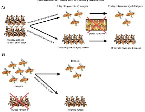
Based upon the fact that honey bees undergo a natural switch from a phase when they work in the hive to another when they forage outside, with behavioral rhythms that, in addition to be primarily age-determined, are generally also socially mediated, a large interactive research has been performed, using “the single cohort colonies” (reviewed by Elekonich & Roberts, 2005) deriving from the original typical colony. These single cohort colonies are based on discrimination of specimens (typically age vs behavior) that allows the collection and observation of precocious foragers (young workers) or reverted nurses (typical aged bees) obtained as a consequence of an unbalanced colony age demography (Fig. 5) (Robinson et al., 1989; Huang & Robinson, 1992; Giray & Robinson, 1994; as cited by Elekonich & Roberts, 2005); investigation on these colonies has shed light on workers-worker interaction determinism of physiological and genetic mechanisms involved during the behavioral transition (Huang & Robinson, 1992).
Honey bee has shown also to be a promising model system for researches that, through useful manipulations, succeed in training the foragers to specific feedings stations and time (Von Frisch, 1964), helping to establish time memory properties of honey bee worker. Others practices were made to obtain colonies characterized by mixed genotypes, that facilitate investigation on genotypic influences on individual versus colony traits (Page et al., 2000), in addition to various pharmacological treatments (Ben-Shahar et al., 2003) or pheromones analogs (Grozinger et al., 2003) that allowed a major efficiency in bee behavior and physiology dissection (reviewed by Elekonich & Roberts, 2005).
Fig. 5. Behavioral development in a Single Cohort Colony (SCC). (A) The typical SCC is obtained from the collection of newly emerged bees (1 day old). It is assumed that only the 10% will follow a precocious development without any connection between the age and the behavior. Thereby, observations are made in presence of a group consisting of a number of young (normal age) nurses and young (precocious) foragers, and later old (over-aged) nurses and old (normal aged) foragers, just in absence of additional newly emerged bees. (B) Reversion SCC, consisting in removing nurses from the group which trigger the reversion of foragers to brood care. Thus, observations will be made in the presence of workers that experienced foraging, divided into foragers reverted to an earlier stage (nursing work) and old (normal aged) foragers (adapted from Elekonich & Roberts, 2005).
1.3.2 The social clock of honey bee
1.3.2.1 Social influences on the ontogeny of circadian rhythm
Such as a representative of a very advanced social insect, each honey bee emerges and develops inside a social environment and in presence of conspecific individuals belonging to all life stages (including eggs to adult workers) which, through an elaborated ways of communication by either direct (trophallaxis) or indirect (pheromones, vibratory, visual or acoustic signals) contact, succeed considerably in driving characteristics of the circadian rhythm (phase, expression or development) of the single individual bees. Such a social entrainment has been deeply studied by many researchers (Southwick & Moritz, 1987; Moritz & Sakofski, 1991; Frisch & Koeniger, 1994; Moritz &
Kryger, 1994) and seems to influence the social behavior in a functionally significant way that guarantees colony efficiency.
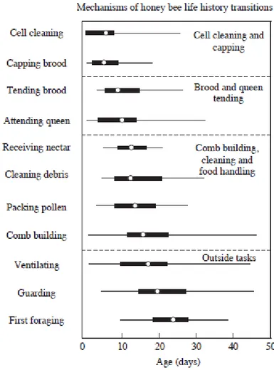
In the same order of ideas, and contrary to other insects, honey bee expresses circadian rhythm only after adult emergence (Moore, 2001), according to a precise postembryonic ontogeny in the development of such a rhythm. In their natural environment, the newly emerged bees remain inside the nest, without any or limited contact with the external environment, and specialize in taking care of the brood and in other “in-hive” activities (like hive maintenance) around the clock, with an attenuated molecular and behavioral rhythm (Crailsheim et al., 1996; Moore et al., 1998). After the first 2 to 3 weeks of adult life, the worker shift to foraging for nectar and pollen outside the hive, according to a strong circadian rhythm which is necessary for the sun compass navigation, for the recruitment waggle dance and for the time memory regulating the timing of foraging to the maximum nectar and pollen reward availability (Fig. 6) (reviewed by Moore, 2001). Interestingly, the nurse bees, which are usually active around the clock when examined inside the colony, have been shown to exhibit a strong circadian oscillations, either in locomotor activity or gene clock expression, once they have been isolated individually from the brood (Shemesh et al., 2007, 2010; as reviewed by Eban–Rothschild & Bloch, 2012) or under constant conditions in the laboratory (Eban-Rothschild et al., 2012), which has been incongruent with the hypothesis suggesting that the circadian clockwork is not functional at young stage of life. Moreover, this consistent behavioral plasticity has been associated with a functional molecular feedback loop in nurse clock, that tends to free run under constant conditions, supporting the idea that the ontogeny of the circadian rhythm in honey bees seems to be socially fine tuned and endogenously defined (Toma et al., 2000; Bloch et al., 2001, 2003, 2004; Shimizu et al., 2001; as reviewed by Bloch, 2010).
Fig. 6. Age-related task performance (“Temporal polytheism”) by worker honey bees (adapted from Winston, 1987; by Elekonich & Roberts, 2005).
In the meanwhile, a wide range of researches aims at identifying whether the social signals that generally mediate the task-related behavioral development could be of efficiency according to the social context to which young workers will be adapted, either at the molecular or behavioral level. To answer such question, many intra- and inter-individual manipulations have been made. Neither the use of biogenic amines or some of their antagonists (reviewed by Bloch et al., 2009) nor the removal of their source of secretion (corpora allata) appeared to affect the age at onset of rhythmicity; this trigged the hypothesis that Juvenile hormone (JH) and/or octopamine could be involved in some non circadian functions, which are originally settled by other period mRNA expressing cells, that are diverse from those cells influencing the diurnal locomotor activity (Bloch, 2010). In the case of nurses that have been monitored individually in the laboratory, soon after having experienced the colony environment after emergence, synchronization with social factors is perhaps the most predominant element influencing the ontogeny of circadian rhythms, as well as the overall locomotor activity or its free-running period (Meshi & Bloch, 2007; Eban–Rothschild
et al., 2012; as reviewed by Eban–Rothschild & Bloch, 2012). Even if little is known about the mechanisms underlying the influence of the post embryonic development of internal circadian rhythms, Eban–Rothschild & Bloch (2012) have been more supportive of the hypothesis that rely the delay in overt circadian rhythm, in the case of isolated young bees outside the colony, to the effect of in-hive environmental conditions, including either the social (as pheromones) or the microenvironment (such as temperature etc.) factors rather than to others (such as physical contact with conspecifcs) or the light dark illumination regime.
1.3.2.2 Social modulation of plasticity in the expression of circadian rhythm
It is well known how honey bees show a specific plasticity in expressing their circadian rhythms, enabling them to switch from working around the clock to diurnal foraging activity with robust circadian rhythms, according to the temporal division of labour (reviewed in Bloch, 2009, 2010; Eban–Rothschild & Bloch, 2012). Such task-related plasticity in the circadian clockwork, that has been well defined as modulated by the social environment, is suggested to be functionally relevant and serves as guarantor of maximal efficiency of the whole colony (Bloch, 2010; Bloch & Grozinger, 2011). In natural conditions, relevant results have already suggested that, while the presence of brood could be involved in the attenuation of the expression of workers circadian rhythms (Shemesh et al., 2010) by acting probably as a masking agent, the isolation of the young nurses from any colony life, either in the laboratory or in broodless comb or outside the hive, promotes the diurnal patterns of expression in locomotor activity and also clock genes expression. Likely, previous studies, that adopted social manipulations leading to uncouple the age and tasks to perform, have shown the reversal of honey bee behavioral rhythms, which linked strongly the synchronization of the molecular clockwork more to the task than to age (Bloch & Robinson, 2001; Bloch et al., 2001). Thus, it seems that at least the brood presence plays an important role in strongly modulating such plasticity in circadian clockwork and, interestingly, it has been demonstrated that the brood social synchronization may strongly be mediated through sensory signals whose detection involves the antennal flagella of the worker (Nagari & Bloch, 2012). The evidence of the involvement of the social signals in modulating plasticity in the circadian rhythms is provided also by other eusocial insects, including ants and bumble bees (Ingram et al., 2009; Yerushalmi et al., 2006). For example, bumble bees follow a similar chronobiological plasticity, overall behavior and molecular patterns, that is highly modulated by size–related division of labor
rather than age-dependent one (Yerushalmi et al., 2006). On the contrary, harvester ants are more unlikely to exhibit an age–related polyethism, associated with developmental regulation of clock genes expression, promoting some similarities between them and honey bees (Ingram et al., 2009). So far, such findings could validate the theory of the “internal temporal order” to which the colony macro-environment of such social groups is associated (cited by Teixeira et al., 2011). Such remarkable natural plasticity in circadian rhythm may represent a conserved selective toolkit adopted by those species and translated through individual adoption of the right endogenous temporal system that occur following a precise phase-relationship, in synchrony with those expressed by others of the community, to guarantee a functional coordination of the “Super– organism” (Moritz & Fuchs, 1998) and consequently the maximal survival in an ecological context.
1.4 Thesis objectives
The aim of the thesis is to emphasize on the study of the circadian regulation of flight muscle tissue in honey bee Apis mellifera. Circadian clocks synchronize behavior and physiological functions to the fluctuating environment (Dunlap et al., 2004). In social insects, the circadian clock is highly influenced by the social interactions between individuals within their colony. In honeybee for example, is thought that the behavior and circadian clockwork exhibit generally profound context-dependent plasticity, enabling adjustments to rapid changes in the social environment (Bloch, 2010). However, little is known about the function and mechanism of such interplay between social needs and sensitivity of the circadian system, in tissues of the body periphery. Due to the parallels between the circadian mechanisms of honeybee and mammals, I hypothesize that a peripheral clocks regulate flight behavior in worker bees. In Chapter 2, I examine whether the main core clock gene undergo differences in the circadian regulation in the flight muscle tissue, from young nurses and adult foragers collected from field colonies under natural LD conditions. I found that the core clock genes are circadianly expressed in a period-dependent fashion in flight peripheral tissue of both workers. Nevertheless, the temporal gene expression differed in the wave form and phase between our witnesses of the transition from nursing to foraging behavior. Consequently, we assessed the endogenous nature of such peripheral clock, when exposed to different cycle of continuous darkness, at different key ages, over the honeybee life span. Additionally and under the same conditions, we verified our hypothesis concerning the fact that
the flight muscle tissue might contain a circadian clock which regulates itself the temporal expression of genes involved in relevant metabolic activities. Comparing the clock gene expression profile between brain and flight muscle tissue at each age sampling, add functional insights related to the nature of the coordination that happen between oscillators, throughout the developmental related circadian regulation of the flight muscle in honeybee. This study sheds new light on the molecular dynamics and social regulation of context-dependent plasticity in the organization of the circadian clockwork of the honeybee.
2. FIELD RESEARCH
2.1 Introductory notes
Circadian rhythms govern the behavior, physiology and metabolism of living organisms (Hall & Rosbash, 1993). These circadian rhythms are driven by a cell-autonomous daily timekeeping mechanism, that allows organisms to adapt to the changing environment and thereby optimize their survival. The key features of circadian clock are its ability: i) to synchronize (entrain) to external daily rhythms of light, temperature and other environmental cues, and ii) to persist (free-run) when placed in constant conditions with close to a 24h (circadian) period.
Genomic analysis of behavioral rhythm in flies has elucidated the molecular and cellular components of the core clock, giving better understanding of the tools by which the molecular circuitry underlying circadian function are conserved in other organisms. In Drosophila, the molecular basis for these rhythms is proposed to consist of molecular feedback loops, which comprise several core clock genes, such as period (Per), timeless (Tim), clock (Clk) and cycle (Cyc). These genes encode PER, TIM, CLK and CYC proteins, respectively, which orchestrate changes in their own steady-state levels and that of their mRNAs. More precisely, the two transcription factors CLOCK (CLK) and CYCLE (CYC) form heterodimers and activate transcription of Per and Tim genes by binding to their E-boxes in their promoter regions. Resulting PER and TIM proteins bind to each other and enter to the nucleus, where they repress their own CLK:CYC complex induced transcription, allowing the cycle to begin anew. The stability and ability to accumulate to levels that are functional of PER and TIM is extensively regulated by CK1ɛ protein DBT and Casein Kinase 2 phosphorylation, that promote their degradation. The CLK:CYC heterodimer also regulates the expression of two transcription factors Vri and Pdpɛ, that form the second feedback loop by regulating the transcription of Clk. Simultaneously, molecular output generated from the central oscillator to overt rhythms is regulated via the induction of rhythmic transcription of an array of Clock controlled genes (ccgs) such as pdf, takeout or lark.
Although the anatomical site of the central circadian clock in insects is the brain area, recent studies about spatial distribution of clock gene expression revealed the presence of peripheral oscillators existing in many tissues in fly bodies. While a persistent Per-driven bioluminescent
oscillations occurred in vivo in the compound eyes, antennae, proboscis, wings, legs in per-luc Drososphila (Plautz et al., 1997), other fly’s tissues as the malpighian tubules in adults (Giebultowicz & Hege, 1997; Hege et al., 1997), the ring gland in pupae (Emery et al., 1997), the alimentary tract, fat bodies, ovaries or testes (Liu et al., 1988) have been reported to free run and to be directly entrained by light when isolated in vitro. Taken together, these findings attest that such peripheral tissue clocks have the potential to function independently from the central clock in the brain, generating local clocks controlling tissue-specific functions. To our knowledge, the organization of the circadian system in social insects, such as bees, is still in its infancy and the possible presence of widespread oscillators outside the brain has never been considered yet.
Bees have specific biological clocks that function in time memory, enabling visiting flowers at time of maximal nectar and pollen availability, time-compensated sun-orientation and recruitment waggle dance (Von Frisch, 1967). The biological clock used in these behaviors of foragers is considered to be based on a circadian system and its endogenous nature has been shown repeatedly for general locomotor activity in individual honey bees (Toma et al., 2000; Bloch et al., 2001, 2004; Rubin et al., 2006; Shemesh et al., 2007, 2010), which rhythm persist (“free-run”) under constant dark (DD) conditions with a period of about 24h. Because bees are social organisms that live in a complex and highly organized society, notion of timekeeping mechanisms should be correlated with division of labor that organizes workers, leading to occurrence of behavioral or physiological processes according to the right moment and social context. In field colonies, honey bee workers showing evident temporal polyethism evolve across their life span, from nursing brood around the clock, with no circadian rhythm during the first 1 to 2 weeks of adult life, to foraging activities later (typically > 3 weeks of age), with a strong circadian rhythms (Kaiser & Steiner-Kaiser, 1983; Moore et al., 1998; Sauer et al., 2003). Indeed, and altough the studies on molecular basis of circadian rhythm in social insects have only started recently, various studies already report how the age related division of labor is highly associated with the differences in the mRNA levels of the main core clock period gene in the brain (Toma et al., 2000; Bloch et al., 2001, 2004). From self–sustained colonies, Per expression tend to oscillate at its highest during the night in brain of rhythmic foragers, contrary to their young sisters that typically do not show any circadian rhythms in clock genes expression (Bloch et al., 2001; Bloch & Meshi, 2007; Meshi & Bloch, 2007) exceptionally till 7 days old, if they do not experience any social environment influences (Shemesh et al., 2007, 2010). This evidences make tempting to investigate
how such circadian system behaves outside the brain in honeybees. Indeed, evaluating such scenarios would be a good starting point for understanding if the central oscillator regulates cellular processes in different tissues or whether there are specific rhythmic functions of cellular processes, driven by functionally separable oscillators, organized with respect to internal temporal order of such social insects and their age related development needs.
Such investigation can be interestingly undertaken in the case of honeybee’s muscle tissue, which undergoes related molecular and biochemical lifetime changes according to the adult behavioral transitions and the consequent physiological capacity needs (reviewed by Roberts & Elekonich, 2005).
The field part of this research, whose results are here reported, simultaneously examined the 24h mRNA expression profiles of two main core clock genes, Per and Cry, and the myogenic factor gene Nautilus (Nau) involved in honeybee muscle metabolism, every 4h in brains and flight muscle isolated from individual workers in field–collected colonies under natural LD conditions. The main objective of the present study was to provide for the first time a comparative description of the expression patterns between foragers and their nurse sisters at the central and flight muscle peripheral level and to verify any correspondence to differences in locomotor activity.
2.2 Materials and methods
All the field experimental part of the research exposed in this report, and the consequent lab work strictly correlated to it, has been carried out at the Department of Biology of the University of Puerto Rico, Rio Pedras campus, where I had the possibility to spend a fruitful study and training period, under the guidance and scientific responsibility of Prof. Tugrul Giray, subdivided in two experimental periods, for totally 6 months between November 2011 and June 2012.
2.2.1 Bees
Triple cohort colonies were kept according to standard beekeeping techniques in the local greenhouse facility of the Department of Biology of the University of Puerto Rico. Climatic