DOI 10.1393/ncc/i2016-16306-x Colloquia: SWGM 2015
Two dynamical crossovers in protein hydration water revealed
by the NMR spin-spin relaxation time
D. Mallamace(1)(∗), S. Vasi(2) and C. Corsaro(2)(3)
(1) Consorzio interuniversitario per lo sviluppo dei Sistemi a Grande Interfase - CSGI Via della Lastruccia 3, 50019 Sesto Fiorentino (FI), Italy
(2) Dipartimento MIFT, Sezione di Fisica, Universit`a di Messina Viale F. Stagno D’Alcontres 31, 98166 Messina, Italy
(3) CNR-IPCF, Istituto per i Processi Chimico-Fisici - Viale F. Stagno D’Alcontres 37 98158 Messina, Italy
received 19 July 2016
Summary. — Hydration water is essential in determining the optimal conditions for the development of the biological activity of biological systems. Indeed the physical properties of hydration water are responsible for and determine the region of biological stability of proteins. By means of Nuclear Magnetic Resonance, we probe some thermodynamical properties of the first hydration shell of lysozyme from 200 K to 360 K. In particular, we study the thermal behavior of the nuclear magnetization and of the apparent spin-spin relaxation time (T2∗). We find the
existence of two thermal borders with two corresponding evident crossovers at low and high temperatures signaling the thresholds of the native state of lysozyme and therefore of its functionality.
1. – Introduction
Nowadays it is consolidated that water can be considered as the 21st amino acid be-cause it is needed by any biological systems to exploit its activity. Therefore, water is even more, simply because it is the only amino acid that whatever biological mechanism cannot lack. Somehow water can be replaced by glycerol or other hydrogen bonding sys-tems but the stability and the corresponding activity will be always of a lesser quality [1]. In fact, the peculiar characteristic that allows water to be so special is its enhanced ability to form long-living Hydrogen Bonds (HBs). In particular, depending on temper-ature and pressure, water molecules can adopt a series of local structures that strongly
(∗) Corresponding author. E-mail: [email protected]
influence the physical chemical properties of the coupled biosystems. In fact, it is well known that a minimum amount of water is requested by the considered biosystem in order to accomplish its proper biological task [2]. For what concerns water-protein systems, the first layer of water covering the protein surface (hydration water), bridges different amino acid residuals by means of hydrogen bonds [3, 4]. The geometry, lifetime and strength of these HBs confer a specific flexibility that corresponds to the peculiar enzymatic activity of proteins. However, these characteristics of the HBs, being a function of temperature, determine the thermal region within which a protein can develop its functionality (na-tive state). Indeed the study of the properties of hydration water is essential to clarify which are the thermodynamical limits and to what extent they correspond to varia-tion in the hydrogen bonding properties of water [5]. Recently, a temperature of about 325 K has been identified as the temperature above which water behaves as a simple liquid because HBs become weaker and with a too short lifetime. Several recent stud-ies have shown how the competition between hydrophilic and hydrophobic interactions controls the properties of hydrated proteins including their folding process [6-12]. An altered protein folding, following an incorrect pathway, provokes the formation of aggre-gates [13] (such as beta amyloid) that of course modify the three-dimensional structures of protein and therefore the corresponding bilogical activity, giving rise to different dis-eases [14, 15]. Indeed, the studies of the physical processes occurring in the region of the reversible folding/unfolding process and at higher temperature to the thermal denat-uration, is fundamental for their deeper comprehension [16]. Of course water (and its thermodynamical properties) is the principal actor of these very important aggregation processes. Note that the temperature of irreversible denaturation for lysozyme has been identified by several studies performed with different experimental techniques to occur at about 345 K [9, 12, 17-23]. Above this temperature water molecules are essentially free and the protein becomes a linear chain of amino acids that are indeed no longer kept together.
On the other hand, even at very low temperature, within the supercooled region of water, other aggregation phenomena take place. Many studies demonstrated that these low-temperature phenomena are related with the water dynamical crossover occurring at about 225 K [24-29]. This crossover corresponds, on decreasing the temperature, to a transition from water local structures characteristic of the so-called High Density Liquid (HDL) phase to those of the Low Density Liquid (LDL) phase [30-35]. The LDL phase seems to be the corresponding phase of the Low Density Amorphous phase of water and is characterized by having a completely developed hydrogen-bonded network. Below the temperature of the dynamical crossover water molecules cannot undergo diffusion motions and only hopping processes contribute to the dynamics. This picture coincides with the theoretical scenario of the Extended Mode Coupling Theory [36] and with that of the Energy Landscape [12, 37, 38]. Furthermore, the temperature of the dynamical crossover for water coincides with the so-called Widom line [39] that is the critical isochore terminating in the hypothesized second critical point [29, 40, 41].
In this paper, we investigate the thermodynamical properties of lysozyme hydration water in a large temperature interval (200 K < T < 360 K) by means of Nuclear Magnetic Resonance (NMR) spectroscopy. In particular, we extend some of our previous results obtained only in the high temperature region (by means of the High Resolution Magic Angle Spinning, HR-MAS, technique) by discussing the thermal behavior of the proton Magnetization and of the apparent spin-spin relaxation time (T2∗). We are able to observe the existence of two dynamic crossovers at about 225 K and 325 K strictly related with the hydrogen bonding ability of water molecules.
2. – Materials and methods
In our study we considered one of the most investigated proteins that is hen egg white lysozyme, a globular protein of 14.4 kDa usually used as anti-inflammatory and antibacterial. Lysozyme samples were obtained from Fluka (L7651 three times crystal-lized, dialyzed, and lyophilized) and used without further purification. Samples were dried, hydrated isopiestically, and controlled by means of a precise procedure [24].
In our study we considered lysozyme powders with only the first hydration shell of water. This corresponds to a hydration level, h, defined as the grams of water with respect to the grams of dry protein, of about 30%.
Hydrated powders of lysozyme were gently pressed inside the 5 mm NMR tubes and standard 90◦ pulse sequences were applied to measure the proton magnetization and the apparent spin-spin relaxation time that is the inverse of the width at the half-maximum (HWHM) [42]. We performed NMR experiments by means of a Bruker Avance spectrometer, operating at 700 MHz, equipped with an Inverse Triple X-Resonance Probe (TXI). We used an hard pulse duration of 22 μs, a spectral width of 100 kHz and we acquired 64 transients with a repetition time of 10 s. The temperature was controlled by a cold flow of N2 against an heating element with the precision of 0.1 K.
In fig. 1 we show a three-dimensional colour-coded representation of the measured NMR spectra in all the studied thermal region 200 K < T < 360 K. Note that, considering the NMR band, red zones refer to higher intensity signals and correspond to the high temperature interval. On the contrary, blue zones refer to lower intensity signals and
Fig. 1. – Three-dimensional colour-coded spectrum of the hydrated lysozyme system. Red zones refer to higher intensity signals (high temperature) whereas blue zones refer to lower intensity signals (low temperature).
correspond to the low temperature regions. In particular a sharp red peak is clearly visible only at the highest temperature, whereas at the lowest temperature the signal intensity stops in the blue zone. In other words, the three-dimensional spectra of hydrated lysozyme in all the studied thermal range clearly show the evolution from the native to the completely unfolded state.
3. – Results and discussions
As shown by fig. 1, the magnetization of the NMR signal of hydrated lysozyme has a strong dependence with the temperature. In fig. 2 we report its value, corrected for the Curie law [12] just as a function of the temperature. Two temperatures can be easily identified as the thermal borders of the native state in which the magnetization is essentially constant by varying the temperature. In details, at low temperature, TK ≈
245 K corresponds to the low temperature limit at which the vitrification process begins. Below this temperature the dynamics of water molecules starts to slow down in a sensible way [43]. The high temperature border is represented by T∗ ≈ 325 K that corresponds to the thermal onset above which water becomes a simple liquid [44] and the unfolding process starts [12].
In order to get a deeper insight into the dynamics of water molecules within the entire studied thermal region, we evaluated the apparent spin-spin relaxation time, T2∗, by the HWHM of the NMR signal. In fact, the proton spin-spin relaxation time, being the time needed by the transverse component of the macroscopic magnetization to vanish in the plane orthogonal to the static field direction, is associated with the interaction of spins belonging to the same species. The vanishing of this transverse component is indeed
Fig. 2. – Proton magnetization of NMR spectra for hydrated lysozyme from 200 K to 360 K corrected by the Curie law. Solid lines indicate the temperatures that define the onset of vitrification (TK at low T ) and unfolding (T∗ at high T ) processes.
related to the dephasing mechanism that each spin experiences by the interaction with other spins. Stronger are these interaction smaller is the value of T2∗.
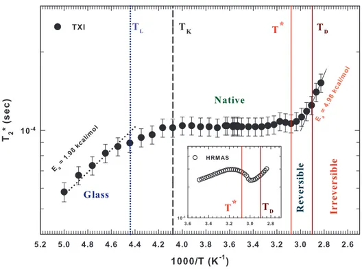
Figure 3 reports in an Arrhenius plot the value of T2∗as a function of the inverse tem-perature. It is noteworthy that a smooth variation of T2∗occurs at the two temperatures identified in fig. 2 (1000/TK = 4.08 K−1 and 1000/T∗ = 3.08 K−1). Instead T2∗ shows
two sharp variations at 1000/TL = 4.44 K−1 (TL ≈ 225 K) and at 1000/TD = 2.9 K−1
(TD ≈ 345 K). The inset of fig. 3 displays a zoom of the high temperature region about
the behavior of T2∗ achieved with the HR-MAS technique where the two relevant tem-peratures, T∗ and TD, are also reported [22, 45].
By means of the Arrhenius representation, we were able to evaluate the mean activa-tion energy of the underlying processes occurring out of the thermal borders character-izing the native state of hydrated lysozyme. In particular, the mean activation energy corresponding to the low-T dynamical crossover is about 1.98 kcal/mol. An activation energy value corresponding to that of hydrogen bonds. On the contrary, the mean acti-vation energy corresponding to the high-T irreversible denaturation is more than twice, being about 4.98 kcal/mol.
The picture arising from our data seems clear enough: the hydrogen bonds dynamics is about constant only within a precise thermal region providing the right condition to the survival of the native states. The low temperature border (TL ≈ 225 K) of this
Fig. 3. – The Arrhenius plot of the thermal evolution of the apparent spin-spin relaxation time (T2∗) for hydrated lysozyme in the range 200 K–360 K. We report all the relevant temperatures
characterizing the changes in the dynamical behavior of water. Furthermore, we show the Arrhenius activation energy characterizing the two observed dynamical crossover at the end of the reversible borders of lysozyme stability. The inset reports a zoom of the high temperature region about the behavior of T2∗achieved with the HR-MAS technique [22, 45].
thermal region coincides with the temperature of the dynamical crossover for water, below which the extended network of hydrogen bonds is fully developed and the dynamics of the system is almost frozen; only hops between different sites are allowed. This is the temperature of the Widom line at atmospheric pressure where the correlation length of water molecules is maximum [46] and indeed they tend to strongly hydrogen bond with each others. The high temperature border (TD ≈ 345 K) instead, coincides with
the temperature of the irreversible denaturation, few degrees above the onset of the folding/unfolding process, that indeed can be reversed for T < TD. We stress that
the onset of the folding/unfolding process coincides with T∗ ≈ 325 K, the threshold temperature below which water anomalies, responsible for its peculiar characteristic, can be observed. Indeed our results confirm that the side chains of the protein enhance too much their mobility above 325K due to the steep decrease of HB strength and lifetime causing the protein structure to unravel.
4. – Conclusions
In this work we have studied by means of NMR spectroscopy some thermodynamical properties of hydration water covering lysozyme surface. In particular, we have studied the behavior of the nuclear magnetization and of the apparent spin-spin relaxation time as a function of the temperature in a large interval (200 K < T < 360 K) including the vitrification and irreversible denaturation processes. Our aim is to determine the thermal thresholds of the stability of the protein native state and to associate them to the dynami-cal variations of the HBs that in particular hydration water performs with the amino acid residuals of lysozyme. The behavior of the magnetization as a function of the tempera-ture (shown in fig. 2) reveals two net changes at two specific temperatempera-tures that indicate, respectively, the beginning of the HB fragility (T∗ at high temperature) and the begin-ning of the vitrification process (TK at low temperature). Both mechanisms provoke for
opposite reasons the end of the protein biological activity and have a clear dynamical implication as shown by T2∗ as a function of T , more evident few degrees beyond them. In particular, from the low temperature side, the transition from the high- to low-density local structures for water (at TL) implies the end of the protein side chains mobility. On
the other hand, from the high temperature side, the small value for the HB strength and lifetime does not allow the formation of stable HBs and the protein irreversibly unravels (at TD). By means of an Arrhenius representation we have obtained the corresponding
activation energy, which is 1.98 kcal/mol for the low-T crossover and 4.98 kcal/mol for the hight-T crossover. Finally, it is noteworthy that the amplitude of both the thermal regions TK− TLand TD− T∗, before vitrification and irreversible denaturation occur, is
of about 20 K.
∗ ∗ ∗
DM’s activity is carried out within the framework of the NANORESTART project which has received funding from the European Union’s Horizon 2020 research and inno-vation programme under grant agreement No. 646063.
REFERENCES
[1] Russo J. and Tanaka H., Nat. Commun., 5 (2014) 3556. [2] Rupley J. A. and Careri G., Adv. Protein Chem., 41 (1991) 37.
[3] Bagchi B., Water in Bilogical and Chemical Processes: From Structure and Dynamics to Function, edited by Saykally R., Zewail A. and King D. (Cambridge University Press, New York) 2013.
[4] Fogarty A. C. and Laage D., J. Phys. Chem. B, 118 (2014) 7715.
[5] Fenimore P. W., Frauenfelder H., McMahon B. H. and Parak F. G., Proc. Natl. Acad. Sci. U.S.A., 99 (2002) 16047.
[6] Ball P., Chem. Rev., 108 (2008) 74.
[7] Mallamace F., Corsaro C., Mallamace D., Vasi C. and Stanley H. E., Faraday Discuss., 167 (2013) 95.
[8] Bellissent-Funel M.-C. et al., Chem. Rev., 116 (2016) 7673. [9] Zhang Y. et al., J. Phys. Chem., 130 (2009) 135101.
[10] Mallamace F. et al., J. Chem. Phys., 141 (2014) 18C504.
[11] Dubou`e-Dijon E., Fogarty A. C.and Laage D., J. Phys. Chem. B, 118 (2014) 1574. [12] Mallamace F. et al., Proc. Natl. Acad. Sci. U.S.A., 113 (2016) 3159.
[13] Ben-Naim A., J. Phys. Chem., 95 (1991) 1437.
[14] Chiti F. and Dobson C. M., Nat. Chem. Biol., 5 (2009) 15. [15] Selkoe D. J., Nature, 126 (1997) 141.
[16] Dagget V., Acc. Chem. Res., 35 (2002) 422.
[17] Mallamace F. et al., J. Chem. Phys., 127 (2007) 045104. [18] Mallamace F. et al., J. Chem. Phys., 141 (2014) 165104.
[19] Salvetti G., Tombari E., Mikheeva L. and Johari G. P., J. Phys. Chem. B, 106 (2002) 6081.
[20] Mallamace F. et al., Front. Phys., 10 (2015) 106104. [21] Mallamace F. et al., J. Chem. Phys., 142 (2015) 215103. [22] Corsaro C. and Mallamace D., Physica A, 390 (2011) 2904. [23] Mallamace D. et al., Physica A, 412 (2014) 39.
[24] Chen S.-H. et al., Proc. Natl. Acad. Sci. U.S.A., 103 (2006) 9012. [25] Lagi M. et al., J. Phys. Chem. B, 112 (2008) 1571.
[26] Camisasca G., De Marzio M., Corradini D. and Gallo P., J. Chem. Phys., 41 (2016) 044503.
[27] Schir`o G., Fomina M.and Cupane A., J. Chem. Phys., 139 (2013) 121102.
[28] Ngai K. L., Capaccioli S. and Shinyashiki N., J. Phys. Chem. B, 112 (2008) 3826. [29] Cerveny S., Mallamace F., Swenson J., Vogel M. and Xu L., Chem. Rev., 116
(2016) 7608.
[30] Stanley H. E. et al., Eur. Phys. J. ST, 161 (2008) 1.
[31] Mallamace F., Proc. Natl. Acad. Sci. U.S.A., 106 (2009) 15097. [32] Debenedetti P. G., J. Phys.: Condens. Matter, 15 (2003) R1669. [33] Gallo P. et al., Chem. Rev., 116 (2016) 7463.
[34] Palmer J. C. et al., Nature, 510 (2014) 385.
[35] Limmer D. T. and Chandler D., J. Chem. Phys., 135 (2011) 134503.
[36] Mallamace F., Corsaro C., Stanley H. E., Mallamace D. and Chen S.-H., J. Chem. Phys., 139 (2013) 214502.
[37] Karplus M., Nat. Chem. Biol., 7 (2001) 401.
[38] Frauenfelder H., Fenimore P. W. and Young R. D., IUBMB Life, 59 (2007) 506. [39] Xu L. et al., Proc. Natl. Acad. Sci. U.S.A., 102 (2005) 16558.
[40] Poole P. H., Sciortino F., Essmann U. and Stanley H. E., Nature, 360 (1992) 324. [41] Kumar P., Phys. Rev. Lett., 97 (2006) 177802.
[42] Abragam A., The Principles of Nuclear Magnetism (Clarendon, Oxford) 1961. [43] Corsaro C. et al., J. Phys. Chem. B, 112 (2008) 10449.
[44] Mallamace F., Corsaro C. and Stanley H. E., Sci. Rep., 2 (2012) 993. [45] Mallamace F. et al., J. Phys. Chem. B, 115 (2011) 14280.
[46] Mallamace F., Corsaro C. and Stanley H. E., Proc. Natl. Acad. Sci. U.S.A., 110 (2013) 4899.