Volume 2013, Article ID 191823,13pages http://dx.doi.org/10.1155/2013/191823
Review Article
Atherosclerosis, Dyslipidemia, and Inflammation:
The Significant Role of Polyunsaturated Fatty Acids
Mariarita Dess
J,
1Annalisa Noce,
2Pierfrancesco Bertucci,
1Simone Manca di Villahermosa,
2Rossella Zenobi,
1Veronica Castagnola,
2Eliana Addessi,
2and Nicola Di Daniele
21Department of Laboratory Medicine, “Tor Vergata” University Hospital, Viale Oxford 81, 00133 Rome, Italy 2Nephrology and Hypertension Unit, Department of System Medicine, “Tor Vergata” University Hospital,
Viale Oxford 81, 00133 Rome, Italy
Correspondence should be addressed to Mariarita Dess`ı; [email protected] Received 1 March 2013; Accepted 9 April 2013
Academic Editors: A. El-Shazly, D. Frommhold, F. M. Kovar, A. Merched, and D. Szukiewicz
Copyright © 2013 Mariarita Dess`ı et al. This is an open access article distributed under the Creative Commons Attribution License, which permits unrestricted use, distribution, and reproduction in any medium, provided the original work is properly cited. Phospholipids play an essential role in cell membrane structure and function. The length and number of double bonds of fatty acids in membrane phospholipids are main determinants of fluidity, transport systems, activity of membrane-bound enzymes, and susceptibility to lipid peroxidation. The fatty acid profile of serum lipids, especially the phospholipids, reflects the fatty acid composition of cell membranes. Moreover, long-chain n-3 polyunsatured fatty acids decrease very-low-density lipoprotein assembly and secretion reducing triacylglycerol production. N-6 and n-3 polyunsatured fatty acids are the precursors of signalling molecules, termed “eicosanoids,” which play an important role in the regulation of inflammation. Eicosanoids derived from n-6 polyunsatured fatty acids have proinflammatory actions, while eicosanoids derived from n-3 polyunsatured fatty acids have anti-inflammatory ones. Previous studies showed that inflammation contributes to both the onset and progression of atherosclerosis: actually, atherosclerosis is predominantly a chronic low-grade inflammatory disease of the vessel wall. Several studies suggested the relationship between long-chain n-3 polyunsaturated fatty acids and inflammation, showing that fatty acids may decrease endothelial activation and affect eicosanoid metabolism.
1. Introduction
Cardiovascular disease is the leading cause of mortality in many economically developed nations accounting for about 30% of all deaths [1] and its incidence is still increasing. Ongoing research aims to investigate and prevent the early development of cardiovascular risk factors such as atheroscle-rosis, hypertension, dyslipidemia, chronic inflammation, and insulin resistance.
The beneficial effects of n-3 polyunsaturated fatty acids (n-3 PUFAs) were proved in several observational and exper-imental studies. The lipid lowering action of n3-PUFAs was detected at the beginning, so these nutrients were used for the treatment of dyslipidemic disorders. Their inflammatory, antithrombotic, antiatherosclerotic, and anti-arrhythmogenic effects were observed later. Low-grade chronic inflammation is now recognized as a prominent pro-cess in the development of atherosclerosis and coronary heart
disease. The induction of inflammation may well provide a link between hyperlipidemia and atherogenesis [2,3].
Atherosclerosis is now considered a “systemic disease” featured by low-grade arterial inflammatory lesions that can develop through the disease progression [4].
In physiological conditions, endothelial cells synthesize and release adequate amounts of nitric oxide (NO) and prostaglandins (such as PGE2 and PGE3) and maintain a downstream balance between pro- and anti-inflammatory molecules. However, in the presence of atherosclerosis this balance disrupts leading towards an increase production of proinflammatory cytokines as interleukins 1, 2, and 6 (IL-1, 2, and 6) and tumor necrosis factor𝛼 (TNF-𝛼), with fur-ther progression of the disease [5]. These pro-inflammatory cytokines can induce oxidative stress by enhancing the production of reactive oxygen species (ROS) by monocytes, macrophages, and leukocytes. PUFAs and their eicosanoid
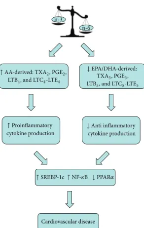
Cardiovascular disease n-6 n-3 ↑ Proinflammatory cytokine production ↓ PPAR𝛼 ↑ NF-𝜅B ↑ SREBP-1c
↑ AA-derived: TXA2, PGE2,
LTB4, and LTC4-LTE4 ↓ EPA/DHA-derived: TXA3, PGE3, LTB5, and LTC5-LTE5 ↓ Anti inflammatory cytokine production
Figure 1: Effects of unbalanced n-6:n-3 dietary fatty acid intake on inflammatory state. EPA: eicosapentaenoic acid; DHA: docas-ahexaenoic acid; AA: arachidonic acid; PPAR-𝛼: peroxisome pro-liferation-activated receptors-𝛼; TX A2: thromboxane A2; TX A3: thromboxane A3; NF-𝜅B: nuclear factor kappa light-chain enhancer of activated B cells; PGE2: prostaglandin E2; PGE3: prostaglandin E3; LTB: leukotriene; SREBP-1c: sterol regulatory binding protein 1c.
derivatives may play a significant role modulating the inflam-matory response (Figure 1) [6].
2. Metabolism of PUFAs
Unsaturated fatty acids are referred to as PUFAs when two or more double bounds are present. There are two PUFAs families, omega-3 (n-3) and omega-6 (n-6) fatty acids. They differ in location of the last double bond relative to the terminal methyl end of the molecule. The human body can produce almost all fatty acids, except linoleic acid (LA, C18:2 n-6, precursor to the n-6 series of fatty acids) and𝛼-linoleic acid (ALA, C18:3n-3, precursor to the n-3 series of fatty acids). These two PUFAs are named “essential fatty acids” because the body cannot synthesize them [7].
Endogenous conversion (elongation and desaturation) of the initial C18 PUFA precursors results in the synthesis of longer-chain counterparts such as eicosapentaenoic acid (EPA) and docosahexaenoic acid (DHA) in the n-3 family and dihomo-𝛾-linoleic acid (DGLA) and arachidonic acid (AA) in the n-6 family [1].
In humans the biochemical pathways converting ALA to EPA and EPA to DHA are limited: 0.2–8% of ALA is converted to EPA (generally more in women) and 0–4% of ALA to DHA [8–12]. Results from pilot studies suggest that ALA conversion is also limited to function as a surrogate for fish consumption [13]. Thus, tissue and circulating EPA and DHA levels are primarily related to their dietary intake. Fish is the major food source of long-chain n-3 PUFAs, including EPA, DHA, and docosapentaenoic acid (DPA), while ALA is a plant n-3 fatty acid mainly found in seeds, nuts, and their oils. Thus, plant sources of n-3 fatty acids cannot currently be considered as a replacement for seafood-derived n-3 PUFAs [14]. This suggests that n-3 fatty acids derived from different sources might have their own specific effects on cardiovascular risk markers. Linoleic acid is thought to decrease the conversion of ALA into EPA and DHA by competing for the n-6-desaturase enzyme [15]. Previous studies showed that genetic variations in this enzyme may be related to cardiovascular disease [16]. Furthermore, because the two fatty acid pathways are mutually exclusive (i.e., n-3 fatty acid cannot become n-6 fatty acid and vice versa), a balanced intake of ALA and LA as precursors or of their longer-chain products EPA, DHA, and AA is required [1].
3. PUFAs and Cardiovascular Disease
There is a vast amount of epidemiologic evidence of a cardio-protective effect of fish oil-derived EPA and DHA and it was confirmed in randomized controlled trials [17–20]. Several intervention studies, such as the “Gruppo Italiano per lo Studio della Sopravvivenza nell’Infarto miocardico” (GISSI)-Prevenzione trial and the Cardiovascular Health Study, have shown that an increased intake of eicosapentaenoic acid (EPA, C20:5n-3) and docosahexaenoic acid (DHA, C22:6n-3) lowers the risk of coronary heart disease (CHD) [21,22]. The first GISSI-Prevenzione trial, a randomized open label study in 11324 Italian patients with recent myocardial infarction, demonstrated that patients had a 15% lower combined risk of mortality, nonfatal myocardial infarction, and stroke upon supplementation for 3.5 years with 850 mg⋅day-1 of LC n-3 PUFA. The relative risk of cardiovascular mortality was also decreased by 30% and that of sudden death by 45%. The GISSI-Prevenzione trial demonstrated that a significant protective effect could be obtained with doses much lower than those previously considered necessary for significant beneficial effects [21].
The second GISSI-HF study, a randomized double-blind placebo-controlled trial in 6975 Italian patients with chronic heart failure, revealed a moderate decrease in both all-cause mortality admissions to hospital for cardiovascular disease upon supplementation for an average of 3.9 years with 1 g of LC n-3 PUFA daily. Again, the beneficial effects were seen in a population already treated with recommended therapies [23]. The “Japan Eicosapentaenoic acid (EPA) Lipid Interven-tion Study,” (JELIS) trial, was performed on 18645 Japanese men and women with hypercholesterolaemia treated with statins. Supplementation with 1.8 g EPA daily decreased major coronary events by 19% over 4.6 years. Non-fatal coro-nary events, rather than CHD death, were decreased [24].
This may be evident in Japanese population where a con-sistent reduction of CHD is observed probably because of a high background seafood intake [25, 26]. In summary, a modest intake of LC n-3 PUFA significantly decreases the risk of fatal CHD. However, higher doses and longer duration of intervention, as reported for the JELIS study, have the potential to protect from non-fatal CHD events [26]. Presently, there is evidence to suggest that the two LC n3-PUFAs—EPA (eicosapentaenoic acid, 20:5N3) and DHA (docosahexaenoic acid, 22:6N3)—exert pleiotropism (akin to those effects which have been reported for certain cardiovascular drugs) by modulating a range of diverse target mechanisms involved in cardiovascular disease development. [27–30]. Since the appearance of purified forms of DHA in the market in the 1990s, researchers have started to investigate the differential effects of EPA and DHA on cardiovascular health. However, the number of human studies is still limited in this field and the independent effects of EPA and DHA on various cardiovascular outcomes are yet to be firmly established [31]. Furthermore, the incorporation of EPA and DHA into the cell membrane influences its organisation, fluidity, and permeability, as well as the activity of transmembrane pro-teins, including receptors, enzymes, and ion channels. Both EPA and DHA modulate K, Na, and Ca channel activities in myocardial cells, regulating myocyte electrical excitability and contractility [32–34]. These effects are concentration dependent and appear to be mediated by the action of EPA and DHA on membrane fluidity [34], although other mech-anisms, such as a direct binding of n-3 LCP to the channel, may be involved [35]. Furthermore, animal studies support the increasing evidence that DHA rather than EPA is prefer-entially incorporated into the myocardial cell membrane [36]. Collectively, these findings help to explain the antiarrhythmic and heart rate (HR)-lowering effects observed by DHA but not by EPA in humans [37]. In addition, incorporation of DHA into the membrane of cardiomyocytes influences the beta adrenergic system to a greater extent than EPA [38], that is, potentially another important mechanism involved in the hypotensive and anti-arrhythmic action of DHA. Further-more, DHA incorporation into the membrane of endothelial cells stimulates ATP release from the endothelium, increasing vasodilation by stimulating nitric oxide (NO) release [39]. The induction of NO release, together with the decrease in noradrenaline levels, may also account for the BP-lowering effect of DHA [39]. A recent meta-analysis including thirty randomized controlled trials showed that prolonged fish oil intake may reduce HR, especially in populations with a high baseline HR [40]. In dyslipidemic males and postmenopausal women, this decrease appears to be mediated by DHA rather than EPA [37, 41, 42]. In contrast, no significant effect of either EPA or DHA on HR was observed in healthy males for similar dosage and treatment duration [43]. HR variability (HRV) is a strong predictor of CVD, including sudden cardiac death, arrhythmic CHD, and atrial fibrillation. Fish oils have shown anti-arrhythmic properties in animal studies [36], and several clinical and epidemiological studies have reported an association between HRV and n-3 LCP blood cell levels and/or fish oil intake [44–46].
The relationship between ALA and cardiovascular dis-ease is less clear. Short-term trials (6–12 week) in healthy individuals showed no or low effect of a daily intake of 1.2– 3.6 g of ALA on blood lipids, LDL-oxidation, lipoprotein (a), and apolipoproteins A-I and B [8,47]. Long-term treatment with high-dose ALA (40 g/daily) showed a beneficial effect by reduction of body weight and blood LDL/total cholesterol ratio [47, 48]. Previous evidence favored recommendation for modest dietary consumption of ALA (2-3 g daily) in the primary and secondary prevention of CHD [19]. The recently published case-control study by Campos et al. [49] shows a strong inverse associations between ALA status-intake and nonfatal MI. Recent data support the assumption that ALA may protect against atherosclerosis [50, 51]. However, data from two recent epidemiologic studies suggest that high tissue ALA is related to an increased rather than decreased risk of fatal cardiovascular events [52] and sudden death [53]. Also, n-3 PUFAs may interfere with CVD with other physiological effects: the reduction of triglyceride (TG) synthesis is well recognized [54]. Moreover, the reduction of hepatic VLDL synthesis may contribute to this protective effect reducing the availability of fatty acids for triglyceride synthesis due to the decreased “de novo” lipogenesis (DNL), increasing the beta-oxidation of fatty acids, reducing the delivery of nonesterified fatty acids to the liver, reducing the activity of TG synthesis by hepatocytes, and finally increasing the hepatic synthesis of phospholipids [54–59].
In both experimental models and human studies, reduc-tion of DNL appears to be prominent [54–60]. A different effect was reported in some studies between EPA and DHA: both EPA and DHA produced a significant reduction of TGs and LDL-cholesterol, raising effects of fish oil [61,62]. EPA showed antiplatelet and anti-inflammatory properties [63,64].
Recently, Buckley and coworkers [65] showed that a four-week supplementation with DHA significantly reduced TG levels in normolipidemic human subjects by 22%, while EPA decreased TG levels by 15% without reaching statistical significance. In another four-week interventional study, both EPA and DHA reduced postprandial TG without affecting fasting TG levels in healthy human subjects [66]. However, EPA and DHA seemed to reduce triglyceridaemia to the same extent when given for a long-enough period [60,61,67–72].
Studies in humans show that an average intake of n-3 PUFAs 3-4 g/daily decreases serum triacylglycerol concentra-tion by 25–30% in a dose-dependent manner. The same intake does not affect total cholesterol but increases LDL cholesterol by 5–10% and HDL cholesterol by 1–3% [73].
The increase in LDL cholesterol is mainly due to a rise of the larger and the potentially less atherogenic LDL particles [74].
Hypertension (AH) and dyslipidemia (DLP) are the most important and frequent risk factors for CVD in the general population, with a prevalence of 50–80% according to different authors [75–79]. The prevalence of DLP is 40% among untreated hypertensive patients. On the other hand almost half of the patients with increased total cholesterol (TC) have systo-diastolic hypertension [80–83].
NF-𝜅B Endothelial dysfunction n-3 PUFA NF-𝜅B Endothelial protection Atherosclerosis Cytokines ROS ↓ Monocyte adhesion ↓ Endothelial inflammation ↓ Thrombogenic mediator secretion ↓ Vasoconstriction
↓ ROS production
↑ Monocyte adhesion
↑ Endothelial inflammation ↑ Thrombogenic mediator secretion ↑ Vasoconstriction ↑ ROS production ↓ Adhesion molecules ↓ Chemokines ↓ Vasoconstrictors ↑ Proinflammatory genes:
cPLA2, COX-2, and INOS
↑ Adhesion molecules:
VCAM-1 and ICAM-1 ↑ Chemokines:
IL-8 and MCP-1
↑ Vasoconstriction: ET-1 ↑ Platelet aggregation:
TF and TXA2
↓ Cytokines
Figure 2: Relationship between inflammation, atherosclerosis, and n-3-PUFA. NF-𝜅B: nuclear factor kappa light-chain enhancer of activated B cells; ROS: reactive oxygen species; cPLA2: cytosolic phospholipase A2; COX-2: cyclooxygenase-2; i-NOS: inducible nitric oxide synthase; VCAM-1: vascular cell adhesion protein 1; ICAM-1: intercellular adhesion molecule-1; IL-8: interlukin-8; MCP-1: monocyte chemoattractant protein-1; TF: tissue factor; ET-1: endothelin-1; TX A2: thromboxane A2.
Modifications of erythrocyte-membrane fatty acids (FA) composition are an early indicator of the development of AH and lipid disorders [79]. Thus, modifications of erythrocyte-membrane FA are fairly subtle indicators of lipid metabolism pathology which manifest themselves much earlier than changes in plasma lipoproteins. Modifications of fatty acids composition of the cell membrane lipid matrix play an important role in the AH pathogenesis. Changes of the fatty acids (FA) composition with decrease of essential PUFAs may result in increase of membrane microviscosity, activation of pro-inflammatory eicosanoids synthesis and increased sensitivity of the smooth muscle cells in artery walls to the influence of vasoconstrictors [84]. That is the reason of the great interest in the study of the spectrum of erythrocyte lipids in hypertensive dyslipidemic patients.
Deficiency of n-3 PUFA is a distinctive feature of the modification of erythrocyte-membrane FA in hypertensive dyslipidemic patients. In particular Novgorodetseva and coworkers observed a significant reduction of 22:5n-3 and 22:6n-3 [79]. Deficiency of endogenous n-3 PUFAs may lead to changes in physicochemical properties of cell membranes, activation of the synthesis of proinflammatory and vaso-constrictive eicosanoids, and finally induction of a systemic inflammatory syndrome [84]. All these conditions may favor the induction and progression of atherosclerosis [85]. Thus, structural and functional changes in erythrocyte cell mem-branes develop in hypertension as a part of the common systemic disorder of lipid metabolism. The FA disorganiza-tion of the cell membrane may cause the development of hyperlipidemia and the progression of hypertensive disease [74]. Thus, an important role of FA in the pathogenesis of cardiovascular disease seems clear.
A large part of the cardioprotective actions of LC n-3-PUFAs is likely to be mediated by the vascular endothelium [28,86–88]. This thin monolayer of cells plays a central role in cardiovascular homeostasis and function via the production of a range of potent autocrine and paracrine biochemical mediators that control the tone of vascular smooth muscle cells [31, 89, 90]. Vascular endothelium is also the site for the inception, progression, and clinical manifestations of atherosclerosis [91–93].
Multifactorial endothelial dysfunction induced by DLP, toxins, cigarette-smoking, and so on may be the early event in the development of atherosclerosis. The endothelium becomes “proadhesive” and induces an increased adhesion of circulating monocytes that subsequently infiltrate the arterial intima. At this level endothelial cells or macrophages release reactive oxygen species that oxidize circulating LDL into oxidized-LDL and create the “lipid streak” [94]. In the evolution from the lipid streak to the atherosclerotic plaque, numerous cytokines that cause infiltration of smooth muscle layer by leukocytes and fibroblasts and promote platelet adhesion are involved in turn [2] (Figures2and3).
During the migration over the vessel wall, monocytes are exposed to various proinflammatory stimuli that contribute to their differentiation into macrophages. After the absorp-tion of lipoproteins, these macrophages become “foam cells.” Other macrophages induce a local inflammatory compart-ment within the vassel wall, that is, continuously powered by cytokines, activated T-helper cells and scavanger receptor activators. The inhibition of this self-maintaining process may reduce atherogenesis as demonstrated in mutant mice deficient for macrophage colony stimulating factor (M-CSF) or macrophage migration inhibitory factor (MIF) [95, 96]. In more advanced lesions, cell debris, apoptotic cells, and
ROS
Circulating LDL
Foam cells
Activation of inflammatory cells
Induction and activation of oxidant generating enzymes
Reduced n-3PUFA
Atherosclerosis
LDL oxidized
Free radicals, ROS, and
LPO-products
Tissue and DNA damage, gene alteration, and increased
NF𝜅B altered cell function
Figure 3: Factors and cells involved in the development of atherosclerosis. ROS: reactive oxygen species; LPO-products: lipo-peroxidation products; NF-𝜅B: nuclear factor kappa light-chain enhancer of activated B cells.
free cholesterol particles accumulate in the lipid-rich necrotic core of the plaque, are sometimes separated from the blood only by a thin fibrous cap. The number of inflammatory cells within the lesion is strongly associated with the risk of rupture of the so-called “vulnerable plaque.” At last, the process may propagate intraluminal thrombosis, thereby distal ischemia of heart or brain tissue [97,98].
Through their positive antithrombotic, lipid-lowering, proendothelial functional activities, n-3 PUFAs play an important role on the mechanisms of atherosclerosis. In fact supplementation of N-3 PUFAs improves flow-mediated arterial dilatation by an increased endothelial synthesis of nitric oxide [86, 99–111]. Several, although not all, clinical trials have also found that consumption of n-3 PUFAs lowers circulating markers of endothelial dysfunction, such as E-selectin, vascular cell adhesion molecule-1, and intracellular adhesion molecule-1 [74, 112, 113]. Thus, normalization of endothelial function could partly mediate the protective effects of n-3 PUFA against CVD. A recent randomized clinical trial established the role of PUFAs on plaque stability
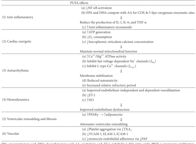
[114]. Patients with carotid artery atherosclerosis were ran-domized to take placebo, fish oil 3 PUFAs), or seed oil (n-6 PUFAs). Subjects treated with n-3 PUFAs showed higher EPA and DHA concentration, reduction of monocytes and macrophages, and a thicker fibrous cap compared to controls and group treated with n-6 PUFAs, all changes that can enhance the stability of atherosclerotic plaques. By contrast, increased assumption of n-6 PUFAs did not affect carotid plaque fatty acid composition or stability. Plaques stability may explain the reductions in nonfatal and fatal cardiovascu-lar events, associated with increased n-3 PUFAs intake [114]. The triglyceride-lowering, antiatherogenic, antithrombotic, and anti-inflammatory effects of n-3 PUFAs were clearly demonstrated in human studies. Conversely, their anti-arrhytmogenic effect was showed only in vitro and in animal experiments, as confirmation in humans has been limited by the absence of reliable physiological measures or biomarkers to quantify antiarrhythmic potential [115]. The main effects exerted by PUFAs in cardiovascular diseases are summarized inTable 1.
Table 1: PUFAs and cardiovascular disease. PUFA effects
(1) Anti-inflammatory
(a)↓NF-𝜅B activation
(b) EPA and DHA compete with AA for COX & 5-lipo-oxygenase enzymatic sites ⇓
Reduce the production of IL-1, IL-6, and TNF-𝛼 (c)↑Anti inflammatory eicosanoids
(2) Cardiac energetic
(a)↑ATP generation (b)↓O2consumption
(c)↓Sarcoplasmic reticulum calcium concentration ⇓
Maintain normal mitochondrial function
(3) Antiarrhythmic
(a)↑Ca2+/Mg2+ATPase activity
(b) Inhibit fast voltage-dependent Na+channels (𝐼Na)
(c) Inhibit L-type Ca2+channels (𝐼Ca,L)
⇓ Membrane stabilization
(d) Reduced automaticity
(e) Increased relative refractory period
(4) Hemodynamics
(a) Improved endothelium-independent and dependent vasodilatation (b)↓ET-1
(c)↑NO
⇓ Improved endothelium dysfunction (5) Ventricular remodeling and fibrosis
(a)↑PPAR𝛾 → ↑adiponectin ⇓ Attenuates ventricular remodeling (6) Vascular
(a)↓Platelet aggregation via ↓TXA2
(b)↓VCAM-1, ELAM-1, ICAM-1
(c)↓monocyte endothelial adherence via ↓PAF
EPA: eicosapentaenoic acid; DHA: docosahexaenoic acid; AA: arachidonic acid; ET-1: endothelin-1; NO: nitric oxide; PPAR-𝛾: peroxisome proliferation-activated receptors-𝛾; TX A2: thromboxane A2; VCAM-1: vascular cell adhesion protein 1; ELAM-1: endothelial leukocyte adhesion molecule-1; ICAM-1:
intercellular adhesion molecule-1; PAF: platelet activating factor; COX: cyclooxygenase.
4. PUFAs and Inflammation
Inflammation is a physiological response to tissue trauma or infection, but leukocytes, which are the effector cells of the inflammatory process, have powerful capabilities of tissue remodelling. Thus, the passage of leukocytes from the bloodstream into inflamed tissue is tightly regulated to ensure their precise localization. Recruitment of circulating neutrophils into the tissue stroma occurs during the early phases of inflammation. In this process, peptide agonists of the chemokine family are assumed to provide a chemotactic stimulus capable of supporting the migration of neutrophils across vascular endothelial cells, through the vessel wall and out into the tissue stroma. Although an initial chemokine stimulus is essential for the recruitment of flowing neu-trophils by endothelial cells, stimulated by the inflammatory cytokine (TNF-𝛼), transit across the endothelial monolayer is regulated by additional stimuli [116].
A new step in the neutrophil recruitment process that relies upon a lipid-mediated signal to regulate the migration of neutrophils across endothelial cells was recently described.
This signal is supplied by the metabolism of arachidonic acid into the eicosanoid prostaglandin-D2 (PGD2) by cyclooxy-genase (COX) enzymes. This step in the neutrophil recruit-ment process was revealed when the EPA was used as an alternative substrate for COX enzymes, leading to the gener-ation of prostaglandin-D3(PGD3) [117–120]. This alternative eicosanoid inhibited the migration of neutrophils across endothelial cells competing for the PGD2receptor [121,122]. PGD2signalling is subordinate to the chemokine-mediated activation of neutrophils, but without the sequential delivery of this signal, neutrophils fail to penetrate the endothelial cell monolayer. The ability of dietary EPA to inhibit this process reveals an unsuspected level of regulation in the migration of inflammatory leukocytes. This may contribute to clarify the interactions between PUFAs and the inflammatory system and direct research on novel therapeutic agents that target the inflammatory system with greater affinity and/or specificity than dietary supplements of n-3-PUFAs [116].
Acute inflammation and its healing are essential processes for tissue protection. The inflammation healing mechanisms
are of main interest, and in recent years, new endoge-nous anti-inflammatory and proresolving lipid mediators generated from PUFAs (lipoxins, resolvins, protectin, and maresin) were uncovered [123]. Lipid mediator metabolomics of self-resolving inflammatory exudates recently highlighted a new family of potent anti-inflammatory and proresolving mediators. Serhan and coworkers identified families of novel bioactive mediators derived from arachidonic acid (AA-derived lipoxins), eicosapentaenoic acid (EPA-(AA-derived E-series resolvins), and docosahexaenoic acid (DHA-derived D-series resolvins, protectin, and maresin) [124–135]. These lipid mediators promote resolution through enhanced clear-ance of apoptotic PMNs, chemokines, cytokines, and micro-bial products by macrophages [136,137].
N-3 PUFAs may act through several mechanisms, for instance, by preventing conversion of the n-6 PUFA arachi-donic acid to pro-inflammatory eicosanoids and by their conversion to potent anti-inflammatory mediators such as resolvins [124–126,138].
A possible biological mechanism underlying the benefi-cial effects of LC n-3 PUFAs on inflammation and endothelial function is that they may compete with n-6 fatty acids for prostaglandin and leukotriene synthesis at the cyclooxyge-nase and lipoxygecyclooxyge-nase levels. LC n-3 PUFAs from fish or fish oil modulate prostaglandin metabolism by increasing the active vasodilator and inhibitor of platelet aggrega-tion prostaglandin E3, the weak platelet aggregator and vasoconstrictor thromboxane A3, and the weak inducer of inflammation leukotriene B5. Otherwise, the production of thromboxane A2, a potent platelet aggregator and vasocon-strictor, and leukotriene B4, an inducer of inflammation and a powerful inducer of leukocyte chemotaxis and adherence is reduced [139]. Another suggested mechanism is that LC n-3 PUFAs may bind reactive oxygen species because of their multiple double bonds and lead to a decreased production of hydrogen peroxide. Hydrogen peroxide is a critical activator of the nuclear factor-𝜅B system of transcription factors that controls the coordinated expression of adhesion molecules and of leukocyte-specific chemoattractants upon cytokine stimulation [140]. He and coworkers [141] found independent inverse associations of LC n-3 PUFAs and nonfried fish with IL-6, CRP and MMP3. The relevance of these biomarkers of inflammatory and endothelial activation in the atherogenic process is well recognized. Previous investigations suggest that both CRP and IL-6, two systemic inflammatory mark-ers, are independent predictors of CVD and may play an important role in atherogenesis [142]. In addition, MMP3 is suggested as an independent prognostic factor in stable coronary artery disease [143]. Several lines of evidence support the main role of MMPs in plaque stability [144].
Soluble intercellular adhesion molecule-1 (sICAM-1) is thought to be a key factor in the adherence of monocytes to the endothelium and subsequent transmigration into the intima (Figure 2). The role of sICAM-1 in the pathogenesis of inflammation and atherosclerosis has been confirmed in experimental models [145]. Previous investigations also indicate that sICAM-1 is an independent predictor of CVD apart of other traditional risk factors [146]. He et al. [141] observed that fried fish but not non-fried fish consumption
was significantly inversely related to sICAM-1. This finding is not expected since the frying process may reduce the content of LC n-3 PUFAs and produce trans-fatty acids. One possible explanation is that fried fish consumption may be a marker of relatively unhealthy lifestyle; those who had high fried fish consumption were more likely to suffer from dyslipidemia and were likely to be under treatment with medications (e.g., statins), which may lower sICAM-1 [147]. However, the inverse association between fried fish intake and sICAM-1 level remained significant when patients taking cholesterol-lowering medications were excluded. Anyway, further investigation is warranted [148].
LC n-3 PUFAs are believed to affect inflammatory pro-cesses mainly through two pathways: endothelial activation and changes in eicosanoid production, or a combination of the two [28]. Two hypotheses may define these molecular mechanisms. The first, predicated on the observation that n-3 PUFAs regulate the transcription of endothelial cell inflammatory genes by downregulating the activity of the nuclear factor-kB, predicts changes in the levels of adhesion receptor and chemokine expression after supplementation with EPA [149–151]. The second hypothesis proposes that upon endothelial cell activation, EPA may compete with the n-6-PUFA arachidonic acid (AA; 20:4n-6) for cyclooxy-genase enzymes (COX1 and COX2) after both fatty acids are released from membrane phospholipids by endogenous phospholipases [152]. The main effects exerted by PUFAs in inflammatory processes are summarized inTable 1.
5. Conclusions
A large amount of investigations suggest, the cardioprotective effects of LC n-3 PUFAs EPA and DHA intake in human subjects, because of their lipid lowering, hypotensive, anti-arrhythmic, and anti-thrombotic properties. Moreover, stud-ies performed in the last twenty years showed heterogeneous effects of different n-3 PUFAs on various cardiovascular outcomes, which may be of paramount relevance in primary and secondary prevention of cardiovascular disease.
Recent in vitro investigations as well as clinical studies also demonstrated that LC-n3-PUFAs significantly interact with inflammation-related mechanisms, such as endothelial activation, modification of eicosanoid metabolism, and reso-lution of the inflammatory process.
Low-grade chronic inflammation is present in several diseases and is characterized by abnormal circulating levels of pro- and anti-inflammatory cytokines. N-3 PUFAs may modulate inflammation, that is, suggested by the reduction of plasma inflammatory cytokines (TNF-𝛼, Il-6) and inflam-matory markers as high sensitive C reactive protein, observed after the intake of EPA and DHA. Low-grade chronic inflam-mation plays a key role in the induction and progression of atherosclerosis and consequently of cardiovascular disease. Taking into consideration the pleiotropic nature of their actions, we suggest that dietary intake of LC n-3 PUFAs may lead to improvements in cardio-metabolic health parameters of their antioxidant, anti-inflammatory, and antiarrhytmic actions.
Abbreviations
n-3 PUFAs: n-3 polyunsaturated fatty acids
NO: Nitric oxide
PGE2; PGE3: Prostaglandins IL-1: Interleukins 1 IL-2: Interleukins 2 IL-6: Interleukins 6
TNF-𝛼: Tumor necrosis factor𝛼 ROS: Reactive oxygen species
LA: Linoleic acid
ALA: 𝛼-Linoleic acid
EPA: Eicosapentaenoic acid
DHA: Docosahexaenoic acid
DGLA: Dihomo-𝛾-linoleic acid
AA: Arachidonic acid
DPA: Docosapentaenoic acid CHD: Coronary Heart Disease HRV: Heart Rate Variability
Tgs: Triglyceride
AH: Arterial Hypertension
DLP: Dyslipidemia,
TC: Total cholesterol
FA: Fatty acids
M-CSF: Macrophage colony stimulating factor MIF: Migration Inhibiting Factor
PGD2: Prostaglandin D2 COX1-COX2: Cycloxygenase 1-2
TXA: Thromboxane A
sICAM-1: Soluble intercellular adhesion molecule-1.
References
[1] M. Y. Abeywardena and G. S. Patten, “Role of𝜔3 longchain polyunsaturated fatty acids in reducing cardio-metabolic risk factors,” Endocrine, Metabolic and Immune Disorders, vol. 11, no. 3, pp. 232–246, 2011.
[2] L. Dei Cas, S. Nodari, and A. Manerba, “Polyunsaturated fatty acids (n-3 PUFAs),” Giornale di Gerontologia, vol. 55, no. 1, pp. 45–57, 2007.
[3] R. Ross, “Atherosclerosis and inflammatory disease,” New
Eng-land Journal Medicine, vol. 340, pp. 115–126, 1999.
[4] F. Montecucco and F. Mach, “Atherosclerosis is an inflammatory disease,” in Seminars in Immunopathology, Springer, New York, NY, USA, 2009.
[5] U. N. Das, “A defect in the activity ofΔ6 and Δ5 desaturases may be a factor in the initiation and progression of atherosclerosis,”
Prostaglandins Leukotrienes and Essential Fatty Acids, vol. 76,
no. 5, pp. 251–268, 2007.
[6] U. N. Das, “Biological significance of essential fatty acids,”
Journal of Association of Physicians of India, vol. 54, pp. 309–319,
2006.
[7] E. Patterson, R. Wall, G. F. Fitzgerald et al., “Health implications of high dietary Omega-6 polyunsaturated fatty acids,” Journal of
Nutrition and Metabolism, vol. 5, pp. 1–16, 2012.
[8] G. Burdge, “𝛼-Linolenic acid metabolism in men and women: nutritional and biological implications,” Current Opinion in
Clinical Nutrition and Metabolic Care, vol. 7, no. 2, pp. 137–144,
2004.
[9] G. C. Burdge, Y. E. Finnegan, A. M. Minihane, C. M. Williams, and S. A. Wootton, “Effect of altered dietary n-3 fatty acid intake upon plasma lipid fatty acid composition, conversion of [13C]𝛼-linolenic acid to longer-chain fatty acids and partitioning towards𝛽-oxidation in older men,” British Journal of Nutrition, vol. 90, no. 2, pp. 311–321, 2003.
[10] G. C. Burdge, A. E. Jones, and S. A. Wootton, “Eicosapentanoic and docosapentanoic acids are the principal products of alpha-linoleic acid metabolism in young men,” British Journal of
Nutrition, vol. 88, pp. 355–363, 2002.
[11] E. A. Emken, R. O. Adlof, and R. M. Gulley, “Dietary linoleic acid influences desaturation and acylation of deuterium-labeled linoleic and linolenic acids in young adult males,” Biochimica et
Biophysica Acta, vol. 1213, no. 3, pp. 277–288, 1994.
[12] R. J. Pawlosky, J. R. Hibbeln, J. A. Novotny, and N. Salem, “Physi-ological compartmental analysis of𝛼-linolenic acid metabolism in adult humans,” Journal of Lipid Research, vol. 42, no. 8, pp. 1257–1265, 2001.
[13] P. L. L. Goyens, M. E. Spilker, P. L. Zock, M. B. Katan, and R. P. Mensink, “Compartmental modeling to quantify𝛼-linolenic acid conversion after longer term intake of multiple tracer boluses,” Journal of Lipid Research, vol. 46, no. 7, pp. 1474–1483, 2005.
[14] U.S Department of Agriculture and U.S Department of Health and Human Services, Dietary Guidelines for Americans, Gov-ernment Printing Office, Washington, DC, USA, 7th edition, 2010.
[15] C. R. Harper, M. J. Edwards, A. P. DeFilipis et al., “Flaxseed oil increases the plasma concentrations of cardioprotective (n-3) fatty acids in humans,” American Society For Nutrition, vol. 137, no. 2816, pp. 83–87, 2007.
[16] H. Truong, J. R. DiBello, E. Ruiz-Narvaez et al., “Does genetic variation in the Delta6-desaturase promoter modify the asso-ciatio between alpha-linolenic acid and the prevalence of metabolic syndrome?” The American Journal of Clinical
Nutri-tion, vol. 89, pp. 920–925, 2009.
[17] S. N. Hansen and W. S. Harris, “New evidence for the cardio-vascular benefits of long chain omega-3 fatty acids,” Current
Atherosclerosis Reports, vol. 9, pp. 434–440, 2007.
[18] W. S. Harris, P. M. Kris-Etherton, and K. A. Harris, “Intakes of long-chain omega-3 fatty acid associated with reduced risk for death from coronary heart disease in healthy adults,” Current
Atherosclerosis Reports, vol. 10, no. 6, pp. 503–509, 2008.
[19] D. Mozaffarian, “Does alpha-linolenic acid intake reduce the risk of coronary heart disease? A review of the evidence,”
Alternative Therapies in Health and Medicine, vol. 11, no. 3, pp.
24–31, 2005.
[20] W. S. Harris, M. Miller, A. P. Tighe, M. H. Davidson, and E. J. Schaefer, “Omega-3 fatty acids and coronary heart disease risk: clinical and mechanistic perspectives,” Atherosclerosis, vol. 197, no. 1, pp. 12–24, 2008.
[21] GISSI Investigators, “Dietary supplementation with n-3 polyun-saturated fatty acids and vitamin E after myocardial infarction: results of the GISSI-Prevenzione trial. Gruppo Italiano per lo Studio della Sopravvivenza nel’Infarto miocardico,” The Lancet, vol. 354, pp. 447–455, 1999.
[22] R. N. Lemaitre, I. B. King, D. Mozaffarian, L. H. Kuller, R. P. Tracy, and D. S. Siscovick, “n-3 polyunsaturated fatty acids, fatal ischemic heart disease, and nonfatal myocardial infarction in older adults: the Cardiovascular Health Study,” American
[23] GISSI-HF investigators, “Effect of n-3 polyunsaturated fatty acids in patients with chronic heart failure (the GISSI-HF trial): a randomised, double-blind, placebo-controlled trial,”
The Lancet, vol. 372, no. 9645, pp. 1223–1230, 2008.
[24] M. Yokoyama, H. Origasa, M. Matsuzaki et al., “Effects of eicosapentaenoic acid on major coronary events in hyper-cholesterolaemic patients (JELIS): a randomised open-label, blinded endpoint analysis,” The Lancet, vol. 369, no. 9567, pp. 1090–1098, 2007.
[25] D. Mozaffarian, “JELIS, fish oil, and cardiac events,” The Lancet, vol. 369, no. 9567, pp. 1062–1063, 2007.
[26] D. Mozaffarian, “Fish and n-3 fatty acids for the prevention of fatal coronary heart disease and sudden cardiac death,”
American Journal of Clinical Nutrition, vol. 87, no. 6, pp. 1991S–
1996S, 2008.
[27] M. N. D. Di Minno, E. Tremoli, A. Tufano, A. Russolillo, R. Lupoli, and G. Di Minno, “Exploring newer cardioprotective strategies: 𝜔-3 fatty acids in perspective,” Thrombosis and
Haemostasis, vol. 104, no. 4, pp. 664–680, 2010.
[28] M. Y. Abeywardena and R. J. Head, “Longchain n-3 polyun-saturated fatty acids and blood vessel function,” Cardiovascular
Research, vol. 52, no. 3, pp. 361–371, 2001.
[29] G. C. Makris, G. Geroulakos, M. C. Makris, D. P. Mikhailidis, and M. E. Falagas, “The pleiotropic effects of statins and omega-3 fatty acids against sepsis: a new perspective,” Expert Opinion
on Investigational Drugs, vol. 19, no. 7, pp. 809–814, 2010.
[30] P. P. Dimitrow and M. Jawien, “Pleiotropic, cardioprotective effects of omega-3 polyunsaturated fatty acids,” Mini-Reviews in
Medicinal Chemistry, vol. 9, no. 9, pp. 1030–1039, 2009.
[31] S. C. Cottin, T. A. Sanders, and W. L. Hall, “The differential effects of EPA and DHA on cardiovascular risk factors,” Dia-betes and Nutritional Sciences Division, School of Medicine, King’s College London, London, UK.
[32] Y. F. Xiao, A. M. Gomez, J. P. Morgan, W. J. Lederer, and A. Leaf, “Suppression of voltage-gated L-type Ca2+ currents by polyunsaturated fatty acids in adult and neonatal rat ventricular myocytes,” Proceedings of the National Academy of Sciences of the
United States of America, vol. 94, no. 8, pp. 4182–4187, 1997.
[33] G. R. Li, H. Y. Sun, X. H. Zhang et al., “Omega-3 polyun-saturated fatty acids inhibit transient outward and ultra-rapid delayed rectifier K+currents and Na+current in human atrial myocytes,” Cardiovascular Research, vol. 81, no. 2, pp. 286–293, 2009.
[34] Y. F. Xiao, D. C. Sigg, and A. Leaf, “The antiarrhythmic effect of n-3 polyunsaturated fatty acids: modulation of cardiac ion channels as a potential mechanism,” Journal of Membrane
Biology, vol. 206, no. 2, pp. 141–154, 2005.
[35] J. X. Kang and A. Leaf, “Evidence that free polyunsaturated fatty acids modify Na+ channels by directly binding to the channel proteins,” Proceedings of the National Academy of Sciences of the
United States of America, vol. 93, no. 8, pp. 3542–3546, 1996.
[36] P. L. McLennan, “Myocardial membrane fatty acids and the antiarrhythmic actions of dietary fish oil in animal models,”
Lipids, vol. 36, supplement, pp. S111–S114, 2001.
[37] T. A. Mori, D. Q. Bao, V. Burke, I. B. Puddey, and L. J. Beilin, “Docosahexaenoic acid but not eicosapentaenoic acid lowers ambulatory blood pressure and heart rate in humans,”
Hypertension, vol. 34, no. 2, pp. 253–260, 1999.
[38] A. Grynberg, A. Fournier, J. P. Sergiel, and P. Athias, “Effect of docosahexaenoic acid and eicosapentaenoic acid in the
phospholipids of rat heart muscle cells on adrenoceptor respon-siveness and mechanism,” Journal of Molecular and Cellular
Cardiology, vol. 27, no. 11, pp. 2507–2520, 1995.
[39] M. Hashimoto, K. Shinozuka, S. Gamoh et al., “The hypotensive effect of docosahexaenoic acid is associated with the enhanced release of ATP from the caudal artery of aged rats,” Journal of
Nutrition, vol. 129, no. 1, pp. 70–76, 1999.
[40] D. Mozaffarian, A. Geelen, I. A. Brouwer, J. M. Geleijnse, P. L. Zock, and M. B. Katan, “Effect of fish oil on heart rate in humans: a meta-analysis of randomized controlled trials,”
Circulation, vol. 112, no. 13, pp. 1945–1952, 2005.
[41] K. D. Stark and B. J. Holub, “Differential eicosapentaenoic acid elevations and altered cardiovascular disease risk factor responses after supplementation with docosahexaenoic acid in postmenopausal women receiving and not receiving hormone replacement therapy,” American Journal of Clinical Nutrition, vol. 79, no. 5, pp. 765–773, 2004.
[42] S. Grimsgaard, K. H. Bønaa, J. B. Hansen, and E. S. P. Myhre, “Effects of highly purified eicosapentaenoic acid and docos-ahexaenoic acid on hemodynamics in humans,” American
Journal of Clinical Nutrition, vol. 68, no. 1, pp. 52–59, 1998.
[43] R. J. Woodman, T. A. Mori, V. Burke, I. B. Puddey, G. F. Watts, and L. J. Beilin, “Effects of purified eicosapentaenoic and docosahexaenoic acids on glycemic control, blood pressure, and serum lipids in type 2 diabetic patients with treated hypertension,” American Journal of Clinical Nutrition, vol. 76, no. 5, pp. 1007–1015, 2002.
[44] J. H. Christensen, H. A. Skou, L. Fog et al., “Marine n-3 fatty acids, wine intake, and heart rate variability in patients referred for coronary angiography,” Circulation, vol. 103, no. 5, pp. 651– 657, 2001.
[45] F. Holguin, M. M. T´ellez-Rojo, M. Lazo et al., “Cardiac auto-nomic changes associated with fish oil vs soy oil supplementa-tion in the elderly,” Chest, vol. 127, no. 4, pp. 1102–1107, 2005. [46] J. H. Christensen, M. S. Christensen, J. Dyerberg, and E. B.
Schmidt, “Heart rate variability and fatty acid content of blood cell membranes: a dose-response study with n-3 fatty acids,”
American Journal of Clinical Nutrition, vol. 70, no. 3, pp. 331–
337, 1999.
[47] J. M. Geleijnse, J. De Goede, and I. A. Brouwer, “Alpha-linolenic acid: is it essential to cardiovascular health?” Current
Atherosclerosis Reports, vol. 12, no. 6, pp. 359–367, 2010.
[48] S. Dodin, S. C. . Cunnane, B. Mˆasse et al., “Flaxseed on cardio-vascular disease markers in healthy menopausal women: a randomized, double-blind, placebo-controlled trial,” Nutrition, vol. 24, pp. 23–30, 2008.
[49] H. Campos, A. Baylin, and W. C. . Willett, “Alpha-linolenic acid and risk of nonfatal acute myocardial infarction,” Circulation, vol. 118, pp. 339–345, 2008.
[50] A. Sala-Vila, M. Cof´an, A. P´erez-Heras et al., “Fatty acids in serum phospholipids and carotid intima-media thickness in Spanish subjects with primary dyslipidemia,” The American
Journal of Clinical Nutrition, vol. 92, pp. 186–193, 2010.
[51] Y. Park, J. Lim, Y. Kwon, and J. Lee, “Correlation of erythrocyte fatty acid composition and dietary intakes with markers of atherosclerosis in patients with myocardial infarction,”
Nutri-tion Research, vol. 29, no. 6, pp. 391–396, 2009.
[52] E. Warensj¨o, J. Sundstr¨om, B. Vessby, T. Cederholm, and U. Ris´erus, “Markers of dietary fat quality and fatty acid desat-uration as predictors of total and cardiovascular mortality: a population-based prospective study,” American Journal of
[53] R. N. Lemaitre, I. B. King, N. Sotoodehnia et al., “Red blood cell membrane𝛼-linolenic acid and the risk of sudden cardiac arrest,” Metabolism, vol. 58, no. 4, pp. 534–540, 2009.
[54] W. S. Harris and D. Bulchandani, “Why do omega-3 fatty acids lower serum triglycerides?” Current Opinion in Lipidology, vol. 17, no. 4, pp. 387–393, 2006.
[55] D. Faeh, K. Minehira, J. M. Schwarz et al., “Effect of fructose overfeeding and fish oil administration on hepatic de novo lipogenesis and insulin sensitivity in healthy men,” Diabetes, vol. 54, no. 1, pp. 907–913, 2005.
[56] S. D. Clarke, “Polyunsaturated fatty acid regulation of gene transcription: a molecular mechanism to improve the metabolic syndrome,” Journal of Nutrition, vol. 131, no. 4, pp. 1129–1132, 2001.
[57] A. A. Rivellese, A. Maffettone, C. Iovine et al., “Long-term effects of fish oil on insulin resistance and plasma lipoproteins in NIDDM patients with hypertriglyceridemia,” Diabetes Care, vol. 19, no. 11, pp. 1207–1213, 1996.
[58] V. Saraswathi, J. D. Morrow, and A. H. Hasty, “Dietary fish oil exerts hypolipidemic effects in lean and insulin sensitizing effects in obese LDLR-/- mice,” Journal of Nutrition, vol. 139, no. 12, pp. 2380–2386, 2009.
[59] D. B. Jump, “Fatty acid regulation of hepatic lipid metabolism,”
Current Opinion in Clinical Nutrition and Metabolic Care, vol.
14, no. 2, pp. 115–120, 2011.
[60] S. Wada, T. Yamazaki, Y. Kawano, S. Miura, and O. Ezaki, “Fish oil fed prior to ethanol administration prevents acute ethanol-induced fatty liver in mice,” Journal of Hepatology, vol. 49, no. 3, pp. 441–450, 2008.
[61] Y. Kobatake, K. Kuroda, and H. Jinnouchi, “Differential effects of dietary eicosapentaenoic and docosahexaenoic fatty acids on lowering of triglyceride and cholesterol levels in the serum of rats on hypercholesterolemic diet,” Journal of Nutritional Science
and Vitaminology, vol. 30, no. 4, pp. 357–372, 1984.
[62] G. S. Rambjør, A. I. W˚alen, S. L. Windsor, and W. S. Harris, “Eicosapentaenoic acid is primarily responsible for hypotriglyc-eridemic effect of fish oil in humans,” Lipids, vol. 31, no. 3, supplement, pp. S45–S49, 1996.
[63] T. Terano, T. Kojima, A. Seya et al., “The effect of highly purified eicosapentaenoic acid in patients with psoriasis,” Advance
Prostaglandin, Thromboxane, and Leukotriene Research, vol. 19,
pp. 610–613, 1989.
[64] A. Hirai, T. Terano, H. Makuta et al., “Effect of oral admin-istration of highly purified eicosapentaenoic acid and docosa-hexaenoic acid on platelet function and serum lipids in hyper-lipidemic patients,” Advance Prostaglandin, Thromboxane, and
Leukotriene Research, vol. 19, pp. 627–630, 1989.
[65] R. Buckley, B. Shewring, R. Turner, P. Yaqoob, and A. M. Mini-hane, “Circulating triacylglycerol and apoE levels in response to EPA and docosahexaenoic acid supplementation in adult human subjets,” British Journal of Nutrition, vol. 92, no. 3, pp. 477–483, 2004.
[66] Y. Park and W. S. Harris, “Omega-3 fatty acid supplementation accelerates chylomicron triglyceride clearance,” Journal of Lipid
Research, vol. 44, no. 3, pp. 455–463, 2003.
[67] S. Grimsgaard, K. H. Bønaa, J. B. Hansen, and A. Nordøy, “Highly purified eicosapentaenoic acid and docosahexaenoic acid in humans have similar triacylglycerol-lowering effects but divergent effects on serum fatty acids,” American Journal of
Clinical Nutrition, vol. 66, no. 3, pp. 649–659, 1997.
[68] S. Egert, F. Kannenberg, V. Somoza, H. F. Erbersdobler, and U. Wahrburg, “Dietary𝛼-linolenic acid, EPA, and DHA have
differential effects on LDL fatty acid composition but similar effects on serum lipid profiles in normolipidemic humans,”
Journal of Nutrition, vol. 139, no. 5, pp. 861–868, 2009.
[69] T. A. Mori, V. Burke, I. B. Puddey et al., “Purified eicosapen-taenoic and docosahexaenoic acids have differential effects on serum lipids and lipoproteins, LDL particle size, glucose, and insulin in mildly hypedipidemic men,” American Journal of
Clinical Nutrition, vol. 71, no. 5, pp. 1085–1094, 2000.
[70] E. Olano-Martin, E. Anil, M. J. Caslake et al., “Contribution of apolipoprotein E genotype and docosahexaenoic acid to the LDL-cholesterol response to fish oil,” Atherosclerosis, vol. 209, no. 1, pp. 104–110, 2010.
[71] R. J. Woodman, T. A. Mori, V. Burke, I. B. Puddey, G. F. Watts, and L. J. Beilin, “Effects of purified eicosapentaenoic and docosahexaenoic acids on glycemic control, blood pressure, and serum lipids in type 2 diabetic patients with treated hypertension,” American Journal of Clinical Nutrition, vol. 76, no. 5, pp. 1007–1015, 2002.
[72] P. Nestel, H. Shige, S. Pomeroy, M. Cehun, M. Abbey, and D. Raederstorff, “The n-3 fatty acids eicosapentaenoic acid and docosahexaenoic acid increase systemic arterial compliance in humans,” American Journal of Clinical Nutrition, vol. 76, no. 2, pp. 326–330, 2002.
[73] W. S. Harris, “n-3 fatty acids and serum lipoproteins: human studies,” The American Journal of Clinical Nutrition, vol. 65, pp. 1645S–1654S, 1997.
[74] B. De Roos, Y. Mavrommatis, and I. A. Brouwer, “Long-chain n-3 polyunsaturated fatty acids: new insights into mechanisms relating to inflammation and coronary heart disease,” British
Journal of Pharmacology, vol. 158, no. 2, pp. 413–428, 2009.
[75] D. C. Goff, A. G. Bertoni, H. Kramer et al., “Dyslipidemia prevalence, treatment, and control in the Multi-Ethnic Study of Atherosclerosis (MESA): gender, ethnicity, and coronary artery calcium,” Circulation, vol. 113, no. 5, pp. 647–656, 2006. [76] K. M. Anderson, W. P. Castelli, and D. Levy, “Cholesterol and
mortality. 30 Years of follow-up from the Framingham Study,”
Journal of the American Medical Association, vol. 257, no. 16, pp.
2176–2180, 1987.
[77] A. A. Liakishev, “Correction of lipid disorders in patients with arterial hypertension,” Russian Medical Journals, vol. 10, no. 19, pp. 878–882, 2002.
[78] J. D. Kobalava and V. V. Tolkachev, “Hypercholesterolemia and arterial hypertension,” Heart, vol. 4, no. 28, pp. 172–176, 2006. [79] T. P. Novgorodtseva, T. A. Kantur, Y. K. Karaman, M. V.
Anto-nyuk, and N. V. Zhukova, “Modification of fatty acids compo-sition in erythrocytes lipids in arterial hypertension associated with dyslipidemia,” Lipids in Health and Disease, vol. 10, article 18, 2011.
[80] D. C. Goff, A. G. Bertoni, H. Kramer et al., “Dyslipidemia prevalence, treatment, and control in the Multi-Ethnic Study of Atherosclerosis (MESA): gender, ethnicity, and coronary artery calcium,” Circulation, vol. 113, no. 5, pp. 647–656, 2006. [81] D. K. Arnett, D. R. Jacobs, R. V. Luepker, H. Blackburn, C.
Armstrong, and S. A. Claas, “Twenty-year trends in serum cholesterol, hypercholesterolemia, and cholesterol medication use: the Minnesota Heart Survey, 1980–1982 to 2000–2002,”
Circulation, vol. 112, no. 25, pp. 3884–3891, 2005.
[82] I. Graham, M. T. Cooney, D. Bradley, A. Dudina, and Z. Reiner, “Dyslipidemias in the prevention of cardiovascular disease: risks and causality,” Current Cardiology Reports, vol. 14, no. 6, pp. 709–720, 2012.
[83] S. Y. Martsevich, “Treatment of lipid disorders in patients with coronary heart disease,” Treating Physician, vol. 5, pp. 42–45, 2005.
[84] E. A. Endakova and T. P. Novgorodtseva, Svetashev VI
Modifica-tion of Blood Fatty Acids ComposiModifica-tion in Case of Cardiovascular Diseases, vol. 296, Dalnauka, Vladivostok, Russia, 2002.
[85] V. N. Titov, “Atherosclerosis as a pathology polien fatty acids,” in Biological Bases Pathogenesis, Diagnostics, Preventive and
Treatment of Atherosclerosis, vol. 750, M:Altus, 2002.
[86] S. Rizza, M. Tesauro, C. Cardillo et al., “Fish oil supplementation improves endothelial function in normoglycemic offspring of patients with type 2 diabetes,” Atherosclerosis, vol. 206, no. 2, pp. 569–574, 2009.
[87] T. A. Mori, “Omega-3 fatty acids and hypertension in humans,”
Clinical and Experimental Pharmacology and Physiology, vol. 33,
no. 9, pp. 842–846, 2006.
[88] S. Wang, A. Q. Ma, S. W. Song, Q. H. Quan, X. F. Zhao, and X. H. Zheng, “Fish oil supplementation improves large arterial elasticity in overweight hypertensive patients,” European
Jour-nal of Clinical Nutrition, vol. 62, no. 12, pp. 1426–1431, 2008.
[89] D. Ait-Yahia, S. Madani, J. L. Savelli, J. Prost, M. Bouchenak, and J. Belleville, “Dietary fish protein lowers blood pressure and alters tissue polyunsaturated fatty acid composition in spontaneously hypertensive rats,” Nutrition, vol. 19, no. 4, pp. 342–346, 2003.
[90] A. T. Erkkil¨a, U. S. Schwab, V. D. F. De Mello et al., “Effects of fatty and lean fish intake on blood pressure in subjects with coronary heart disease using multiple medications,” European
Journal of Nutrition, vol. 47, no. 6, pp. 319–328, 2008.
[91] S. Taddei, L. Ghiadoni, A. Virdis, D. Versari, and A. Salvetti, “Mechanisms of endothelial dysfunction: clinical significance and preventive non-pharmacological therapeutic strategies,”
Current Pharmaceutical Design, vol. 9, no. 29, pp. 2385–2402,
2003.
[92] C. L. Stebbins, J. P. Stice, C. M. Hart, F. N. Mbai, and A. A. Knowlton, “Effects of dietary decosahexaenoic acid (DHA) on eNOS in human coronary artery endothelial cells,” Journal of
Cardiovascular Pharmacology and Therapeutics, vol. 13, no. 4,
pp. 261–268, 2008.
[93] R. De Caterina, M. Massaro, E. Scoditti, and M. Annunziata Carluccio, “Pharmacological modulation of vascular inflamma-tion in atherothrombosis,” Annals of the New York Academy of
Sciences, vol. 1207, pp. 23–31, 2010.
[94] R. Ross, “Phathogenesis of atherosclerosis a prospective for the 1990s,” Nature, vol. 362, pp. 801–809, 1993.
[95] J. H. Qiao, J. Tripathi, N. K. Mishra et al., “Role of macrophage colony-stimulating factor in atherosclerosis: studies of osteo-petrotic mice,” American Journal of Pathology, vol. 150, no. 5, pp. 1687–1699, 1997.
[96] J. Bernhagen, R. Krohn, H. Lue et al., “MIF is a noncognate ligand of CXC chemokine receptors in inflammatory and atherogenic cell recruitment,” Nature Medicine, vol. 13, no. 5, pp. 587–596, 2007.
[97] P. Libby, “Vascular biology of atherosclerosis: overview and state of the art,” American Journal of Cardiology, vol. 91, no. 3, pp. 3A– 6A, 2003.
[98] F. M. van der Valk, D. F. van Wijk, and E. S. G. Stroes, “Novel anti-inflammatory strategies in atherosclerosis,” Current
Opinion in Lipidology, vol. 23, pp. 532–539, 2012.
[99] A. Stirban, S. Nandrean, C. G¨otting et al., “Effects of n-3 fatty acids on macro- and microvascular function in subjects with
type 2 diabetes mellitus,” American Journal of Clinical Nutrition, vol. 91, no. 3, pp. 808–813, 2010.
[100] F. Dangardt, W. Osika, Y. Chen et al., “Omega-3 fatty acid sup-plementation improves vascular function and reduces inflam-mation in obese adolescents,” Atherosclerosis, vol. 212, no. 2, pp. 580–585, 2010.
[101] J. Goodfellow, M. F. Bellamy, M. W. Ramsey, C. J. H. Jones, and M. J. Lewis, “Dietary supplementation with marine omega-3 fatty acids improve systemic large artery endothelial function in subjects with hypercholesterolemia,” Journal of the American
College of Cardiology, vol. 35, no. 2, pp. 265–270, 2000.
[102] M. M. Engler, M. B. Engler, M. Malloy et al., “Docosahexaenoic acid restores endothelial function in children with hyperlipi-demia: results from the early study,” International Journal of
Clinical Pharmacology and Therapeutics, vol. 42, no. 12, pp. 672–
679, 2004.
[103] M. Haberka, K. Mizia-Stec, M. Mizia et al., “N-3 polyunsat-urated fatty acids early supplementation improves ultrasound indices of endothelial function, but not through NO inhibitors in patients with acute myocardial infarction. N-3 PUFA sup-plementation in acute myocardial infarction,” Clinical Nutrition, vol. 30, no. 1, pp. 79–85, 2011.
[104] V. Schiano, E. Laurenzano, G. Brevetti et al., “Omega-3 polyun-saturated fatty acid in peripheral arterial disease: effect on lipid pattern, disease severity, inflammation profile, and endothelial function,” Clinical Nutrition, vol. 27, no. 2, pp. 241–247, 2008. [105] H. Tagawa, H. Shimokawa, T. Tagawa, M. Kuroiwa-Matsumoto,
Y. Hirooka, and A. Takeshita, “Long-term treatment with eicosapentaenoic acid augments both nitric oxide-mediated and non-nitric oxide-mediated endothelium-dependent fore-arm vasodilatation in patients with coronary artery disease,”
Journal of Cardiovascular Pharmacology, vol. 33, no. 4, pp. 633–
640, 1999.
[106] S. N. Doshi, K. K. Naka, N. Payne et al., “Flow-mediated dilatation following wrist and upper arm occlusion in humans: the contribution of nitric oxide,” Clinical Science, vol. 101, no. 6, pp. 629–635, 2001.
[107] W. S. Harris, G. S. Rambjor, S. L. Windsor et al., “N-3 fatty acids and urinary excretion of nitric oxide metabolites in humans,”
The Amrican Journal of Clinical Nutrition, vol. 65, pp. 459–464,
1997.
[108] Y. Okuda, K. Kawashima, T. Sawada et al., “Eicosapentaenoic acid enhances nitric oxide production by cultured human endothelial cells,” Biochemical and Biophysical Research
Com-munications, vol. 232, no. 2, pp. 487–491, 1997.
[109] M. Omura, S. Kobayashi, Y. Mizukami et al., “Eicosapentaenoic acid (EPA) induces Ca2+-independent activation and translo-cation of endothelial nitric oxide synthase and endothelium-dependent vasorelaxation,” FEBS Letters, vol. 487, no. 3, pp. 361– 366, 2001.
[110] T. U. Singh, K. Kathirvel, S. Choudhury, S. K. Garg, and S. K. Mishra, “Eicosapentaenoic acid-induced endothelium-dependent and -inendothelium-dependent relaxation of sheep pulmonary artery,” European Journal of Pharmacology, vol. 636, no. 1–3, pp. 108–113, 2010.
[111] D. L´opez, M. M¨oller, A. Denicola et al., “Long-chain n-3 polyunsaturated fatty acid from fish oil modulates aortic nitric oxide and tocopherol status in the rat,” British Journal of
Nutrition, vol. 100, no. 4, pp. 767–775, 2008.
[112] J. G. Robinson and N. J. Stone, “Antiatherosclerotic and antithrombotic effects of omega-3 fatty acids,” American Journal
[113] P. M. Kris-Etherton, W. S. Harris, and L. J. Appel, “Fish consumption, fish oil, omega-3 fatty acids, and cardiovascular disease,” Circulation, vol. 106, no. 21, pp. 2747–2757, 2002. [114] F. Thies, J. M. C. Garry, P. Yaqoob et al., “Association of
n-3 polyunsaturated fatty acids with stability of atherosclerotic plaques: a randomised controlled trial,” The Lancet, vol. 361, no. 9356, pp. 477–485, 2003.
[115] D. Mozaffarian and J. H. Wu, “Omega-3 fatty acids and car-diovascular disease: effects on risk factors, molecular pathways, and clinical events,” Journal of the American College of
Cardiol-ogy, vol. 58, no. 20, pp. 2047–2068, 2011.
[116] S. P. Tull, C. M. Yates, B. H. Maskrey et al., “Omega-3 fatty acids and inflammation: novel interactions reveal a new step in neutrophil recruitment,” PLoS Biology, vol. 7, no. 8, Article ID e1000177, 2009.
[117] T. Hamada, S. Tsuchihashi, A. Avanesyan et al., “Cyclooxygen-ase-2 deficiency enhances Th2 immune responses and impairs neutrophil recruitment in hepatic ischemia/reperfusion injury,”
Journal of Immunology, vol. 180, no. 3, pp. 1843–1853, 2008.
[118] V. L. Gonc¸alves De Moraes, B. B. Vargaftig, J. Lefort, A. Meager, and M. Chignard, “Effect of cyclo-oxygenase inhibitors and modulators of cyclic AMP formation on lipopolysaccharide-induced neutrophil infiltration in mouse lung,” British Journal
of Pharmacology, vol. 117, no. 8, pp. 1792–1796, 1996.
[119] J. E. Beesley, J. D. Pearson, and A. Hutchings, “Granulocyte migration through endothelium in culture,” Journal of Cell
Science, vol. 38, pp. 237–248, 1979.
[120] J. D. Pearson, J. S. Carleton, and J. E. Beesley, “Granylocyte adhesion to endothelium in culture,” Journal of Cell Science, vol. 38, pp. 225–235, 1979.
[121] Y. Fujitani, Y. Kanaoka, K. Aritake, N. Uodome, K. Okazaki-Hatake, and Y. Urade, “Pronounced eosinophilic lung inflam-mation and Th2 cytokine release in human lipocalin-type prostaglandin D synthase transgenic mice,” Journal of
Immunol-ogy, vol. 168, no. 1, pp. 443–449, 2002.
[122] R. Rajakariar, M. Hilliard, T. Lawrence et al., “Hematopoietic prostaglandin D2 synthase controls the onset and resolution of acute inflammation through PGD2 and 15-deoxyΔ12-14 PGJ2,”
Proceedings of the National Academy of Sciences of the United States of America, vol. 104, no. 52, pp. 20979–20984, 2007.
[123] Y. Isobe, T. Kato, and M. Arita, “Emerging roles of eosinophils and eosinophil derived lipid mediators in the resolution of inflammation,” Frontiers in Immunology, vol. 3, article 270, 2012. [124] C. N. Serhan, C. B. Clish, J. Brannon et al., “Novel func-tional sets of lipid-derived mediators with antiinflammatory actions generated fromomega-3fatty acids via cyclooxygenase2-nonsteroidal antiinflammatory drugs and tran-scellular pro-cessing,” The Journal of Experimental Medicine, vol. 192, pp. 1197–1204, 2000.
[125] M. Arita, F. Bianchini, J. Aliberti et al., “Stereochemical assign-ment, antiinflammatory properties, and receptor for the omega-3 lipid mediator resolvin E1,” Journal of Experimental Medicine, vol. 201, no. 5, pp. 713–722, 2005.
[126] E. Tjonahen, S. F. Oh, J. Siegelman et al., “Resolvin E2: iden-tification and anti-inflammatory actions. Pivotal roleof human 5-lipoxygenasein resolvin Eseries biosynthesis,” Chemistry and
Biology, vol. 13, no. 11, pp. 1193–1202, 2006.
[127] C. N. Serhan, S. Hong, K. Gronert et al., “Resolvins: a family of bioactive products of omega-3 fatty acid transformation circuits initiated by aspirin treatment that counter proinflammation signals,” Journal of Experimental Medicine, vol. 196, no. 8, pp. 1025–1037, 2002.
[128] Y. P. Sun, S. F. Oh, J. Uddin et al., “Resolvin D1 and its aspirin-triggered 17R epimer: stereochemical assignments, anti-inflammatory properties, and enzymatic inactivation,” Journal
of Biological Chemistry, vol. 282, no. 13, pp. 9323–9334, 2007.
[129] M. Spite, L. V. Norling, L. Summers et al., “Resolvin D2 is a potent regulator of leukocytes and controls microbial sepsis,”
Nature, vol. 461, no. 7268, pp. 1287–1291, 2009.
[130] N. Chiang, G. Fredman, F. B¨ackhed et al., “Infection regulates pro-resolving mediators that lower antibiotic requirements,”
Nature, vol. 484, pp. 524–528, 2012.
[131] S. Hong, K. Gronert, P. R. Devchand, R. L. Moussignac, and C. N. Serhan, “Novel docosatrienes and 17S-resolvins generated from docosahexaenoic acid in murine brain, human blood, and glial cells: autacoids in anti-inflammation,” Journal of Biological
Chemistry, vol. 278, no. 17, pp. 14677–14687, 2003.
[132] V. L. Marcheselli, S. Hong, W. J. Lukiw et al., “Novel docosanoids inhibit brain ischemia-reperfusion-mediated leukocyte infiltra-tion and pro-inflammatory gene expression,” The Journal of
Biological Chemistry, vol. 278, pp. 43807–43817, 2003.
[133] C. N. Serhan, K. Gotlinger, S. Hong et al., “Anti-inflammatory actions of neuroprotectin D1/protectin D1 and its natural ster-eoisomers: assignments of dihydroxy-containing docosatri-enes,” The Journal of Immunology, vol. 176, pp. 1848–1859, 2006. [134] C. N. Serhan, R. Yang, K. Martinod et al., “Maresins: novel macrophage mediators with potent antiinflammatory and proresolving actions,” Journal of Experimental Medicine, vol. 206, no. 1, pp. 15–23, 2009.
[135] C. N. Serhan, J. Dalli, S. Karamnov et al., “Macrophage prore-solving mediator maresin1 stimulates tissuere generation and controls pain,” The FASEB Journal, vol. 26, pp. 1755–1765, 2012. [136] A. Ariel, G. Fredman, Y. P. Sun et al., “Apoptotic neutrophils and T cells sequester chemokines during immune response resolution through modulation of CCR5 expression,” Nature
Immunology, vol. 7, no. 11, pp. 1209–1216, 2006.
[137] J. M. Schwab, N. Chiang, M. Arita, and C. N. Serhan, “Resolvin E1 and protectin D1 activate inflammation-resolution pro-grammes,” Nature, vol. 447, no. 7146, pp. 869–874, 2007. [138] S. F. Oh, P. S. Pillai, A. Recchiuti, R. Yang, and C. N. Serhan,
“Pro-resolving actions and stereoselective biosynthesis of 18S E-series resolvins in human leukocytes and murine inflamma-tion,” Journal of Clinical Investigation, vol. 121, no. 2, pp. 569–581, 2011.
[139] A. Leaf and P. C. Weber, “Cardiovascular effects of n-3 fatty acids,” New England Journal of Medicine, vol. 318, no. 9, pp. 549– 557, 1988.
[140] R. De Caterina and A. Zampolli, “n-3 fatty acids: antiatheroscle-rotic effects,” Lipids, vol. 36, supplement, pp. S69–S78, 2001. [141] K. He, K. Liu, M. L. Daviglus et al., “Associations of Dietary
Long-Chain n-3 Polyunsaturated Fatty Acids and Fish With Biomarkers of Inflammation and Endothelial Activation (from the Multi-Ethnic Study of Atherosclerosis [MESA]),” American
Journal of Cardiology, vol. 103, no. 9, pp. 1238–1243, 2009.
[142] P. M. Ridker, C. H. Hennekens, J. E. Buring, and N. Rifai, “C-reactive protein and other markers of inflammation in the prediction of cardiovascular disease in women,” New England
Journal of Medicine, vol. 342, no. 12, pp. 836–843, 2000.
[143] T. C. Wu, H. B. Leu, W. T. Lin, C. P. Lin, S. J. Lin, and J. W. Chen, “Plasma matrix metalloproteinase-3 level is an independent prognostic factor in stable coronary artery disease,” European
[144] S. Nanni, G. Melandri, R. Hanemaaijer et al., “Matrix metallo-proteinases in premature coronary atherosclerosis: influence of inhibitors, inflammation, and genetic polymorphisms,”
Trans-lational Research, vol. 149, no. 3, pp. 137–144, 2007.
[145] M. C. Bourdillon, R. N. Poston, C. Covacho, E. Chignier, G. Bricca, and J. L. McGregor, “ICAM-1 deficiency reduces atherosclerotic lesions in double-knockout mice (ApoE(-/-)/ICAM-1(-/-)) fed a fat or a chow diet,” Arteriosclerosis,
Throm-bosis, and Vascular Biology, vol. 20, no. 12, pp. 2630–2635, 2000.
[146] P. M. Ridker, C. H. Hennekens, B. Roitman-Johnson, M. J. Stampfer, and J. Allen, “Plasma concentration of soluble inter-cellular adhesion molecule 1 and risks of future myocardial infarction in apparently healthy men,” The Lancet, vol. 351, no. 9096, pp. 88–92, 1998.
[147] K. K. Ray, D. A. Morrow, A. Shui, N. Rifai, and C. P. Cannon, “Relation between soluble intercellular adhesion molecule-1, statin therapy, and long-term risk of clinical cardiovascular events in patients with previous acute coronary syndrome (from PROVE IT-TIMI 22),” American Journal of Cardiology, vol. 98, no. 7, pp. 861–865, 2006.
[148] C. Weber, W. Erl, A. Pietsch, U. Danesch, and P. C. Weber, “Docosahexaenoic acid selectively attenuates induction of vas-cular cell adhesion molecule-1 and subsequent monocytic cell adhesion to human endothelial cells stimulated by tumor necrosis factor-𝛼,” Arteriosclerosis, Thrombosis, and Vascular
Biology, vol. 15, no. 5, pp. 622–628, 1995.
[149] E. S. R. Collie-Duguid and K. W. J. Wahle, “Inhibitory effect of fish oil n-3 polyunsaturated fatty acids on the expression of endothelial cell adhesion molecules,” Biochemical and
Biophysi-cal Research Communications, vol. 220, no. 3, pp. 969–974, 1996.
[150] D. I. Shaw, W. L. Hall, N. R. Jeffs, and C. M. Williams, “Comparative effects of fatty acids on endothelial inflammatory gene expression,” European Journal of Nutrition, vol. 46, no. 6, pp. 321–328, 2007.
[151] P. C. Calder, “N-3 polyunsaturated fatty acids, inflammation and immunity: pouring oil on troubled waters or another fishy tale?” Nutrition Research, vol. 21, no. 1-2, pp. 309–341, 2001. [152] K. H. Ruan, V. Cervantes, and S. P. So, “Engineering of a novel
hybrid enzyme: an anti-inflammatory drug target with triple catalytic activities directly converting arachidonic acid into the inflammatory prostaglandin E2,” Protein Engineering, Design