Alma Mater Studiorum – Università di Bologna
Dottorato di Ricerca in
“ECOLOGIA MICROBICA E PATOLOGIA VEGETALE”
Ciclo XXV
Settore concorsuale di afferenza: 07/D1 Patologia Vegetale ed Entolomogia Settore scientifico disciplinare: AGR/12 Patologia Vegetalein co‐tutela con l’Università di Strasburgo
TITOLO TESI:Molecular mechanisms involved in
the pathogenesis of beet soil‐borne viruses
Presentata da:Dott.ssa Alice Delbianco
Coordinatore Dottorato: Relatore Università di Bologna: Prof. Paolo Bertolini Dott.ssa Concepcion Rubies Autonell Relatore Università di Strasburgo: Prof. David Gilmer Co‐Relatore: Dott. Claudio Ratti Esame finale anno 2013
Alice Delbianco a été membre de la promotion Jane Goodall du Collège Doctoral Européen de l’Université de Strasbourg pendant la préparation de sa thèse de novembre 2010 au mars 2013. Elle a bénéficié des aides spécifiques du CDE et à suivi un enseignement hebdomadaire, au cours de ses séjours à Strasbourg, sur les affaires européennes dispensé par des spécialistes internationaux. Ses travaux de recherche ont été effectués dans le cadre d’une convention de cotutelle avec l’Université de Bologne (Italie) et l’Université de Strasbourg. Alice Delbianco was a member of the European Doctoral College of the University of Strasbourg during the preparation of her PhD from November 2010 to March 2013, class name: Jane Goodall. She has benefited from specific financial supports offered by the College and, along with her mainstream research, has followed a special course on topics of general European interests presented by international experts. This PhD research project has been led with the collaboration of two universities: the University of Bologna (Italy) and the University of Strasbourg (France).
Alice Delbianco è stata membro della promozione Jane Goodall del Collège Doctoral Européen dell’Università di Strasburgo durante la preparazione della tesi da novembre 2010 a marzo 2013. Ha beneficiato dei sostegni del CDE e ha seguito una formazione settimanale, durante il suo soggiorno a Strasburgo, sugli affari europei impartita da specialisti internazionali. I suoi lavori di ricerca sono stati svolti nel quadro di una convenzione di cotutela tra l’Università di Bologna e l’Università di Strasburgo (Francia).
Acknowledgments
First of all I’d like to thank Imma and Claudio for their continuous support and the great opportunity to carry on this research. I also thank very much David who accepted me as a PhD student in cotutelle and for all the helpful and interesting discussions.
I want to thank the members of the jury, Dr. Sylvie German‐Retana, Pr. Mario Keller, Pr. Alessandra Zambonelli and Pr. Laura Mugnai, for evaluating my work.
I really want to thank all my past and present collegues of the lab in Bologna for their friendship and support: Macs, Chiara L., Chiara M., Roberta Bi., Federica, Roberta Be. and…last but not least…Mattia!!! ;‐)
Thanks also to Carlo, Annamaria, Rino and all the mycologists: Antonio, Paola, Stefano, Marta and Dima.
I also want to thank the members of the group of the IBMP for their support and assistence during my period in Strasbourg where I learned so much: Kamal, Salah, Elodie, Véronique and Danièle.
Table of Contents
PhD Thesis Summary ...
IGENERAL INTRODUCTION ... 1
1.Sugar beet ... 2 2. Benyviruses ... 5 2.1 The genus ... 5 2.2 The Rhizomania disease and Beet necrotic yellow vein virus ... 5 2.3 Beet soil‐borne mosaic virus ... 10 2.4 The vector Polymyxa betae ... 11 3. Relationship and interaction between BNYVV and BSBMV ... 14 4. Aim of the study ... 16 References ... 18CHAPTER 1: Construction of BNYVV and BSBMV agroinfectious clones .. 23
1. Introduction ... 24 1.1 The reverse genetic approach ... 24 1.2 In vitro transcription ... 25 1.3 Agroinfection ... 26 2. Construction of BSBMV agroclones ... 29 2.1 Construction of agroclones ... 29 2.2 Agroinfection of N. benthamiana and B. macrocarpa with BSBMV agroclones ... 31 2.3 Discussion ... 323. Agroinoculation of Beet necrotic yellow vein virus cDNA clones results in plant systemic infection and efficient Polymyxa betae transmission ... 35 References ... 43
CHAPTER 2: Production of chimeric isolates of BNYVV and BSBMV ... 46
1. Introduction ... 47 2. BNYVV RNA‐5 is replicated in the presence of BSBMV RNA‐1 and ‐2 ... 49 3. BSBMV and BNYVV RNAs‐1 trans‐replicate BNYVV and BSBMV RNAs‐2 in C. quinoa protoplasts ... 514. BoStras12 and StrasBo12 chimeras move cell‐to‐cell in C. quinoa leaves but induce different symptoms ... 53
5. Discovery of a biological link between Benyviruses’ p14s action and RNAs‐1 ... 56 6. BoStras12 chimera moves systemically in N. benthamiana test plants ... 60 7. Discussion ... 65 References ... 69
Table of Contents
CHAPTER 3: Characterization of Benyvirus p14s ... 71
1. Introduction ... 72
2. The Benyvirus RNA silencing suppressor is essential for long distance movement, requires both Zn‐domain and NoLS basic residues but not a nucleolar localization for its silencing suppression activity ... 77 3. BSBMV p14 binds the “coremin” sequence ... 92 4. Discussion ... 97 References ... 99
GENERAL DISCUSSION AND CONCLUSIONS ... 103
References ... 109APPENDIX A – Material and Methods ... 110
A.1 Organisms ... 111 A.2 RNA and DNA processing ... 112 A.3 Cloning ... 114 A.4 Viral infection ... 117 A.5 Western Blot ... 119 A.6 Northern blot ... 120 A.7 Yeast transformation using the LiAc method ... 122 A.8 Biolistic transformation of BY‐2 cells... 123 A.9 Protoplast preparation ... 124 A.10 Confocal microscopy ... 124APPENDIX B – Primer sequences ... 125
Introduction
The genus Benyvirus includes the most important and widespread sugar beet viruses transmitted through the soil by the plasmodiophorid Polymyxa betae. In particular Beet necrotic yellow vein virus (BNYVV), the leading infectious agent that affects sugar beet, causes an abnormal rootlet proliferation known as rhizomania. BNYVV is widespread in the sugar beet growing areas in Europe, Asia and America (for review see Peltier et al., 2008). According to nucleotide sequences analysis the existence of two types of BNYVV was revealed: A type (found in most European countries, Iran, North America, China and Japan) and B type (present in France, Germany, Sweden, China, United Kingdom and Lithuania). Additional types, characterized by the presence of 5 RNAs were identified in France (P type), Kazakhstan, United Kingdom, China and Japan. Beet soil‐borne mosaic virus (BSBMV) is also a member of the Benyvirus genus (Lee et al., 2001; Gilmer & Ratti, 2012), is widely distributed in the United States and, up to date has not been reported in others countries. It was first identified in Texas as a sugar beet virus morphologically similar but serologically distinct to BNYVV. Subsequent sequence analysis of BSBMV RNAs evidenced similar genomic organization to BNYVV but sufficient molecular differences to distinguish BSBMV and BNYVV in two different species (Lee et al., 2001).
Benyviruses field isolates usually consist of four RNA species. RNAs‐1 contains a single long ORF encoding for RNA‐dependent RNA polymerases (RdRp) and helicases. RNAs‐2 contains six ORFs encoding for the capsid protein (CP), the read‐through protein (RT), the triple gene block proteins (TGB) required for viral cell‐to‐cell movement and for a small protein of 14KDa that is a suppressor of post‐transcriptional gene silencing. RNAs‐3 are involved in disease symptoms and in viral long distance movement, whereas RNAs‐4 are essential for viral transmission through the vector P. betae. RNA‐5 is only retrieved in some BNYVV field isolates that appear to be more aggressive.
BSBMV RNA‐3 and ‐4 can be trans‐replicated and trans‐encapsidated by the BNYVV helper strain (RNA‐1 and ‐2) allowing long distance movement and viral transmission through the vector (Ratti et al., 2009; D’Alonzo et al., 2012).
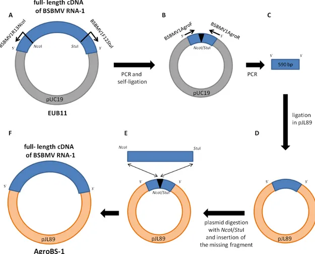
My PhD project aims to investigate molecular interactions between BNYVV and BSBMV and the mechanisms involved in the pathogenesis of these viruses. BNYVV full‐length infectious cDNA clones were available (Quillet et al., 1989) as well as full‐length cDNA clones of BSBMV RNA‐1, ‐2 (unpublished), ‐3 (Ratti et al., 2009) and ‐4 (D’Alonzo et al., 2012). Handling of these cDNA clones in order to produce in vitro infectious transcripts need sensitive and expensive steps, so I developed agroclones of BNYVV and BSBMV RNAs, as well as viral replicons allowing the expression of different proteins.
Chenopodium quinoa and Nicotiana benthamiana plants have been infected with in vitro transcripts and agroclones to investigate the interaction between BNYVV and BSBMV RNA‐1 and ‐2 and the behavior of artificial viral chimeras. Simultaneously I characterized BSBMV p14 and demonstrated that it is a suppressor of post‐transcriptional gene silencing sharing common features with BNYVV p14.
Results and Discussion
Agroinfection (Grimsley, 1986) is a useful technique that appears to be less expensive and more reproducible for plant infection compared to the use of in vitro transcripts. This method consists in tissue infiltration with a suspension of Agrobacterium tumefaciens cells carrying binary plasmids harboring full‐length cDNA copy of a viral genome component. A plant‐functional promoter and the cDNA of a viral RNA are transferred by the way of a T‐DNA from A. tumefaciens into plant cells. The T‐DNA transcription leads to the production of biologically active RNAs able to initiate the viral infection.
Full‐length cDNA of BNYVV and BSBMV RNAs as well as viral replicons expressing different proteins and the green fluorescent protein (GFP) have been introduced in the pJL89 binary vector downstream of the Cauliflower mosaic virus 35S promoter. These plasmids were transferred by electroporation into A. tumefaciens cells (strain C58C1).
Agroclones were used to agroinfect N. benthamiana and Beta macrocarpa plants, showing local and systemic symptoms. Viral RNAs and proteins were detected by northern and western blot. Furthermore, replicon‐mediated GFP
shape particles have been observed through a Transmission Electron Microscope (TEM) in all tissues including roots. The capability of our clones to generate viral RNAs able to complete the viral cycle in B. macrocarpa plants, from replication to the transmission through the vector, has been demonstrated. Such results evidenced that our agroclones are perfectly functional and agroinfection represents indeed a useful strategy to carry on further experiments.
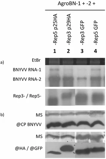
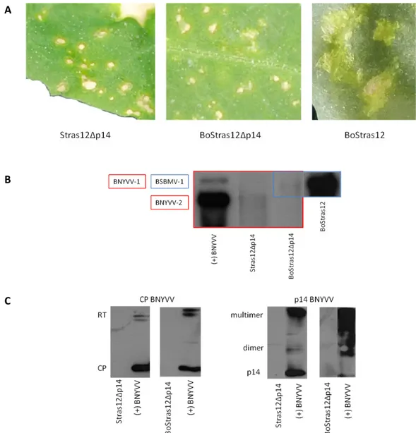
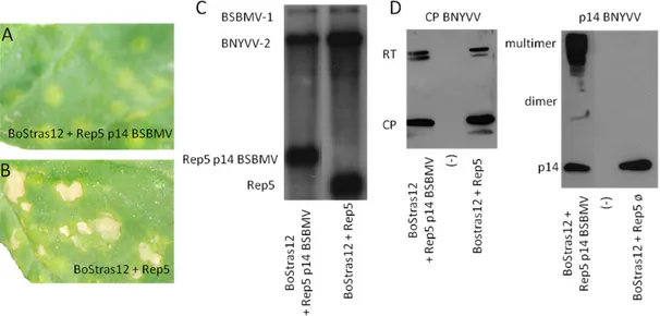
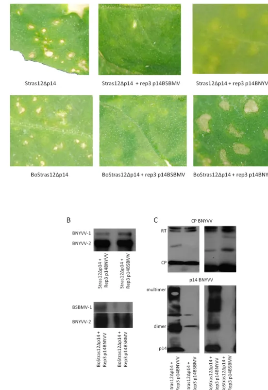
Agroclones, as well as infectious transcripts, have been used to investigate interaction between BNYVV and BSBMV in viral chimeras. Plants of C. quinoa have been infected with BNYVV and BSBMV RNAs infectious transcripts in different combinations named Stras12 (BNYVV RNA‐1 and ‐2, control), Bo12 (BSBMV RNA‐1 and ‐2, control), BoStras12 (BSBMV RNA‐1 and BNYVV RNA‐2) and StrasBo12 (BNYVV RNA‐1 and BSBMV RNA‐2). The combinations Stras12, Bo12 and StrasBo12 showed chlorotic lesions, while BoStras12 induced severe necrotic lesions, probably due to hypersensitive response of the plant. The necrosis disappeared when the plant was co‐inoculated with BoStras12 together with a viral replicon expressing the BSBMV p14. Necrotic lesions arose even in N. benthamiana plants agroinfected with BoStras12, both in the infiltrated and not infiltrated leaves. These results evidenced a possible interaction between BSBMV p14 protein and the RNA‐1 of the virus that requires further investigations. BNYVV p14 is known to be a suppressor of post‐transcriptional gene silencing and it has been characterized in the research group of Prof. David Gilmer (IBMP, University of Strasbourg) but the function of BSBMV p14 has never been investigated. I therefore started the study of BSBMV p14 testing its ability to suppress the PTGS through agroinfiltration of N. benthamiana (line 16C) plants constitutively expressing the Green Fluorescent Protein (GFP) transgene. Challenged N. benthamiana 16C plants challenged with the GFP silencing trigger and BSBMV p14 retained the fluorescence in the infiltrated leaves whereas fluorescence disappeared in the controls. Tissue content analyses evidenced the presence of GFP mRNA and strong reduction of siRNAs, the hallmark of the RNA silencing pathway. Taken together, these results demonstrate that the BSBMV p14 is an efficient silencing suppressor protein (SSP).
I then wanted to investigate at which level the Benyviruses p14s interfere in the post‐transcriptional gene silencing. Agroinfiltration of N. benthamiana plants with different constructs encoding the GFP target, an hairpin GFFG trigger and SSPs showed a normal amount of primary siRNA and a reduced amount of secondary siRNA suggesting that the p14s act downstream of the Dicer proteins without interfering with the transitivity.
Within my PhD project I furthermore demonstrated, through bombardment of N. tabacum BY‐2 cells, that the Benyvirus p14s are localized in the nucleolus and in the cytoplasm. Moreover using the FLIM and yeast two‐hybrid I proved that the BSBMV p14 forms dimers. As for BNYVV p14, I demonstrated that BSBMV p14 can interact with an RNA sequence required for the long distance movement of the virus, name “coremin” sequence. The 20 nucleotides long “coremin” sequence is present in BNYVV RNA‐3 and ‐5 as well as in the BSBMV RNA‐3 and ‐ 4. BNYVV RNA‐3 “coremin” is part of the “core region” and therefore necessary for the systemic movement of the virus within the plant (Lauber et al., 1998) but is also essential for the production and stabilization of the ncRNA‐3 which is required as well for long‐distance movement (Peltier et al., 2012).
Conclusions and prospect
During my PhD program, I developed BNYVV and BSBMV agroinfectious clones useful to investigate Benyviruses biology, protein expression and virus‐vector interactions. Agroinfection will widely enhance BNYVV/BSBMV research. Moreover, it represents a starting point to develop an innovative test to assay Rhizomania resistance of sugar beet cultivars in large scale experiments (Delbianco et al., 2013).
Agroinfection and in vitro transcription have been used to investigate BNYVV/BSBMV RNA‐1 and ‐2 interactions and the behavior of viral chimeras in C. quinoa and N. benthamiana plants. The chimera StrasBo12 showed normal chlorotic lesions whereas the combination BoStras12 induced severe necrotic lesions that disappeared if a viral replicon expressing BSBMV p14 was added to the inoculum. Moreover, the properties of BSBMV p14 have been investigated. This protein, as well BNYVV p14, is a suppressor of post‐transcriptional gene
transitivity. It is localized in the nucleolus and cytoplam, forms dimers and interact with the “coremin” sequence (Chiba et al., 2012).
The results obtained and tools developed by my study will allow new researches about biology, interaction and suppression of post‐transcriptional gene silencing of Benyviruses.
The role of p14 and its discovered interaction with RNA‐1 in the viral chimera BoStras12 open new hypothesis on molecular mechanisms involved in the pathogenesis of Benyviruses which need to be further investigated. BNYVV/BSBMV chimeras will be therefore tested on B. macrocarpa plants, a natural host of Benyviruses.
The characterization of the post‐transcriptional gene silencing suppression activity of p14s will be carried on through immunoprecipitation. Moreover, its interaction with the “coremin” sequence, and therefore with the ncRNA‐3, has to be further investigated. The mode of action of the ncRNA‐3, together with p14, is yet to be discovered but its overproduction could be a way to saturate the silencing machinery of the host, as proposed for the human adenovirus VA‐RNA (Andersson et al., 2005; Peltier et al., 2012).
References
Andersson M. G., Haasnoot P. C., Xu N., Berenjian S., Berkhout B., Akusjarvi G. (2005). Suppression of RNA interference by adenovirus virus‐associated RNA. J. Virol. 79, 9556–9565.
Chiba S., Hleibieh K., Delbianco A., Klein E., Ratti C., Ziegler‐Graff V., Bouzoubaa S., Gilmer D. (2012). The Benyvirus RNA silencing suppressor is essential for long distance movement, requires both Zn‐finger domain and NoLS basic residues but not a nucleolar localization for its silencing suppression activity. Molecular plant‐microbe interactions, Vol.26, No.2, pp.168‐181.
D’Alonzo M., Delbianco A., Lanzoni C., Rubies Autonell C., Gilmer D., Ratti C. (2012). Beet soil‐
borne mosaic virus RNA‐4 encodes a 32 kDa protein involved in symptom expression and in virus
transmission through Polymyxa betae. Virology, 423, pg.187‐194.
Delbianco A., Lanzoni C., Klein E., Rubies Autonell C., Gilmer D., Ratti C. (2013). Agroinoculation of
Beet necrotic yellow vein virus cDNA clones results in plant systemic infection and efficient Polymyxa betae transmission. Molecular Plant Pathology, in press.
Gilmer D., Ratti C. (2012). Benyvirus. In A.M.Q. King, M.J. Adams, E.B. Carstens, and E.J. Lefkowitz (ed.), Virus taxonomy: classification and nomenclature of viruses: Ninth Report of the International Committee on Taxonomy of Viruses. Elsevier, San Diego, p. 1133‐1138.
Grimsley N., Hohn B., Hohn T., Walden R. (1986). Agroinfection, an alternative route for viral infection of plants by using the Ti plasmid. Proc. Natl. Acad. Sci. USA 83, 3282‐3286.
Lauber E., Guilley H., Tamada T., Richards K.E., Jonard G. (1998). Vascular movement of Beet
necrotic yellow vein virus in Beta macrocarpa is probably dependent on an RNA3 sequence
domain rather than a gene product. Journal of General Virology 79, 385‐393.
Lee L., Telford E.B., Batten J.S., Scholthof K.B., Rush C.M. (2001). Complete nucleotide sequence and genome organization of Beet soil‐borne mosaic virus, a proposed member of the genus
Benyvirus. Arch. Virol. 146, 2443‐2453.
Peltier C., Hleibieh K., Thiel H., Klein E., Bragard C., Gilmer D. (2008). Molecular Biology of the
Beet necrotic yellow vein virus. Plant Viruses 2, 14‐24.
Peltier C., Klein E., Hleibieh K, D'Alonzo M., Hammann P., Bouzoubaa S., Ratti C., Gilmer D. (2012).
Beet necrotic yellow vein virus subgenomic RNA3 is a cleavage product leading to stable non‐
coding RNA required for long‐distance movement. J. Gen. Virol. 93 (Pt 5), 1093‐102.
Quillet L., Guilley H., Jonard G., Richards K. (1989). In vitro synthesis of biologically active Beet
necrotic yellow vein virus RNA. Virology 172, 293‐301.
Ratti C., Hleibieh K., Bianchi L., Schirmer A., Rubies Autonell C., Gilmer D. (2009). Beet soil‐borne
mosaic virus RNA‐3 is replicated and encapsidated in the presence of BNYVV RNA‐1 and ‐2 and
Ratti (2012). Beet soil borne mosaic virus RNA‐4 encodes a 32 kDa protein involved in symptom expression and in virus transmission through Polymyxa betae. Virology, 423, pg.187‐194. S. Chiba*, K. Hleibieh*, A. Delbianco*, E. Klein, C. Ratti, V. Ziegler‐Graff, S. Bouzoubaa, D. Gilmer (2012). The Benyvirus RNA silencing suppressor is essential for long distance movement, requires both Zn‐finger domain and NoLS basic residues but not a nucleolar localization for its silencing suppression activity. Molecular plant‐microbe interactions, Vol.26, No.2, pp.168‐181. *Authors contributed equally to this work.
A. Delbianco, M. D’Alonzo, E. Klein, D. Gilmer, C. Rubies Autonell, C. Ratti (2012). Agroinfection: a new tool for Beet necrotic yellow vein virus study and for rhizomania‐resistance assessment. In Proceedings of the Eighth Symposium of the International Working Group on Plant Viruses with Fungal Vectors, Louvain la Neuve (Belgium), 6‐8 july 2011, pg.7. M. D’Alonzo, A. Delbianco, C. Lanzoni, C. Rubies Autonell, D. Gilmer, C. Ratti (2012). Essential role of BSBMV RNA‐4’s p32 on virus transmission by Polymyxa betae in Beta vulgaris plants. In Proceedings of the Eighth Symposium of the International Working Group on Plant Viruses with Fungal Vectors, Louvain la Neuve (Belgium), 6‐8 july 2011, pg.52.
A. Delbianco, C. Lanzoni, E. Klein, C. Rubies Autonell, D. Gilmer, C. Ratti (2013). Agroinoculation of Beet necrotic yellow vein virus cDNA clones results in plant systemic infection and efficient Polymyxa betae transmission. Molecular Plant Pathology, DOI: 10.1111/mpp.12018.
SCIENTIFIC CONGRESS COMUNICATION ‐ POSTER
A. Delbianco, E. Klein, M. D’Alonzo, Gilmer, C. Rubies Autonell and C. Ratti. Agroinfection: a new approach for Beet necrotic yellow vein virus study. In: Abs. Book of XIIIemes Rencontres de Virologie Végétale, Aussois, 16‐20 january 2011, pg.52.
K. Hleibieh, A. Delbianco, A. Flabinus, S. Chiba, E. Klein, D. Scheidecker, C. Ratti, S. Bouzoubaa, V. Ziegler‐Graff, D. Gilmer. BNYVV p14 RNA silencing suppressor and viral RNA3 coremin sequence are implicated in the long distance movement.” In: Abs. Book of XIVemes Rencontres de Virologie Végétale, Aussois, 13‐17 january 2013.
SCIENTIFIC CONGRESS COMUNICATION ‐ ORAL COMUNICATION
A. Delbianco, E. Klein, M. D’Alonzo, D. Gilmer, C. Rubies Autonell, C. Ratti. Agroinfection: a new approach for Beet necrotic yellow vein virus study. Séminaire de Microbiologie de Strasbourg, Amphi VLES, IPCB, Strasbourg, 31 march 2011.
A. Delbianco, M. D’Alonzo E. Klein, D. Gilmer, C. Rubies Autonell and C. Ratti. Agroinfection: a new tool for Beet necrotic yellow vein virus study and for Rhizomania‐resistance assessment. In: Abs. Book of VIIIth International Working Group of Plant Viruses with Fungal Vectors, Louvain La Neuve, 6‐8 july 2011, pg.9.
A. Delbianco, M. D’Alonzo, C. Lanzoni, E. Klein, D. Gilmer, C. Rubies Autonell and C. Ratti. Agroinfection: a new approach for Beet necrotic yellow vein virus study. In Abs. Book of XVII National Congress of the Italian Society for Phytopathology (SiPAV), Bologna, 12‐14 september 2011, pg.15.
A. Delbianco, K. Hleibieh, M. Dall’Ara, C. Rubies Autonell, D. Gilmer, C. Ratti. Characterization of post‐trascriptional gene silencing suppressor proteins of Benyvirus. In Abs. Book of 11th National Congress of the Italian Society for Virology (SIV), Orvieto, 17‐19 september 2012, pg.17.
A. Delbianco, M. Dall’Ara, K. Hleibieh, E. Klein, C. Rubies Autonell, D. Gilmer and C. Ratti. Post‐transcriptional gene silencing suppression study of Beet soil‐borne mosaic virus: characterization of p14 and production of chimeric isolates of Benyviruses. In Abs. Book of XIVemes Rencontres de Virologie Végétale, Aussois, 13‐17 january 2013, pg.19.
General Introduction Before the presentation of the aim of my PhD project, I would like to place the current knowledge and the existing relationships of the main actors used in this research work leading to the most devastating viral disease of sugar beet.
1. Sugar beet
Beta vulgaris ssp. vulgaris belongs to the genus Beta of the Chenopodiaceae family and it descends from Beta vulgaris ssp. maritima or Beta maritima, an halophytic plant adapted to salty environments, native of the Mediterranean area (Francis, 2006). Sugar beet is agriculturally important due to its capability to accumulate a great quantity of sugar in its taproot. It is a biennial plant and its vegetative development is mainly divided into three phases: shoot growth, storage root growth (“tuberization”) and sugar storage (“ripening”) (Milford, 2006).Sugar beet has been cropped and eaten as a spinach‐like vegetable since ancient historical times, but only in the second part of the eighteen century as industrial crop developed in Europe for sugar production, in order to compete with sugar cane (Saccharum officinarum) coming from South American colonies (Francis, 2006).
Nowadays, sugar beet occupies globally a cultivated area of 4,7 million hectares in 51 countries and provides about 25% of world sugar consumption (Rush et al., 2006; FAO, 2010). Recent plant breeding has contributed to improve sugar concentration from 12% of the fresh root to the current value of the 20% (Draycott, 2006). In Europe, Germany, France and Ukraine cultivate the widest area but roots production per hectare is quite variable with France, Spain and Belgium obtaining the highest yields (Table I.A, FAO 2010). Recently, the interest in this plant increased thanks to bioethanol production, as a replacement of fossil fuels in transports’ sector.
However, sugar beets are susceptible to many pathogens such as nematodes, fungi, bacteria and viruses, attacking crops and leading to the reduction of both taproot size and sugar content (Whitney and Duffus, 1986) (Table I.B). Among viruses, the Beet necrotic yellow vein virus and its associated Rhizomania disease is the most pathogenic and worldwide distributed (Stevens et al., 2006).
Table I.A: World sugar beet production. Area harvested, production and yield in all productive countries of the world (adapted from FAO, 2010).
General Introduction Table I.B: Principal sugar beet diseases. Disease Causal agent Viruses Rhizomania Beet necrotic yellow vein virus (BNYVV) Rhizomania related virus Beet soil‐borne mosaic virus (BSBMV) Beet soil‐borne virus (BSBV) Beet virus Q (BVQ) Beet oak leaf virus (BOLV) Beet black scorch virus (BBSV) Beet mosaic Beet mosaic virus (BtMV) Beet yellows Beet yellows (BYV) Beet mild yellows (BMYV) Beet western yellows (BWYV) Beet chlorosis virus (BChV) Curly top Beet curly top virus (BCTV) Bacteria Bacterial vascular necrosis and rot Erwinia carotovora spp. betavasculorum Bacterial leaf spot or leaf blight Pseudomonas syringae Yellow wilt Rickettsia‐like organism Fungi Cercospora leaf spot Cercospora beticola Alternaria leaf blight Alternaria alternata, Alternaria brassicae Powdery mildew Erysiphe betae Downy mildew Peronospora schachtii (farinosa) Fusarium yellows / Fusarium root rot Fusarium oxysporum f. sp. betae Rhizoctonia solani Root rots Pythium spp. Phoma betae Southern sclerotium root rot Sclerotium rolfsii Black root / Black leg Aphanomyces cochlioides Nematodes Cyst nematode Heterodera schachtii Root‐knot nematode Meloidogyne spp.
2. Benyviruses
2.1 The genus
The genus Benyvirus, type member Beet necrotic yellow vein virus (BNYVV), was accepted by the International Committee of Taxonomy of Viruses (ICTV) in 1997 after a revision of the genus Furovirus (Rush, 2003). This genus include the BNYVV, the Beet soil borne mosaic virus (BSBMV), Rice stripe necrosis virus (RSNV) and the tentative member Burdock mottle virus (BdMV) (Gilmer and Ratti, 2012). These viruses have non‐enveloped rod‐shaped and helically constructed particles. Their multipartite genomes consist of positive and single stranded RNA fragments with 5’ m7G cap, 3’ polyadenylated sequence and post translational cleavage of the viral replicase (Hehn et al., 1997; Lee et al., 2001, Peltier et al., 2008).
Benyviruses have limited host ranges. Beta vulgaris is the natural host of BNYVV and BSBMV, and experimentally these viruses can infect some species of Chenopodium genus that allow local infection only and Nicotiana benthamiana, Spinacia oleracea and Beta macrocarpa where the viruses can move systemically. Both BNYVV and BSBMV are naturally transmitted by the protozoa Polymyxa betae and RSNV by P. graminis (Tamada, 1975; Lee et al., 2001; Rush, 2003).
This genus shares conserved residues within the coat protein and similar viral particle morphology with the Virgaviridae family (furo‐, peclu‐, pomo‐, hordei‐, tobra‐ and tobamoviruses). Benyviruses movement strategy involves triple gene block cluster similarly to pomo‐, peclu‐ and hordeiviruses (Verchot et al, 2010). Conversely, replication proteins domains display high degree of similarity to those of the Togaviridae family and, interestingly, to the human hepatitis virus E (Gilmer and Ratti, 2012). Thereby, benyviruses could be proposed as a distinct family thanks to its particular genome specificities.
2.2 The Rhizomania disease and Beet necrotic yellow vein virus
The Beet necrotic yellow vein virus has been identified as the causal agent of Rhizomania disease in the early seventies (Tamada and Baba, 1973). In 1959, Antonio
General Introduction
Canova working at the “Istituto di Patologia Vegetale” of the University of Bologna published the first report about a disease affecting sugar beet roots in the Padan Plain of Italy. Later, this syndrome has been named “rizomania” (Canova, 1966), a term composed by the Latin words “rhizo” and “man ̆ıa” meaning “root madness”, the principal character of this disease (Biancardi, 2005). Since the middle of the twenty century, Rhizomania has been reported in all the sugar beet growing areas all around the world (McGrann et al., 2009).
Infected plants show characteristic proliferation of lateral rootlets with consequently stunting of the tap‐root and browning of the vascular system. The size of the tap‐root can be strongly reduced (Brunt and Richards, 1989; Putz et al., 1990). The inefficient nutrient uptake causes yellowing in leaves (Stevens et al., 2006). Occasionally, BNYVV spread systemically and leaves show necrosis and yellowing in the leaf veins, these symptoms provided the name for the virus (Tamada and Baba, 1973; Tamada, 2002) (Fig. I.1).
Rhizomania causes severe losses to the sugar beet crop, due to serious decreases in root yield, sugar content and juice purity. The sugar yield reduction can reach the 80% (McGrann et al., 2009). Fig I.1: Rhizomania symptoms caused by BNYVV infection on sugar beet. (A) Yellowing and necrosis on the leaves veins. (B) Lateral rootlet proliferation with browning of the vascular system and constriction of the main root. (C) Symptoms on fields. The genome of Beet necrotic yellow vein virus consists of four to five (+) ssRNAs. RNA‐1 and ‐2 carry “house‐keeping” genes involved in virus replication, assembly, cell‐to‐cell movement and suppression of post‐transcriptional gene silencing (Tamada, 1999; Dunoyer et al., 2002). These RNAs are necessary and sufficient for viral infection when
inoculated to the leaves of Beta macrocarpa, B. vulgaris, Chenopodium quinoa and Tetragonia expansa plants (Koenig et al., 1986; Pelsy and Merdinoglu, 1996; Tamada et al., 1989). A replicative strain named “Stras12” was obtained extracting viral RNA from leaves of C. quinoa and T. expansa infected with in vitro transcripts of BNYVV RNA‐1 and ‐2 full‐length cDNA clones (Quillet et al., 1989). However, natural infection requires RNA‐3 and ‐4 encoded proteins directly involved in the pathogenesis and viral transmission through the vector P. betae, respectively (Lemaire et al., 1988; Koenig et al., 1991).
RNA‐1 is 6,746 nucleotides (nts) long and encodes for a single open reading frame (ORF) generating a polypeptide of 237 kDa that, after translation, is processed by an autocatalytic cleavage in two proteins of 150 kDa and 66 kDa. The first one contains conserved motifs of methyltransferase (MTR) and helicase (HEL) while the second displays RNA‐dependant RNA polymerase (RdRp) motif, which is essential for virus replication (Bouzoubaa et al., 1987; Hehn et al., 1997). The proteolytic cleavage of the replicase distinguishes Benyvirus from all other virus with rod‐shaped particles, which have their replication‐associated proteins encoded in two ORFs (Gilmer and Ratti, 2012).
RNA‐2 (4,609 nts) contains six ORFs. The viral Coat Protein (CP) of 21kDa is encoded by the first ORF and is followed by an in‐frame region of 54 kDa read‐through (RT) domain. CP and RT are fused in a 75 kDa protein during translation when the internal leaky UAG stop codon is bypassed by ribosomes (Ziegler‐Graff et al., 1985; Niesbach‐ Klosgen et al., 1990). The N‐terminal of this protein is involved in viral assembly whereas the C‐terminal is required for viral transmission through the vector P. betae (Schmitt et al., 1992; Tamada and Kusume, 1991). The three subsequent ORFs show typical motifs of the “triple gene block” (TGB) movement proteins and encode for p42, p13 and p15 proteins that are expressed by the way of two subgenomic RNAs (Gilmer et al., 1992). The last ORF is also expressed by a subgenomic RNA and encodes for a cysteine‐rich protein of 14kDa, which is a suppressor of post‐transcriptional gene silencing (Dunoyer et al., 2002; this study: Chiba et al, 2012).
RNA‐3 (1,774 nts) encodes the pathogenicity protein p25 and two other proteins of 6.8 kDa (N) and 4.6 kDa. Functions of the last two small proteins are not well documented and the last one has never been detected. Expression of p25 is linked to the
General Introduction
development of the rhizomania symptoms in sugar beet roots and it is able to induce bright yellow local lesions in C. quinoa plants and abnormal root branching in transgenic Arabidopsis thaliana (Tamada et al., 1990; Koenig et al.,1991; Tamada et al., 1989; Jupin et al., 1992; Peltier et al., 2010). Variability in different amino acid positions of p25 has been associated with an increased pathogenicity and the capability to overcome rhizomania resistance conferred by the Rz1 resistance gene derived by the ‘Holly’ source (Schirmer et al., 2005; Acosta‐Leal et al., 2008; Lewellen et al., 1987; Chiba et al., 2008; Koenig et al., 2009). RNA‐3 is required for viral long distance movement in B. macrocarpa (Tamada et al., 1989). The observation that deletion of nts 1033‐1257, named “core region”, inhibit the vascular movement, suggested that RNA‐3 sequence, rather than RNA‐3 encoded protein, was required for BNYVV long distance movement in B. macrocarpa (Lauber et al., 1998). Ratti et al., (2009) identified a stretch of 20 nts named “coremin”, inside the “core region”, which appears to be responsible of the long distance movement. “Coremin” is also present in BNYVV RNA‐5, BSBMV RNA‐3 and ‐4 and in other viral species of the genus Cucumovirus. Interestingly, this sequence is present in the 5’ leader ORF‐less regions of subgenomic CMV RNA‐5, BNYVV and BSBMV RNA‐3. A recent study demonstrated that BNYVV subRNA‐3 is, in fact, a cleavage product leading to stable non‐coding RNA (ncRNA‐3) required for long distance movement. Mutagenesis revealed the importance of “coremin” sequence both for long distance movement and ncRNA‐3 stabilization (Peltier et al., 2012).
RNA‐4 (1,467 nts) is necessary for the viral transmission through the protozoa P. betae (Tamada and Abe, 1989). The encoded protein (p31) is required for efficient transmission and is able to suppress post‐transcriptional gene silencing in N. benthamiana roots (Andika et al., 2005; Rahim et al., 2007). Finally, RNA‐5 is present only in some field isolates that appears to be more aggressive (Tamada et al., 1996). Such RNA encodes for another pathogenicity protein of 26 kDa that probably acts in a synergistic manner with RNA‐3 p25 (Kiguchi et al., 1996; Link et al., 2005) (Fig. I.2).
. Fig. I. 2 : Sc h ematic represe n ta tion of BNYV V/BSB M V gen o mic orga nization
General Introduction
According to restriction length polymorphism, single strain conformation polymorphism patterns and phylogenetic relationships, three different types of BNYVV have been described and named A, B and P (Koenig et al., 1995; Kruse et al., 1994; Schirmer et al., 2005).
A‐type is worldwide distributed, while B‐type has been found mainly in France, Germany and Japan and it has been reported even in United Kingdom and Sweden (Koenig et al., 1995; Miyanishi et al., 1999; Lennefors et al., 2000). Nucleotide identity score between these two variants is very high, ranging between 96%‐99% (Saito et al., 1996; Koenig and Lennefors, 2000; Meunier et al., 2003).
No pathogenecity differences have been reported between A‐ and B‐type in contrast with P‐type that contain RNA‐5 and seems to be more aggressive (Heijbroek et al., 1999). P‐type has been isolated in Pithiviers (France) (Koenig et al., 1995) but isolates containing five RNAs have been described also in Japan, United Kingdom and Kazakhstan (Tamada et al., 1989; Koenig and Lennefors, 2000; Harju et al., 2002;). P variant seems to move more rapidly in plants, resulting in a higher level of infection of tap root compared to A‐ and B‐type, even in partially resistant cultivars (Hejibrowk et al., 1999). 2.3 Beet soil‐borne mosaic virus
Beet soil‐borne mosaic virus was discovered in Texas in 1988 as a sugar beet virus morphologically similar but serologically distinct to BNYVV (Liu and Duffus, 1988). BSBMV was completely sequenced by Lee et al. (2001) demonstrating that BSBMV and BNYVV have identical genomic organization but sufficient molecular differences to be considered two separated species.
BSBMV is widely distributed only in the United States and up to now it has not been reported in other countries (Rush, 2003; Ratti et al., 2009). Sugar beet infected roots often appears asymptomatic whereas leaves show slight distortion, faint general mottling and yellow vein banding, which can progress to chlorosis (Heidel and Rush, 1994).
Rush et al. (1994) demonstrated that PCR primers designed on the 3’ end of BNYVV RNAs amplify homologous species of BSBMV RNAs. These PCR products, used as
northern blot probes, weakly hybridized BNYVV RNAs. In contrast, PCR specific primers designed on the 5’ end of BNYVV RNAs were not able to amplify BSBMV (Heidel et al., 1997). Ratti et al (2009) showed that 5’ terminus of BSBMV and BNYVV RNA‐3s share common structures. BSBMV genome consists of four capped and polyadenylated RNAs (Lee et al., 2001). RNA‐1 (6,683 nts) contains a single long ORF encoding for a 239kDa protein that share amino acids homology with known viral RNA‐dependent RNA polymerases (RdRp) and helicases.
RNA‐2 is 4,615 nts long and contains six ORFs. The first ORF encode for the Coat Protein of 21 kDa and is followed by a leaky UAG stop codon whose suppression leads to the expression of a 74 kDa protein (RT, read‐through). These two genes are followed by three ORFs encoding movement proteins showing typical motifs of the Triple Gene Block with predicted masses of 42kDa, 13kDa and 15kDa. The last ORF encodes for a 14kDa cysteine‐rich protein similar to BNYVV p14, but so far it has not been deeply investigated (Hehn et al., 1995; Lee et al., 2001). RNA‐3 (1,730 nts) encodes for a 29 kDa protein that share only the 23% amino acids identity score with the p25 of BNYVV RNA‐3 (Ratti et al., 2009). RNA‐4 species described by Lee et al. (2001) is 1,203 nts long encoding a protein of 13 kDa, considerably smaller than the BNYVV RNA‐4 p31. This species could correspond to a shortened form of BSBMV RNA‐4 produced after serial mechanical inoculation on C. quinoa leaves as already described for BNYVV RNA‐3 and ‐4 by Bouzoubaa et al. (1991). Recently, a new species of BSBMV RNA‐4 has been characterized which is 1,733 nts long and encodes for a protein of 32 kDa necessary for the viral transmission through the vector P. betae (D’Alonzo et al., 2012) (Fig. I.2).
2.4 The vector Polymyxa betae
BNYVV and BSBMV are both vectored by the plasmodiophorid Polymyxa betae. Traditionally, plasmodiophorids have been considered as fungi in phylum Plasmodiophoromycota, but recently they have been reclassified within the Protozoa (Braselton, 1995; Rush, 2003).
General Introduction
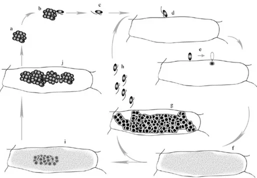
P. betae is only weakly pathogenic and its host range is restricted to roots of Chenopodiaceae, Amaranthaceae, Caryophyllaceae and Portulaceae (Rush, 2003). Life cycle of P. betae includes several stages (Fig. I.3 and I.4). Clusters of thick‐walled resting spores, named sporosori, are liberated into the soil during senescence of infected plant roots. Sporosori are able to survive in soil for years, however in the presence of a susceptible host and suitable conditions of temperature and humidity, these resting spores germinate and release primary zoospores. Zoospores encyst in rootlets and inject their cytoplasmic content inside the root cells, inducing the formation of a multinucleated plasmodium. In this phase the viral particles can be transferred to the host or acquired by the vector. The plasmodium can differentiate in a zoosporangium, leading to the production of secondary zoospores that can infect new roots, or in a sporosorus with the production of resting spores (Keskin, 1964; Adams, 1991; Barr and Asher, 1992).
Lubicz et al. (2007) observed that BNYVV proteins accumulate inside P. betae resting spores and zoospores. Association of viral replication and movement proteins with sporangial and sporogenic stages suggests that the virus resides in the vector more than one life cycle, advancing the hypothesis that P. betae may have an additional role as a host. However, in the absence of negative sense viral RNA detection, the replication of benyviruses in their vector is still controversial.
Fig. I.3: Schematic representation of the P. betae life cycles and its developing states. (a) sporosore; (b)
germinating zoospore; (c) swimming zoospore to a (d) cortical or epidermal cell; (e) the zoospore encyst on the cell and injects its contents through the cell wall and the cellular membrane; (f) developing plasmodium that will tend to a zoosporangium (g) that will issue either (h) the secondary zoospores able to infect new cells or (i) to the sporogenous plasmodium (j) leading to new sporosores. Such sporosores will be further released in soil after root decomposition (adapted from Peltier et al., 2008). Fig. I.4: (A) and (B) Scanning Electron Microscope and (C) optical microscope images of P. betae resting spores in sugar beet cortical root cells.
General Introduction
3. Relationship and interaction between BNYVV and BSBMV
BNYVV and BSBMV have similar host range and particle morphology, present identical genomic organization and share the same vector P. betae. Based on the degree of nucleotide and amino acid sequence identity between the two viruses, it has been concluded that BNYVV and BSBMV are distinct species but more closely related to each other than to other multipartite rod‐shaped viruses with “fungal” vectors included in the original genus Furovirus (Lee et al., 2001). Moreover, the first characterization of BSBMV demonstrated that its capsid protein (CP) is serologically distinct to that of BNYVV (Wisler et al., 1994).
Sequence alignments shows that BNYVV and BSBMV RNA‐1 share 77% nucleotide sequence identity (Lee et al., 2001). The large and unique ORF encode for replication proteins (83% amino acid identity), in contrast with the two ORFs presents in other rod‐shaped viruses with fungal vectors (Koonin and Doljia, 1993).
BSBMV RNA‐2 share the 67% nucleotide sequence identity with the BNYVV RNA‐2. The coat protein and the 74 kDa read‐through translation product show amino acid sequence identity of 56%, whereas the triple gene block proteins and p14 have 74%, 81%, 65% and 32% amino acid identity, respectively (Lee et al., 2001).
BSBMV RNA‐3 sequence has 60% identity with the BNYVV RNA‐3 and the encoded p29 shared just the 23% amino acid identity with p25. It has been recently reported that BSBMV p29 sequence is closer to BNYVV RNA‐5 p26 than to RNA‐3 p25 (Ratti et al., 2009).
BSBMV and BNYVV RNA‐4 share the 47% nucleotide identity score and the encoded proteins of 32 and 31 kDa share a significant amino acid identity of 49,8% (D’Alonzo et al., 2012).
Comparisons between 5’ and 3’ UTRs of all BNYVV and BSBMV RNAs showed highly conserved regions of 7 and 70 nucleotides, respectively (Lee et al., 2011). The 5’ UTRs of BNYVV and BSBMV RNA‐1 and ‐2 share 92% and 81% nucleotide identity sequence, respectively, in contrast with the 38% and 50% of RNA‐3 and ‐4. The 3’UTRs of RNA‐1, ‐ 2, ‐3 and ‐4 have a nucleotide identity score of 66%, 67%, 79% and 64%, respectively (Lee et al., 2001).
Conserved cis‐acting elements, essential for replication, are present in 5’ and 3’ UTRs of BNYVV RNA‐3 and ‐4 (Bouzoubaa et al., 1991). These conserved regions can fold in double hairpin secondary structure that presumably is recognized by the viral replicase during initiation of minus strand synthesis (Lauber et al., 1997). Recently Ratti et al. (2009) observed that both BNYVV and BSBMV RNA‐3 show strong sequence and structure similarities in 5’ and 3’ UTRs. In the same work the authors demonstrated that BSBMV RNA‐3 can be replicated and encapsidated by BNYVV RNA‐1 and ‐2 and allows long distance movement in B. macrocarpa. Moreover expression of different proteins through a BSBMV expression vector (Rep III), containing 5’ and 3’ UTRs of RNA‐3, was possible in the presence of BNYVV RNA‐1 and ‐2. However, Rep III cannot be replicated in the same cell together with BNYVV RNA‐3 or its derived replicon leading to the competitive loss of one of the molecules within the local lesion (Ratti et al., 2009).
Similarly, BSBMV RNA‐4 can be amplified by BNYVV RNA‐1 and ‐2 and can complement BNYVV RNA‐4 in virus transmission through the vector P. beta in B. vulgaris plants (D’Alonzo et al., 2012).
In the United States BNYVV and BSBMV are often present in the same field, sometimes in the same plant and therefore interactions such as cross protection have been investigated. Cross protection is a mechanism that occurs when a plant infected by one virus (protecting) is then protected by the infection of a second virus (challenging). This phenomenon usually occurs between two strains of the same virus but sometimes among different viruses.
Mahmood and Rush (1999) showed a high degree of reciprocal cross protection between BNYVV and BSBMV in greenhouse experiments on Beta vulgaris seedlings inoculated with protecting virus on roots and with challenging virus on leaves through sap of C. quinoa infected leaves. The degree of cross‐protection was increased by longer inoculation intervals between the first and the second inoculum. Moreover, RNA of both viruses was detected in doubly infected plants but capsid protein of the BNYVV was undetected by serological tests, suggesting that BSBMV CP is involved in some mechanisms able to avoid superinfection in cross‐protection tests.
However, distinct results were obtained using different approaches. Experiments performed with soils naturally infested with P. betae zoospores carrying BNYVV and
General Introduction
BSBMV seem to demonstrate that BNYVV is able to suppress BSBMV in mixed infections (Wisler, 2003). When BSBMV was present in mixed infections with BNYVV, its level was strongly reduced, even when BNYVV titer was very low, particularly in Rhizomania‐resistant cultivars. Furthermore, the Rz allele of rhizomania‐resistance does not provides resistance to BSBMV. The significant reduction of BSBMV in the presence of BNYVV may be due to several factors such as competition for host infection sites by virouliferous P. betae, BNYVV infected zoospores could be more aggressive or one virus may have a competitive advantages once inside the cell (Wisler, 2003).
However, these studies about BNYVV/BSBMV interactions have been conducted under different experimental conditions that have to be considered (Rush, 2003). Sugar beet plants vortexed in a liquid inoculum become entirely infected, in contrast to infection through P. betae that usually remains localized into the roots and rarely goes systemic. With the vortex method, the first virus becomes established and interferes with subsequent infection of a second virus. Whereas, in natural infection through P. betae zoospores, the virus with the highest inoculum usually colonize the majority of the roots and it will predominate. Moreover in such experiments the initial inoculum density should be determined and soil temperature must be manipulated in order to obtain repeatable results. In fact, BSBMV usually predominate at temperature <20°C, whereas BNYVV at >25°C (Rush, 2003).
4. Aim of the study
Beet necrotic yellow vein virus and Beet soil‐borne mosaic virus belong to the Benyvirus genus and are both vectored by P. betae. These viruses infect roots of Chenopodiaceae plants, share the same genomic organization but have sufficient molecular differences to be distinguished in two different species (Lee et al., 2001; Peltier et al., 2008). However, BSBMV RNA‐3 and ‐4 can be replicated and encapsidated by BNYVV RNA‐1 and ‐2, allowing long distance movement and transmission through the vector(Ratti et al., 2009; D’Alonzo et al., 2012). In the United States these viruses are frequently present in the same field affecting the same plant but no chimeric forms have been described in nature so far.
The aim of this PhD project is to further investigate molecular interactions between BNYVV and BSBMV and the mechanisms involved in the pathogenesis of these viruses. One purpose of my thesis was to study the possible synergism or antagonism effect between BNYVV and BSBMV. To perform this study, plant host leaves have to be coinoculated with different ratio of both viruses. BNYVV full‐length infectious cDNA clones are available (Quillet et al., 1989) as well as full‐length cDNA clones of BSBMV RNA‐1, ‐2 (D’Alonzo, 2011), ‐3 (Ratti et al., 2009) and ‐4 (D’Alonzo et al., 2012). Handling of these cDNA clones in order to produce in vitro infectious transcripts need sensitive and expensive steps. I decided to develop alternative tools to carry on my experiments so I produced agroclones of BNYVV and BSBMV RNAs, as well as viral replicons allowing the expression of different proteins that are described in Chapter 1. Chapter 2 is dedicated to the study of the relationship between BNYVV and BSBMV. I checked the capability of BSBMV RNAs to replicate BNYVV RNA‐5 and the behavior of artificial viral chimeras between BNYVV and BSBMV RNA‐1 and ‐2 in test plants, such as Chenopodium quinoa and Nicotiana benthamiana. If BNYVV RNA‐1 and ‐2 are known to support BSBMV RNA‐3 and ‐4 replication to transmission, nothing is known about the properties of chimera exchanging one of the largest RNAs, that could possibly arise from the co‐infection described in Chapter 1.
Chapter 3 presents the study about BSBMV p14 demonstrating that it is a suppressor of post‐transcriptional gene silencing sharing common features with BNYVV p14 and its implications in viral long distance movement.
Finally, a general discussion and conclusion summarizes the results and present some prospects about Benyviruses research.
General Introduction
References
Acosta‐Leal R., Fawley M.W., Rush C.M. (2008). Changes in the intraisolate genetic structure of beet necrotic yellow vein virus populations associated with plant resistance breakdown. Virology 376, 60‐68.
Adams M. (1991). Transmission of plant viruses by fungi. Ann. Appl. Biol., 118, 479‐92.
Andika I.B., Kondo H., Tamada T. (2005). Evidence that RNA silencing‐mediated resistance to beet necrotic yellow vein virus is less effective in roots than in leaves. Molecular plant‐microbe interactions, 18, 194‐204. Barr D., Asher M.J.C. (1992). The host range of Polymyxa betae in Britain. Plant Pathol.41, 64‐88. Biancardi E. (2005). Brief history of sugar beet cultivation. In BiancardiE., Campbell L.G., Skaracis G.N. & De Biaggi (eds.) “Genetics and breeding of sugar beet”. Science Publishers, Inc., Enfield, NH, USA, 3‐9. Bouzoubaa S., Quillet L., Guilley H. (1987). Nucleotide sequence of beet necrotic yellow vein virus RNA‐ 1. Journal of General Virology, 68, 615‐626. Bouzoubaa S., Niesbach‐Klosgen U., Jupin I., Guilley H., Richrds K.E., Honard G. (1991). Shortened forms of beet necrotic yellow vein virus RNA‐3 and ‐4: internal deletions and a subgenomic RNA. Journal of General Virology, 72, 259‐266. Braselton J. (1995). Current status of the plasmodiophorids. Curr. Rev. Microbiol., 21, 263‐275. Brunt A.A., Richards K.E. (1989). Biology and Molecular Biology of Furoviruses. Adv. Virus Research, 36, 1‐32. Canova A. (1959). Appunti di patologie della barbabietola. Informatore fitopatologico, 9, 390‐396. Canova A. (1966). Si studia la rizomania della bietola.Informatore fitopatologico, 16, 235‐239. Chiba S., Miyanishi M., Andika I.B., Kondo H., Tamada T. (2008). Identification of amino acids of the beet necrotic yellov vein virus p25 protein required for induction of the resistance response in leaves of Beta vulgaris plants. Journal of General Virology, 89, 1314‐1323. Chiba S., Hleibieh K., Delbianco A., Klein E., Ratti C., Ziegler‐Graff V., Bouzoubaa S., Gilmer D. (2012). The Benyvirus RNA silencing suppressor is essential for long distance movement, requires both Zn‐finger domain and NoLS basic residues but not a nucleolar localization for its silencing suppression activity. Molecular plant‐microbe interactions, Vol.26, No.2, pp.168‐181.
D'Alonzo M., Delbianco A., Lanzoni C., Autonell C.R., Gilmer D., Ratti C. (2012). Beet soil‐borne mosaic virus RNA‐4 encodes a 32 kDa protein involved in symptom expression and in virus transmission through Polymyxa betae. Virology; 423:187‐94.
Draycott A.P. (2006). Introduction. In Sugar Beet, pp.1‐8. Edited by A.P. Draycott. Oxford: Blackwell Publishing Ltd.
Dunoyer P., Pfeffer S., Fritsch C., Hemmer O., Voinnet O., Richards K.E. (2002). Identification, subcellular localization and some properties of a cysteine‐rich suppressor of gene silencing encoded by peanut clump virus. Plant Journal, 29, 555‐567.
FAO (2010). FaoSTAT Agricolture Statistics Database. Web: http://faostat.fao.org/site/567/default.aspx‐ ancor.
Francis S.A. (2006). Development of sugar beet. In Sugar beet, 9‐29. Edited by A.P. Draycott. Oxford: Blackwell Publishing Ltd.
Gilmer D., Bouzoubaa S., Henh A., Guilley H., Richards K.E., Jonard G. (1992). Efficient cell‐to‐cell movement of bet necrotic yellow vein virus requires 3’ proximal genes located on RNA‐2. Virology, 189, 40‐47.
Gilmer D., Ratti C. (2012). Benyvirus. Pages 1133‐1138 in: Virus taxonomy: classification and nomenclature of viruses: Ninth Report of the International Committee on Taxonomy of Viruses.
Harju V.A., Mumford R.A., Blockley A., Boonham N., Clover G.R.G., Weekes R., Henry C.M. (2002). Occurrence in the United Kinfdom of beet necrotiv yellow vein virus isolates which contain RNA5. Plant pathology, 51, 811.
Hehn A., Bouzoubaa S., Bate N., Twell D., Marbach J., Richards K.E., Guilley H., Jonard G. (1995). The small cysteine‐rich protein p14 of beet necrotic yellow vein virus regulates accumulation of RNA‐2 in cis and coat protein in trans. Virology, 210, 73‐81.
Hehn A., Fritsch C., Richards K.E., Guilley H., Jonard G. (1997). Evidence for in vitro and in vivo autocatalytic processing of the primary translation product of beet necrotic yellow vein virus RNA‐1 by a papain‐like proteinase. Archives of Virology, 142, 1051‐1058.
Heidel G.B., Rush C.M. (1994). Distribution of beet necrotic yellow vein virus, beet distorsion mosaic virus and unnamed soilborne sugar beet virus in Texasand New Mexico. Plant disease, 78, 603‐606.
Heidel G.B., Rush C.M., Kendall T.L., Lommel S.A., French R.C. (1997). Characteristics of beet soil borne mosaic virus, a furo‐like virus infecting sugar beet. Plant disease, 81, 1070‐1076.
Heijbroek W., Musters‐Van Oorschot P.M.S., Schoone A.H.L. (1999). Variation in pathogenicity and multiplication of beet necrotic yellow vein virus (BNYVV) in relation to the resistance of sugar beet cultivars. European Journal of Plant Pathology, 105, 397‐405.
Jupin I., Guilley H., Richards K.E., Jonard G. (1992). Two proteins encoded by beet necrotic yellow vein virus RNA‐3 influence symptom phenotype on leaves. EMBO Journal, 11, 479‐488.
Kiguchi T., Saito M., Tamada T. (1996). Nucleotide sequence analysis of RNA‐5 of five isolates of beet necrotic yellow vein virus and the identity of a deletion mutant. Journal of General Virology, 77 (Pt 4), 575‐580.
Keskin, B. (1964). Polymyxa betae n.sp., ein Parasit in den Wurzeln von Beta vulgaris Tournefort, besonders während der Jugendentwicklung der Zuckerrübe. Archives of Microbiology 49, 348‐373.
Koenig R., Burgermeister W., Weich H., Sebald W., Kothe C. (1986). Uniform RNA patterns of beet necrotic yellow vein virus in sugar beet roots, but not in leaves from several species. Journal of General Virology, 67, 2043‐2046.
Koenig R., Jarausch W., Li Y., Commandeur U., Burgermeister W., Gehrke M., Luddecke P.( 1991). Effect of recombinant beet necrotic yellow vein virus with different RNA composition on mechanically inoculated sugar beets. Journal of General Virology, 72, 2243‐2246.
Koenig R., Luddecke P., Haeberle A.M. (1995). Detection of beet necrotic yellow vein virus strains, variants and mixed infections by examining single strand conformation polymorphisms of immunocapture RT‐PCR products. Journal of General Virology, 76, 2051‐2055.
Koenig R., Lennefors B.L. (2000). Genus Benyvirus. In Van Regenmortel MHV, Fauquet CM & Bishop DHL (eds.) Virus Taxonomy. 7th Report of the International Committee on Taxonomy of Viruses. Academic Press, San Diego, CA, pp. 917‐922.