Alma Mater Studiorum
Alma Mater Studiorum
Università degli Studi di Bologna
Università degli Studi di Bologna
Tesi di Dottorato:
Stem Cells as a therapy for myocardial
infarction in animal models
Dottorato di Ricerca in Biochimica
XX ciclo
Settore scientifico disciplinare BIO/10
Presentata da:
Dott.ssa Emanuela Fiumana
Relatore: Coordinatore:
Chiar.mo Prof. Carlo Guarnieri Chiar.mo Prof. Giorgio Lenaz
Esame Finale Anno 2008
Esame Finale Anno 2008
"I heard that scientists are growing
human organs in lab dishes."
"Is this true?"
ABSTRACT
Advances in stem cell biology have challenged the notion that infarcted myocardium is irreparable. The pluripotent ability of stem cells to differentiate into specialized cell lines began to garner intense interest within cardiology when it was shown in animal models that intramyocardial injection of bone marrow stem cells (MSCs), or the mobilization of bone marrow stem cells with spontaneous homing to myocardium, could improve cardiac function and survival after induced myocardial infarction (MI) [1, 2]. Furthermore, the existence of stem cells in myocardium has been identified in animal heart [3, 4], and intense research is under way in an attempt to clarify their potential clinical application for patients with myocardial infarction. To date, in order to identify the best one, different kinds of stem cells have been studied; these have been derived from embryo or adult tissues (i.e. bone marrow, heart, peripheral blood etc.). Currently, three different biologic therapies for cardiovascular diseases are under investigation: cell therapy, gene therapy and the more recent “tissue-engineering” therapy .
During my Ph.D. course, first I focalised my study on the isolation and characterization of Cardiac Stem Cells (CSCs) in wild-type and transgenic mice and for this purpose I attended, for more than one year, the Cardiovascular Research Institute of the New York Medical College, in Valhalla (NY, USA) under the direction of Doctor Piero Anversa. During this period I learnt different Immunohistochemical and Biomolecular techniques, useful for investigating the regenerative potential of stem cells.
Then, during the next two years, I studied the new approach of cardiac regenerative medicine based on “tissue-engineering” in order to investigate a new strategy to regenerate the infarcted myocardium. Tissue-engineering is a promising approach that makes possible the creation of new functional tissue to replace lost or failing tissue. This new discipline combines isolated functioning cells and biodegradable 3-dimensional (3D) polymeric scaffolds. The scaffold temporarily provides the biomechanical support for the cells until they produce their own extracellular matrix. Because tissue-engineering constructs contain living cells, they may have the potential for growth and cellular self-repair and remodeling. In the present study, I examined whether the tissue-engineering strategy within hyaluron-based scaffolds would result in the formation of alternative cardiac tissue that could replace the scar and improve cardiac function after MI in syngeneic heterotopic rat hearts. Rat hearts were explanted, subjected to left coronary descending artery occlusion, and then grafted into the abdomen (aorta-aorta anastomosis) of receiving syngeneic rat. After 2 weeks, a pouch of 3
mm2 was made in the thickness of the ventricular wall at the level of the post-infarction scar. The
hyaluronic scaffold, previously engineered for 3 weeks with rat MSCs, was introduced into the pouch and the myocardial edges sutured with few stitches. Two weeks later we evaluated the cardiac function by M-Mode echocardiography and the myocardial morphology by microscope analysis.
We chose bone marrow-derived mensenchymal stem cells (MSCs) because they have shown great signaling and regenerative properties when delivered to heart tissue following a myocardial infarction (MI). However, while the object of cell transplantation is to improve ventricular function, cardiac cell transplantation has had limited success because of poor graft viability and low cell retention, that’s why we decided to combine MSCs with a biopolimeric scaffold.
At the end of the experiments we observed that the hyaluronan fibres had not been substantially degraded 2 weeks after heart-transplantation. Most MSCs had migrated to the surrounding infarcted area where they were especially found close to small-sized vessels. Scar tissue was moderated in the engrafted region and the thickness of the corresponding ventricular wall was comparable to that of the non-infarcted remote area. Also, the left ventricular shortening fraction, evaluated by M-Mode echocardiography, was found a little bit increased when compared to that measured just before construct transplantation. Therefore, this study suggests that post-infarction myocardial remodelling can be favourably affected by the grafting of MSCs delivered through a hyaluron-based scaffold
1. Orlic D, Kajstura J, Chimenti S, Limana F, Jakoniuk I, Quaini F, et al. Mobilized bone marrow cells repair the infarcted heart, improving function and survival. Proc Natl Acad Sci U S A 2001;98(18):10344-9
2. Orlic D, Kajstura J, Chimenti S, Jakoniuk I, Anderson SM, Li B, et al. Bone marrow cells regenerate infracted myocardium. Nature 2001;410:701-5
3. Beltrami A, Barlucchi L, Torella D, Baker M, Limana F, Chimenti S, Kasahara H, Rota M,
Musso E, Urbanek K Adult cardiac stem cells are multipotent and support myocardial
regeneration.bCell. 2003 Sep 19;114(6):763-76
4. Nadal-Ginard B , Anvera P, Kajstura J, Leri A Cardiac stem cells and myocardial regeneration. Novartis Found Symp 2005; 265: 142-54
TABLE OF CONTENTS
LIST OF ABBREVIATION 6
INTRODUCTION 8
1. Heart Failure 9
2. Cardiac regenerative medicine: the Stem Cells Therapy 11
3. Stem Cells 12
3.1 Cardiac Stem Cell 17
3.2 Bone Marrow Mesenchymal Stem cells 20
3.2.1 Bone Marrow-MSC and cardiac repairing 24
4. Cell delivery in cardiac stem cell therapy 26
5. Tissue-engineering: a new approach for cell delivery in cardiac regenerative medicine 30 5.1 Materials as scaffolds for cells transplantation in cardiac tissue-engineering 33
6. Strategies, challegenges and further directions 37
EXPERIMENTAL SECTION 40
Aim of the study 41
Materials&Methods 42 Results 53 Discussion 65 Conclusion 68 APPENDIX 69 REFERENCES 80
LIST OF ABBREVIATION
CCT = Cardiac Cell Therapy MI = Myocardial Infarction ICM = Inner Cell Mass ES = Embrionic Stem cells
HSCs = Hematopoietic Stem Cells SP cells = Side Population cells CSCs = Cardiac Stem Cells
MSCs = Mesenchymal Stem Cells or Mesenchymal Stromal Cells
BM-MSCs or BMSCs = Bone Marrow derived Mesenchymal Stem or Stromal Cells MAPC = Multipotent Adult Progenitor
TCSC = Tissue Committed Stem Cells CFU = Colony Forming Units
FACS = Fluorescent-Activated Cell Sorting SDF-1 = Stromal cell-Derived Factor-1 GFP = Green Fluorescent Protein
VEGF = Vascular Endothelial Growth Factor HGF = Hepatocyte Growth Factor
AM = AdrenoMedullin
IGF-1 = Insulin Growth Factor-1
MCP-1 = Monocyte Chemeattractant Protein-1
MR and MRI = Magnetic Resonance and Magnetic Resonce Imaging ECM = Extra Cellular Matrix
PLA = PolyLactic Acid PGA = PolyGlicolic Acid PCL = PolyCaproLactone GAGs = GlycosAminoGlycans
PLGA = PolyLactic-co-Glycolic Acid PIPAAm = poly(N-isopropylacrylamide) PVDF = Poly(Vinylidene difluoride) HA = Hyaluronic Acid
ICAM-1 = IntraCellular Adhesion Molecule-1 HASs = Hyaluronic Acid Synthases
HYALs = Hyaluronidases
HABPs = Hyaluronic Acid Binding Proteins
RHAMM = Receptor for Hyaluronan Mediated Mobility expressed protein LYVE-1 = Lymphatic Vessel Endothelial Hyaluronan Receptor-1
COX-2 = Cycloxygenase-2
HYAFF® 11 = Hyaluronan biopolymer produced by Fidia Advanced Biopolymer
BSA = Bovine Serum Albumin FBS = Fetal Bovine Serum PBS = Phosphate Buffered Salin DAPI = 4',6-Diamidino-2-Phenylindole
SEM = Scanning Electron Microscopy TEM = Transmission Electron Microscopy
CFDA-SE = CarboxyFluorescein DiAcetate Succinimydil Ester MTT = (3-(4,5-dimethylthiazol-2-yl)-2,5-diphenyltetrazolium bromide LM = Light Microscopy
BrdU = 5- Bromo- 2’ DeoxyUridine LAD = Left Artery Discendent FS = Fractional Shortening
LVDd or LVDs = Left Ventricular Diameter in diastole or systole H&E = Hematoxylin and Eosin
cTnI = cardiac Troponin I MP = Phagocyte Marker CM = cardiomyocytes
ODC =Ornithine decarboxylase α-MHC = α-Myosin Haevy Chain
1. Heart failure
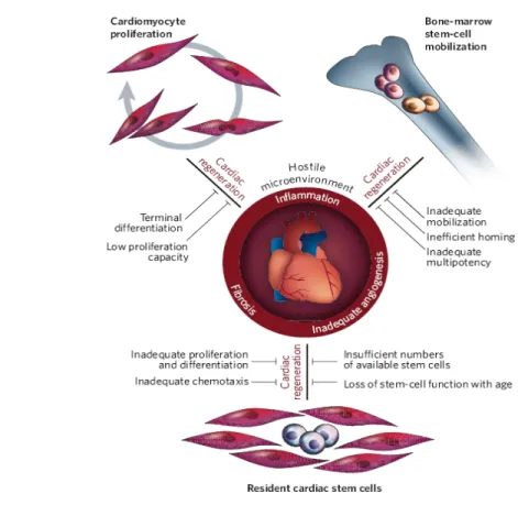
Heart failure is a major cardiovascular health problem worldwide. Despite advances in medical therapy and coronary revascularization strategies, ischemic heart disease remains the leading cause of congestive heart failure and cardiovascular mortality in industrialized countries. For example in USA about 5.2 million patients have heart failure, and the lifetime risk of developing chronic heart failure for both men and women is 1 in 5 [1] . Coronary artery disease is the most common cause of heart failure, followed by idiopathic dilated cardiomyopathy and valvular heart disease [2, 3]. Obstruction of coronary artery leads to myocardial infarction (heart attack) with associated necrosis of the myocardium, followed by infiltration of inflammatory cells. Then a scar forms, leading to the loss of cardiac function, ventricular remodelling and progressive dysfunction, and, finally, congestive heart failure [4-6].
Current therapy of heart failure is limited to the treatment of already established disease and is predominantly pharmacological, aiming primarily to inhibit the neurohormonal axis that results in excessive cardiac activation through angiotensin- or norepinephrine-dependent pathways. For patients with end-stage heart failure treatment options are extremely limited, with fewer than 3,000 being offered cardiac transplants annually due to the severely limited supply of donor organs [7, 8], and implantable left ventricular assist devices being expensive, not proven for long-term use, and associated with significant complications [9-11]. Therefore, there is a need to develop more effective, less invasive therapeutic strategies for heart failure. Because the self-renewal capacity of adult cardiomyocytes is limited, the development of strategies to regenerate damaged myocardium and improve heart function represents a major challenge. Analysis of cardiac myocyte growth during early mammalian development indicates that cardiac myocyte DNA synthesis occurs primarily in utero, with proliferating cells decreasing from 33% at midgestation to 2% at birth [12]. While ventricular karyokinesis and cytokinesis are coupled during fetal growth, resulting in increases in mononucleated cardiac myocytes, karyokinesis occurs in the absence of cytokinesis for a transient period during the postnatal period, resulting in binucleation of ventricular myocytes without an overall increase in cell number. A similar dissociation between karyokinesis and cytokinesis characterizes the primary adult mammalian cardiac response to ischemia, resulting in myocyte hypertrophy and increase in nuclear ploidy rather than myocyte hyperplasia [13, 14]. Throughout life a mixture of young and old cells is present in the normal myocardium. Although most myocytes seem to be terminally differentiated, there is a fraction of younger myocytes (15– 20%) that retains the capacity to replicate [15].
Moreover, recent observations suggest that some human ventricular cardiomyocytes also have the capacity to proliferate and regenerate in response to ischemic injury [16, 17]. The dividing myocytes can be identified on the basis of immunohistochemical staining of proliferating nuclear structures such as Ki67 and cell surface expression of specific surface markers, including c-kit (CD117). Whether these cells are derived from a resident pool of cardiomyocyte stem cells or from a renewable source of circulating bone marrow derived stem cells that home to the damaged myocardium remains to be determined. More importantly, the signals required for homing, in situ expansion, and differentiation of these cells are at present unknown. Gaining and understanding of these issues would open the possibility of manipulating the biology of endogenous cardiomyocytes to augment the healing process after myocardial ischemia [18].
Over the past several years a number of studies have suggested that stem cells can be used to generate cardiomyocytes and endothelial cells ex vivo and in vivo [19-23] and so Cardiac Cell Therapy (CCT) should represent a new approach to replacing damaged myocardium.
The cell therapy for heart failure could has the potential to restore cardiac function by inducing neovascularization, and regenerating and protecting cardiomyocytes [4]. Replacement and regeneration of functional cardiac muscle after an ischemic insult to the heart could be achieved by either stimulating proliferation of endogenous mature cardiomyocytes or resident cardiac stem cells or by implanting exogenous donor-derived or allogeneic cardiomyocytes. The newly formed cardiomyocytes must integrate precisely into the existing myocardial wall to augment contractile function of the residual myocardium in a synchronized manner and avoid alterations in the electrical conduction and syncytial contraction of the heart, potentially resulting in life-threatening consequences. In addition, whatever the source of the cells used, it is likely that concurrent myocardial revascularization must also occur to ensure viability of the repaired region and prevent further scar tissue formation [18].
2. Cardiac regenerative medicine: the Stem Cells Therapy
It’s widely agreed that regenerative capacity of human myocardium is grossly inadequate to compensate for the severe loss of heart muscle presented by catastrophic myocardial infarction or other myocardial diseases.
The ideal therapy for heart failure would have the following activities; it would minimize loss of cardiomyocytes by reducing cell death, promote return of stunned and hibernating myocardium to normal function, stimulate revascularization of the ischemic region by enhancing angiogenesis, and regenerate viable cardiomyocytes to replace those lost to the initial ischemia thereby preserving contractile function and reducing the opportunity for scarring [4]. Recent advances in biotechnology and in the understanding of tissue regeneration have allowed development of novel therapeutics with the potential to approximate this ideal: the stem cell therapy.
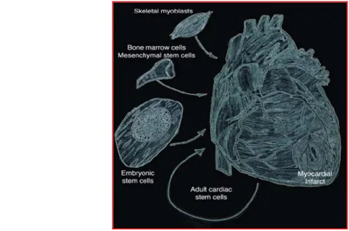
Cardiac stem cell therapy involves delivering a variety of cells into hearts following myocardial infarction or chronic cardiomyopathy [Fig.1]. Suitable sources of cells for cardiac transplant will depend on the types of diseases to be treated. For acute myocardial infarction, a cell that reduces myocardial necrosis and augments vascular blood flow will be desirable. For heart failure, cells that replace or promote myogenesis, reverse apoptopic mechanisms and reactivate dormant cell processes will be useful.
Figure 1. Stem cells as applied for the treatment of myocardial infarcts. The main purpose of stem cell therapies for the
treatment of myocardial infarcts is the prevention and or regeneration of dying muscle. A variety of cell types have been used for such a treatment. These cells include skeletal myoblasts, bone marrow-MSCs, cardiac resident stem cells, and embryonic stem cells including their differentiated progeny (Figure from Christoforou N and Gearhant JD, 2007).
Stem cells might play a role in cardiac repairing becoming a specialized cell. One important type of cell that can be developed is the cardiomyocyte, the heart muscle cell that contracts to eject the blood out of the heart's main pumping chamber (the ventricle). Two other cell types are important to a properly functioning heart are the vascular endothelial cell, which forms the inner lining of new blood vessels, and the smooth muscle cell, which forms the wall of blood vessels. The heart has a large demand for blood flow, and these specialized cells are important for developing a new network of arteries to bring nutrients and oxygen to the cardiomyocytes after a heart has been damaged. The potential capability of both embryonic and adult stem cells to develop into these cells types in the damaged heart is now being explored as part of a strategy to restore heart function to people who have had heart attacks or have congestive heart failure.
3. Stem Cells
In the last few years, great emphasis has been placed on the isolation, characterization and potential therapeutic uses of stem cells. Stem cells are defined as cells capable of both self-renewal and commitment to differentiation into one or more mature cell types. However different types of stem cells can be distinguished on the basis of their developmental potential.
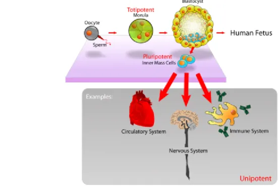
The real totipotent stem cells are the fertilized oocyte (the zygote) and its descendants of the first two divisions. These cells are indeed able to form the embryo and the trophoblasts of the placenta. After about 4 days, these totipotent cells begin to specialize, forming a hollow ball of cells, the blastocyst, and a cluster of cells called the inner cell mass (ICM) from which the embryo develops. The ICM cells are considered to be pluripotent, namely able to differentiate into almost all cells that arise from the three germ layers, but they are unable to give rise to the placenta and supporting tissues. ICM cells can be maintained in culture and the cell lines derived, known as embryonic stem (ES) cells, are also considered to be pluripotential [Fig.2]. In the adult, most tissues have multipotential stem cells, defined a cells capable of producing a limited range of differentiated cell lineages appropriate to their location. Also adult stem cells are heterogeneous with respect to developmental potential. For example, small intestinal stem cells can produce all four indigenous lineages (Paneth, goblet, absorptive columnar, and enteroendocrine), while central nervous system (CNS) stem cells have tri-lineage potential, giving rise to neurons, oligodendrocytes, and astrocytes. At the bottom of the stem cell hierarchy are unipotential stem cells, capable of generating one
specific cell type. Examples are epidermal stem cells in the basal layer that produce only keratinized squames and certain adult hepatocytes that have long-term repopulating ability [24].
Tissue-specific stem cells appear to be present in most organs of the body, and share some common properties:
(1) Stem cells are a self-maintaining population.
This is achieved if, on average, each stem cell division gives rise to one replacing stem cell and one transit-amplifying cell (asymmetrical division). Equally well, stem cell numbers would remain constant if only symmetrical divisions occurred, provided that each time a stem cell gave rise to two daughter transit amplifying cells, another stem cell gave rise to two daughter stem cells.
(2) Stem cells are a small percentage of the total cellularity.
In the mouse small intestine, there are perhaps 4–5 stem cells in a ring near the bottom of the crypt [25] out of a total crypt population of about 250 cells. Likewise, in skeletal muscle, satellite cells comprise about 5% of all nuclei. In the bone marrow, the multipotential hematopoietic stem cell (HSC) is even more rare, with a frequency of perhaps 1 in 104-105 bone marrow cells.
Figure 2. Pluripotent, embryonic stem cells originate as inner mass cells within a blastocyst. The stem cells
can become any tissue in the body, excluding a placenta. Only the morula's cells are totipotent, able to become all tissues and a placenta.
(3) Stem cells are undifferentiated.
In most tissues, stem cells do not have the specialized functions of the progeny that they originate.
(4) Stem cells are slowly cycling but highly clonogenic.
In theory, it would seem prudent to restrict stem cell division because DNA synthesis can be error-prone. Thus, in many tissues we see that stem cells divide less frequently than transit-amplifying cells. In the intestine, stem cells cycle less frequently than transit-transit-amplifying cells, located more luminally [26] and in human epidermis, integrin-bright cells have a lower level of proliferation as compared to other basal cells. In hair follicles, the hair shaft and its surrounding sheaths are produced by the hair matrix, which is itself replenished by the bulge stem cells. The bulge cells divide less frequently, but are more clonogenic than the transit-amplifying cells of the hair matrix [27] thus showing an extensive proliferative ability.
In many tissues and organs, the identity of the stem cells has remained either elusive or at least equivocal [Fig.3]. However, in the bone marrow the identification of cells with the properties of self-renewal and multi-lineage differentiation potentialities well advanced. Indeed, such cells were functionally defined in the mouse back in 1961 by Till and McCulloch [28] as cells that, upon transplantation, were able to form multilineage hematopoietic colonies in the spleen of lethally irradiated animals [colony forming units – spleen (CFU-S)].
In human bone marrow, the sialomucin CD34 is a hematopoietic cell surface antigen that has been extensively exploited for the selection of long-term repopulating cells with multi-lineage potential, although not all HSCs express this. Nowadays, murine HSCs are empirically recognized on the basis of their immunoprofile and known as KLS cells (selected using several markers; Kit
+/Lin¯/Sca-1+). An alternative method for enriching HSCs exploits the fact that some cells have
evolved a cellular protection mechanism against toxic metabolites and xenobiotics. This mechanism involves the activity of efflux pumps that belong to the ATP-binding cassette (ABC) superfamily of membrane transporters, and such cells are able to efflux a combination of Hoechst 33342 and Rhodamine 123, thus appearing at the bottom left corner of a dual parameter FACS analysis – hence called the side population (SP) [29]. SP cells have been found in many other tissues, and the association between SP phenotype and stemness seems to be true in most of these tissues.
In the central nervous system, neural stem cells and probably their transit-amplifying descendants express both the intermediate filament nestin and a 39 kD RNA-binding protein known as Musashi1 [30,31]. Musashi was first identified in Drosophila and thought to be responsible for the asymmetric divisions of sensory organ precursor cells [32]; it may also be a marker for intestinal crypt stem cells.
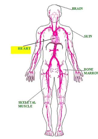
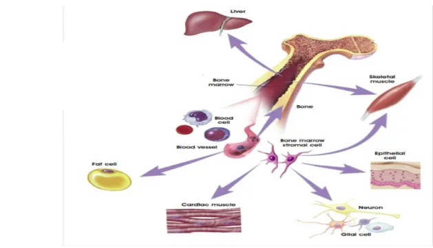
A tissue that has been traditionally considered completely post-mitotic is the heart. However, in the last few years a number of studies suggested the presence of a cardiac stem cell population capable of (re)generating the cardiac tissue throughout life, raising the possibility to speculate about new potentially therapeutic strategies for cardiac repair. Initial evidence, though in part controversial, has been presented that bone marrow cells injected into damaged myocardium or mobilized into the circulation may transdifferentiate into heart cell types [33-36]. Additional, exciting approaches for cardiac repair have been raised by the remarkable discovery that the heart contains a reservoir of stem and progenitor cells.
HEART
Comparing embryonic and adult stem cells, it’s clear that both of them have advantages and disadvantages regarding potential use for cell-based regenerative therapies. Of course, adult and embryonic stem cells differ in the number and type of differentiated cells types they can become. Embryonic stem cells can become all cell types of the body because they are pluripotent. Adult stem cells are generally limited to differentiating into different cell types of their tissue of origin. However, different scientific evidences suggest that adult stem cell plasticity may exist, increasing the number of cell types a given adult stem cell can become. A great potential advantage of using stem cells from an adult is that the patient's own cells could be expanded in culture and then reintroduced into the patient. The use of the patient's own adult stem cells would mean that the cells would not be rejected by the immune system. This represents a significant advantage as immune rejection is a difficult problem that can only be circumvented with immunosuppressive drugs. Moreover, the use of adult stem cells overcomes the ethical problem of embryo “destruction”; that’s why the interest of researchers about stem cells derived from adult tissues is increasing.
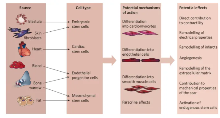
Actually, the most stunning aspect of current progress towards cardiac regeneration is the wide variety of cell types that have been considered as candidates for therapeutic delivery in humans [Fig. 4]. This myriad of cell types reflects the unmet medical need for treating heart disease, and hence the large amount of experimental effort being put into devising cell-based therapies. It also points to the lack of mechanistic understanding at many levels. The ideal cell type has not yet emerged, and few clinical studies are still comparing different adult stem-cell types such as skeletal myoblasts, endogenous cardiac stem cells, bone-marrow-derived cells (i.e. haematopoietic stem cells, endothelial progenitor cells, mesenchymal stem cells etc.) but also embryonic stem cells.
Some of these tested stem cells have been shown to improve cardiac function through various mechanisms, including the formation of new myocytes, endothelial cells and vascular smooth muscle cells, as well as trough paracrine effects.
Different experimental data have shown that, for cardiac regenerative medicine, cardiac stem cells (CSCs) and bone marrow- derived mesenchymal stem cells (BMSCs) have shown the greatest regenerative potential.
3.1 Cardiac stem cells
The adult heart was said for many years to be a postmitotic organ [12, 37, 38]. Of course, it was known that the endothelial, smooth muscle, and fibroblast cells of the heart do proliferate. But the cells that make up the meat of the heart—the myocardium—were thought to be terminally differentiated and therefore had lost their proliferative capacity [12, 37-39]. A major implication of this belief is that the myocytes comprising the adult myocardium are present at birth and are not replenished during an individual’s life span. This traditional viewpoint was supported by several lines of evidence. First, studies examining mitosis in the heart showed that the number of myocytes undergoing proliferation was either very low or nonexistent in the adult myocardium. When comparing the heart to other tissues that have obvious high levels of cellular regeneration (e.g., the bone marrow, liver, etc.), it was reasonable to conclude that the myocardium was postmitotic. Second, it has been well established that the myocardium responds to increased load, due to physiological and pathological stress, principally by cellular hypertrophy (increase in cell size) without obvious cellular hyperplasia (increase in cell number). Third, in response to severe injury of the myocardium, as occurs in an infarct, the myocardium is unable to restore functional cardiac muscle within the damaged area [12, 39, 40].
Figure 4. Different cell types and mechanisms proposed for cardiac stem cell therapy (Figure
Throughout the years, there were several reports suggesting that cells within the myocardium undergo cell division [41, 42], thus challenging the prevailing concept that the adult myocardium is postmitotic. However, the idea that the adult heart contains at least some newly regenerated myocytes has only been taken seriously during the past five years [43-45]. In 2003, in fact, several independent groups reported the discovery of resident progenitor cells in heart tissue, which would have the capacity to regenerate sections of the healthy or injured myocardium [46-48].
In one such study, a Lin- c-Kit+ cell was isolated from the adult rat heart, and it was reported to be
self-renewing, clonogenic, and multipotent, being able to give rise to cardiomyocytes, smooth muscle cells, and endothelial cells [49]. The cells were injected into ischemic hearts and were reported to reconstitute up to 70% of the myocardium, forming both new vessels and cardiomyocytes. More importantly, the authors reported a significant increase in cardiac function of the animals that had received these cells. In a similar study, the same cell type was delivered to the coronary arteries of rats that had had a myocardial infarct, via a catheter positioned into the aortic root [50]. The cells were reported to induce myocardial regeneration through the decrease of the infarct size by 29% and also to improve the function of the injured heart.
At about the same time, a different type of adult cardiac stem cell was reported by Oh et al [51]. The cells, isolated from adult mouse hearts, were Sca-1+ and initiated in vitro cardiac gene
expression after treatment with the DNA demethylating agent 5-azacytidine. A mouse model of ischemia/ reperfusion injury was used to assay the in vivo capacity of the Sca-1+ cardiac stem cells
to regenerate the infarcted myocardium. Transplanted cells initiated production of cardiac functional proteins and were identified in the mouse hearts 2 weeks after injection. Fusion between the donor cells and the host cardiomyocytes was evident in about 50% of the identified cells, whereas the other 50% had differentiated into cardiomyocytes.
Martin et al [52] reported on the identification of a resident cardiac population of adult stem cells with the unique expression of the Abcg2 transporter protein (Side Population Cells-SPCs). The Abcg2+ cells were reported to differentiate into alpha actinin-positive cells when cocultured with
adult cardiomyocytes; however, when cultured individually in methylcellulose, they gave rise to hematopoietic colonies. Finally, gene expression analysis revealed a unique transcriptional signature similar to that of endothelial and hematopoietic progenitor cells. Cell-based cardiac therapies using the Abcg2+ cells are underway.
Islet-1, a LIM homeodomain transcription factor, is uniquely expressed in the adult heart by the fourth identified cardiac stem cell population. This marker is expressed by cardiac progenitor cells
in the secondary cardiac field, a structure present during early development, which contributes to most cells in the heart [53]. During development, proliferating progenitor cells in the outflow tract, the right ventricle, and the atrium express isl1, without which they cannot contribute to the heart. Isl1+ cells were identified in the postnatal rat, mouse, and human myocardium [54].
A technique of conditional genetic marking was used to identify the isl1+ population in a
temporal/spatial manner. This allowed the authors to selectively isolate the isl1+ cells at a particular
developmental stage. When cocultured with isolated cardiac mesenchymal cells, the isl1+ cells
maintained isl1 expression and proliferated in culture without differentiating. The isolated cells could be induced to differentiate in culture into cardiomyocytes after exposure to 4-OH-TM or after coculture with neonatal cardiac myocytes. No cell-based cardiac therapy experiments have yet to be reported involving the isl1+ cardiac stem cells.
The four adult cardiac stem cell populations reported isolated are Lin-/c-Kit+, Sca-1+, Abcg2+, and
isl1+ cells. When these cell types were examined for markers expressed by the rest of the stem cell
populations, it was found that the c- Kit+ cells did not express Sca-1, the Sca-1+ cells did not express
c-Kit, and the isl1+ cells did not express c-Kit or Sca-1. The Abcg2+ cells were reported to be
approximately 50% Sca-1+ and only about 2% c-Kit+ (no data were given for isl1).
Therefore, cardiac stem and progenitor cell types characterized so far exhibit significant differences in their immunophenotypic, developmental and biological properties. Thus, the heart probably contains various types of stem and progenitor cells. This is in agreement with the emerging consensus that more than one stem cell may be present in a particular tissue [55], and with recent evidence that heart endothelial, cardiac and muscle cells may arise from a hierarchy of multipotent/bipotent stem progenitor cells [56-58, Fig.5]. The developmental origin(s) of stem and progenitor cells of the adult heart has been studied only to a very limited extent [59]. Mouse chimera experiments suggest that most (at least 95%) cardiomyocytes are derived from a relatively small embryonic founder population, ruling out any major contribution to the adult cardiomyocyte population from “immigrant” non-cardiac stem cells, at least under physiological conditions [60]. On the other hand, several findings, including the presence of cardiac chimerism in patients receiving allogeneic bone marrow transplantation, suggest that immigrant bone marrow-derived cells could behave, under pathological conditions leading to heart damage, as precursors of cardiac stem cells [61]. Thus, the important issue of extra-cardiac sources of CSCs, following cardiac damage, still needs to be addressed. If extra-cardiac cells can be induced, not only to migrate to the
heart, but also to reconstitute a pool of extensively replicating CSCs, exciting therapeutic possibilities may arise.
Moreover, if these Lin-/c-Kit+, Sca-1+, Abcg2+, and isl1+ described progenitor/stem cell
populations reside in the myocardium, it would be informative to examine why under normal circumstances they do not regenerate the myocardium and how can we stimulate them to do so.
3.2 Bone marrow mesenchymal Stem Cells
About 40 years ago Friedenstein described stromal cells in the bone marrow that were spindle shaped and proliferate to form colonies [62]. These cells attach to plastic and are able to differentiate under defined in vitro conditions into multiple cell types present in many different tissues, e.g. osteoblasts, chondroblasts, adipocytes and, more recently, cardiomyocytes and vascular endothelial cells [21, 63, Fig.6]. Later on these cells, obtained from postnatal bone marrow, were called mesenchymal stem cells (MSCs) or stromal stem cells [64, 65]. The term mesenchymal stem cells (MSC) was popularized by Caplan [66], in reference to work by Friedenstein and Owen [67], describing a plastic-adherent fibroblastic cell isolated by Percoll density centrifugation, reactive with monoclonal antibodies SH2 and SH3. The adjective ‘mesenchymal’ is fraught with some ambiguity since ‘mesenchyme’ describes tissue of mesodermal origin, the middle embryological germ layer, giving rise to the musculoskeletal, blood, vascular and urinogenital systems, and to
connective tissue (including dermis). Thus, developmentally speaking, the term ‘mesenchymal’ should include both blood and connective tissue cells. In practice however, only the latter cells are usually described as being derived from MSC and considered distinct from haematopoietic stem cells (HSC), which are responsible for the development, maintenance, and regeneration of blood forming tissues [68]. It is quite possible that MSC and HSC have a common precursor in the elusive ‘‘haemangioblasts’’ [69], in the cells identified by the group of Verfaillie originally termed ‘‘mesodermal progenitor’’ [70], later ‘‘multipotent adult progenitor’’ (MAPC) [71] cells, or in ‘‘pluripotent stem cells” [72] , or ‘tissue committed stem cells’ (TCSC) [73]. However, this is contentious and the physiological relevance of these cells remains to be demonstrated.
Recently the presence of somewhat similar cells has been demonstrated in many other tissues too. In fact, different studies have shown that MSCs reside not only in bone marrow but also in other tissues such as adipose tissue [74] , synovium [75] , periosteum [76] , muscle [77] , dental pulp [78], periodontal ligament [79] , placenta [80] and umbilical cord blood [81]. A recent study suggests that MSC reside in virtually all postnatal organs and tissues, and may be localized to vessel walls [82]. Bone marrow-derived MSC, which are most investigated, are a rare subpopulation of bone marrow cells (approximately 0.001-0.01%) [21].
So far the nomenclature is not consistent. Designations for cells with nonhaematopoietic multipotency have included ‘‘colony-forming-unit-fibroblasts’’, ‘‘stromal (stem) cells’’, ‘‘bone marrow (stromal) cells’’, ‘‘skeletal stem cells’’, ‘‘mesodermal progenitor cells’’, ‘‘non-haematopoietic stem cells’’, ‘‘(bone marrow) stem cells’’, ‘‘mesenchymal progenitor cells’’ and others [71, 83]. There is also an understandable tendency to designate such cells as ‘‘pre-(lineage-under-investigation)’’ cells (e.g. pre-osteoblast etc.). It has also been suggested that MSC are simply pericytes [84]. Some of the inconsistencies surrounding the identification of MSC arise from the fact that specific markers have not yet been agreed on. In the absence of a universal antigenic indication (analogous to CD34+ for HSC) and an universal assay (analogous to the repopulation assays for
HSC) MSC are often identified simply by testing a cultures’ differentiation potential into colony forming units (CFU) indicative of proliferative capacity and into several lineages of mesenchymal tissue as defined above [21]. Also, the ability to adhere to tissue culture plastic and a fibroblastlike morphology are taken as characteristic markers for MSC [66]. Recently, different surface markers have been associated with MSC including D7fib [85], Stro1 [86], CD45 and glycophorin A [21, 70, 87], BMPR1a [83, 88].
Further complications arise when different sources, extraction and cultivation methods are used. Even when narrowing sources to bone marrow, the site of extraction is reported to influence cell behaviour: e.g. MSC from alveolar bone show less chondrogenic and adipogenic potential compared to iliac bone [89] . Isolation is usually conducted by density centrifugation (sometimes enhanced by gradient solutions) to obtain the mononuclear fraction of marrow cells and by using the widely reported ability of MSC to adhere to tissue culture plastic [90]. Newer methods employ magnetic beads [86] or FACS sorting [91] in conjunction with antibodies to the proposed MSC markers above. Additionally, widely differing standards regarding serum composition, culture conditions, and growth factor application in MSC cultivation exist. Differing conditions can lead to enrichment of different subsets of MSC with differing clonogenic potential. All these potential deviation points in current methods are summarised in Table 1.
Table 1
Extraction sites (Prinz et al., 1999)
Bone marrow (live donors—partial samples only): hip, sternum, broken bones (rare) Bone marrow (cadaver donors): all sites (rare)
Other tissue: teeth, fat, muscle, cartilage, synovial fluid, skin Developmental tissue: foetal, umbilical, placenta
Dissociation method Trypsination Scraping Suspension culture Marker combinations CD10+, CD13+, CD34+, CD56+, CD90+, MHC-1+ (Young et al., 1999) CD10_, CD13+, CD31_, CD34_, CD44+, CD45_, CD90+, CD105+, CD133_
Wnt2+ ,Wnt4+, Wnt5a+, Wnt11+, Wnt16+, Fz2+, Fz3+, Fz4+, Fz5+, Fz6+ (Etheridge et al., 2004)
VCAM+, STRO-1+, CD73+, CD105+ (Tuli et al., 2003)
GlyA_, CD45_ (Reyes et al., 2001)
D7-FIB+, CD13+; CD45_, GPA_, LNGFR+ (Jones et al., 2002)
SH2+; SH3+; CD14_; CD29+, CD34_, CD44+; CD45_, CD71+, CD90+, CD106+, CD120a+,
CD124+ (Pittenger et al., 1999)
Identification
Adherence to plastic Magnetic bead FACS cell sorting
Other factors
Cell line or ex vivo Donor age
Donor sex
Donor disease status Point of first analysis Seeding density Feeder cells used
Culture conditions (temperature, motion, etc.) Differentiation agent
Medium composition First medium change
Frequency of medium change
Listed are some variables in MSC description where differing standards reportedly or likely result in influencing cell behaviour or otherwise lead to variant data
Since MSC can be rather easily isolated from the bone marrow and can also be expanded in vitro they have become a prime target for researchers of tissue regeneration. These cells have now been extensively used for transplantation experiments in animals and also for some therapeutic trials in humans. However, much new research is needed to learn enough on the molecular mechanisms of MSC differentiation to evaluate their full capacity for tissue regeneration. Mesenchymal stem cells have been studied in great detail and scientists have advanced knowledge about how to grow these cells in culture. Unlike most other human adult stem cells, mesenchymal stem cells can be obtained in quantities appropriate for clinical applications, making them good candidates for use in tissue repair. Techniques for isolation and amplification of mesenchymal stem cells in culture have been established and the cells can be maintained and propagated in culture for long periods of time, without loosing their capacity to form all the above cell types.
3.2.1 Bone Marrow-MSC and cardiac repairing
In vitro studies have demonstrated that bone marrow stem cells (BMSC) can differentiate not only into adipocytes and osteocytes, but also into cardiomyocytes and vascular endothelial cells in
vivo and in vitro [21, 63, 92-94]. Conversely, it has also been reported that the differentiation of
BMSC into cardiomyocytes occurs rarely, if at all [95-97]. There is no consensus on the plasticity of BMSC, including their differentiation into functional cardiomyocytes; however, experimental studies and clinical trials have demonstrated that the implantation of bone marrow-derived cells can improve regional perfusion and cardiac function of the injured heart, by rebuilding the damaged myocardium and cardiac vessels [98-100]. Thus, autologous BMSC are still one of the most studied cell sources for myocardial repair.
Different researchers tested the in vitro capacity of mouse bone marrow–derived MSCs to differentiate into cardiomyocytes in cultures containing either BMSC alone, treated with 5-azacytidine or a cocktail of growth factors and in co-culture with cardiomyocytes [101, 102]. The in vitro differentiation of BMSC into cardiomyocytes was first reported in 1999 by Makino et al. [92]. The authors induced the cells to differentiate by treating them with 5-azacytidine: a global DNA demethylating agent that acts as a cytosine analog capable of altering expression of certain genes that may regulate differentiation. The morphology of about 30% of the treated cells changed within a week of treatment, and by the second week, the cells were spontaneously contracting and expressing cardiac specific proteins. Bittira et al [103] also reported the isolation of rat bone
marrow–derived MSCs. LacZ labeled cells received either 5-azacytidine treatment or no treatment and were subsequently injected into the cryoinjured myocardium of isogenic rats. The authors reported that 4 to 8 weeks postinjection, the treated cells appeared myotube-like while expressing the cardiac marker troponin I-C. The data from both studies suggest that cell treatment with a DNA demethylating agent is necessary for the differentiation of MSCs into cardiomyocytes.
The effect of bone marrow–derived MSCs on cardiac function after myocardial infarction was also examined. A study reported by Shake et al [104] focused on the implantation of autologous MSCs in a swine myocardial infarct model. Labelled cells were administered 2 weeks postmyocardial infarction in the infarcted area through direct injection. The authors reported a significant attenuation in the degree of contractile dysfunction in the transplanted animals with reduced wall thinning in the infarcted region of the myocardium. In the second study, bone marrow MSCs were injected into the tail vein of rats that had had a myocardial infarct (MI) [105]. The MSCs were reported to be recruited to the injured heart through the expression of the stromal cell-derived factor-1 (SDF-1), enhance angiogenesis, and improve cardiac function. The same authors also reported in another study that lacZ labelled rat bone marrow–derived MSCs were injected into the tail vein. The cells were able to home to the injured area of the heart and were found at high concentrations in the peri-infarct region of the myocardium [106].
Toma et al [107] reported the injection of lacZ labelled human bone marrow– derived MSCs into the left ventricle of mice. One week postinjection, only a limited number of cells had survived; however, these cells were reported to express cardiac-specific markers similarly to the host myocardium. Hattan et al [108] used a transgene that allowed expression of GFP under a ventricular-specific promoter. After MSC differentiation, GFP+ cells were sorted and transplanted
into the adult mouse myocardium with a reported long-term survival. In a most recent report, MSCs cultured in the presence of cardiogenic growth factors were injected into the myocardium of dogs that had had a myocardial infarct (coronary artery ligation) 8 weeks prior to the injections. The authors report significant functional recovery of the transplanted hearts [109]. Bone marrow–derived MSCs are reported to have the ability to home to the areas of the heart that have sustained an injury as a result of a myocardial infarct. They have also been shown to express cardiac markers in the myocardium independent of 5-azacytidine treatment. One of their advantages is that they are an autologous cell source obviating the need for immunosuppression therapy [110].
However, the time needed for MSCs to proliferate in culture to a sufficient cell number needed for the transplantation is far longer than the short amount of time the patient has postinfarction for
the injection of these cells as shown by Bittira et al [103]. The capacity of these cells to completely regenerate the infarcted myocardium has not been proven yet; so far it’s clear that the presence of the cells in the myocardium decreases the potential infarct size but the mechanism is still unclear and poorly understood.
More recently, it has been shown that MSC should exert their effect on cardiac regeneration not only by differentiation into specific cell types, but also through paracrine actions. In vitro studies have demonstrated that MSC can secrete a variety of angiogenic, antiapoptotic and mitogenic factors such as vascular endothelial growth factor (VEGF), hepatocyte growth factor (HGF), adrenomedullin (AM) and insulin-like growth factor-1 (IGF-1) [111, 112]. Interestingly, administration of conditioned medium obtained from MSC culture exerted cytoprotective effects on the myocardium in an animal model of myocardial infarction [113]. Ohnishi et al [114] demonstrated that cultured cardiomyocytes were injured in response to monocyte chemoattractant protein-1 (MCP-1), which plays an important role in myocarditis, whereas this effect was significantly attenuated by conditioned medium derived from MSC culture. These results suggest a cardioprotective effect of MSC acting in a paracrine manner, demonstrating the importance of secreted factors in cardiac repair.
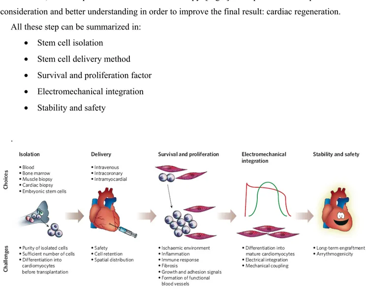
4. Cell delivery in cardiac stem cell therapy
Functional benefits of cell therapy for cardiovascular applications may arise from induction of angiogenesis, cardiomyogenesis or mechanical interstitial support. The former two modes of benefit may result from site-specific transdifferentiation of administered cells or by secretion of paracrine factors that may stimulate endogenous repair mechanisms. On this basis, improvements in regional myocardial perfusion, systolic function, diastolic function and adverse ventricular remodelling would be predicted. The additional mechanical interstitial support provided by cell administration may itself impact beneficially on the ventricular remodelling process. Clearly these potential benefits need to be weighed against the potential toxicity from cell therapy, such as exacerbation of atherosclerosis, arrhythmogenesis, inappropriate calcification and local or ectopic tumor formation.
The optimal cell preparation for each of the variety of potential cardiovascular applications remains to be determined. It cannot be assumed that one cell preparation will be equally efficacious for all clinical applications, and different cell preparations may have varying toxicity profiles .
Indeed, it is unclear if administration of a highly selected cell population is preferable to a heterogeneous unselected or combination cell product [115].
Other unresolved issues include determination of the optimal number of cells to be delivered, timing of cell administration, importance of growth factor preconditioning of cellular products prior to administration, how to follow transplanted cells once they are in the body, effects of ex vivo cell expansion and prolonged cell culture prior to administration, and the use of allogeneic rather than autologous cell populations [Table 2].
Table 2
Comparison of allogeneic and autologous cell preparations (from R de Silva and RJ Lederman, 2004)
About the sources of cell for cardiac transplant, cardiac and bone marrow stem cells are described by different researchers as the cells with the best potentiality in cardiac regeneration. Very little data is available to guide cell dosing. Scientific data suggests that there is a dose-dependent improvement in function but it is still unclear which is the best dose to warranty a real functional cardiac improvement.
Determining optimal delivery methods raise issues not only of dose, but also of timing. Moreover, assessing the fate of injected cells is critical to understanding mechanisms of action.
For therapeutic purposes, cells should be delivered to the target tissue of interest in sufficient number to confer functional benefit with minimal toxicity to the recipient.
For cardiovascular applications, modes of cell delivery may be broadly categorized as systemic or local [Table 3]. Systemic delivery may consist of intravenous infusion of cells or cytokine mobilization of cells into the circulation. The homing of systemically administered cells to the target tissue of interest requires appropriate chemokine signalling from the target tissue. The intensity of these homing signals appears to decline with time following an acute injury and may determine the time window during which systemic cell administration may confer functional benefit. Studies in rodents have demonstrated the ability of human progenitor cells from G-CSF-mobilized leukapheresis products [99] and rat allogeneic mesenchymal stromal cells [116] to home to regions of acute myocardial infarction following intravenous administration. In the former study, improvements in ejection fraction and attenuation of adverse cardiac remodelling were noted on the basis of echocardiography. Other animal studies have demonstrated that cytokine mobilization may confer hemodynamic benefits in a murine model of acute coronary artery ligation [117]. However, scientific and clinical data suggest that systemic cell delivery may not be optimal in the setting of acute or chronic coronary syndromes, although further studies are required.
Table 3
Local delivery is theoretically more attractive than systemic in that larger numbers of cells may potentially be administered to specific regions of interest within the target organ. Local delivery systems should be biocompatible with the cell preparation to be administered, such that there is minimal loss of cell number and cell viability as a consequence of passage through the delivery system. Clinically, local cell delivery can be achieved by direct injection at the time of open heart surgery or by percutaneous catheter-guided intracoronary infusion or intramyocardial injection.
For example, surgical delivery is excellent for performing intramyocardial cell injection targeted to infarct borders. Using this method the cells are directly transplanted close to the damaged area in order to facilitate cell grafting and the regenerative process. Others local delivery methods are represented by catheter delivery. Percutaneous catheter-based approaches are available for delivery of cells to the myocardium via intracoronary infusion, endomyocardial injection, transcoronary vein intramyocardial injection, transcoronary sinus retrograde infusion and intrapericardial injection. Intracoronary infusions can be performed down the infarct-related artery [100] and endomyocardial injections can be localized to peri-infarct regions using an electromechanical mapping system [118,119]. Targeted cell delivery may also be achieved using real-time magnetic resonance imaging (MRI) [120, 121] techniques and cells labelled with particles [122], which appear as signal voids on the magnetic resonance (MR) image. Using this sophisticated technology, cell delivery can be targeted in real time precisely to infarct borders.
Anyway, there are several major unresolved issues in cell delivery. The optimal delivery route has not been established. There are few quantitative data addressing cell distribution and cell retention as a function of the mode of cell delivery. Biodistribution studies using technetium-99m labeled BM-derived mesenchymal stromal cells in recently infarcted rats suggest that, following intravenous infusion, the vast majority of infused cells are entrapped in the lungs with little distribution to the heart [123]. The number of cells in the heart was increased by infusion of cells directly into the left ventricular cavity [123]. Retention of cells following direct intramyocardial injection was not assessed in this study. Other pre-clinical data suggest that at best only 30–40% of particulate material is retained within the myocardium following a successful endomyocardial injection [124]. Clearly, significant improvements in cell retention and its quantification are required. Furthermore, the biocompatibility of interventional devices with therapeutic cell preparations needs further assessment. Variability in cell number, viability, migratory, proliferative and differentiation capacity as a function of cell handling and interaction with the delivery system may be important determinants of the efficacy of cardiac stem cell therapy.
5. Tissue-engineering: a new approach for cell delivery in cardiac regenerative medicine
The poor survival of grafted cells has been a concern of researchers. Given the poor vascular supply after a heart attack and an active inflammatory process, grafted cells survive with difficulty. As mentioned above, the efficacy of cell engraftment by injection of a cell suspension is generally very low as more than 90% of the cells injected is lost and does not engraft [125]. It is necessary to provide cells an environment that is suitable to proliferation and differentiation for the induction of tissue regeneration.
Thus, much effort is now conveyed to the development of alternative cell delivery system. One of these new strategies is the tissue-engineering strategy, in which biomatrices are used to generate three-dimensional cell constructs able to provide a physiological support to the cells, allowing to successfully engrafting new cells into the myocardium.
Tissue-engineering combines cells, engineering and materials methods, and suitable biochemical and physio-chemical factors to improve or replace biological functions. While most definitions of tissue engineering cover a broad range of applications, in practice the term is closely associated with applications that repair or replace portions of or whole tissues (i.e., bone, cartilage, blood vessels, heart etc...).
Often, the tissues involved require certain mechanical and structural properties for proper function. The term has also been applied to efforts to perform specific biochemical functions using cells within an artificially-created support system.
A commonly applied definition of tissue engineering, as stated by Langer and Vacanti, is "an interdisciplinary field that applies the principles of engineering and life sciences toward the development of biological substitutes that restore, maintain, or improve tissue function or a whole organ" [126]. Tissue engineering has also been defined as "understanding the principles of tissue growth, and applying this to produce functional replacement tissue for clinical use” [127].
Scientific advances in biomaterials, stem cells, growth and differentiation factors, and biomimetic environments have created unique opportunities to fabricate tissues in the laboratory from combinations of engineered extracellular matrices ("scaffolds"), cells, and biologically active molecules. Tissue-engineering utilizes living cells as engineering materials, the scaffold for cell proliferation and differentiation, and, eventually, growth factors which has the potential to accelerate tissue regeneration.
• Autologous cells are obtained from the same individual to which they will be reimplanted.
Autologous cells have the fewest problems with rejection and pathogen transmission, however in some cases might not be available. Moreover since this category of cells needs to be harvested from the patient, there are also some concerns related to the necessity of per-forming such surgical operations that might lead to donor site infection or chronic pain. Autologous cells also must be cultured from samples before they can be used: this takes time, so autologous solutions may not be very quick. Recently there has been a trend towards the use of mesenchymal stem cells from bone marrow and fat. A large number of cells can be easily and quickly isolated from fat and bone marrow.
• Allogenic cells come from the body of a donor of the same species. While there are some
ethical constraints to the use of human cells for in vitro studies, the employment of dermal fibroblasts from human foreskin has been demonstrated to be immunologically safe and thus a viable choice for tissue engineering of skin.
• Xenogenic cells are those isolated from individuals of another species. In particular animal
cells have been used quite extensively in experiments aimed at the construction of cardiovascular implants.
• Syngeneic’ or isogenic cells are isolated from genetically identical organisms, such as twins,
clones, or highly inbred research animal models.
• Stem cells are undifferentiated cells with the ability to divide in culture and give rise to
different forms of specialized cells. According to their source stem cells are divided into "adult" and "embryonic" stem cells, the first class being multipotent and the latter mostly pluripotent some cells are totipotent, in the earliest stages of the embryo. While there is still a large ethical debate related with the use of embryonic stem cells, it is thought that stem cells may be useful for the repair of diseased or damaged tissues, or may be used to grow new organs.
Cells are often implanted or 'seeded' into an artificial structure capable of supporting three-dimensional tissue formation. These structures, typically called scaffolds, are often critical, both ex
vivo as well as in vivo, to recapitulating the in vivo milieu and allowing cells to influence their own
microenvironments. Scaffolds usually serve at least one of the following purposes:
• Allow cell attachment and migration
• Deliver and retain cells and biochemical factors
• Enable diffusion of vital cell nutrients and expressed products
• Exert certain mechanical and biological influences to modify the behavior of the cell phase
• Protect trasplanted cells from the immunological attack.
It is necessary for tissue regeneration to increase the number of cells constituting the tissue as well as reconstruct a structure of extracellular matrix (ECM) to support the proliferation and differentiation of cells for regeneration induction. In addition, it is necessary to allow cells of high potential to proliferate as well as to maintain their biological function.
To achieve the goal of tissue reconstruction, scaffolds must meet some specific requirements. The materials should be well deliquesced with contiguous tissues after transplantation without or with a very low inflammatory reaction. They must have a high porosity and an adequate pore size, necessary to facilitate cell seeding and diffusion throughout the whole structure of both cells and nutrients. Biodegradability is often an essential factor since scaffolds should preferably be absorbed by the surrounding tissues without the necessity of a surgical removal. The rate at which degradation occurs has to coincide as much as possible with the rate of tissue formation: this means that while cells are fabricating their own natural matrix structure around themselves, the scaffold is able to provide structural integrity within the body and eventually it will break down leaving the neotissue, newly formed tissue which will take over the mechanical load.
Many different materials (natural and synthetic, biodegradable and permanent) have been investigated. Most of these materials have been known in the medical field before the advent of tissue-engineering as a research topic, being already employed as bioresorbable sutures. Examples of these materials are collagen or some linear aliphatic polyester.
New biomaterials have been engineered to have ideal properties and functional customization: synthetic manufacture, biocompatibility, non-immunogenicity, transparency, nano-scale fibers, low concentration, resorption rates, etc.
A commonly used synthetic material is polylactic acid (PLA). This is a polyester which degrades within the human body to form lactic acid, a naturally occurring chemical which is easily removed
from the body. Similar materials are polyglycolic acid (PGA) and polycaprolactone (PCL): their degradation mechanism is similar to that of PLA, but they exhibit respectively a faster and a slower rate of degradation compared to PLA. Scaffolds may also be constructed from natural materials: in particular different derivatives of the extracellular matrix have been studied to evaluate their ability to support cell growth. Proteic materials, such as collagen or fibrin, and polysaccharidic materials, like chitosan or glycosaminoglycans (GAGs), have all proved suitable in terms of cell compatibility, but some issues with potential immunogenicity still remains. Among GAGs hyaluronic acid, possibly in combination with cross linking agents (e.g. glutaraldehyde, water soluble carbodiimide, etc...), is one of the possible choices as scaffold material. Functionalized groups of scaffolds may be useful in the delivery of small molecules (drugs) to specific tissues.
5.1 Materials as scaffolds for cells transplantation in cardiac tissue-engineering
One of the first materials used for tissue-engineering of the heart was based on hydrolytically degradable biocompatible polymers composed of polyactic acid (PLA), polyglycolic acid (PGA) and their copolymer polylactic-co-glycolic acid (PLGA) [128]. Subsequently, researchers realized that the mechanical properties of the material used had to be adapted to the elastic properties of the heart tissue. Therefore, most research is focusing on the use of scaffold made of different synthetic and/or natural polymers. Table 4 gives an overview on different materials used in the past few years for the regeneration of the heart [125]. It has to be pointed out that only a few materials were tested in humans, while many animal studies were carried out in rats and dogs.
Table 4
Several groups are currently working with scaffold materials composed of natural polymers such as collagen [125, 129, 130], this latter being the major constituent of the cardiac extracellular matrice (ECM). Promising results in the development of collagen-based grafts or ‘patches’ containing beating cardiomyocytes were obtained in Canada [129] and Germany [130]. These studies comprised the application of cardiomyocyte-seeded collagen strings that were cyclically stretched, thus providing patches with improved morphology and contractile function. Zimmermann
et al [125] demonstrated that these collagen patches could survive and beat for up to eight weeks
after engraftment on the heart of immunosuppressed rats.
Similar approaches and results were obtained using alginate-based scaffolds by Cohen et al. in Israel [131, 132]. Alginate, a negatively charged polysaccharide from seaweed which forms hydrogels in the presence of calcium, offers the advantage of detecting ECM formation by cardiac cells to follow not only the proliferation and migration of the cells, but also the kinetics of ECM formation. After implantation into the infarcted rat myocardium, the alginate-biografts were shown to stimulate intense neovascularization and to attenuate left ventricular dilatation and failure, compared with control rat hearts [132].
Composites of natural and synthetic polymers were also developed; for example, sponges based on caprolactone-co-L-lactide reinforced with knitted poly-L-lactide fabric (PCLA), gelatin or PGA. Using rat aortic smooth muscle cells, an increased colonization of the right ventricular outflow tract was obtained using gelatin or PCLA, but not with PGA-reinforced grafts [133]. Another interesting synthetic material is based on 1,3-trimethylene carbonate and D,L-lactide copolymers [134, 135], which have the ability to be reabsorbed over a ten month period and to sustain the cyclic loading of the heart muscle under physiological conditions. However, as yet no animal studies have been carried out with this amorphous material. The most fascinating approach to the regeneration of heart has been proposed by Shimizu and co-workers [136], who used materials to create electrically communicating three-dimensional cardiac tissue layers [Fig. 7, panel c]. In this case, cells were adhered on tissue-culture plates previously coated with poly(N-isopropylacrylamide) (PIPAAm), a temperature-sensitive polymer. At 37°C PIPAAm is hydrophobic, enabling cell adhesion and access to the binding sites offered on this modified surface; at a lower temperature such as 32°C, the surface becomes hydrophilic and inappropriate for cell adhesion due to the rapid hydration and swelling of PIPAAm. Using poly(vinylidene difluoride) (PVDF) membranes, which are hydrophobic, the detaching cell layers can be collected and handled, providing up to four conducting layers of synchronously beating cardiomyocytes. When these patches were implanted on rats with
induced myocardial infarction, an improved myocardial contractility was observed, concomitant with the appearance of a vascular network within a few days after implantation [137]. Figure 7 summarizes three of the major approaches to cardiac engineering described above, based on the use of collagen, hydrogel or multiple layers.
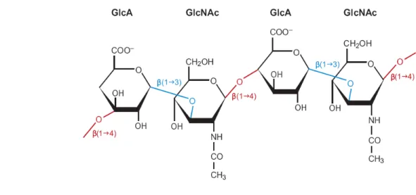
More recently researchers are using another natural component of ECM as support for cardiac stem cell transplantation: hyaluronic acid (HA). HA is a naturally occurring polysaccharide, and is one of the basic components of the ECM of connective tissue. HA is biocompatible and biodegradable and exhibits an unusual and distinctive set of physicochemical and biological properties, enabling it to regulate and control several physiological functions of tissue [138]. It plays a major role in tissue growth and remodelling, interacting specifically with endogenous receptors such as CD44 and intracellular adhesion molecule-1 (ICAM-1) to guide and control cellular migration, growth and adhesion [139]. It also regulates macromolecular traffic within the interstitial space in joints contributing to lubrication and to the strength in compression of soft tissue [140].
Twenty years after the initial discovery of HA, Meyer’s laboratory determined the exact chemical structure of HA, a non sulfated, highmolecular-weight glycosaminoglycan composed of
Figure 7. Scheme of the present major strategies of cardiac tissue engineering using (a) collagen strings, (b) biodegradable gels or (c)
cardiac cell sheets. The incorporation of growth factors and/or cytokines (triangles) may have a crucial role to support cell differentiation, engraftment and survival, both within the scaffolds and in vivo, thus improving the overall cardiac function. (Figure from (Zammaretti and Jaconi, 2004)