PhD Course
in
Mind, Gender and Language
XXXI° cycle
The effect of cognitive training on subsequent
sleep characteristics
PhD Supervisor: PhD Candidate:
Prof. Gianluca Ficca Dr. Mariangela Cerasuolo
PhD Coordinator:
Prof. Dario Bacchini
3
The effect of cognitive training on
subsequent sleep characteristics
Abstract
Introduction: Several studies have consistently shown that pre-sleep learning produces changes in sleep structure. Whereas the majority of these studies has mainly focused on post-training changes in sleep states (namely REM and NREM sleep amount) and, more recently, in specific electrophysiological features (e.g., sleep spindles, slow wave activity), very little attention has been paid to the hypothesis that pre-sleep learning might improve sleep quality, as expressed by sleep continuity, stability and cyclic organization measures. Furthermore, studies addressing the relationship between sleep and learning usually employ purely declarative or procedural tasks, neglecting that everyday life learning processes depend on the simultaneous activation of different memory systems. Recently, we have reported that a complex ecological learning task (requiring the simultaneous activation of several cognitive functions), intensively administered at bedtime, improves daytime sleep continuity and stability, possibly as a result of ongoing memory processes. To follow up our previous study, here we aimed to extend these findings to a night paradigm and to test whether a similar post-training sleep improvement may be obtained in a sample of individuals with sleep complaints. Specifically, our focus was on post-training changes in objective and subjective sleep quality. Furthermore, we compared overnight performance changes with those obtained over a wake retention period, in order to address the possible differential effect of sleep and wake on memory processes.
Method: After a habituation night, twenty-one subjects (F=15, mean age: 27.5±7.7 years, all bad sleepers according to the Pittsburgh Sleep Quality Index) underwent conventional polygraphic recording under three conditions: 1) BL, baseline night sleep; 2) post-active control sleep (AC), a sleep episode preceded by a non-learning control task; 3) post-training sleep (TR), a sleep episode preceded by a complex ecological task. The same task as in TR was administered in a Wake condition (W), in which the retention period between training sessions corresponded to the duration of the subject’s baseline sleep time. Subjects underwent AC, TR and W conditions in balanced order.
4 The complex cognitive task consisted in a slightly modified version of the famous word game “Ruzzle”. In this game, the player has two minutes to form as many words as possible and reach the highest score achievable with the 16 letters available in a 4x4 grid on an iPad screen. Performance measures were WORDS%, i.e., the number of detected words over total available words, and R-SCORE%, i.e., the global score achieved, depending on the number of words found, on their length and on the ability to use the coloured bonus letters which multiply letter or word values.
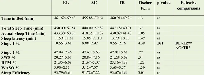
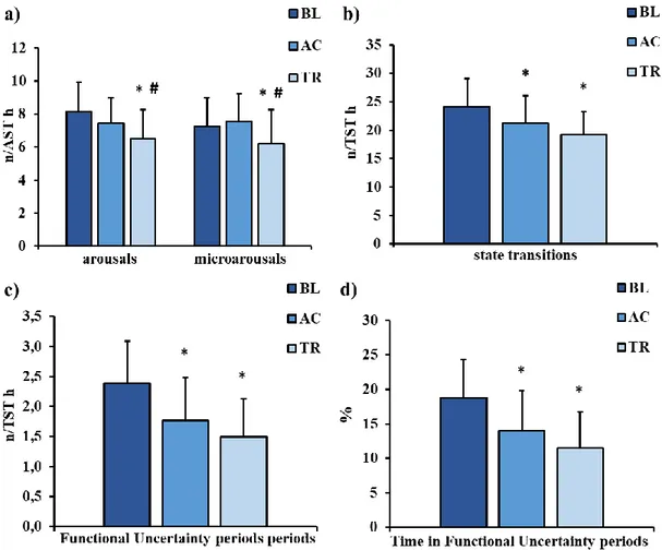
Results: Post-training sleep (TR) showed a reduction in Stage 1 proportion (F=4.39, p=.021; TR<BL and AC) and a significant improvement in sleep continuity, stability and organization, as expressed by: a decrease of total (F=4.90, p=.014, BL>TR and AC) and brief awakenings frequency (F=5.89, p=.007, BL>TR and AC), decreased frequency of arousals (F=6.25, p=.005; TR<BL and AC), microarousals (F=3.63, p=.050; TR<BL), state transitions (F=10.16, p<.001; BL>TR and AC) and functional uncertainty (FU) periods (F=14.23, p<.001; BL>TR and AC), as well as a reduction of time spent in FU periods (F=515.33, p<.001; BL>TR and AC); an increase in the number of NREM-REM cycles (F=4.51, p=.019; TR>BL and AC), and of time spent in cycles (F=4.77, p=.015; TR>BL and AC). This improvement in objective sleep quality was paralleled by that in subjective ratings, assessed through the Self-Rating Scale for Sleep and Awakenings Quality (χ2=9.13, p=.010; TR<BL). No other sleep measure displayed significant changes between conditions. Furthermore, the comparison of R-SCORE% changes between the TR and W conditions yielded a significant sleep effect (t=5.38, p<.001; TR>W), while the opposite effect emerged for the R-WORDS% (t=-2.96, p=.01; W>TR).
Conclusions: Our results extend previous findings on post-training changes in sleep continuity, stability and organization to a sample of bad sleepers; also, they show that objective sleep improvement may be reflected in subjective sleep quality perception. Interestingly, the active control task also produced improvements in some of these features, prompting future investigations on the contribution to post-training sleep changes of additional factors not specifically linked to learning processes. As for performance, the finding of a significant sleep effect for the more complex performance measure (R-SCORE%) suggests that sleep preferentially promotes effective learning of elaborate cognitive strategies rather than that of simpler cognitive processes. In conclusion, in light of the importance of non-pharmacological treatments for sleep disturbances, this study offers the
5 possibility to further explore planned cognitive training as a low-cost treatment strategy to improve sleep quality.
6
Index
Chapter 1 – Principles of sleep phenomenology: sleep organization and
regulation... 8
1.1. Sleep: definition and main constituents ... 8
1.2. Methods to study sleep ... 9
1.2.1. Behavioural observation ... 10
1.2.2. Subjective questionnaires and scales ... 10
1.2.3. Actigraphy ... 11
1.2.4. Polysomnography... 11
1.2.5 New methods to study sleep ... 13
1.3. Organizational levels of sleep ... 14
1.3.1 First organizational level: the sleep episode... 15
1.3.2 Second organizational level: the NREM-REM cycle ... 17
1.3.3 Third organizational level: NREM and REM sleep states ... 17
1.3.4 Fourth organizational level: the intra-state events... 19
1.3.4.1. NREM sleep intra-state phasic events ... 19
1.3.4.2. REM sleep intra-state phasic events ... 23
1.4. Models of sleep regulation ... 24
1.4.1 Circadian factors ... 25
1.4.2 Homeostatic factors ... 26
1.4.3 The two process model of sleep regulation ... 27
1.4.4 Reconsidering the sleep regulation model... 29
Chapter 2 - How sleep is modified by previous cognitive activity: insights from
sleep and memory literature ... 32
2.1. The role of wake intensity in sleep regulation ... 32
2.2. The relationship between sleep and memory: overview of concepts and findings ... 34
2.2.1. Main experimental paradigms in the sleep-memory field ... 37
2.3. An overview of learning-dependent sleep changes ... 38
2.3.1. Sleep duration and propensity ... 39
2.3.2. Wake after sleep onset and sleep efficiency ... 40
2.3.3. Behavioural awakenings, arousals, state transitions... 41
2.3.4. Sleep organization: NREM-REM cycles and sleep state sequences ... 42
2.3.5. NREM sleep ... 43
2.3.6. REM sleep ... 47
2.3.7. NREM sleep intra-state phasic events: sleep spindles, slow oscillations, sharp-wave ripples, Cyclic Alternating Pattern ... 48
2.3.8. REM sleep intra-state phasic events: rapid eye movements and pontine waves ... 51
2.3.9. Subjective sleep quality ... 52
7
Chapter 3 - The experimental study: the effect of cognitive training on
subsequent sleep characteristics ... 59
3.1. Introduction ... 59
3.2. Materials and method ... 62
3.2.1. Participants ... 62
3.2.2. Procedure ... 63
3.2.3. Cognitive tasks ... 64
3.2.4. Sleep measures ... 67
3.2.5. Performance measures ... 68
3.2.6. EEG automatic analyses ... 68
3.2.7. Statistical analysis ... 69
3.3. Results ... 69
3.3.1. Quantitative sleep variables ... 70
3.3.2. Sleep continuity... 70
3.3.3. Sleep stability ... 72
3.3.4. Sleep organization ... 73
3.3.5. EEG power ... 74
3.3.6. Spindle density ... 75
3.3.7. Subjective sleep quality ... 75
3.3.8. Sleepiness ... 76 3.3.9. Fatigue... 76 3.3.10. Cognitive performance ... 76 3.3.11. Sleep-memory correlation ... 78 3.4. Discussion ... 79 3.5. Conclusion ... 86
References ... 87
8
Chapter 1 – Principles of sleep phenomenology:
sleep organization and regulation
Before going deep in the “heart” of our research project, I will introduce some theoretical and methodological principles of sleep phenomenology. Specifically, starting from a general definition of sleep, this chapter provides a brief overview of basic sleep physiology, with a focus on sleep organization, which represents an important theoretical construct underlying our study. Finally, I will describe in detail what has been considered as the most prominent model of sleep-wake rhythm regulation, introducing our hypothesis on the need of reconsidering and updating it in light of recent evidence.
1.1. Sleep: definition and main constituents
According to the “Oxford English Dictionary”, sleep is defined as “a condition of body and mind which typically recurs for several hours every night, in which the nervous system is inactive, the eyes closed, the postural muscles relaxed, and consciousness practically suspended”. This definition stresses some of the most evident characteristics of sleep, easily recognizable through behavioural observation. However, even though some of its statements are commonly accepted, this definition appears in certain respects inexact and/or incomplete. For instance, sleep typically occurs every night, but it could be dislocated to another time of day or be repeated in case of polyphasic sleep or naps. Also, the idea that “the nervous system is inactive” during sleep is in disagreement with relatively recent literature showing the importance of sleep for cognitive and memory processes (Conte and Ficca, 2013; Rasch and Born, 2013) and that external information processing while sleeping is still possible (Atienza et al., 2001; Hennevin et al., 2007). Finally, since sleep is a complex and dynamic phenomenon, a useful and complete definition must take into account the behavioural and physiological changes that characterize sleep and allow us to distinguish it from wake. Overtime, many authors attempted this challenge, emphasizing one or the other sleep feature at a time.
9 One of the latest and largely accepted definition of sleep has been introduced by two Italian scientists, Fagioli and Salzarulo (1995), who described sleep as “a state of the organism characterized by a reduced reactivity to the environment provoking a temporary suspension of relational activities: (this state) occurs spontaneously and periodically, is self-limited in time and reversible”. Here, the authors synthesized some of the key features of sleep. The reduced responsiveness to stimuli, which is in contrast with the general idea of a perceptual disengagement from the environment, refers to the elevated threshold of stimulus perception. The relative inactivity and suspension of relational activities is accompanied by a loss of consciousness of the outer word. The last sentence highlights the fact that sleep is a natural (spontaneous) and reversible state, meaning, on one side, that no specific external event is needed to induce sleep and that, on the other, stimuli of elevated intensity may interrupt it. Finally, as will be exhaustively explained later, the “periodical” occurrence of sleep and its limited duration in time, refer to the rhythmic alternation between sleep and wake and the mechanisms beyond this organization.
Actually, in humans, sleep is not restricted to a two states “wake-sleep” system. Sleep architecture consists of three behavioural states, defined on the basis of their characteristics: wake, REM sleep and NREM sleep (Comte et al., 2006). Shortly, NREM sleep, composed of four stages (Stages 1, 2, 3 and 4), gradually occurring as sleep becomes deeper, is characterized by a brain slowed down in a movable body; instead, REM sleep, marked by rapid eye movements, is described as a stage where the brain is intensively active in a paralyzed body, due to muscle atonia.
1.2. Methods to study sleep
Following Fagioli and Salzarulo’s definition (1995), sleep is not simply a “condition” but it is considered as a “state” and specifically a “behavioural state”: a “combination of variables that occurs several times and has a stability in time” (Prechtl, 1974). In order to identify the “constellation” of variables characterizing human sleep physiology and, inside sleep, its macro and microstructure, a multidimensional approach based on different methods and instruments would be more appropriate. However, in most cases, researchers and clinicians cannot administer all of them.
10 Depending on the instruments used, chosen according to their objectives, it is possible to recognize and study different aspects of sleep that, when considered together, allow us to catch the entire sleep phenomenon, with its complex time-dynamics.
Three are the main methods used in sleep medicine and research: behavioural observation, subjective scales and questionnaire and objective methods such as polysomnography and actigraphy. A brief paragraph is devoted to new recent methods used in sleep research, derived from nuclear and functional medicine.
1.2.1. Behavioural observation
Behavioural observation in sleep studies consists in systematically observing specific behaviour that characterizes a sleeping person (such as eyes, position and type and intensity of movements of the subject, responsiveness to internal and external environment) and taking note of them “live”, which means when the target event happened, or deferred based on video-recordings, by using guidelines, checklists and time grids.
Because of its multiple advantages over more physiological methods to study sleep (lower costs, the non-invasive nature, paralleled by the increased compliance and the possibility to use it in natural contexts), it is frequently used in psychophysiological research (Bliwise et al., 1990).
Behavioural observation has been often considered as a useful method to study sleep during ontogenetic development and aging, when it is often difficult to identify objective indicators of sleep and/or wakefulness, and recently, also as a possible screening tool in the diagnosis of some sleep disorders (Ipsiroglu et al., 2015).
1.2.2. Subjective questionnaires and scales
An important possibility to easily obtain information related to previous or habitual sleep characteristics consists in asking the subject questions about his sleep and/or the sleep-wake rhythms. Self-administered sleep questionnaires and sleep diaries are often used in sleep studies because they allow us to gather information on different populations in the short term.
Sleep diaries are usually administered in order to collect specific information about sleep for a long period of time. Questions typically include bedtime, wake time, sleep latency, daytime activities and information about sleep quality (number and duration
11 of awakenings and sensation of feeling rested in the morning and so on). Subjects are asked to fill it immediately after final awakening, referring to the night before.
Compared to sleep diaries which are often administered for several days, sleep questionnaires usually consist in a series of structured items about habitual sleep-wake behaviours, such as usual bedtimes and rise times, daytime sleepiness, sleep quality and so on. An example of this kind is the Pittsburgh Sleep Quality Index (PSQI) by Buysse et al. (1989), assessing sleep quality and duration over a 1-month time interval. The questionnaire measures seven “components”: subjective sleep quality, sleep latency, sleep duration, sleep efficiency, sleep disturbances, use of sleep medications, and daytime dysfunction. The global score, allows us to distinguish between good (PSQI<5) and bad sleepers (PSQI≥5).
Sleepiness scales are administered to evaluate sleepiness or vigilance levels at different time points. A well-known example is the Karolinska Sleepiness Scale (Åkerstedt and Gillberg, 1990), where the subject is asked to indicate his/her sleepiness level on a self-administered 9-point scale.
1.2.3. Actigraphy
The actigraph is a computerized device, similar to a watch, which provides information on sleep-wake rhythms, based on the recording of the subject’s body movements. It is based on the assumption that the profile of body movements represents a sufficiently accurate index of sleep and wake states. In particular, the outcome measures are: time in bed, actual sleep time, actual wake time, sleep efficiency, number of awakenings, sleep fragmentation and movement indexes. Actigraphy has the advantage to be a low cost, non-invasive tool, useful to provide objective information on sleep-wake behaviour for a long period of time in the subject’s natural environment.
Despite the high concordance between actigraphy and polysomnography for sleep scoring (Kripke et al., 1978; Cole and Kripke, 1988), the concurrent use of sleep diaries is recommended because this technology seems to overestimate sleep in some sleep disordered individuals and during specific daily activities (Martin and Hakim, 2011).
1.2.4. Polysomnography
Transitions between wakefulness and sleep and, inside sleep, between different sleep stages, are accompanied by physiological and electrical changes in the brain. Thanks
12 to Hans Berger (1930), it became possible to measure and record them using an instrument that detects and amplifies electrical activity from the human scalp. After almost one century, electroencephalography (EEG) is still a widely used technique to investigate brain activity through electrodes specifically positioned over the scalp, using a standardized method (10-20 International System). However, in order to define the main stages of sleep, the collection of other biosignals is also required. Polysomnography, the “gold standard” objective assessment of sleep, is based on the simultaneous recording of different physiological parameters during sleep. In addition to the EEG, other important measures are: the electrooculogram (EOG) for eye movements, the electromyogram (EMG) for tonic muscle activity, the electrocardiogram (ECG) for cardiac activity, the pneumogram (for respiratory parameters). Finally, in the clinical field most polysomnographic recordings also rely on the measurement of body temperature, a crucial index of circadian rhythmicity. Once sleep has been recorded through a polygraph, the next step is sleep scoring, i.e., the identification of sleep stages. After the historic R & K’s manual (Rechtschaffen and Kales, 1968), collecting standardized rules for visual sleep scoring, a more up-to-date manual has been introduced by the American Academy of Sleep Medicine (AASM, Iber et al., 2007), which is still subject to continuous revisions based on new experimental and clinical data.
Another well-established method for the analysis of EEG signals is spectral analysis. This mathematical approach uses the fast Fourier transform to automatically decompose EEG signals into its constituting frequency components. In the field of sleep medicine and research, five are the brain rhythms that are easily recognizable at sleep onset and during sleep itself: beta (16-40 Hz), sigma (12-16 Hz), alpha (8-12 Hz), theta (4-8 Hz), delta (.5-4 Hz). The power spectrum analysis determines the relative amounts of given frequencies in the waveform over the analysed time segment. The idea is that EEG waves can be split into an infinite number of pure sinusoidal components, each of a different frequency, that when summed together reconstitute the original waveform. However, a faithful representation of the original signal is only possible when the signal is stationary; instead, the EEG signal has waves that are not stable or even appear intermittently (Campbell, 2009). Therefore, it is recommended not to include in the analysis abrupt variations, such as those due to drowsiness and alerting. Another important drawback of spectral analysis concerns the scarce ability
13 to recognize artefacts, which may lead to misinterpretation of the power spectra and the entire EEG signal (Campbell, 2009).
Besides the fact that polysomnography is the best instrument to obtain information about sleep architecture both in the sleep laboratory and the natural environment, the main disadvantages are high costs and the high participant burden (Martin and Hakim, 2011).
1.2.5 New methods to study sleep
Although the EEG and polysomnographic assessment remain the best method in the sleep field, new methods have been more recently used to study brain functions in discrete neural areas not accessible to surface EEG and during specific states of consciousness. Specifically, functional neuroimaging techniques applied in the study of sleep allow clinicians and researchers to determine which neuroanatomic areas are activated during sleep. For instance, a wide body of imaging studies, using functional Magnetic Resonance Imaging (fMRI) and/or Positron Emission Tomography (PET), have shown that during REM sleep there is a greater activation of the thalamus, the brainstem and basal forebrain, as well as of the limbic and paralimbic cortex (Braun et al., 1997; Nofzinger, 2004), whereas the sleeping brain during NREM sleep is less active, showing reduced blood flow and metabolism in several brain areas (i.e., dorsal pons and mesencephalon, cerebellum, thalami, basal ganglia, basal forebrain/hypothalamus, prefrontal cortex, anterior cingulate cortex, precuneus and the mesial temporal lobe) (Nofzinger et al., 2002; Nofzinger, 2004).
Interestingly, neuroimaging techniques can be combined with EEG procedures for greater specificity and to overcome some of the drawbacks that each method has when used alone. For example, EEG combined with PET and with fMRI techniques allow researchers to investigate both the brain network that support sleep (top-down control) and the brain circuitry supporting processes, function and behaviour associated with sleep (bottom-up control) (Picchioni et al., 2014). Furthermore, recently greater attention has been paid to the use of Non-Invasive Brain Stimulation (NIBS) methods combined with polysomnography. In their study, Massimini and co-workers (2005) used transcranial magnetic stimulation (TMS, a non-invasive tool for manipulating neuronal excitability by stimulating the cerebral cortex) together with high-density EEG in order to study cortical connectivity during quiet wakefulness and sleep. The authors showed that during NREM sleep, there was a breakdown of cortical
14 connectivity, in that after stimulation of the premotor area, there was no propagation of the cortical response beyond the stimulation site (Massimini et al., 2005).
1.3. Organizational levels of sleep
The discovery of REM sleep thanks to Aserinsky and Kleitman (1953) may be considered as a Copernican Revolution in sleep research and medicine. It did not simply give us hints about sleep structure, but it has changed the way we look at the sleep phenomenon.
For a long time, it was believed that sleep was a stable and quite homogeneous state, simply a pause between wakefulness states. Nowadays, sleep is considered as a biological state with a complex internal organization. Four organizational levels may be identified, hierarchically proceeding from the alternation between sleep and wake to the regular and multiple transitions from NREM to REM (the NREM-REM cycle), to the occurrence of the sleep stages, up to the phasic events appearing within them, such as rapid eye movements during REM sleep or sleep spindles and K-complexes during NREM sleep (Salzarulo et al., 1998; Conte and Ficca, 2013).
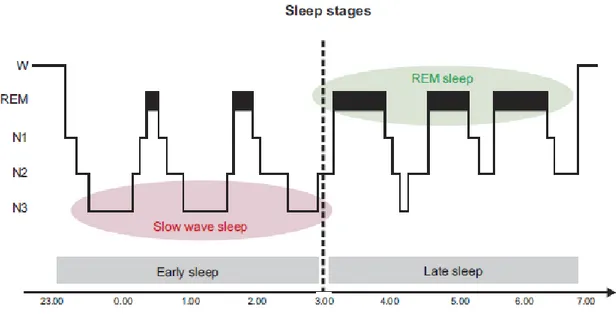
Figure 1 shows the levels of sleep organization from the macro-structural to the microstructural one.
15 Figure 1. Levels of sleep organization (adapted from Salzarulo et al., 1998).
1.3.1 First organizational level: the sleep episode
The sleep-wake cycle represents the highest hierarchical level of sleep organization. Across the 24 hours, sleep and wake alternate in a cyclic manner and, as will be better explained later, in most mammals this alternation is regulated by an endogenous circadian pacemaker and a homeostatic (sleep-wake dependent) process.
The nocturnal sleep episode of a healthy young good sleeper is predominantly made up of NREM sleep, which accounts for the 75-80% of the sleep episode, whereas REM sleep takes up the remaining 20-25%. In particular, the percentage of occurrence of each NREM sleep stage is: about 5% for Stage 1; at least 50% for Stage 2; the remaining sleep (20-25%) is composed of sleep stages 3 and 4, namely slow wave sleep (SWS). However, sleep stage percentages, while providing a global measure of sleep architecture, are less useful to get hints on how really sleep goes on throughout the night. Therefore, the first level of sleep organization does not only include the way sleep and wake interact with each other, in terms of “when” sleep and wake will most likely occur (i.e., its beginning and end over the 24 hours), but also how the sleep episode starts, proceeds and terminates in a more or less continuous and stable way. The analysis of sleep time dynamics is receiving more attention both in clinical practice and in research, since it provides more useful information about the sleep process beyond traditional sleep variables (Norman et al., 2006; Kishi et al., 2017).
16 Measures referring to sleep quality may capture the temporal dynamic of sleep-wake transition. However, despite its clinical importance, an ultimate definition of sleep quality is still lacking. This is somehow surprising since sleep quality has been associated with a wide range of positive outcomes, such as daytime wellbeing, mood and performance (Hyyppa and Kronholm, 1989). One possibility may be to look at the phenomenon from different points of view.
From a subjective perspective, sleep quality seems to rely on the perception of easy falling asleep (Kecklund and Åkerstedt, 1997; Kecklund et al., 2003), total sleep time (Bastien et al., 2003), tiredness on waking and throughout the day, feelings of being rested, restored and refreshed at awakening and the number of intra-sleep awakenings experienced during the night (Harvey et al.,2008).
Following a more “structural” perspective, objective sleep quality seems to be associated with reduced light sleep (Stage 1) and increased sleep depth (SWS) (Harvey et al., 2008). Besides sleep duration and sleep stage percentages, what seems to be determinant for a good sleep is the continuity and stability of the sleep episode throughout the night. Several measures have been used in order to address sleep fragmentation, such as wake after sleep onset and sleep efficiency. However, these metrics provide just a quantitative overall measure of the entire sleep episode, while neglecting the temporal and dynamic distribution of overnight events. For these reasons, besides classical sleep variables, sleep continuity has been extended with the assessment of the frequency and mean duration of awakenings. In this sense, sleep quality might depend on any event reversing the natural build-up of the sleep episode, such as the frequency of arousals and state transitions (Conte and Ficca, 2013). In our view, it is most likely that the quality of intra-night wakefulness and the frequent and continuous passage from one state to another, affects the judgement of sleep quality more than sleep duration and structure. It is worth nothing that in real-life situations, individual, relational and environmental factors may influence the subsequent sleep characteristics; on the other hand, the way sleep proceeds during the night affects subsequent wakefulness. Therefore, the first organizational level, besides the theoretical models proposed overtime (see 1.4 paragraph), stresses the strict relationship between sleep and wake, so that any specific event occurring within the sleep-wake rhythms depends on what precedes it and will influence what is coming next (Conte and Ficca, 2013).
17 1.3.2 Second organizational level: the NREM-REM cycle
The second level of organization concerns the regular alternation of NREM and REM sleep, within the basic functional unit of the sleep cycle. During the nocturnal sleep episode in the healthy young adult, sleep cycles occur, in average, 4-6 times per night, each lasting from 90 to 120 minutes (Carskadon and Dement, 2005). Also, NREM and REM sleep sequences are different across the night, with the cycles in the middle of the sleep episode being longer than the remainders, following a curvilinear trend (Feinberg and Floyd, 1979). As for NREM-REM cycle composition throughout the night, SWS prevails in the first two sleep cycles and is clearly reduced or even disappears after that in favour of Stage 2 (Feinberg et al., 1980; Carskadon and Dement, 2005). In contrast, REM sleep usually becomes longer and more frequent towards morning, mainly alternating with Stage 2 sleep during later cycles.
Since NREM-REM alternation within cycles survives in basically all pathological conditions (Feinberg and Floyd, 1979) and in light of its increases during early development (Ficca and Salzarulo, 2004), several authors suggest that cycles could have a relevant functional meaning.
More recently, as we will see in the next chapter, a crucial role of NREM-REM cycles has been hypothesized for cognitive functions, and specifically for offline memory consolidation processes (Mazzoni et al., 1999; Ficca et al., 2000; Ficca and Salzarulo, 2004).
1.3.3 Third organizational level: NREM and REM sleep states
With the notion of sleep architecture, we generally refer to the progression and continuity of sleep through the sleep episode and, within it, through different sleep states, namely NREM and REM sleep. As recently shown by Markov and co-workers (2012), there are a lot of physiological differences between them, which are accounted for by the balance of the autonomic nervous system drives.
From an organizational perspective, NREM sleep is, on its turn, divided in 4 different stages, characterized by specific frequency and amplitude of EEG waves and EOG and EMG patterns. Stage 1 sleep is the shallowest sleep stage, typically appearing at sleep onset, during the transition between relaxed wakefulness (characterized by a predominant alpha rhythm) and “deeper” Stage 2 sleep. For this reason, this stage usually constitutes 2-5% of total sleep; however, Stage 1proportion increases in case of disrupted sleep and, specifically, anytime there is an arousal during Stage 2 or REM
18 sleep, usually signalled by the appearance of body movements on the EMG channel (Rechtschaffen and Kales, 1968; Iber, 2007). In normal conditions, Stage 1 is scored when waking alpha rhythm is replaced by theta activity, occupying more than 50% of the epoch, along with slow eye movements and decreased muscle tone. Stage 2 sleep is known as the other lighter sleep stage, with increased arousal thresholds and sleep depth compared to Stage 1. This stage is still characterized for the most part of theta frequencies, along with particular electrographic elements such as sleep “spindles” and “K complexes”.
As sleep goes by in the night, the EEG starts to be gradually occupied by high-voltage (at least 75 μV) and low-frequency slow wave activity (SWA, i.e., delta activity, in the .5-4 Hz frequency band). Stages 3 and 4 are known as the deepest stages of sleep, with higher arousal thresholds than the other NREM sleep stages. Scoring of stages 3 and 4 is based on the percentage of delta activity in asleep epoch: 20-50% for Stage 3, more than 50% for Stage 4 (Rechtschaffen and Kales, 1968). For this reason, stages 3 and 4 are collectively referred to as slow wave sleep (SWS) and the American Academy of Sleep Medicine (AASM) guidelines no longer distinguish between the two (Iber, 2007). The EOG shows no eye movements during Stage 2 and SWS, while on the EMG channel muscle tone continues to decline with the deepening of NREM sleep stages.
After staying in SWS for about 20 minutes during the first sleep cycle, the EEG pattern starts to become desynchronized, with low-voltage, mixed-frequency brain wave activity. The transition from NREM to REM sleep is usually rapid, with all the variables characterizing one state suddenly modifying to leave place to the one that follows. However, in some clinical conditions, this transition can be prolonged and expressed by a period of “functional uncertainty” (Salzarulo et al., 1997), in which the characteristics of one well defined state occur only for short intervals, oscillating continuously between different states.
REM sleep is scored when rapid eye movements, either isolated or in bursts, appear on the EOG channels and muscle atonia on the EMG one, along with a desynchronized Stage 1-like EEG pattern and characteristic “sawtooth” wave forms. In addition, REM sleep is often accompanied by other physiological phenomena such as intense neurovegetative modifications (globally named “neurovegetative storm”), including an increased heart rate variability with arrhythmias, changes in respiratory rate and in blood pressure, and remarkable alterations of thermoregulatory mechanisms.
19 The profile of a sleep episode is generally displayed by means of a “hypnogram”. As shown in Figure 2, SWS is predominant in the first part of the night (early sleep), especially in the first two cycles, whereas REM sleep dominates the second half (late sleep).
Figure 2. Hypnogram of a healthy adult sleep episode (from Rasch and Born, 2013).
1.3.4 Fourth organizational level: the intra-state events
The fourth organizational level concerns sleep microstructure, i.e., the intrinsic organization of specific intra-state elements. In the next paragraphs we will describe the main field potential oscillations of brain activity observed during NREM and REM sleep.
1.3.4.1. NREM sleep intra-state phasic events
The main electrical field potential rhythms occurring during deeper NREM sleep are slow oscillations, sharp-wave ripples and sleep spindles, strictly interacting with each other (Figure 3).
20 Figure 3. Main field potential oscillations during NREM sleep (modified from Rasch and Born, 2013).
Neocortical slow oscillations predominate during human SWS and are defined as the part of SWA with an EEG power density <1 Hz and a peak frequency of .8 Hz (Mölle et al., 2002; Rasch and Born, 2013). They originate in the neocortex and reflect the interaction between cortical and thalamic networks (Rasch and Born, 2013). Slow oscillations are characterized by alternating “down-states”, periods of membrane hyperpolarization during which cortical neurons remain silent, and “up-states”, with increased wake-like neuronal firing and membrane depolarization (Steriade et al., 2001). One of the main function attributed to slow oscillations is the synchronization of large neuronal populations in the neocortex, during the transition from down-to-up phases, and at the hippocampal and thalamic level, therefore coordinating the activity of other intra-state events: sharp-wave ripples and sleep spindles (Mölle et al., 2002; Diekelmann and Born, 2010).
When hippocampal sharp waves (fast, depolarizing waves originating in the hippocampus) are overlaid with ripple activity (100-300 Hz local field potential oscillations), they are called Sharp Wave-Ripple complexes (Sw-Rs). Sw-Rs are considered as the most synchronous microstructural events in the mammalian brain, occurring during SWS, but also during non-exploratory wakefulness.
Since these features typically accompany the reactivation of neuronal ensembles active during previous wakefulness, it is hypothesized that Sw-Rs may be a mechanism for the transfer of information from the hippocampus to the neocortex during sleep (Buzsáki, 2015).
Sw-Rs are often coupled to spindles, leading to the formation of “spindle-ripple events”, which may constitute an important mechanism of cortico-hippocampal communication during sleep (Siapas and Wilson, 1998; Rasch and Born, 2013).
21 The sleep spindle, the hallmark of Stage 2, is a waxing and waning EEG oscillation in the 12-16 Hz frequency range (sigma power) lasting .5-3 sec, predominant over centro-parietal EEG derivations (De Gennaro and Ferrara, 2003). According to their frequency and topographical distribution, sleep spindles are classified into: slow spindles, with a frequency range between 12 and 14 Hz and distributed over frontal regions, and fast spindles (14-16 Hz) which have a posterior distribution. It is still a matter of debate whether these two kind of spindles have the same generator or reflect the activity of different neural networks (Mascetti et al., 2011; Rasch and Born, 2013). Spindles originate from the interplay between reticular thalamic and cortical neurons and for this reason they are often called thalamo-cortical spindles (Steriade and McCarley, 2005).
From an organizational standpoint, spindles generally follow a curvilinear U-shaped pattern, with few spindles during early sleep, peaking in the middle and finally dropping off at the end of the sleep episode (Silverstein and Levy, 1976). Also, there is an intra-cycle variation in the density (i.e., frequency) of spindles, which is lower in the middle of the sleep cycle compared to the initial and the final part of the same cycle (Himanen et al., 2002). According to the authors, this phenomenon occurs only in the first four sleep cycles, apparently in association with stage transitions, and not in the last ones, which are instead more stable, probably due to a reduced homeostatic sleep pressure at the end of the night (Himanen et al., 2002).
Sleep spindles may also occur during SWS, although their density is lower than during Stage 2 sleep. An inverse overnight relationship between spindles and SWA has been proposed, in that while SWA is higher during early sleep and progressively decreases across the night, spindle activity increases during late sleep (Åeschbach and Borbély, 1993; Fogel and Smith, 2011).
Besides spindles typical variations during the night and within sleep cycles, intra-individual spindle density remains stable across different nights, so much so that they have been considered as an “electrophysiological fingerprint” (De Gennaro et al., 2005). On the other hand, the variation in spindle density among individuals and over the lifespan (Nicolas et al., 2001) supports the notion that spindles may be a physiological index of intellectual ability (Fogel and Smith, 2011). For instance, in two studies Nader and Smith (2001, 2003) showed that the number of sleep spindles positively correlated with general intellectual potential as measured through IQ scores.
22 Sleep spindles have usually been attributed two main functions, which may not be mutually exclusive. EEG and neuroimaging studies demonstrated that spindles provide an inhibitory thalamic response to internal and external stimuli, suggesting a role for spindles in protecting overnight sleep stability and maintenance (Cote et al., 2000; Dang-Vu et al., 2010, 2011). Recently, it has been suggested that spindle activity may be a neurophysiological vulnerability factor predisposing to stress-related sleep disturbance in the face of precipitating events (Dang-Vu et al., 2015).
The protective role of spindles on sleep maintenance has been recently related to another important function attributed to sleep spindles, that is their role in brain plasticity and sleep-dependent memory consolidation (Gais et al., 2002; Morin et al., 2008; Fogel and Smith, 2011). In this view, spindles may contribute to consolidation processes by protecting sleep and allowing the undisturbed development of biological mechanisms required for learning (Dang-Vu et al., 2011; Conte and Ficca, 2013). According to the last edition of the AASM (Iber et al., 2007), Stage 2 sleep is scored when at least one sleep spindle and/or K complex occur in the first half of the epoch or in the second part of the previous one. Therefore, the K complex is the other phasic hallmark feature of Stage 2. A K-complex is characterized by a well-defined negative sharp wave followed by a high amplitude positive component, occurring spontaneously or elicited by auditory stimuli (Colrain, 2005; Cash et al., 2009). The specific function of this feature is not well understood. Since it is often followed by an arousal-related EEG event or a body movement, some authors suggest that it may represent a cortical arousal response to internal or external stimuli that are not intense enough to provoke a full awakening (Kokkinos et al., 2013). However, other studies propose a role for K complexes in protecting and promoting sleep maintenance (Nicholas et al., 2002; Cash et al., 2009). Recently, in a study by Kokkinos and co-workers (2013), the authors reported that the oscillations appearing during the K-complexes may reflect arousing processes, whereas the K-K-complexes down-state, which represent periods of neuronal silence, may have a role in sleep protection. Therefore, the two hallmarks of Stage 2 sleep, namely K complex and sleep spindles, may both exert a protective role on sleep maintenance and in brain information processing.
23 Finally, another important microstructural feature of NREM sleep is the “cyclic alternating pattern” (CAP). The CAP is a periodic EEG activity of NREM sleep characterized by repeated spontaneous sequences of transient events (phase A) with an abrupt frequency/amplitude variation from the ongoing sleep stage, recurring at intervals up to 60 sec long, followed by a return to background activity (Phase B) (Terzano et al., 2001). Phase A has been divided into 3 subtypes: A1 subtype, characterized by synchronized slow-waves, A3 subtype in which prevailed EEG fast rhythms, and A2 subtype defined by a combination of both EEG patterns (A2 subtype). It has been proposed that these A subtypes subserve different sleep functions: while A2 and A3 may have a role in maintaining arousability, the A1 subtype, mostly composed of slow waves, is involved in the build-up and maintaining of deep NREM sleep stability (Ferri et al., 2008).
1.3.4.2. REM sleep intra-state phasic events
According to Ktonas et al. (1990), measures of rapid eye movements (REMs), the hallmark of REM sleep detectable on the EOG channel, may be distinguished into “first order parameters” (e.g., number of REMs, REM density), and “second order parameters”, which refer to their clustering and to the characteristics of REM bursts or “bouffeès” (e.g., number of REMs occurring in bursts, duration of REM bursts, probability burst-to-burst, which indexes the tendency of REMs to cluster in bursts). The presence of REMs occurring in bursts appears to be an important organizational aspect, which in turn depends on the central nervous system (CNS) development: REMs are higher during maturation (Ktonas et al., 1990) and impaired with aging (Ficca et al., 1999; Vegni et al., 2001).
In animals, REMs occurrence is closely associated to another phasic bioelectrical potential characterizing REM sleep, called P-wave in rats and PGO waves in cats and nonhuman primates, since in the latter these waves originate in the pons (P) and propagate to the lateral geniculate nucleus (G) and the occipital cortex (O) (Datta, 2006; Bourdiec et al., 2010). PGO/P-waves are monophasic negative waves of 100-150 mV amplitude and of short duration (around 75-100-150 msec), occurring isolated or in bursts during the transition from SWS to Paradoxical Sleep (PS, corresponding to REM sleep in many animal species) and during PS (Datta, 2006). Several studies suggest that PGO waves may also occur during human sleep (Peigneux et al., 2001; Lim et al., 2007).
24 In addition to the total amount of REM sleep, some of its phasic events, such as rapid eye movements (REMs) and PGO waves, have been originally related to the dreaming state. However, the early hypothesis that REMs reflected the dreamer’s exploratory activity during the oneiric scene, known as “the scanning hypothesis”, can be hardly reconciled with inconsistent results (Arnulf, 2011).
Some authors suggest that REMs might represent an index of sleep satiation. In fact, REM density seems to be related to prior sleep time (Aserinsky, 1973; Lucidi, 1996) and it is significantly reduced in recovery nights after sleep deprivation (Reynolds et al., 1993).
Furthermore, according to Barbato et al. (1994), there might be a close relationship between REM density and arousal level. In an extended sleep paradigm, the authors found that REM periods terminating with awakenings showed higher REM density than those not interrupted by wakefulness, probably as a result of a reduced sleep pressure (Barbato et al., 1994).
Recently, intra-state phasic events of REM sleep have received strong attention in relation to a possible role for memory consolidation processes (Conte and Ficca, 2013), since human and animal studies showed significant changes in number of REMs and density after the administration of cognitive and learning tasks during wake (Smith and Lapp, 1986; Smith et al., 2004a). Similarly, PGO waves have been proposed as a mechanism supporting synaptic plasticity and memory processing during post-training REM sleep (Datta, 1999, 2006).
1.4. Models of sleep regulation
In order to explain why and how sleep and wake cyclically alternate, which represent the highest hierarchical level of behavioural states organization, many theories have been proposed overtime. The one of the “multiple oscillators” (Aschoff and Wever, 1976; Kronauer et al., 1983) was the most widely accepted model of sleep regulation up to the Eighties. This model was based on the evidence that during a free-running period, i.e., in the absence of environmental time cues, the sleep-wake rhythm and that of core body temperature dissociate (Wever, 1979a). Therefore, it was believed that circadian biological rhythms were controlled by at least two endogenous pacemakers
25 (Aschoff and Wever, 1976): a stronger one would determine core body temperature, urine volume, urinary excretion of potassium and REM sleep; and a weaker one responsible of the control of sleep-wake alternation, SWS, GH secretion and urinary calcium excretion. According to this model, the latter, in the absence of time cues or zeitgebers, would tend to synchronize with the former.
However, evidence of the existence of more than one circadian oscillator in mammals are still lacking. Furthermore, these models have the great limit of not taking into account the homeostatic aspect of sleep regulation, which in turn depends on the characteristics of previous sleep and wake (Borbély and Achermann, 1992). Just a few years later, these limits have been overcome when Borbély (1982) proposed his sleep regulation model, which is still considered as the major conceptual framework in sleep research. Before dealing with the “two-process model of sleep regulation” (Borbély, 1982), a detailed separate description of the two major components of the model, namely circadian and homeostatic factors, is provided below.
1.4.1 Circadian factors
A circadian rhythm is a biological rhythm whose periodicity is approximately 24 hours, oscillating between a maximum peak (“zenith”) and a minimum level (“nadir”). The best-known circadian rhythm is the sleep-wake cycle, whose biological pacemaker is located in the suprachiasmatic nucleus (SCN) of the hypothalamus (Moore and Eichler, 1972). Indeed, lesions of this structure abolish and disrupt rhythmicity of the sleep-wake cycle and increase total sleep time, suggesting that the SCN has an active role in facilitating wake initiation and maintenance and in opposing sleep drive in primates (Edgar et al., 1993). However, in rodents and humans, the circadian clock seems to actively promote both wake and sleep at different phases of the circadian cycle (Dijk et al., 1999; Mistlberger, 2005).
The wake-promoting system begins to decline at bedtime, in order to enhance sleep initiation (sleep gates), until it reaches a minimum around 6:00 am, which coincides with the temperature nadir, followed by a subsequent increase in correspondence of the rising slope of the temperature curve (Dijk and Czeisler, 1994). In fact, the biological pacemaker regulates the circadian rhythms of a great number of variables, including in particular body temperature (Eastmann et al., 1984; Moore, 1999), which is thus considered as the marker of the circadian rhythm.
26 The study of sleep in the absence of environmental time cues (“free-running” protocol) is considered as one of the best methods to investigate the sleep-wake timing. As a matter of fact, the endogenous “clock” has to be synchronized to environmental cues (zeitgebers), the most powerful of which is the light-dark cycle (Lu and Zee, 2010). The process of entrainment occurs when light inhibits melatonin secretion through a pathway leading from the retina to the pineal gland and the hypothalamus (Czeisler, 1994). Under free-running conditions, the sleep-wake cycle not only is desynchronized from the external light-dark cycle, revealing that the endogenous circadian rhythm spans for longer than 24 hours (Wever, 1979b; Czeisler et al., 1999), but also from the internal rhythm of body temperature (Zulley et al., 1981; Daan et al., 1984). This phenomenon suggests that sleep duration and sleep structure are determined by the interaction of a circadian oscillator and a sleep-wake oscillator (Zulley et al., 1981; Daan et al., 1984).
The role of the circadian pacemaker in the regulation of the ultradian NREM-REM sleep cycle was investigated using a forced desynchrony protocol, in which subjects were scheduled to activity-rest cycles outside the circadian range, e.g., 20 or 28 hours (Wyatt et al., 1999; Dijk and Czeisler, 1995). As a result, the rhythm of SWS and SWA was almost totally independent from the circadian phase, with a minimum peak in the early morning hours when, from a circadian perspective, sleepiness is at its highest (Dijk and Czeisler, 1995). In contrast, sleep spindles and REM sleep showed a strong circadian modulation, with their maximum activity coinciding with the melatonin rhythm in the former and occurring 1-2 hours after the temperature nadir in the latter (Dijk and Czeisler, 1995; Dijk et al., 1997).
Therefore, forced desynchrony studies have suggested that the biological clock differently influences the rhythmicity of sleep states and that of intra-state elements. Accordingly, SWA seems to be primarily regulated by time spent awake, and it is considered the electrophysiological correlate of sleep homeostasis.
1.4.2 Homeostatic factors
The concept of “homeostasis” relies on the physiological tendency of a system to continuously regulate and control its condition, in order to maintain an internal stability despite changes. Many experimental approaches, such as sleep deprivation, which increase sleep pressure, and sleep extension studies, which lead to a reduction
27 of the sleep drive, have been used to investigate whether sleep is homeostatically regulated.
Early studies (Berger and Oswald, 1962; Webb, 1969; Webb and Agnew, 1971) consistently showed that sleep deprivation increases sleep intensity in the recovery night, suggesting that SWS is a physiological indicator of sleep homeostasis, depending on previous wake length: the longer the wake period, the higher is the amount of deep sleep, especially in the beginning of the sleep episode (Webb and Agnew, 1971). Therefore, the homeostatic drive to sleep builds up during sustained wakefulness and is dissipated during sustained sleep through SWA. Similarly, a daytime nap, which reduces wake length and counteracts the rising trend of sleep propensity, attenuates SWA in the subsequent night-time sleep episode (Werth et al., 1996).
The existence of a prominent homeostatic sleep mechanism has been interpreted as proof of a restorative function of sleep. It seems that wakefulness causes an accumulation of a factor whose dissipation is necessary for the subject’s wellbeing. Feinberg’s homeostatic model of delta sleep (1974) suggested that NREM sleep counteracts the effect of waking on the brain and that SWA amplitude is a function of this reversal process. Also, REM sleep is considered as a co-factor allowing NREM sleep to continue until optimal levels of homeostasis have been attained (Feinberg, 1974; Feinberg and March, 1988, 1995). However, it seems that the homeostatic control is mainly exerted on SWS rather than REM sleep, which serves just to optimize the reversal process of delta sleep. This view appears rather simplistic, since there are studies showing that REM sleep, despite its depending mainly on the circadian clock, is under partial homeostatic control as well (Benington and Heller, 1994; Barbato and Wehr, 1998). However, in contrast to the homeostatic SWA drive, rebounds of REM sleep may only occur after longer periods of sleep deprivation (Deboer, 2015). Also, it is still to be clarified whether it is homeostatically regulated as a function of prior waking or of prior NREM sleep (Benington and Heller, 1994; Franken, 2002).
1.4.3 The two process model of sleep regulation
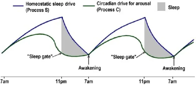
In 1982, Alexander Borbély included both circadian and homeostatic factors inside a mathematical model, which has represented the leading model of sleep regulation ever since. The two-process model of sleep regulation describes the sleep-wake rhythm as a result of the interaction between the sleep-independent circadian process (“process
28 C”) with the sleep-dependent homeostatic process (“process S”), whose combined action determines sleep timing, propensity and depth. Process C is controlled by the internal pacemaker and behaviourally reflects circadian modulation of fatigue and alertness, serving to counteract homeostatic sleep pressure at specific times of the day. Core body temperature and melatonin rhythms are markers of process C.
Process S accounts for an increase in sleep drive as a function of prior waking time and for a recovery process occurring during sleep and specifically during the first part of the night. While the former appears to be expressed by the increase in theta activity during the day, the latter is reflected by the increase in SWA during the first hours of sleep, followed by an exponential decline (Borbély and Achermann, 1999).
Figure 4 displays the interplay between process C and S throughout a 24 hours’ sleep-wake cycle. The build-up of the homeostatic sleep drive throughout the day is countered and moderated by the circadian drive for arousal. In the late evening, the circadian drive falls off, melatonin production increases and the homeostatic sleep drive becomes dominant, promoting sleep gates opening. During the night, process S rapidly dissipates until early morning, when melatonin production stops and process C is rising again. When the two curves meet, final awakening will occur (Borbély, 1982).
Therefore, while the homeostatic process maintains the duration and intensity of sleep within certain boundaries, the circadian rhythm determines the temporal organization of sleep and wake.
Figure 4. The “two-process model” of sleep regulation. When daytime S approaches the upper boundary and encounter the falling curve of C, it triggers sleep; when curves of S and C meet after
29 homeostatic process dissipation and during the rising phase of process C alerting system, awakening may likely occur (redrawn from Borbély, 1982).
1.4.4 Reconsidering the sleep regulation model
The two-process model (Borbély, 1982) represented a major breakthrough in sleep research during the Eighties and it is still the prevalent conceptual model. In the last thirty years, it has been applied in studies on fatigue and performance and on age-related and intra-individual differences in sleep regulation. The model successfully predicts sleep timing and intensity in several experimental paradigms, such as sleep deprivation and fragmentation, forced desynchrony protocols and in “natural” manipulations of sleep-wake rhythms, i.e., in shift-workers, long/short sleepers, early/late chronotypes (Daan et al., 1984; Borbély and Achermann, 1999; Borbély et al., 2016). Also, recently a growing line of research is focused on the possible impact of the model in the clinical field, and specifically, in promoting new treatment strategies for mood disorders (Borbély et al., 2016). In fact, the model contributes to the development of new non-pharmacological treatments in psychiatry, based on circadian and sleep manipulations and light exposure (Wu et al., 2009; Benedetti et al., 2014; Echizenya et al., 2014; Borbély et al., 2016).
Nevertheless, the two-process model has been continuously revised and re-updated. For instance, just a few years after Borbély’s proposal, Achermann and colleagues incorporated in the model the ultradian NREM-REM sleep cycle, representing the alternation of the two basic sleep states within the sleep episode (Achermann and Borbély, 1990; Achermann et al., 1990).
Importantly, at the beginning it was believed that the homeostatic process was independent of the circadian clock and that they interacted together only at sleep onset and final awakening (Borbély, 1982). Later evidence suggests instead a mutual and continuous interaction between the two processes. For instance, forced-desynchrony protocols showed that the circadian rhythm of several neurobehavioral functions was modulated by the homeostatic sleep drive (Dijk et al., 1992; Dijk and Czeisler, 1995). On the other hand, it was shown that the circadian clock influences the build-up and decay of process S, in that the amount of SWA may depend on the time of day when waking occurs (Franken et al., 1991; Deboer, 2009). Finally, evidence in favour of a strict interaction between homeostatic and circadian processes arose from molecular and genetic studies (Curie et al., 2015).
30 In the attempt to explain the physiological phenomenon occurring immediately after awakening characterized by reduced alertness and impaired cognitive performance, known as sleep inertia (Tassi and Muzet, 2000), Folkard and Åkerstedt proposed the “three-process model”: in addition to the previously described basic processes (C and S), the authors include a “process W” (“Process Wake”), which accounts for the quantity and quality of wake after sleep (Folkard and Åkerstedt; 1987; Åkerstedt and Folkard, 1997). In fact, sleep inertia is not predicted by the original model since it occurs when the homeostatic sleep drive is already dissipated and the circadian alerting system becomes stronger. The main outcome of this model was the “nomogram”: a tool developed by the same authors in order to predict vigilance level, through the sum of C and S functions (Åkerstedt and Folkard, 1995, 1997).
Another aspect not included in the classical sleep regulation model is the evidence that sleep homeostasis is not simply a global brain phenomenon, running in parallel in the entire brain, but has a local cortical component (Krueger and Obal, 1993). Several studies have suggested that SWA rebound during NREM sleep may be induced not simply by a longer wake but also by a use-dependent local cortical mechanism. Kattler and colleagues (1994) showed that the stimulation of a specific cortical area during wake results in an increase in SWA in subsequent sleep over the cortical area which was stimulated while awake. More recently, Huber and co-workers’ findings (2004) confirmed the idea that local SWA changes during NREM sleep may be triggered by an intensive cognitive training performed during previous waking involved the activation of the same cortical area. Combining the sleep homeostasis hypothesis with function of sleep theories, Tononi and Cirelli (2003, 2006, 2014) have proposed “the synaptic homeostasis” model, which suggests that synaptic and cellular processes enhanced during wake are re-established during sleep, again through SWA, favouring synaptic plasticity. However, from our point of view, it is more plausible that the effects of wake intensity may not only result in subsequent SWS rebound but may trigger several macro-structural and microstructural sleep changes, influencing the entire sleep episode.
Finally, it is worth noting that, as discussed by Webb (1988), there are many psychological and contextual factors able to modulate sleep regulation, exerting their influence beyond the main physiological influences (homeostatic drive, circadian placement) usually studied in the laboratory setting. Examples are: family life
31 organization, work hours, emotional states, living conditions. To assess these factors, experiments in real life contexts are required.
32
Chapter 2 - How sleep is modified by previous cognitive activity:
insights from sleep and memory literature
The main idea underlying our research project is that wake intensity (i.e., the quantity and quality of waking cognitive activity) deeply affects sleep structure. In this sense, we believe that the classical model of sleep regulation, enabling us to predict sleep timing based on circadian and homeostatic factors, should be complemented by considering the effects of wake content on sleep features. In fact, wake intervals of the same duration can profoundly differ in their content, i.e., the quality and the quantity of the stimuli (both internal and environmental) to elaborate and respond to, and consequently in their intensity. The bulk of research addressing the relationship between sleep and memory has produced massive data on the way sleep might be modified by wake intensity changes.
This chapter deals with the theoretical background of our research project. First, we will introduce the importance of wake intensity and its effects on sleep features; then, we will devote a paragraph to the role of sleep in memory and learning, explaining the main experimental paradigms conceived in sleep and memory research to investigate the “sleep effect” phenomenon; finally, in the third part (which is the result of a systematic review we are working on), we will review the bulk of data on the effects of cognitive activity on sleep characteristics, with the aim of clarifying to what extent different sleep features are affected by wake intensity. Predicting which sleep variables are actually modified by cognitive activity, and in which direction, would not only expand our comprehension of sleep regulation mechanisms but also provide insight on how to manipulate these processes in order to improve sleep quality with a meaningful applicative fall-out for sleep medicine.
2.1. The role of wake intensity in sleep regulation
Sleep and wakefulness are behavioral states characterized by a tight interdependence: as the quantity and quality of previous sleep affects subsequent wakefulness, likewise wake characteristics influence sleep of the following night.
33 Taking into account a restorative role for sleep, the influence of its characteristics on subsequent wake has been largely studied in the last century, so that it is now generally accepted that a good night sleep is a crucial requirement for the effectiveness of a wide variety of daytime cognitive processes (Diekelmann, 2014).
The inverse research question, i.e., the effect of wake characteristics on sleep, has been mainly addressed in the frame of the classical “two-process” model of sleep regulation (Borbély, 1982). As shown in the previous chapter, this model, based on previous wake duration and circadian factors, allows us to predict “when” sleep will most likely occur (i.e., its beginning and end over the 24 hours) and, though only partially, “how” it will be, i.e., some of its structural features - essentially the amount of Slow Wave Sleep (SWS).
Actually, already a few years before Borbély’s model (1982), it had been suggested that sleep is also modulated by the intensity of waking brain activity, measured through the cerebral metabolic rate, which would in turn depend on the quantity and quality of physical and cognitive activity carried out during wake (Feinberg, 1974). According to Feinberg (1974), the cerebral metabolic rate represented the physical substrate of the homeostatic factor accumulating during wake; subsequent sleep, especially delta sleep, has the function to reverse the consequences of this “intense” brain activity (Feinberg, 2007). This idea received support from a conspicuous body of work showing, in rats, massive increases of NREM delta sleep following experimentally induced increments of the waking brain metabolic rate (Feinberg and Campbell, 1993; Campbell and Feinberg, 1996a, 1996b). Another animal study (Meerlo et al., 1997) showed that the exposition to a social stressor accelerated the build-up of Process S, resulting in an increase in subsequent SWA. The same authors stated that “sleep intensity may, thus, not only depend on the duration of prior wakefulness but also on the nature of the waking experience” (Meerlo et al., 1997).
More recently, the notion of “wake quality” was reintroduced by Franken (2007), commenting on Huber et al.’s findings (2007) of an increase of delta sleep in rats that had been subjected to an acute dark condition, thus augmenting exploratory behavior at the expense of quiet waking (Huber et al., 2007). All these works have the merit to underline that sleep-wake reciprocal influences cannot be fully enlightened without taking into account wake “content” alongside its duration. In other words, as sleep quality and quantity significantly influence the quality and quantity of wake, the same is to be held true the other way round.
Although this idea has been quite convincingly expressed in the past, an ultimate definition of “wake intensity” is still lacking. In general terms, an “intensive” day is usually
34 characterized by a greater amount of physical and cognitive activity. However, it has been suggested that the latter is intrinsically involved in the first. For instance, as suggested by Horne (2013) exercise in everyday life is actually inseparable from cognition, in that “physical activity intrinsically implies cognitive challenges and demands triggered by multisensory encounters, curiosity and interactions with novel environments”.
The relationship between cognitive processes and sleep has been largely studied in the frame of sleep-dependent consolidation models (Conte and Ficca, 2013). In fact, a vast source of evidence comes from the bulk of research linking sleep to consolidation processes, showing significant sleep changes after bedtime training sessions both in animal and human studies (Peigneux et al., 2001; Conte and Ficca, 2013).
Which kind of sleep changes are triggered by learning and cognition? Before trying to answer this question, I will briefly describe concepts and evidence in support of the idea that sleep actively benefits cognitive and memory processes, starting from the first historical “steps” taken and going through new recent findings produced so far in the field.
2.2. The relationship between sleep and memory: overview of
concepts and findings
The relationship between sleep and memory has been the object of scientific interest since the discovery of the “sleep effect” on memory (i.e., a better memory recall when the retention period is followed by a period spent in sleep). In the first half of the 20th
century, two psychologists of memory, Jenkins and Dallenbach, in the attempt to demonstrate that forgetting was due to the interference that newly learned information exerts on old memory traces, carried out an experiment that is still considered a milestone in sleep-memory literature. By comparing retention periods filled either with sleep or wakefulness, they showed that recall of non-sense syllables was higher after a retention period spent asleep, regardless of its duration (either 1, 2, 4 and 8 hours) (Jenkins and Dallenbach, 1924).
Over the following decades, this phenomenon was consistently replicated by numerous authors, using different experimental paradigms and learning materials, confirming the positive effect of sleep on memory (Lovatt and Warr, 1968; Benson and Feinberg, 1975; Ekstrand et al., 1977; Grosvenor and Lack, 1984).