Master's Degree Programme in
Sustainable Chemistry and Technologies
Final Thesis
Characterization of ceramic
biomaterials for dental implant
applications
Supervisor
Ch. Prof. Giuseppe Pezzotti
Ch. Prof. Alvise Benedetti
Graduand
Michele Santini
Matriculation Number 850235
Academic Year
2018 / 2019
2
Abstract
Nowadays, zirconia is an attractive material in dental implant applications due to its high mechanical properties and natural white colour which make it the ideal candidate material for the production of strong and aesthetically attractive implants. However, in addition to its aesthetic and mechanical properties, zirconia is considered to be an inert biomaterial, therefore it has limited interactions with biological environments and has limited osseointegration. This characteristic has allowed the development of dental implants with modified surfaces, which speed up the osseointegration process but, at the same time, increase the possibility of peri-implantitis or the adhesion of bacteria on the surface and zirconia does not have antibacterial properties. Therefore, an alternative was proposed to reduce the above problem through the use of an active bioceramic material: silicon nitride. Specifically, a coating of silicon nitride was applied to the surface of zirconia, thus maintaining the mechanical properties of zirconia, but stimulating osteointegration and making the material antibacterial thanks to the properties of silicon nitride. Silicon nitride is a ceramic biomaterial that has bioactive effects, such as antibacterial properties and promotes cell proliferation, which cannot be obtained with oxide materials. In this work, concepts such as advanced ceramic materials, biomaterial and an analysis of the properties of Si3N4 and ZrO2 were introduced first. Then the concept of bone
tissue was introduced to give a detailed insight into the process of cellular osseointegration and defined the action of bacteria on dental prosthesis. In addition, the epidermal staphylococcal bacteria and human osteosarcoma (SaOS-2) cell lines that have been used for this work are described. Four types of samples were analysed: polished zirconia; rough zirconia; zirconia coated with silicon nitride and stoichiometric silicon nitride as positive control. First, the morphology and surface properties were evaluated through a pre-characterization of the samples, using spectroscopic and microscopic techniques such as: Laser microscopy, Raman, FT-IR, XPS, SEM and cross-sectional analysis. The results showed that silicon nitride coating is a biphasic composite in which Si3N4 hard particles are dispersed in a nano crystalline and/or
amorphous silicon matrix. In addition, the coating led to an increase in surface roughness, increasing the possibility of interaction with biological tissues. Subsequently, the surfaces of the samples were treated in vivo with staphylococcus epidermidis bacteria to allow the study of antibacterial effect and with osteoblastic cells of the SaOS-2 line to stimulate cell proliferation.
3
Various biological and bacterial markers were used to allow in vivo characterization. Bacterial analysis was performed using Raman, Fluorescence microscopy and WST techniques, while the analysis of SaOS-2 cell lines was characterized using Fluorescence microscopy, WST, Raman and SEM techniques. Bacterial tests were performed after 12, 24, 48 hours of exposure and showed low antibacterial activity for all samples. This is a predictable characteristic in zirconia, which by nature has no antibacterial activity, while for the silicon nitride coated zirconia sample it is probably due to a low concentration of nitride on the surface of the sample. The tests with SAOS-2 solutions were performed after 10 days of exposure and led to the formation of bone tissue with high collagen maturity, high carbonate/phosphate ratios and good levels of tissue mineralization, but less cell proliferation than stoichiometric Si3N4. Bone tissue
quality parameters measured with Raman spectroscopy were comparable to healthy human bone tissue.
4
Summary
Chapter 1. Introduction ... 7
Chapter 2. Materials ... 11
2.1 Biomaterials ... 11
2.2 Advanced ceramic materials ... 12
2.3 Silicon Nitride ... 13 2.3.1 Property ... 13 2.3.2 Production Methods ... 17 2.3.3 Biocompatibility ... 19 2.3.4 Antibacterial activity ... 20 2.4 Zirconia ... 21 2.4.1 Property ... 22 2.5 Samples ... 25 2.5.1 Silicon Nitride ... 25 2.5.2 Zirconia ... 26
2.6 Bone tissue properties and characteristics ... 26
2.6.1 Hydroxyapatite (HA) ... 28
2.6.2 Bases of osteointegration ... 30
2.6.3 Factors that influence osteointegration ... 31
2.7 Bacterial inflammation on prosthesis ... 32
2.9 Cellular and bacteria test ... 33
2.9.1 Osteosarcoma cells (SAOS-2) ... 33
2.9.2 Staphylococcus epidermidis bacteria ... 34
Chapter 3. Methods ... 36
3.1 Raman and Fourier-Transform Infrared Spectroscopy ... 36
3.2 Raman Spectroscopy ... 40
3.2.2 Raman theory ... 41
5
3.3 Fourier Transform Infrared Spectroscopy ... 46
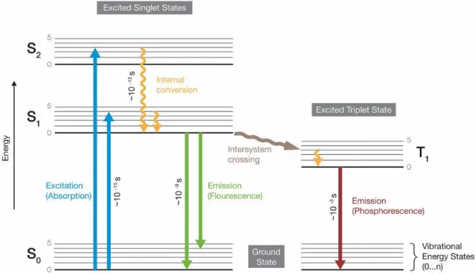
3.4 Fluorescence Microscopy (FM) ... 48
3.4.1 Introduction of Fluorescence ... 48
3.4.2 Instrumentation... 50
3.4.3 Markers and Staining techniques ... 51
3.5 Scanning Electron Microscopy (SEM) ... 53
3.6 XPS (x-ray photoelectron spectroscopy) ... 56
3.7 Laser Microscopy ... 59
3.8 Experimental procedure ... 59
3.8.1 Cellular Treatment: Osteosarcoma SAOS-2 ... 59
3.8.2 Bacteria protocol: Staphylococcus epidermidis ... 59
3.8.3 WST essay ... 60
3.8.4 Raman Spectroscopy ... 60
3.8.5 Fourier Transform Infrared Spectroscopy ... 61
3.8.6 Fluorescence Microscope ... 61
3.8.7 Scanning electron microscopy ... 62
3.8.8 X-Ray Photoelectron Spectroscopy ... 62
3.8.9 Laser Microscopy ... 62
Chapter 4. Result ... 63
4.1 Substrates characterization ... 63
4.1.1 Laser microscopy ... 63
4.1.2 Fourier Transform Infrared Spectroscopy ... 66
4.1.3 Raman Spectroscopy ... 67
4.1.4 X-Ray Photoelectron Spectroscopy ... 69
4.1.5 Scanning electron microscopy ... 71
4.1.6 Cross-sectional analysis ... 72
4.2 Bacteria characterization: Staphylococcus epidermidis ... 73
4.2.1 RamanSpectroscopy ... 73
4.2.2 Fluorescence microscope ... 75
4.2.3 WST ... 77
4.3 Cell characterization: SaOS-2 ... 79
4.3.1 Fluorescence microscopy ... 79
4.3.2 WST ... 81
6
4.3.4 Quality index ... 84
4.3.5 Scanning electron microscopy ... 88
Chapter 5. Discussion ... 91
Chapter 6. Conclusion ... 94
Chapter 7. Bibliography ... 95
7
Chapter 1
Introduction
A dental implant allows the rehabilitation of edentulous areas of the jaws by surgical insertion into the bone of a dental prosthesis. The first rudimentary dental implant ever found dates back to about 600 B.C. and was made from shell fragments embedded in the mandible, an example of the ingenuity of the Maya population. The Etruscans at about 500 B.C. used fragments of animal bone in substitution of missing teeth. The first uses of ceramic materials for making dental prostheses date back to the end of the 18th century. Until then, the most common prostheses were made of hippopotamus ivory or reused natural teeth. One example was George Washington who brought one of the first mobile dental implants that was made of lead and animal teeth. The main problem that were encountered was the rejection of the implant with the organism. For the implant to be successful, the external body and bone must fuse together through a process known as osseointegration. Later in 1952, an orthopaedic surgeon discovered the first boneintergration process with a titanium cylinder that had fused with the bone of a rabbit's femur. Then in 1965, Brånemark successfully inserted the first titanium dental implant into a human: the "modern implantology" [1] [2] was born. The success of the first titanium dental implant quickly led to a significant development of the process. In particular, today, a titanium alloy screw with a machined surface (rough or smooth or coated) is used, which helps to improve the osseointegration process. The favourable properties have made titanium the material of choice for dental implant applications. In fact, modern oral implantology is predominantly based on titanium due to its properties of high biocompatibility, favourable tissue response and adequate corrosion resistance. The main problem that were encountered was also good mechanical properties, such as a relatively high elastic modulus (Young's modulus), which allows good loading stress during mastication. All these features have made titanium implants with the longest traceable record of predictable clinical performance, with a cumulative success rate of 98.8% for 15 years [3] and an increase in implant types. Currently, 98% of the dental implants produced worldwide are titanium implants. Titanium, however, is
8
no longer considered a completely bio-inert material, but can be an allergen [4] [5] [6]. Indeed, high concentrations of titanium have been found in the vicinity of oral implants [7], in regional lymph nodes [8], in serum and in urine [9], which are potentially dangerous for the human body. In addition, the dark grey titanium implant is aesthetically unpleasant, there is also the problem of the taste of the metal [10] And, since some patients are allergic to specific metals, they require to be treated exclusively with dental implants without metal [11].
Today, the main features that allow a good implant, in addition to the osteointegration process, are: a prevention of bacterial adhesion that can cause perimplantitis with the progressive loss of bone and consequently of the prosthesis; resistance to mechanical stress; pleasant sensation in the mouth and good aesthetics. Zirconium oxide or "Zirconia" (ZrO2) is among the most
widely used ceramic biomaterials on the market today for the fabrication of fixed partial dentures and crowns [12]. The natural white colour of zirconium and its high mechanical properties make it the ideal candidate material for the production of strong and aesthetically attractive implants [13]. Technological development has allowed the introduction of new biomaterials, in particular advanced ceramic materials with metal-free properties and mechanical properties similar to those of titanium. Zirconium oxide or "Zirconia" (ZrO2) is
among the strongest ceramic biomaterials on the market today for the fabrication of fixed partial dentures and crowns [14] [15]. Zirconia at room temperature is stable in the monocline form, which has relatively low mechanical properties [16]. At temperatures above 1170 °C, the monoclinic zirconia transforms into the more compact tetragonal phase, which then, if not stabilized, disintegrates by cracking upon cooling. In order to maintain the integrity of tetragonal zirconia compounds, it is stabilized by the addition of oxides, thus avoiding t-m transformation during cooling [16]. Tetragonal zirconia at room temperature has a high fracture toughness which is associated with a stress-induced t-m transformation that hinders crack propagation. Nevertheless, the fracture toughness of zirconia is compromised in aqueous or heavily moist environments [17] [18]. In addition to its aesthetic and mechanical properties, zirconia is considered an inert biomaterial, which means that it has limited interactions with biological environments. In fact, in vitro and in vivo tests on zirconia have shown no mutagenic or carcinogenic compatibility [19] and a low affinity with bacterial plaque [20]. However, zirconium exhibits limited bone integration, in particular with adhesion to biological tissues, in particular bone [21]. Many types of zirconia surface treatments have been performed to improve biological activity and promote its integration into existing biological tissues: bonding with active phases such as hydroxyapatite [22], coatings [23], surface laser modifications [24] and texturing [25]. In this work, an innovative laser coating treatment with Si3N4 powders was
9
applied to the surface of the zirconia. Silicon nitride is a ceramic biomaterial that presents bioactive effects [26] [27] [28] that cannot be obtained with oxide based materials, as antibacterial properties and promotes cell proliferation[26]. However, Si3N4 has too high
mechanical properties (hardness, toughness, resistance to cyclic load, etc.) that do not make it the most attractive for dental implant applications. In fact, Si3N4 has a high elastic modulus
(young's modulus) which requires higher processing costs and in addition the stress load resulting from mastication could damage the bone or break the implant. The silicon nitride coating on zirconia was applied to preserve the mechanical properties of zirconium oxide but, at the same time, to stimulate osteointegration and antibacterial properties due to the properties of silicon nitride. The resistance to bacterial colonization, the ability to stimulate osteoblast differentiation and bone tissue production of Si3N4, has already been successfully applied for
the application of prosthetic implants, as in the case of spinal fusion cages [29] [30] [31]. Several studies have shown [32] [26] [27] [28] [33] [34] [35] [36] [37] that the beneficial effects of silicon nitrides are due to the presence of nitrogen in the crystalline lattice, and in particular the formation and release of NH4+ nitrogen species. These species stimulate cell proliferation
and damage common bacterial strains [38] [39] and silicon ions that actively contribute to the formation of mineralized bone tissue [32] [37] [35] [33]. The present work is intended to be a possible new improvement for dental prosthesis technology and also represents a further study in the understanding the mechanisms of cell proliferation and the antibacterial effect. The stoichiometric Si3N4 is used as a reference that will allow an unequivocal understanding of the
role of each element. The thesis paper is divided into six chapters respectively:
1- Introduction
2- Materials: This chapter gives an introduction to the meaning of Biomaterials and Advanced
ceramic materials. Then silicon nitride is introduced and respectively the production methods, general properties and the meaning of biocompatibility and antibacterial activity are described. Subsequently zirconia is introduced and the properties are described. Afterwards bone tissue is described with particular reference to Hydroxyapatite (HA), bases of osteointegration and factors that influence osteointegration and the concepts of bacterial inflammation on prosthesis are introduced. Finally, cellular and bacteria treatments for osteosarcoma cells (SAOS-2) and staphylococcus epidermidis bacteria are exposed.
3- Methods: This chapter describes in detail the instruments used. Initially an introduction of
general Raman and IR spectroscopy is made, then Raman spectroscopy is introduced through a theory and instrumentation, subsequently FT-IT spectroscopy is exposed. Afterwards fluorescence microscopy is described through an introduction of fluorescence microscopy and
10
its instrumentation, moreover the marker and Stain used to analyse FM images are described in detail. Then the instruments SEM (scanning electron microscope), XPS (x-ray photoelectron spectroscopy) and laser Microscopy are introduced. Finally, all the experimental procedures are listed.
4- Result: This chapter describes the results of the experiments and can be divided into three
sub-chapters. In the first sub-chapter the results of substrate characterization are presented, through the use of technical techniques: laser microscopy, FT-IR, Raman, XPS, SEM and Cross-sectional analysis. The second sub-chapter presents the results of the characterization of Staphylococcus epidermidis bacteria through the use of Raman, fluorescence microscopy and WST techniques. Finally, the third sub-chapter presents the results of characterization of SaOS-2 line cells through the use of fluorescence microscopy, WST, Raman, quality index and SEM techniques.
5- Discussion: This chapter discusses the results obtained. 6- Conclusion
11
Chapter 2
Materials
2.1 Biomaterials
According to the II International Consensus Conference on Biomaterials hold in Chester, Great Britain, in 1991 a biomaterial can be defined “as a material intended to interface with biological systems to evaluate, treat, augment or replace any tissue, organ or function of the body” [40]. First of all, in order to consider the biomaterial, there are two important properties that need to be taken into account, first of all to consider the biomaterial:
- Biocompatibility: it consists in the characteristic of establishing "not unfavourable" interactions with the organisms with which it comes into contact;
- Biofunctionality: it consists in the characteristic that a device must be able to reproduce a certain function, from a physical and mechanical point of view.
The subcategories of biocompatible material are numerous and are classified mainly in two distinct orders, called biostable and biodegradable, on the basis of the consequences on the material after grafting. The former includes materials that, once placed in situ, do not undergo substantial chemical and/or physical changes over time (dental prostheses). On the other hand, the latter includes materials that, once implanted, undergo substantial chemical and/or physical transformations that cause them to disappear over time.
This is not the only possible classification, another one considers instead the material-organism interaction, hence the definitions of biotoxic, bioactive, bioinert, bioreabsorbable.
In particular, the term biotoxic refers to materials that cause a rejection reaction by the biological tissue due to chemical and/or galvanic processes.
Bioinert materials are chemically and physically stable materials and have minimal interactions
with surrounding tissues. These materials allow a good coexistence between organism and implant. In dental implants, the most common materials are: Ti, ZrO2 and Si3N4.
12
Bioactive groups together all those materials that favour direct biochemical interactions with
the biological tissue, which can grow on the surface of the material itself. All this allows the establishment of a solid mechanical bond between the natural tissue and the prosthetic implant. Typical examples of bioactive materials are some ceramic materials, such as hydroxyapatite and Si3N4 [41].
Lastly, bioreabsorbable materials undergo a progressive degradation within the biological system, without causing rejection reactions or toxic effects. The calcium phosphates belong to this class as the tricalcium phosphate, the porous hydroxyapatite and some bioglass. These are generally bioactive and are gradually replaced by biological tissue. Because of these characteristics, they are particularly useful when the replacement prosthesis has to occupy a limited space but they are also widely used for the controlled drug delivery.
Finally, a final distinction is made on the basis of the chemical nature: polymeric biomaterials, metal, ceramic and composite [42] .
2.2 Advanced ceramic materials
The common use of the term ceramic includes all non-metallic inorganic materials, consisting of metallic and non-metallic elements bound together by ionic or covalent bonds or, usually, by a hybrid of these. They are generally obtained by heating processed raw materials to obtain a rigid structure. These include advanced ceramic materials that are obtained from highly selected and pure raw materials and include: oxides, carbides, nitrides, silicide. Some of the most important advanced ceramic materials for structural uses are alumina (Al2O3), silicon nitride
(Si3N4), silicon carbide (SiC) and zirconia (ZrO2), combined with other refractory oxides [43].
Advanced ceramics are materials made in such a way as to offer unique characteristics through the control of composition and microstructure. The physical-mechanical properties of a polycrystalline ceramic therefore depend on the microstructure that is determined by the processing phases (synthesis), and the choice of raw materials. Essential is the way in which they are processed and fired, which are all factors that can influence the properties of the material [16]. Furthermore, the properties of ceramic materials are generally controlled by the atomic order, ordered if the structure is crystalline, disordered if amorphous, and on a larger scale, by the shape and arrangement of the grains and phases, and by the size and volume fraction of pores it contains [44].
13
2.3 Silicon Nitride
Silicon nitride (Si3N4) is a non-oxidic ceramic very rare in nature because it was found only in
meteorite rock particles formed when the atmosphere was chemically reducing and rich in ammonia, the crust contained large quantities of silicon and other nitrides [45]. Synthesized by Deville and Wöhler in 1859 [45] and ignored until the 1950s, when it was used for various refractory applications. In the 1980s, the potential of Si3N4 ceramics was recognised due to its
properties such as mechanical strength, thermal shock resistance, high temperature stability, hardness and wear resistance [46]. As a result, it is now one of the most studied ceramics. Today's major industrial applications include high performance bearings, turbine blades and glow plugs, i.e. applications requiring a material with extreme strength and toughness and high fracture resistance, strength and low wear [47] [48]. Si3N4 is also used in the automotive,
turbomachinery and power industries, where it has a significant advantage due to its low density (half that of bearing steel), low friction, corrosion resistance and reliable performance under extreme conditions [49]. In aircraft and spacecraft high performance, where the operating conditions of very demanding bearings such as high vacuum (<10-6 torr), extreme temperatures (e.g., 230 to 150 °C). Si3N4 finds its space also used in the manufacture of bearings subjected
to extreme performance: large temperature differences, long service life (about 10-15 years without maintenance and subjected to stress and fatigue) and low friction [50].
The material properties of Si3N4 have been developed in medical fields as well, as it has proven
to be biocompatible [51] [52]. It is used in spinal casting implants and is being developed for the coupling of components of hip and knee joint prostheses. In addition, Si3N4 has been tested
in surgical screws and plates as a starting material [29] [30] [31]. The constraints of the widespread use of technology concern the production of most technical ceramics: i.e. the cost of materials and processing, the need for reproducibility, reliability and precision in production.
2.3.1 Property
There are three different crystallographic structures of silicon nitride (Si3N4), α, β and γ [53].
The α and β structures are the most common forms of Si3N4 and can be produced under ordinary
14
temperatures (2100 K) [54]. The forms α and β-Si3N4 have hexagonal structures, which consist
of Si3N4 tetrahedra at a shared angle [55].
Respectively α-Si3N4 presents the lattice parameters of a=0.775 nm; c=0.52 nm and an ABAB..
sequence, while β-Si3N4 presents the lattice parameters of a=0.76 nm; c=0.29nm (about half of
α) and an ABCDABCD.. sequence. The AB planes are the same for α and β, while the CD planes for β are conceptually similar to the AB sequence except for a 180° rotation along the c-crystallographic axis (Fig. 2.0).
The form α is converted into β at a high temperature above 1400°C, while the reverse process is energetically unfavourable (irreversible transformation) [3]. Therefore, the transformation from α to β is permanent.
Silicon nitride has a predominantly covalent Si-N chemical bond (the silicon atom is surrounded by four nitrogen atoms). This type of bond is very strong and extremely directional and determines the characteristics of hardness and resistance to wear and the low coefficient of thermal expansion.
The tribological properties of Si3N4 are divided into mechanical (friction and abrasion) and
chemical properties. Like most metal and other non-oxide materials, Si3N4 is protected by a
thin layer of oxide (SiO2) about 2-5nm thick [56]. When this coating is removed by erosion or
wear, the surface quickly repairs itself through re-oxidation, thus limiting degradation. The surface passivation layer occurs due to its thermodynamic instability in oxidizing or humid environments. In addition, the surface in contact with Si3N4 is subject to oxidative degradation
especially in the presence of moisture. This phenomenon is also present at low temperatures either in liquid water or in humid conditions [57] [58] [59] [60] [61] [62] [63]. it is hypothesized that it can also be operative for in vivo joints [64] [65] [66]. Chemical wear occurs through the following Si3N4 surface reactions [67]:
15
Si3N4 (s) + 6O2 (g) → 3SiO2 (s) + 2N2 (g) ΔG (298 K) = - 1922 [KJ/mol] (2.0)
Si3N4 (s) + 6H2O (l) → 3SiO2 (s) + 4NH3 (g) ΔG (298 K) = - 565 [KJ/mol] (2.1)
However, Si3N4 has a more complex surface chemistry, since Si-N, Si-N-O and Si-O bonds
[56] [68] [69] [70] [71] [72]are present in the near-surface region. By moving towards the surface, the passivated layer is also present and oxygen enters the lattice. Both Si-N-O and Si-O bonds are usually present in the outermost surface layers. In addition, when the Si-O layer increases due to oxidation, the layer becomes chemically equivalent to silica (SiO2). This
process can happen when Si3N4 is exposed to moisture or air the Si-N bonds react to form
neutral (Si-NH2, Si-OH) or ionic (Si-NH3+, Si-OH2+, Si-O-) functional groups [69] [73] [74]
[75] [76] The chemical reactions describing these surface motions are [69] [74]:
Si3N4(s) + 2H2O (l) → Si-NH2(s) + Si-OH(s) (2.2)
Si-NH2(s) + H2O(l) → Si-OH(s) + NH3 (g) (2.3)
The strong smell of ammonia gas detected while working in a wet environment with Si3N4 is a
confirmation of these reactions. The dissociation of function groups (Si-NH2 and Si-OH) in
water occurs through acid-base reactions [69] [77] [78]:
Si-OH2+(s) ↔ Si-OH(s) + H+(aq) (2.4)
Si-NH3+(aq)↔ Si-NH2(s) + H+(aq) (2.5)
Si-OH(s) ↔ Si-O-(s) + H+(aq) (2.6)
The main reactions at homeostatic pH are (2.5) and (2.6), while protonation of one of a silanol group (2.4) is unlikely to occur. These reactions are dependent on the pH variable and the balance can be shifted to both right and left. The pH at the point where the net charge of the considered species is equal to zero is called the isoelectric point (IEP) [79]. The IEP of pure Si3N4 is pH ∼ 9.3 - 9.7 due to the presence of high surface concentration of amine (Si-NH2)
[70] [80], while pure SiO2 has an isoelectric point of 2-3. Ammine has a reduction potential that
requires high pH and Si3N4 rarely has a negative charge. On the other hand, SiO2 has a highly
favourable reduction potential (6) [81]. IEP, it has been demonstrated, as the surface thickening SiO2 oxide layer, defined as "passivation layer". In Si3N4 the pH change leads to a different
composition of the passivation layer, in particular: ∼ 9 pH only pure Si3N4 to ∼2 pH only pure
SiO2 [70] [76]. There is therefore a correlation, through IEP measurement it is possible to
16
In tribo-chemical wear, the passivated film acts as a solid lubricant for the joint components. The Si-OH and Si-O oxide film is quite effective in protecting the joint surfaces, which results in low material wear.In particular, the joint surfaces are very smooth and are generated by the colloidal film which causes low friction between the components [82].
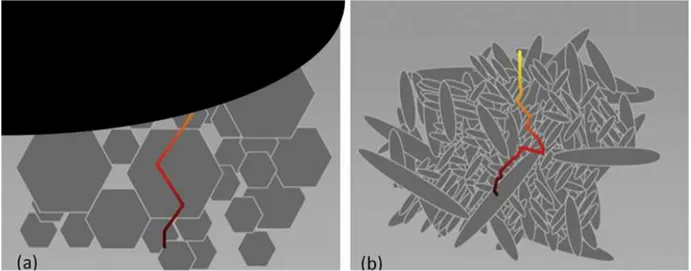
Si3N4 may be produced from interlaced fibrous grains with a completely dense microstructure,
called "in situ toughening", which causes a change in the reticular microstructure. The modification of the in situ hardening microstructure occurs through the formation of an elongated or whiskers-like structure of the grains, is of great interest. The precise composition of the additives used and the heat treatment during the manufacture of the Si3N4 material also
promotes the growth of elongated grains (Fig. 2.1). This generates a self-reinforcing effect due to greater cohesion in the structure. These structural changes promote the formation of elongated grains in Si3N4 situ toughening, which provide hardening mechanisms that are limited
or absent [83]. Therefore, elongated grains cause greater fracture toughness due to mechanisms such as crack deflection, micro-fracture, and grain extraction and binding actions [84]. Fig. 2.1 shows how the propagation of a crack through the in-situ toughened material becomes more tortuous. In addition, the propagation of a crack in Si3N4 in situ toughening (rod elongated
grains) deflects the crack (crack deflection) along the edges of the elongated grains and has been shown to provide energy dissipation mechanisms that reduce the tendency of the crack to grow rapidly. Mechanical strength and toughness are significantly increased.
This material has an optimal combination of flexural strength, wear resistance and fracture toughness for surgical applications such as: total joint arthroplasty (joint reconstruction) [85]
Figure 2.1: Illustrations show the differences in crack propagation modes for (a) conventional ceramics and (b) in situ toughened silicon nitride ceramics.
17
[86] [87] [88], or in arthrodesis devices and bone scaffolds using highly porous constructs [89] [90] [91] [92].
2.3.2
Production Methods
Si3N4 can be synthesized in several methods using Si3N4 powders as the starting reagent. In
order to be suitable, it must be submicrometric with variable granulometry. So that it can guarantee an excellent packaging. A final product can be made by "coarse" particles with a size of 10-15 µm; "medium" particles of 1-2 µm; "fine" particles of 0,1-0,5 µm [93] [94]. In order to obtain Si3N4 powders the main synthesis techniques are:
Direct Si nitridation process: Silicon dust (Si) is heated in N2 (nitrogen atmosphere) to a
temperature in the range 1200-1400 °C, where the Si reacts with N2 to form Si3N4 and binds
the particles together [95]:
3 Si (g) + 2 N2 (g) → Si3N4 (2.7)
During the reaction, the weight of the silicon sample increases progressively due to the chemical combination of silicon and nitrogen. The reaction is facilitated by the use of transition metals as catalysts [96] [97] [98] and coarse particles and agglomerates are generally produced which require grinding to obtain a suitable powder. The resulting powders are α-Si3N4, which are
converted to β-Si3N4 only during densification [94] [96] [97] [98].
Carbothermal reduction/nitridation of silica: Silica and carbon are reacted in the presence of
nitrogen; first silica reduction and then nitriding. The reaction is conducted at a temperature in the range 1250-1300 °C. was the first method used for the production of Si3N4 [99].
SiO2 (g) + 2 N2 (g) +6 C → Si3N4 + 6 CO (g) (2.8)
This method is now considered the most economical industrial method for the production of high purity α-Si3N4 powders.
Vapor-phase at high temperatures: SiCl4 (silicon tetrachloride) and ammonia are made to react
and reaction can be conducted in gaseous phase or in solution.
3SiCl4(g) + 4NH3(l) → Si3N4 + 12HCl(g) (2.9)
The calcination temperature is fundamental; at about 1420 °C α-Si3N4 equiaxial grain is
produced, while, calcination at temperatures above 1460 °C produces β Si3N4 whiskers.
The general process of powder densification is the sintering process, which consists in submitting the form to the green state to a temperature and pressure program.
18
The process is described by Frenkel's ball model according to which two adjacent particles are joined through diffusion mechanisms. The reaction mechanism is related to the Gibbs free energy reduction ∆GT = ∆Gv + ∆Gb + ∆Gs; in which ∆Gs = γ ∆A is the term referred to the surface reduction and is the predominant term. The terms ∆Gv and ∆Gb refer to volume variation and grain edge variation respectively. Additives (metal oxides) are added to the Si3N4
powder. The process involves an interaction between the additives with the passivated layer (SiO2) on the surface of Si3N4. The additives react by forming a liquid phase that surrounds the
Si3N4 particles, thus promoting the densification process. Upon cooling, the liquid phase
solidifies to form an amorphous (glassy) or partially crystallized glassy phase at the grain boundaries of Si3N4.
At the sintering temperature there is the dissolution of phase α and the precipitation of phase β. At the end of the process there is a β structure with elongated grains in a glassy matrix that will form the grain edges. The most common metal oxide additives are magnesia (MgO), alumina (Al2O3) and yttrium (Y2O3) [7] [8] [47], because they have thermal properties very similar to
Si3N4 which avoid problems such as internal stress and different thermal expansions. In fact,
through the use of additives the main modified properties of silicon nitride are the thermal properties. This is because, during the cooling process, the liquid phase (passive phase) that was formed during sintering solidifies as crystalline silicon yttrium, yttrium and oxygenated aluminium, which is called: SiYAlON. In addition, the presence of additives modifies the surface chemistry of Si3N4, because the hydrolysed functional groups Al-OH and Y-OH are
formed in passive regions.
During the sintering process, the powder and additives are subjected to pressure, thus avoiding porosity problems. In fact, an ambient pressure sintering process has a porosity of 15-25 % which results in low mechanical properties. The common pressure sintering methods used in Si3N4 are hot pressing (HP) and hot isostatic pressing (HIP). HP subjects the ceramic material
to pressures up to 40 MPa and temperatures of 1600°C. About 5% of additives are added. The result is a piece with a porosity of 2 %. However, this process is only suitable for obtaining simple shapes and therefore the subsequent machining is carried out on the workpiece. HIP the material is subjected to pressures of 150-200 MPa and temperatures of about 2000°C. The result is a workpiece with a perfect homogeneity and density with an addition of only 1 % of additives.
19
2.3.3
Biocompatibility
Ceramics such as ZrO2 and Al2O3 are the main combined materials in orthopaedic applications
thanks to their excellent biocompatibility, low friction and better wear quality, which has allowed a technological development compared to the Co-Cr alloy [100] [101]. Nowadays, however, the interaction of living tissue at the molecular level with bioceramics (ZrO2 and
Al2O3) is well known and established [102]. The new frontiers of technological development
for new biomaterials with more advanced properties has introduced the study of silicon nitride. Several studies have tested the biocompatibility of Si3N4 through different cell lines (MG-63,
SAOS-2, L929) [103] [104] [105] and in vivo/vitro experiments [106]. In addition, Si3N4 has
been shown to be bioceramic and it can promote cell adhesion, normal proliferation and differentiation [107]. This makes silicon nitride biocompatible [108] [109] [110] [111] [112] [113] [114] and attractive for various orthopaedic applications [115] [116] [117] [118] [102]. The biocompatibility of an orthopaedic implant depends on its surface characteristics, such as chemistry, topography (roughness or smoothness) and biophysical properties (e.g. surface energy, surface load, degree of hydration). These characteristics can have a strong effect on the response of cells, tissues and bacteria. An efficient orthopaedic implant must be able to adapt to bone defects or an unusual anatomy of the patient with a dense, porous architecture or composed of a graduated porosity to suit specific applications [103]. In addition, surface characteristics can strongly influence the performance of an in vivo implant [119].
Si3N4 bioceramics can be sintered with smooth or micro-rough surface topography with an
oxidized surface layer of silicon oxide equivalent composition (Si-O-N). In addition, heat or chemical treatment can be used to vary the chemical composition of the surface to SiO2 that changes the hydrophilic [120] [121] [122]. This different surface topography allows Si3N4 to
make orthopaedic implants with a microrough surface that provides greater osteointegration and smooth surface for a greater antibacterial effect.
It has been shown that in osteointegration, silicon is incorporated into hydroxyapatite (HA) by assisting, in the synthesis of glycosaminoglycans and proteoglycans, by ion replacement [32]. In fact, hydroxyapatites replaced by silicon, porous silicon and silicon/silica nanoparticles have been developed to accelerate the osteointegration process. These materials share the common mechanism of bioactivity enhanced by Si3N4 [26] [27] [28]. In protein synthesis, nitrogen is
essential for bone growth and tissue repair [33]. Cell metabolism consists of transforming H2O,
di-20
nitrogen (N2) into more complex bio-molecules [34]. Silicon nitride (used as a biomaterial) has
been shown to have H4SiO4, NH4+, NO3-, and N2 abundantly and readily available during
biological interactions [35]. The modulable chemistry of silicon nitride [36] provides it with metabolic interactions between eukaryotic and prokaryotic cells (bacteria) and prokaryotic cells on its inorganic surface [37] [35].
2.3.4
Antibacterial activity
Silicon nitride ceramic material (Si3N4) has been used in spinal cord implants showing
antibacterial effects against both Gram-positive and Gram-negative bacteria [123]; however, physical-chemical interactions between Si3N4 and bacteria are not yet fully investigated. In
order to comprehend the antibacterial effect, it is first necessary to understand the mechanisms of bacterial adhesion to the Si3N4substrate. These mechanisms are complex and depend on
many factors, such as the morphological and chemical characteristics of the surfaces. Surface topography has a fundamental role in the bacteria/substrate interaction that influences both adhesion and biofilm formation. In general, rough surfaces promote adhesion and colonisation. The surface chemistry of Si3N4 has demonstrated the existence of a limited diffusion
thermodynamic driving force, which converts silicon nitride to silicic acid Si(OH)4 and
ammonia (NH3). NH3 on the surface increases the local pH; i.e. from ∼ 5.5 to ∼ 8.5 [38] and
highly alkaline environments can delay or prevent bacterial adhesion and biofilm formation. An increase in pH, reduces the ability of the bacteria to adhere to the substrate, which leads to a deterioration in the maturation of the biofilm. In particular, the pH influences the most abundant polyanions in cytoplasmic membranes lipids (lipoteichoic acid and teichoic acid) that are linked to peptidoglycan (PG) [39] [38].
In addition, the change in pH involves chemical interactions between the substrate and the metabolic components that lead to the autolysis (cellular self-destruction) of PG. In particular, activation of autolysin, leading to peptidoglycan hydrolases, which strongly alters peptidoglycan turn over, is promoted. PG degrades and causes a decompensation to the osmotic cell balance from a respiring to a depolarized status [38] [39]. An example of cell lysis for beating Staphylococcus epidermidis is shown in Fig. 2.2 [39].
21
2.4 Zirconia
Zirconia (ZrO2), one of the most studied ceramic materials, is used in various sectors, especially
for its thermal and mechanical properties. In the metallurgical field it is used for the manufacture of refractory materials, opacifiers for ceramics and foundry sand [124].
With its interesting physical properties, i.e. hardness, resistance to wear and refractory (resisting high temperatures for long periods without reacting chemically with other materials) zirconia has been developed for applications such as extrusion dies, machine wear parts and piston liners.
Another important property related to the control of the composition and modification of the microstructure is the "phase transformation toughness". Toughness is the ability of a material to absorb energy and deform plastically before breaking; defined as energy density (J/m3) [125]. The main limitation to applications in ceramic materials is their low toughness. The possibility of increasing the toughness of zirconia by phase transformation, i.e. the resistance they are able to oppose to crack propagation, has led to numerous technological developments. In the 1990s, zirconium began to find its place in prosthetic dentistry, particularly in implantology [126] [127] due to its properties as a ceramic material, specifically for its colour and translucency [128] biocompatibility and elastic modulus (Young's modulus).
Figure 2.2: (a) Schematic model of S. epidermidis bacterial membrane in osmotic equilibrium and (b) in depolarized status after being affected by peptidoglycan hydrolase [39].
22
Studies were developed to investigate the bone-implant interaction, which proved to be better than alumina-covered implants, even after some long time using [129]. Currently, research in Implantology is introducing new materials that can facilitate osseointegration, hindering instead bacterial colonization and thus preventing peri-implantitis.
2.4.1
Property
The unique property of zirconia is its ability to create polymorphic structures (Fig. 2.3).. At atmospheric pressure, these are of three types: the monocline phase (m-ZR), the tetragonal phase (t-ZR) and the cubic phase (c-ZR);
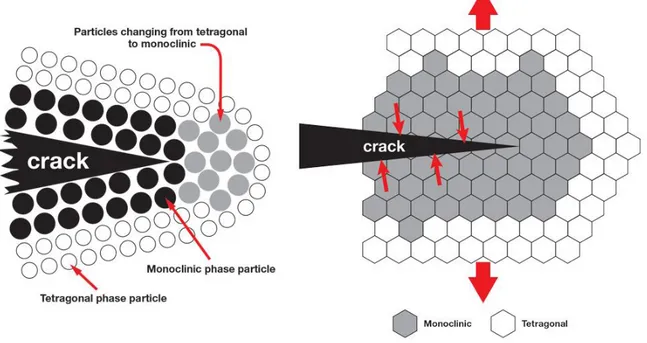
The stable crystalline structure for pure zirconia at room temperature and atmospheric pressure is monocline (m). The monocline phase is transformed into tetragonal (t) at about 1170°C, and the transformation is accompanied by a volume contraction of about 5% during heating and an equivalent expansion during cooling [16]. As the temperature increases, at about 2370°C zirconia transforms from tetragonal to cubic (c), with a volume variation of about 2.3% [131], and melts at 2716°C [132] [133]. These lattice transformations are athermic (they do not transmit heat), adiffusional (without atomic diffusion), and involve a lattice deformation [134]. The volume variations associated with these transformations are sufficient to cause the formation of cracks that compromise the stability of ceramics, so pure zirconia components are unsuitable for structural applications. However, the control of the transformations, and in particular that from t-ZR to m-ZR, is the basis of the tenacity mechanism of zirconium-based
23
ceramics and can lead to significant increases in mechanical properties. The grains of t-ZR, when embedded within a densely sintered structure, cannot expand freely and become m-ZR. As a result, the grains remain at room temperature in the metastable tetragonal phase. The transformation from tetragonal to monocline can then be induced by the application of external stresses ("stress induced transformation") that tend to dilate the structure of the crystalline lattice. Immediately [135] [136] [17] [18] after a crack begins to form, the grains of t-ZR close to the crack are able to expand and transform, returning to the m-ZR phase. The expansion adjacent to the crack compresses the crack and arrests the process (Fig. 2.4 [135]). The final result is that, in the propagation of a crack inside the material, part of the energy is dissipated to induce the phase transformation from tetragonal to monocline. In this way, the fracture toughness of the material is definitely improved. The "energy absorption" during the tetragonal-monocline transformation at room temperature in partially stabilized zirconia was recognized as a tenacity mechanism in 1975 [136].
Toughened zirconia has been developed in three different forms: FSZ fully stabilized zirconia; PSZ Partially Stabilized Zirconia; TZP Tetragonal Zirconia Polycrystals [137].
The three materials have in common the presence of stabilized tetragonal zirconia and the fact that the increase in toughness is related to the tetragonal-monocline transformation.
What distinguishes them is the mechanism of stabilization of the tetragonal phase. In PSZ and TPZ t-ZR is stabilised by the addition of oxides, usually magnesium oxide (MgO), yttrium
Figure 2.4: Schematic representation of the area subjected to stress due to the propagation of a crack. Zirconia changes from the tetragonal phase to the monocline phase.
24
oxide (Y2O3), calcium oxide (CaO) and cerium oxide (Ce2O3). The addition of oxides,
disadvantages the formation of tensioned monocline phase at room temperature, and makes stable (metastable) the crystalline cubic and tetragonal structures, more symmetrical [138]. These metastable phases have a structure similar to that of pure zirconia, but have doping ions substituted at the Zr4+ atomic sites and have a fraction of oxygen sites vacant to maintain charge
neutrality [139]. Since the mobility of cations in zirconia is rather low and oxygen vacations are locally ordered, the cubic and tetragonal metastable phases have prolonged stability at room temperature [140].
In FSZ the zirconia is the cubic phase with the addition of 8% of Y2O3, normally used for
oxygen sensors, and fuel cell electrolytes. In PSZ Partially Stabilized Zirconia 2-5 mol% of Y2O3 is added, depending on the grain size [140]. PSZ zirconia is therefore characterized by
the addition of lower concentrations of dopants than FSZ, hence the designation "partially stabilized". PSZ zirconia is heated to form zirconia in the cubic phase only, then cooled in a controlled way to develop a dispersion of tetragonal zirconia precipitates within the cubic matrix. Normally, the t-ZR should turn into the monocline shape during cooling, but to do so it should expand. The high strength of the surrounding cubic zirconia prevents this expansion, so that the tetragonal shape is maintained up to room temperature. The tetragonal structure is subject to transformation into the monoclinic phase when a stress force is applied, for example by an advancing crack. TZP zirconia essentially has tetragonal zirconia as the only metastable phase in the self-tenacity prerogative for the transformation of the tetragonal-monocline phase after the application of a load.
Factors that may cause structural conversion from tetragonal to monocline may be abrasive processes that can induce compressive stresses to a depth of several microns below the surface. Or, the t-m transformation may be caused by the roughness process that increases the volume of monoclinic grains as a result of the expulsion of some debris from the surface of the material. Moreover, a structural conversion from tetragonal to monocline is a spontaneous process known as aging and occurs in the temperature range between 200◦ and 300◦C in the presence of water vapour [17] [18]. The three types of zirconia FSZ, PSZ, TZP have high mechanical resistance and high toughness. A ceramic material with an internal mechanism capable of inhibiting crack propagation was made available, limiting immediate brittle rupture.
25
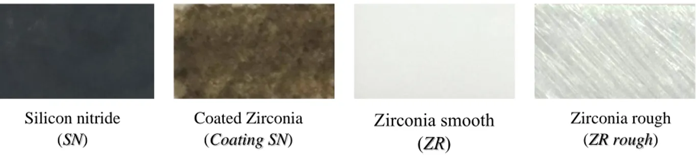
2.5 Samples
In this work, zirconia and silicon nitride were used as substrates. The zirconia samples were: polished zirconia (ZR) and rough zirconia (rough ZR). The rough zirconia samples were prepared using a diamond tip to create oriented scratches on the surface of the material.
Silicon nitride was used as a bulk ceramic. Silicon nitride was used as a bulk ceramic silicon nitride (SN) and as a cladding on the surface of a zirconia substrate (Coating SN). The coating was done through a laser. The work of this thesis does not analyses the coating method.
2.5.1
Silicon Nitride
The silicon nitride samples were in the form of discs (diameter 12 mm, thickness 1 mm) and were supplied by Sintx Corp. (Salt Lake City, Utah, USA). The material consisted of a two-phase microstructure comprising β- Si3N4 acicular grains separated by a continuous
sub-micrometric film of Si-Y-Al-O-N. In order to obtain the ceramic powder, the bulk samples were mechanically ground to a powder with an average particle size of 15 μm. The conditions applied to get a Si3N4-cladding on zirconia were the following: laser wavelength 1064 nm, maximum
pulse energy: 70 joules, peak power 17 kW, voltage range 400 V, pulse time 4 ms, and spot size 2 mm. The device operated under a constant flow of nitrogen gas to limit the decomposition and oxidation of Si3N4. It was necessary to repeat the operation three times in order to obtain a
homogeneous coating with thickness, t = 15 ± 5 μm, over the entire surface of the substrate. A motorized x-y stage with a lateral accuracy of 10 μm was used to align the sample with the laser source. Zirconia smooth (ZR) Zirconia rough (ZR rough) Coated Zirconia (Coating SN) Silicon nitride (SN)
26
2.5.2
Zirconia
The material consisted in stabilized zirconium oxide samples containing 3% yttrium and were obtained from a commercial producer (Kyocera, Kyoto, Japan). To produce a "roughening effect" on otherwise smooth zirconia substrates, abrasions were carried out over the entire surface using a diamond cutting disc (tip diameter: 25 μm) under an applied load of 20 ± 5 N. Once the surface was covered with unidirectional scratches, the direction of scratching was rotated about 90° and the operation was repeated again.
2.6 Bone tissue properties and characteristics
Bone tissue is a connective tissue that is physically identified by the combination of inorganic and organic substance, combined to form a fundamental amorphous substance. The components of the organic part are collagen I, ossein and a glycoprotein called osteo-mucoid; derived from extracellular matrix. In the inorganic substance, which in adults accounts for about 60-70% of the total bone mass, are calcium phosphate (86%) in the form of hydroxyapatite crystals Ca10(PO4)6(OH)2, calcium carbonate Ca3(PO4)2 (12%), magnesium phosphate (1.5%), calcium
fluoride (0.5%) and iron oxide (traces). Elasticity and tensile strength are provided by the organic component, while stiffness and hardness are provided by the mineral component (due to the important presence of hydroxyapatite): these characteristics make the fabric ideal for structural and support functions. From the point of view of mechanical properties, an increase in the mineral content makes the bone brittle and brittle, a decrease makes it softer and more deformable [141].
Collagen represents 65-80% of the dry mass of specialized connective tissues (similar to tendons, skin, joint capsules and cartilage), but it is the only protein with significant tensile strength-resistance properties. This characteristic derives from the molecular configuration of the collagen macromolecule, one of the largest molecules of our organism with a filamentous structure and entirely made up of molecules of tropocollagen. Biological and environmental factors influence its typically three-dimensional configuration. Using transmission electron microscopy (SEM),it can be seen the fibrils are assembled with each other with a phase shift of a quarter of their length. Each individual unit of tropocollagen consists of three peptide chains, the intracellular synthesis of which occurs independently. Each chain contains
27
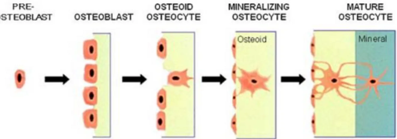
approximately 1000 amino acids. Most of the chains (called alpha chains) are ordered precisely and with frequent repetition of the glycine-proline-hydroxyproline, glycine-proline-x or glycine-x-proline sequences where x is another amino acid. Collagen by nature has a high percentage of glycine, proline and hydroxyproline. Among these three is glycine which, being smaller in size, allows its compact arrangement necessary to assemble the three alpha chains in the tropocollagen. Among the organic components there are other protein components Osteocalcin (OC), Osteonectin, Osteopontin (OPN) that have the function of modulating the mineralization and adhesion between the cells and the bone matrix. [142]. The bone tissue cells that provide for the growth, production and resorption of bone tissue are: Osteo-progenitor,
Osteoblasts, Osteocytes and Osteoclasts (Fig. 2.6) [143].
Osteo-progenitor cells are of mesenchymal origin with stem properties: they can proliferate and
differentiate into osteoblasts. Normally they are used to be found in the periosteum and endosteum: when reactivated, they provide the formation of new bone tissue.
Osteoblasts are mature cells that release extracellular matrix to form new bone tissue. Osteoblasts are massive cells, rich in alkaline phosphatase and highly polarized towards the deposition front, where the collagen and protein fibres of the bone matrix (including OP, OPN) secrete. The new organic matrix deposited and not yet mineralized is called osteoid. Its average thickness is of the order of 10mm with a growth rate of 0.5mm / day. The initial stages of deposition are rather fast. Subsequently, the deposition slows down and the osteoblasts progressively modify the form only from high and voluminous cells to horizontal cells with less and less cytoplasm. As the deposition progresses, other osteogenic cells reach maturation and begin to release extracellular matrix, embedding the osteoblasts on the osteoid in the matrix that the new cells deposit.
Osteocytes are the osteoblasts included in the extracellular matrix. As a result, the new osteocytes are subjected to the mineralization process. Osteocytes maintain contact with osteoblasts by cytoplasmic prolongations, which have numerous gap junctions, creating a cytoplasmic network. They have an oval-shaped body, with a major axis parallel to the bone surface. Numerous cytoplasmic connections emerge from the body and are immersed in the mineralized matrix. The neo formed osteocytes continue to release extracellular matrix within their own bone gap. The cell volume decreases by one third and the cell becomes dormant.
Osteoclasts are the cells that specialize in removing the bone matrix. They are not cells that live
in bone tissue, but are formed when the bone matrix has to be removed and disappear at the end of the process. The principle of function is in contact with the bone matrix and via an acidic pH (5.2) they erode the surface. Through an acid catalysis promoted by the enzyme acid hydrolase,
28
and through the hormone calcitonin, produced by the thyroid, the activity of the osteoclasts is inhibited [144].
2.6.1
Hydroxyapatite (HA)
Since the Hydroxyapatite (HA) is the main component of the tooth, it is essential to deeply know its entire structure. HA is a natural mineral whose chemical formula Ca10(PO4)6(OH)2 has
a crystalline structure with hexagonal symmetry and spatial group P63/m with lattice parameters
a = b = 9.432 Å, c = 6.881 Å [145] [146]. As shown in Figure 2.7.
Figure 2.6: Example of bone growth through cells: osteo-progenitor, osteoblasts, osteocytes.
29
The structure contains tetrahedra of PO4 held together by Ca and OH- ions; calcium ions are
present four as Ca(I) and six as Ca(II). HA, Ca10(PO4)6(OH)2, for bones and teeth is used as a
model, however, apatites are rarely found in pure stoichiometric form because they can incorporate a wide variety of impurities. Depending on the impurities it is classified into three types: the first has Ca10(PO4)6X2, X = F-, CO32-, octacalcium phosphate Ca8H2(PO4)6·5H2O
(OCP), tetracalcium phosphate Ca4(PO4)2O (TTCP). The second type includes polymorphic
forms of tricalcium phosphate (TCP), Ca3(PO4)2. Finally, the third type is Ca-PO4
sheet-containing compounds which includes dehydrated dicalcium phosphate CaHPO4.2H2O
(DCPD), anhydrous dicalcium phosphate CaHPO4 (DCPA) and monocalcium phosphates,
Ca(H2PO4)2·H2O and Ca(H2PO4)2 [147].
HA is thermally unstable, depending on its stoichiometry at temperatures from about 800-1200°C it begins to decompose. Stoichiometry is very significant if the work of
osteoblastic cells is studied or thermal processing of the material takes place. The stoichiometric molar ratio of calcium and phosphorus in HA is 1.67. This ratio, Ca/P, if it is higher than the stoichiometric value you are in an acidic environment and the higher the solubility of HA will be [148].
HA is the inorganic phase of teeth and bone [149] [150], and for biomedical applications it is an essential and attractive material [151] [152] [153] [154]. In HA, replacements to phosphate groups, calcium ions or the hydroxyl group can occur. These replacements help living tissue to maintain a normal metabolism. The main substitution takes place with CO32- which is a
variation between 3 and 8%. Replacements occur mainly with ions such as Mg2+, Na+, K+, Cl-, F- and many other ions [155] [156] [157]. The replacement rate varies depending on the age of the HA [158]. Recent studies have shown that the concentration of CO32- increases with age,
while the concentration of HPO42- decreases. These substitutions also change properties such
as lattice parameters, mechanical properties, morphology, magnetic properties and stability [159] [160] [161] [162] [163] [164] [165] [166] [167] [168]. For instance, substitution with F
-to OH- ions decreases the solubility in acids [169]. While carbonate ions interact with the ionic
interactions that establish the structure of apatite. Carbonate ions exchange with hydroxyl (type A carbonate), or PO43- tetrahedra (type B carbonate) [170] [171].
30
2.6.2
Bases of osteointegration
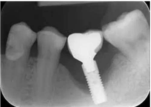
The field of rehabilitation of edentulous patients was enriched by the innovation introduced in the mid-twentieth century by a Swedish professor, Per-Ingvar Brånemark, who had the merit of having developed an implant method based on well-defined criteria and reliable predictions. He is responsible for the concept of osteointegration, defined as "the direct structural and functional union between bone and surface of an implant under load" and the start of "modern implantology" (Brånemark, 1985) [1] [2]. In the early 1960s, after three decades of research and development, consisting of clinical and laboratory studies, an dental implant technique "capable of replacing missing natural teeth with elements similar to dental roots" was established [1]. The discovery that led to the definition of osseointegration was a study of bone by in vivo optical microscopy: a metal optical chamber, made of titanium, was incorporated into the tibia of a rabbit. After the consequent regeneration of the bone, the chamber was removed and it was noticed that it had been "incorporated" into the bone. There was also perfect adhesion between the bone tissue and the irregularities of the titanic surface. Subsequent experiments led to the fabrication of dental implants using titanium screws (Fig. 2.8). There is an affinity of bone with titanium and the oxide layer that forms on its surface. Different bonds, such as van der Waals forces, hydrogen bonds and local chemical bonds, have the ability to unite the biomolecules to the TiO2 layer [2] [172]. Since the 90's, the experimentation of new possible biocompatible materials with the human body has been started. Given the negative effects of TiO2, such as the retraction of the gum towards the prosthesis therefore an effect not aesthetically pleasing; problem of the metal-taste; problem of a possible migration of metal particles in the human body. New possible ways for more efficient osseointegration have been studied using metal-free materials such as Zirconia ZrO2 in the case of dental prostheses [173].
31
2.6.3
Factors that influence osteointegration
The process of osseointegration on an implant surface involves rather complex stages and the involvement of many factors. Among the most important are the microstructure and surface chemistry, which influence the process both quantitatively and qualitatively. Studies by Osborn and Newesley [1] have shown that bone neoformation occurs through two phenomena, distance osteogenesis and contact osteogenesis. In the first case, the release of the extracellular matrix and the subsequent mineralization made by the osteoblasts takes place in a direction that goes from the bone to the implant, i.e. the bone gradually surrounds the screw. In the second process, osseointegration occurs in the opposite direction, from the implant to the bone [2]. Once the cells have polarized, extracellular matrix production begins, with the aim of giving a precise structure to the bone-implant interface, which, after calcification, is transformed into an osteoid matrix and finally into bone tissue. Methods of roughening the surface have been developed to ensure proper and satisfactory osseointegration. Rough surfaces are preferred to smooth surfaces because they absorb more of the biomolecules involved in the processes described above. A rough surface favours cell adhesion and consequently proliferation. An example of this is the difference between climbing a smooth or rough mountain. If the wall is smooth it is almost impossible to climb, while if it is rough and therefore with handholds the climbing becomes easy. Cell adhesion works in the same way, the rougher the wall, the greater the cell adhesion.
32
Osteointegration is also linked to the concepts of osteo-induction and osteoconduction. The first definition indicates the stimulation of osteoprogenitor cells to the osteoblastic differentiation, a phenomenon that gives rise to osteogenesis, then "induces" it. Osteoconduction, on the other hand, concerns the growth of bone on a surface, and therefore implies the existence of more or less osteoconductive surfaces, i.e. surfaces capable of better or worse promoting the adhesion and adaptation of cells to the implant site. The success of a good osseointegration , i.e. the adhesion between implant and new bone), is the concrete result of a previous osteoinduction and osteoconduction [174].
2.7 Bacterial inflammation on prosthesis
The long-term maintenance of osteointegration depends on the state of health of the peri-implant tissues. Inflammation of peri-implant tissues due to bacterial inflammation may lead to marginal bone resorption. Oral hygiene aims to eliminate pathogenic bacteria that can cause inflammation of the peri-implant mucosa and loss, even partial, of osseointegration. The behaviour of the peri-implant mucosa depends on the quality of the soft tissues, the depth to which the implant is immersed, the type of surface material of which the implant is made and the morphology of its surface (rough or smooth) [175].
Inflammatory lesions that develop in the tissues around implants are recognized as peri-implant diseases. Peri-implant disease comprises two forms: Peri-implant mucositis corresponding to gingivitis; defined as a reversible inflammatory reaction of the soft tissues surrounding a working implant. periimplantitis (Fig. 2.9) corresponding to peri-implant disease; definite as inflammatory reactions associated with loss of supporting bone tissue around a working and irreversible implant [176] [177]. Therefore the periimplantitis is an inflammatory disease where, the bacteria cause an infection of the soft and hard peri-implant tissues that lead to a progressive retraction of the bone until then the implant is expelled. The implant becomes unstable and has to be removed. Periimplantitis usually occurs after 5 to 6 to 7 even 9 years. This disease also causes biological damage, both to the jawbone and the mandible. The treatment of periimplantitis is prevention, there is no treatment. Periimplantitis forms more easily if the surface is rough because the bacteria adhere more easily to the surface. Consequently, the only possible treatment is to do a small operation removing the bacteria and consequently smoothening the implant surface, so as to avoid future bacterial adhesion. The
33
aim is to transform the rough surface into a smooth surface that has much less adhesion to bacteria. This is the treatment, however, is only effective in 50% of cases. The reason for using rougher surfaces than is to ensure a faster osseointegration, thus a faster patient rehabilitation [178] [179].
2.9 Cellular and bacteria test
2.9.1
Osteosarcoma cells (SAOS-2)
SAOS-2 is a human tumour cell line first isolated in 1973 by Fogh [180], specifically tumour Osteoblastic cells. This allows a large number of cells to be obtained in a very short time; they can also easily differentiate the same way in which osteoblasts naturally differentiate themselves. These characteristics allow the use of SAOS-2 for the study of bone tissue growth. Furthermore, SAOS-2 is ideal for the study of bone tissue development in vitro because of its ability to produce an extracellular matrix competent for mineralization [181] [182] [183].
Figure 2.9: Effect of peri-implantitis; the image shows bone loss caused by bacterial inflammation.
34
SAOS-2 has been shown to have a collagen structure very similar to that synthesized by human osteoblasts, while lysine is in a state of hydroxylysine caused by the enzyme lysyl hydroxylase (modification of the protein structure: glycosylation of lysine) [184] [185] .
Another important feature is that the activity of alkaline phosphatase which is still high over time. The alkaline phosphatase present in the extracellular matrix completes the process of bone mineralization, promoting the deposition of the mineral material on the organic matrix of the bone being formed. As regards growth factors and cytokines, the values of the SAOS-2 cell line are comparable to those of normal osteoblasts [186].
2.9.2
Staphylococcus epidermidis bacteria
Staphylococcus epidermidis bacteria were used to analyse the interaction between the surface of the ceramic material and the bacteria. Staphylococcus epidermidis bacteria were used to analyses the interaction between the surface of the ceramic material and the bacteria, but not only, a detailed analysis is also carried out on the antibacterial activity of this type of ceramic surface. Staphylococcus epidermidis are gram + bacteria belonging to staphylococci family (cluster growth), diameter 8-1.5μm with production of colonies of 2-3mm. They are usually present on the human skin and in the mucous membranes. They are potentially pathogenic and manage to be dangerous as they can lead to infection if they can penetrate inside the human body (during surgery or due to wounds).
Staphylococcus epidermidis need to be cultivated like other staphylococci in common culture media and the optimal growth temperature is around 25-40 °C with an optimum between 30 and 37 °C. Staphylococcus growth is usually favoured at neutral pH between 6-0-7.0. The pH of the saliva is between 6.7 -7.4 [187].
S. epidermidis is able to adhere to the synthetic surfaces of biomaterials and to form a stable biofilm that leads to persistent infections [188] [189]. They are therefore able to adhere also dental implants of Ti or ZrO2 [190]. The bacteria during the formation of the biofilm, secrete
extracellular polymeric substances that include lipids, proteins, polysaccharides and others. These substances protect the bacterial colony from external attacks [191] [192]. Treatment of biofilm-related infections is extremely difficult. Moreover, the immune system is inefficient in eliminating bacterial inflammation that requires extreme doses of antibiotics. Biofilm contains
35
bacteria up to 1000 times more resistant to bactericidal drugs than their planktonic predecessors [193].
In addition, bacteria released from biofilm often trigger remote secondary reactions to some patients who have compromised immune systems. The need therefore arises to use biocompatible materials capable of eliminating the serious problem of bacterial contamination.
![Figure 2.8: X-ray image of a dental bone implant integrated in the jawbone [174]](https://thumb-eu.123doks.com/thumbv2/123dokorg/2735085.126/32.892.260.675.95.395/figure-ray-image-dental-bone-implant-integrated-jawbone.webp)