Adult stem cell plasticity: Neoblast repopulation in non-lethally irradiated planarians
Alessandra Salvetti
a,⁎
, Leonardo Rossi
a, Lucia Bonuccelli
a, Annalisa Lena
a, Claudio Pugliesi
b,
Giuseppe Rainaldi
c, Monica Evangelista
c, Vittorio Gremigni
a,⁎
a
Dipartimento di Morfologia Umana e Biologia Applicata, Università di Pisa, Pisa, Italy
b
Dipartimento di Biologia delle Piante Agrarie, Sezione di Genetica, Università di Pisa, Pisa, Italy
c
Istituto di Fisiologia Clinica, Laboratorio di Terapia Genica e Molecolare, CNR, Pisa, Italy
a b s t r a c t
a r t i c l e i n f o
Article history:Received for publication 5 June 2008 Revised 17 December 2008 Accepted 23 January 2009 Available online 30 January 2009 Keywords:
Planarian Adult stem cells Neoblast X-ray irradiation Substance P Spantide
Planarians are a model system for studying adult stem cells, as they possess the neoblasts, a population of pluripotent adult stem cells able to give rise to both somatic and germ cells. Although over the last years several efforts have been made to shed light on neoblast biology, only recent evidence indicate that this population of cells is heterogeneous. In this study we irradiated planarians with different non-lethal X-ray doses (1–5 Gy) and we identified subpopulations of neoblasts with diverse levels of tolerance to X-rays. We demonstrated that a dramatic reduction of neoblasts occurred soon after non-lethal irradiations and that de-novo proliferation of some radioresistant cells re-established the primary neoblast number. In particular, a strong proliferation activity occurred at the ventral side of irradiated animals close to the nervous system. The produced cells migrated towards the dorsal parenchyma and, together with some dorsal radioresistant cells, reconstituted the entire neoblast population demonstrating the extreme plasticity of this adult stem cell system.
© 2009 Elsevier Inc. All rights reserved.
Introduction
The ability to remain in an undifferentiated state and the capability, on cue, to undergo an asymmetric division and generate a cell with a stem cell fate and a daughter cell that differentiates, are unique for stem cells in multicellular organisms. Embryonic stem (ES) cells are pluripotent and so capable of giving rise to cells that make up every organ in the adult. Mammalian ES properties became progressively restricted during development and, in the adult, stem cells are generally multipotent or unipotent and are most often used for the maintenance or repair of the tissue in which they reside. This tissue-specificity has now been questioned by evidence suggesting that, under certain conditions, adult stem cells may possess potential for tissue regeneration (Loebinger and Janes, 2007; Zech et al., 2007). More in vivo studies are necessary to understand adult stem cell biology. Unfortunately, researches on adult stem cells have not been pursued vigorously as adult stem cells are generally few in number and there are not many animal systems that enable in vivo studies of these cells. Freshwater planarians (Platyhelminthes) provide an example of one of these suitable model systems. Indeed, these organisms possess a population of adult pluripotent stem cells, the neoblasts, able to give rise to both somatic and germ cells. This
advantage, coupled with the successful application of molecular, cellular and genomic approaches, as well as the possibility to perform loss of function studies by the RNA interference (RNAi) technique, makes planarians a sound model system for in vivo investigations on adult stem cell biology (Saló, 2006; Sánchez Alvarado, 2007; Rossi et al., 2008). Neoblasts are small cells (5– 10μm of diameter), with a high nucleus/cytoplasm ratio, scattered throughout the parenchyma and absent in the most anterior part of the head and the pharynx. Neoblasts also accumulated in clusters along the midline and the dorso-lateral parenchyma (Salvetti et al., 2000; Orii et al., 2005). Neoblasts are involved in regeneration giving rise to regenerative blastema as well as in tissue homeostasis, and they are the only proliferating cells in asexual planarians (Rossi et al., 2008). For this reason, neoblasts are destroyed a few days after treatment with lethal doses of X-rays, while differentiated cells are unaffected over the same time period and with the same doses (Wolff and Dubois, 1948). Dissimilar to what was observed in planarians, it has been recently demonstrated that in Macrostomum lignano high X-ray doses (40 and 80 Gy) resulted in a lack of somatic stem cell proliferation, while S-phase cells were still visible in the gonads (Pfister et al., 2007).
Neoblast population is composed of distinct subpopulations of cells, as demonstrated by the identification of a Dugesia japonica homologue of the Drosophila Piwi gene (DjPiwi-1) that is exclusively expressed in a group of neoblasts distributed along the dorsal body midline (Rossi et al., 2006). Moreover, the recent identification of a nanos-related gene (Djnos) in D. japonica allowed to identify a second subpopulation of ⁎ Corresponding authors.
E-mail addresses:[email protected](A. Salvetti),[email protected] (V. Gremigni).
0012-1606/$– see front matter © 2009 Elsevier Inc. All rights reserved. doi:10.1016/j.ydbio.2009.01.029
Contents lists available atScienceDirect
Developmental Biology
neoblasts (Sato et al., 2006). nanos-positive cells are located along the dorso-lateral parenchyma (the presumptive testes forming regions) and in two ventral spots (the presumptive ovary forming regions), which are considered to be the germline stem cells (Sato et al., 2006; Handberg-Thorsager and Saló, 2007; Wang et al., 2007). Heterogeneity among neoblasts is also confirmed by the existence of two different subpopulations of irradiation-sensitive neoblast-like cells (X1 and X2) identified by X-ray treatments and FACS analysis (Reddien et al., 2005b; Hayashi et al., 2006). These groups of neoblasts diverge in both expression of proliferation markers and in ultrastructural features and they have been classified as type A (concentrated in the X1 fraction) and type B (concentrated in the X2 fraction) cells (Higuchi et al., 2007). So, the term neoblasts is not referred to a single population of stem cells but to a compound of different subpopulations of stem cells each of which expresses particular molecular markers and, consequently, could have different biological properties.
Here, we utilize X-ray treatments to investigate in vivo the planarian adult stem cell system and we provide further evidence that neoblasts are heterogeneous. We also demonstrate that the early reduction of the neoblast number that occurs after non-lethal X-ray irradiation is followed by the activation of cell pro-liferation that allows the reconstitution of the complete neoblast system.
Materials and methods Animals
Planarians utilized in this work belong to the species D. japonica, asexual strain GI (Orii et al., 1993). Animals were kept in autoclaved stream water at 18 °C and starved at least for 2 weeks before being used in the experiments. Regenerating fragments were obtained by transection between auricles and pharynx or tail and pharynx. X-ray irradiation
Intact planarians were exposed to 1, 2, 3, 5, 10, 15 and 30 Gray (Gy) single-dose of hard X-rays (200 kVp, 12 mA, 1 Gy/min) using a Stabilipan 250/1 instrument with a 2 mm aluminum (Al) filter (Siemens, Gorla-Siama, Milan, Italy) equipped with a Radiation Monitor 9010 dosimeter (Radcal Corporation, Monrovia, CA, USA). The animals were sacrificed at: a) 1, 2, 3, 4, 7, 8, 9, 10, 11, 14, 23, 27 or 40 days after irradiation for in situ hybridization experiments; b) 4, 7, 11 or 14 days after irradiation for immunohistochemistry; c) 11 days after irradiation for TEM analysis; d) 1, 2, 3, 4, 5, 6, 7, 9, 14, 25 or 30 days after irradiation for BrdU staining and mitosis count; e) 1, 3, 7, 12 days after irradiation for FACS analysis. For regeneration experi-ments, animals were transected 60 h after a treatment with 5 Gy of X-rays and sacrificed 15, 21 or 28 days after the transection for whole mount in situ hybridization. In some experiments, planarians were treated with 5 Gy of X-rays, kept in stream water for 30 days and irradiated again with an additional X-ray dose of 5 Gy. After this second radiation treatment, animals were sacrificed 2, 4, 7 or 14 days for whole mount in situ hybridization. Untreated animals with correspondent time of starvation and/or regeneration were utilized as controls.
BrdU labelling and detection
X-ray-irradiated or untreated (control) planarians were injected with a 10 mM BrdU solution using the Nanoject Microinjectior (Drummond). For detection of BrdU on macerates, 6 h after injection planarians were dissociated in 250μl of a glycerol/acetic acid/distilled water solution (1:1:13) for 20 h at 4 °C. Twentyμl of cell suspension was placed on a microscope slide and dried for a couple of hours. Slides were immediately re-hydrated with two washes of PBS plus
0.5% Triton X-100 (PBST 0.5) for 5 min each, treated with 1 N HCl-PBST 0.5 solution for 30 min and rinsed three times in PBST 0.5 for 5 min each. After treatment with 20μM proteinase K in PBS plus 0.1% Triton X-100 (PBST 0.1), slides were washed in PBST 0.1 for 1 min and then blocked in 10% non-fat dry milk (Bio-Rad) in PBST 0.5 for 10 min. After blocking, slides were rinsed in PBST 0.1 for 1 min and then incubated with 1:50 dilution of anti-BrdU antibody (Abcam) in 1% dry milk in PBST 0.1 for 30 min. Following 3 washes of 10 min each in PBST 0.5 slides were rinsed in PBST 0.1 and incubated in 1% dry milk in PBST 0.1 solution containing 1:200 dilution of fluorescein isothiocynate-conjugated anti-mouse antibody (Molecular Probes) for 30 min. Following two washes in PBST 0.5 and one in PBS, slides were mounted for microscope analysis. Slides were scored with a Zeiss Axioplan photomicroscope to count the BrdU-positive nuclei. About 8 × 10⁴nuclei were examined for each preparation and only nuclei
withfine grained bright florescence through the nuclear matrix and bright perinuclear chromatin staining were taken as positive. Tenμl of cell suspension was used in a hemocytometer to count the number of total cells. The relative number of BrdU-positive nuclei was calculated dividing the absolute number of positive nuclei by the number of total cells. Three independent samples were analyzed for each experi-mental condition and two different counting were carried out for each sample.
For immunofluorescent detection of BrdU-positive cells on tissues sections, non-irradiated animals and 5 Gy-treated planarians at the 4th and 7th day after the X-ray treatment were injected as described above. Twenty-two hours following microinjection, speci-mens werefixed in relaxant solution (Kobayashi et al., 1998) for 17–24 h at 4 °C and then included in paraffin. After rehydratation in ethanol series, sections were treated with 5μg/ml of Proteinase K (Sigma) for 5-15 min at 37 °C and then incubated in 1 N HCl in PBST for 5 min at 55 °C. After washing in PBST, samples were firstly incubated in 10% goat serum for 30 min at room temperature and then incubated with 1/100 anti-BrdU monoclonal antibody (BD Pharmigen) in 10% goat serum for 1 h. After washing, samples were incubated with 1:100 dilution of HRP conjugated anti-mouse antibody and the signal was amplified by using Tyramide Signal Amplification Kit according to the manufacturer instructions (Molecular Probes).
Mitosis analysis on cell macerates
X-ray-irradiated (5 Gy) and non-irradiated (control) planarians were treated in 3‰ colchicine in stream water for 6 h, dissociated in 250μl of a glycerol/acetic acid/distilled water solution (1:1:13) for 20 h at 4 °C and then stained with 20 μg/ml Hoechst No.33342. Twentyμl of cell suspension was placed on a microscope slide, dried for a couple of hours and then mounted for microscope analysis. Slides were scored with a Zeiss Axioplan photomicroscope to count the number of mitoticfigures. About 8×10⁴nuclei were examined for
each preparation. Tenμl of cell suspension was used in a hemocyt-ometer to count the number of total cells. The mitotic index was calculated dividing the number of mitosis by the number of total cells in which mitosis were counted. Three independent samples were analyzed for each experimental condition and two different counting were carried out for each sample.
In situ hybridization
Whole mount in situ hybridization was performed as described in
Rossi et al. (2007). Section in situ hybridization was performed according toKobayashi et al. (1999).
DNA templates for DjPiwi-1 was prepared as described inRossi et al. (2006); DjMCM2 was prepared according to Salvetti et al. (2000); DjPum was prepared according toSalvetti et al. (2005); CIP29 Gi 32900868 was prepared according toRossi et al. (2007). Djnos
templates were obtained by PCR using the following forward and T7 promoter adapted reverse primers: Djnos forward (5 ′-CTTTGGCAATCGGTAACTTC-3′) and Djnos reverse (5′-TAATACGACT-CACTATAGGGAGAGAAGAATTTGAAGAGATAGGAC-3′). Purified ampli-fication products or digested plasmids were transcribed in vitro to obtain DIG-labelled RNA probes using the DIG-RNA labelling kit (Roche).
Immunohistochemistry
Immunohistochemistry on tissue sections was performed as previously described by Sato et al. (2006) using a mouse anti-synapsin (3C11, Developmental Studies Hybridoma Bank; dilution 1:20) and/or a rabbit anti-PCNA (Proliferating Cell Nuclear Antigen; dilution 1:100) antibodies. As secondary antibodies we used a 1:200 dilution offluorescein isothiocyanate-conjugated mouse or anti-rabbit (Molecular Probes) or 1:200 dilution of rhodamine-conjugated anti-mouse (Molecular Probes) in blocking solution (10% goat serum in PBS). Control sections were incubated in blocking solution with no primary antibody.
Immunohistochemistry on tissue sections was also performed on animals treated with 3‰ colchicine in stream water for 6 h, 14 days after X-ray (5 Gy) irradiation. All slides were dipped in 1 μg/ml Hoechst No.33342 (Sigma-Aldrich) for the counterstaining of nuclei, mounted and observed under the Zeiss Axioplan fluorescence microscope.
After whole-mount in situ hybridization some selected specimens were processed for immunostaining with the anti-synapsin antibody, as previously described (Cebrià and Newmark, 2005). The anti-synapsin was used at a 1:20 dilution and thefluorescein isothiocya-nate-conjugated anti-mouse secondary antibody (Molecular Probes) was used at 1:200.
Transmission electron microscopy
Transmission electron microscopy (TEM) was performed as previously described in Salvetti et al. (2005). Briefly, planarians werefixed with a 2.5% glutaraldehyde solution in 0.1 M cacodylate buffer and post-fixed with 2% osmium tetroxide. Ultrathin sections were stained with uranyl acetate and lead citrate and observed with a Jeol 100 SX transmission electron microscope.
FACS analysis
Intact X-ray-treated and untreated animals were dissociated and analyzed by FACS as previously described (Salvetti et al., 2005; Hayashi et al., 2006). Briefly, after filtration through a 20 μm nylon mesh, cells were incubated with calcein AM (0.5μg/ml, Sigma), fixed in 70% ethanol and then incubated for 30 min at room temperature in PBS containing: propidium iodide (PI, 50 μg/ml, Roche), RNAse (6.25μg/ml, Roche) and IGEPAL CA-630 (0.5% v/v Sigma-Aldrich). FACS analysis of calcein staining versus PI incorporation was performed by a FACScalibur cytofluorimeter (Becton Dickinson) and data were analyzed by CELL Quest analysis software (Becton Dickinson). Cellular debris was excluded by forward-angle light scatter (FSC) and side-angle light scatter (SSC) analysis.
RNA interference (RNAi) experiments
Double strand RNA (dsRNA) of DjPiwi-1 was obtained as described inRossi et al. (2006). Intact planarians were injected with DjPiwi-1 for four consecutive days and then irradiated with an X-ray dose of 5 Gy. One week after thefirst round of injections, irradiated animals were re-injected for 1 day and then fixed for whole mount in situ hybridization 2, 4 or 7 days after the irradiation. Negative controls were done by injection ofβ-Gal-dsRNA or water.
Spantide treatment
Stock solution of spantide (Sigma-Aldrich) was prepared in 2 × 10− 4 M PS (NaCl 6 mM; CaCl2 0.048 mM; KCl 0.1 mM; MgCl2
0.1 mM; NaHCO30.12 mM, pH 7.4). To test the effect of spantide on cell
proliferation, intact animals were kept in a spantide solution 10− 7M for 3 days and then irradiated with an X-ray dose of 5 Gy. After irradiation animals were left in the spantide solution for 4 or 7 days and then processed for mitosis count. Several controls were used: non-irradiated animals or animals left in PS solution and then irradiated as well as non-irradiated animals treated with substance P (Sigma-Aldrich) and spantide were used. Statistical significance was assessed by Student's t test.
Results
Assessment of the minimal lethal X-ray dose
Planarians belonging to D. japonica species were irradiated with 1, 2, 3, 5, 10, 15 or 30 Gy of X-rays. All specimens treated with 30 Gy as well as 70% (35/50) of the planarians treated with 15 Gy died within 4 weeks. In all the animals that survived to a 15 Gy dose of X-rays, we observed lesions in the dorsal parenchyma close to the pharynx that appeared about 2–3 weeks after the irradiation and were later completely rescued. However, all planarians treated with 1, 2, 3, 5 or 10 Gy did not die. Thus, in our experimental condition 15 Gy can be defined as the minimum lethal dose of X-rays for D. japonica. Although planarians survived to an X-ray dose of 10 Gy, all treated animals showed a darkened body area at the pharynx level. On the contrary, in animals treated with 5 Gy this abnormal pigmentation was observed only rarely (1/50). In all cases the animals rescued the normal pigmentation within 4 weeks from irradiation. On the basis of these findings the non-lethal X-ray dose of 5 Gy was used in the following experiments.
Effects of the 5 Gy treatment on planarian regeneration
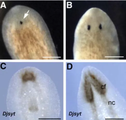
Following transection, 5 Gy-treated animals experienced a regen-eration delay and showed cephalic morphogenetic defects i.e. an abnormal head with a single eye usually centrally located (Figs. 1A, B). The expression pattern analysis of Djsyt, the pan-neural marker
Fig. 1. Effect of X-ray treatment (5 Gy) on regenerating D. japonica. (A, B) Dorsal view, anterior is towards the top. (C, D) Ventral view, anterior is towards the top. (A) Brightfield image of an X-ray-treated planarian regenerating the head 15 days after transection. The arrow indicates a single eye. (B) Brightfield image of an untreated planarian regenerating the head, 15 days after transection. Djsyt expression visualized by whole mount in situ hybridization 15 days after transection in a 5 Gy-treated planarian (C) and untreated planarian (D). cf, cephalic ganglia; nc, nerve cords. Scale bar is 400μm.
synaptogamin (Tazaki et al., 1999), revealed that 15 days after transection the cephalic ganglia of 5 Gy-treated posterior regenerants were smaller than those of untreated animals (Figs. 1C, D). Normal sized cephalic ganglia were observed in animals within 3–4 weeks from transection and, at the same time, all the specimens rescued morphogenetic defects (data not shown).
Effects of the 5 Gy treatment on neoblasts
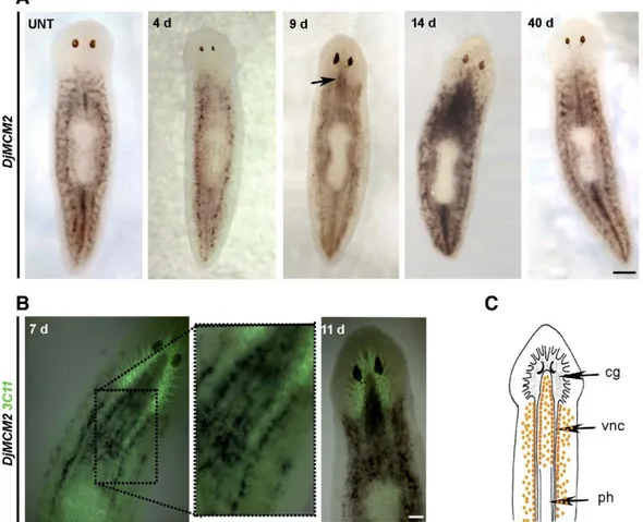
To investigate what happened to neoblasts in intact planarians after irradiation, we performed whole mount in situ hybridization experiments using the neoblast molecular marker DjMCM2 as probe (Salvetti et al., 2000) (Fig. 2). Although the time course of the expression pattern might change depending on individual variability, during the first days after irradiation we usually found that the number of DjMCM2-positive cells spread all over the parenchyma as well as those clustered along the body midline anterior to the pharynx were reduced with respect to untreated planarians (Fig. 2A). At day 4, residual clusters of DjMCM2-positive cells were still visible in the dorso-lateral parenchyma as well as along the body midline posterior to the pharynx. Seven days after X-ray treatment, the hybridization signal increased in intensity in the ventral side of treated animals and it appeared mainly distributed in parallel rows close to the nerve cords (Fig. 2B). In the following days, cells expressing DjMCM2 became abundant in both the ventral and dorsal parenchyma and also accumulated between the two dorsal cephalic ganglia close to the eyes where DjMCM2-expressing cells were undetectable in untreated planarians. The typical DjMCM2 expression pattern was re-established
only 40 days after X-ray treatments (Fig. 2A). The irradiation procedure did not select a population of radioresistant cells as in planarians re-irradiated with an additional 5 Gy dose DjMCM2 expression pattern underwent the same spatial changes described above (data not shown).
To better analyze the neoblast fate after X-ray treatments, we performed in situ hybridization on sections corresponding to different body levels of irradiated animals. Although 2 days after irradiation DjMCM2-expressing cells were strongly reduced in number with respect to controls, positive cells were still detectable in the dorsal as well as in the ventral parenchyma (Figs. 3A–D). At the 4th day, the number of DjMCM2-positive cells in the dorsal parenchyma was similar to that observed 2 days after irradiation (compareFigs. 3C, D withFigs. 3E, F). By contrast, a slight increase of DjMCM2-positive cells occurred in the ventral parenchyma close to nervous system (Figs. 3E, F). In the following days, dorsal and ventral DjMCM2-expressing cells increased in number and a marked accumulation of the hybridization signal was detectable close to and between the two ventral nerve cords (Figs. 3G, H). Successively, a progressive reduction of positive cells in the ventral parenchyma was coupled with a sustained increase in the number of these cells in the dorsal parenchyma with large clusters of cells also detectable between the gut branches (Figs. 3I, J). Twenty-three days after irradiation the hybridization signal was mainly accumulated at the level of the dorsal parenchyma where the number of positive cells still appeared greater than that of untreated planarians (Figs. 3K, L). These events were observable anterior to the pharynx, as well as at the pharynx and tail level. Moreover, clusters of DjMCM2-positive cells were also detectable anterior to the eyes and at
Fig. 2. Analysis of DjMCM2 expression in X-ray-treated (5 Gy) D. japonica by whole mount in situ hybridization. Dorsal view, anterior is towards the top. (A) DjMCM2 expression in an untreated animal (UNT) and in 5 Gy-treated animals at different days (d) after radiation treatment. Arrow indicates DjMCM2-positive cells between the cephalic ganglia. (B) Neuronal synapses visualized by immunostaining with the anti-synapsin (3C11) antibody (green) and DjMCM2 expression in 5 Gy-treated animals. (C) Schematic drawing of a planarian showing the distribution of DjMCM2-positive cells (orange dots) 11 days after irradiation. The planarian nervous system consists of two anterior cephalic ganglia (cg), and two ventral nerve cords (vnc); ph, pharynx. Scale bar is 400μm in A; 200 μm in B.
the eye level, while only rare positive cells were detectable in the controls at the same body levels (Supplementary Fig. 1).
Further evidence for the ability of planarians to re-establish the neoblast population after a 5 Gy treatment was obtained by FACS analysis of the irradiation-sensitive cell fractions (X1 and X2) previously identified (Reddien et al., 2005b; Hayashi et al., 2006). FACS analysis revealed that the number of X1 cells was strongly reduced 1 day after X-ray, while the number of X2 cells was similar to that of untreated planarians (Fig. 3M). At the 3rd day, both X1 and X2 cell subpopulations were reduced, similarly to what occurred in 30 Gy-treated animals. In the following days, X1 and X2 cell fractions were repopulated and 12 days after irradiation the number of events exceeded that of untreated planarians (Fig. 3M).
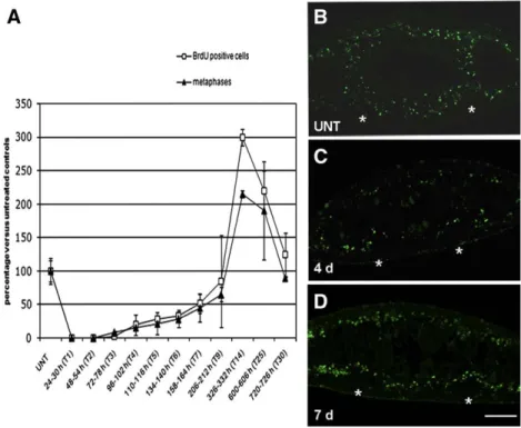
We also followed the neoblast fate after 5 Gy treatments by counting the number of cells able to enter the S or M phase of the cell
cycle in a temporal window of 6 h. We found that in X-ray-treated planarians, 1 day (T1) after irradiation, the number of S-phase cells was dramatically reduced with respect to that of untreated planarians. Starting from the 4th day (T4), the number of S-phase cells gradually increased and 14 days after irradiation (T14) it became significantly higher than that of control (Fig. 4A). Four weeks after irradiation (T30) the number of S-phase cells was comparable with that of untreated animals. The analysis of mitotic figures confirmed the results obtained with BrdU incorporation (Fig. 4A).
BrdU labelling was also used to confirm the distribution of proliferating cells in 5 Gy-treated animals. In untreated planarians 22 h after incubation in BrdU, S-phase cells were found spread all over the parenchyma (Fig. 4B) and only rare dorsal lateral clusters of few neoblasts were detected (data not shown). Four days after irradiation, we found a drastic reduction of proliferating cells with respect to Fig. 3. Analysis of DjMCM2 expression in X-ray-treated (5 Gy) D. japonica detected by in situ hybridization on transverse sections obtained from the body region anterior to the pharynx. Dorsal epithelium is towards the top. DjMCM2 expression in an untreated (UNT) (A) and in 5 Gy-treated animals sacrificed at different days after an X-ray treatment (C, E, G, I, K). (B, D, F, H, J, L) Magnification of boxed region in A, C, E, G, I and K, respectively. Arrows indicate dorsal radioresistant clusters of DjMCM2-positive cells. Arrowhead indicates DjMCM2-positive cells between the gut branches. g, gut; nc, nerve cords. (M) FACS profile of propidium iodide (PI) and calcein labelled cells. X1 (blue) and X2 (red) populations are irradiation-sensitive, Xis (black) is an X-ray insensitive cell fraction. Scale bar is 150μm in A, C, E, G, I and K; 25 μm in B, D, F, H, J and L.
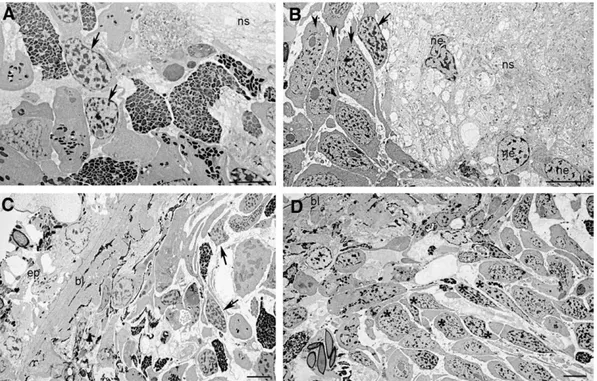
control, but we always detected some BrdU-positive cells in both the dorsal and ventral parenchyma (Fig. 4C). The number of S-phase cells was increased 7 days after irradiation and they were mainly distributed close to the nerve cords in the ventral side of treated animals (Fig. 4D). An accumulation of neoblasts close to the nervous system was also showed by co-immunolocalization experiments performed with an antibody directed against the proliferating cell nuclear antigen (i.e. the neoblast marker DjPCNA;Orii et al., 2005) and an antibody directed against the nervous system protein synapsin (3C11) which is a marker of synaptogenesis and stains the nerve terminals (Fig. 5andSupplementary Fig. 2). In addition, TEM analysis revealed the presence of several neoblasts located close to the nervous system (Figs. 6A, B). Some of these neoblasts showed the features
typical of high proliferative type A neoblasts according toHiguchi et al. (2007). Large groups of these neoblasts appeared oriented towards the dorsal parenchyma (Fig. 6D). By contrast, scarce neoblasts with large patches of heterochromatin were present in the same region of untreated specimens where no groups of neoblasts oriented towards the dorsal surface were observable (Fig. 6C). To understand whether a relationship between the nervous system and neoblast repopulation might occur in 5 Gy-treated animals, we analyzed the release of mitogen neuropeptide substance P (SP) after X-ray treatment. We addressed this issue studying the effects on cell proliferation of spantide (SPA), a substance P antagonist, in irradiated animals. As previously observed by Baguñà et al. (1989), we found that the addition of SPA alone to non-irradiated animals caused a slight Fig. 4. Analysis of proliferating cells in non-irradiated and X-ray-treated (5 Gy) D. japonica. (A) Analysis of S-phase and mitotic cells in untreated (UNT) and X-ray-treated (5 Gy) D. japonica. The number of cells able to enter the S or M phase of the cell cycle in a temporal window of 6 h was analyzed. Values are expressed as percentage with respect to untreated planarians to which the arbitrary value of 100% has been given. Each value is the mean ± standard deviation of at least four independent samples counted in duplicate. (B–D) Distribution of BrdU-positive cells in a temporal window of 22 h analyzed by immunofluorescence on transverse sections obtained from the body region anterior to the pharynx. Dorsal epithelium is towards the top. The asterisks indicate (⁎) nervous system. Scale bar is 150 μm.
Fig. 5. Analysis of DjPCNA expression in X-ray-treated (5 Gy) D. japonica visualized by immunohistochemistry on transverse sections obtained from the region anterior to the pharynx. Distribution of DjPCNA-positive cells in a irradiated animal sacrificed 11 days after treatment (A) and in an untreated animal (B). Distribution of the nervous system is revealed by the use of the anti-synapsin (3C11) antibody. Nuclei are visualized by the nuclear dye Hoechst. Arrows indicate epidermis nuclei. Scale bar is 20μm.
stimulation of cell proliferation while the addition of SPA together with SP produced a reduction of SP-induced cell proliferation. In 5 Gy-irradiated intact animals treated with SPA alone, a significant reduction of the mitotic activity with respect to controls was observed 4 and 7 days after the irradiation (Fig. 7).
Effects of the 5 Gy treatment on distinct neoblast subpopulations To explore the effects of X-ray treatments on specific neoblast subpopulations, we investigated the expression pattern of DjPiwi-1, (Fig. 8A). Two days after treatment, no hybridization signal was detected along the dorsal body midline. On the 4th day after irradiation some DjPiwi-1-positive cells appeared in the ventral
parenchyma and at 7 days this hybridization signal increased in intensity and appeared organized in parallel rows close to the nerve cords. Successively, DjPiwi-1-positive cells were again detectable in the dorsal parenchyma and, 14 days after treatment, the typical DjPiwi-1 expression pattern was re-established in 60% of the irradiated specimens (30/50). In the remaining specimens we detected a stronger and more diffuse hybridization signal with respect to controls (Fig. 8A). The evident ventralization of DjPiwi-1 hybridization signal following X-ray treatments, prompted us to investigate the role of this gene during the cell repopulation by RNAi. The expression pattern of DjMCM2 was analyzed 2, 4 and 7 days after irradiation in DjPiwi-1 RNAi animals. The results indicated that DjPiwi-1 silencing did not alter DjMCM2-positive cells repopulation in X-ray-treated animals (data not shown).
We also analyzed the expression pattern of other neoblast molecular markers such as Djnos (Sato et al., 2006), DjPum (Salvetti et al., 2005) and the planarian homolog of CIP29 (Rossi et al., 2007). After irradiation, the number of Djnos expressing cells was drastically reduced in number with respect to untreated animals (Fig. 8B). However, their spatial distribution remained unchanged and we never detected Djnos-expressing cells close to the ventral nerve cords. On the contrary, we found that DjPum and CIP29 expression pattern in X-ray-treated planarians was similar to that of DjMCM2 (data not shown).
Effects of 1, 2 and 3 Gy treatments on neoblasts
To investigate neoblast radiosensitivity in worms irradiated to a lower dose than 5 Gy of X-rays, we performed whole mount in situ hybridization experiments on planarians treated with 1, 2 and 3 Gy of X-rays (Supplementary Fig. 3). Whatever the X-ray dose, the DjMCM2-positive cells spread all over the parenchyma were reduced in number 1 day after treatments (Supplementary Figs. 3A–C). However, the reduction of the cell number expressing DjMCM2 was proportional to the X-ray dose. Indeed, in 1 Gy-treated worms only a slight reduction of DjMCM2-positive cells was detected in comparison to both 2 and Fig. 6. Micrographs of untreated and 5 Gy-treated D. japonica sacrificed 11 days after irradiation. (A) Neoblasts close to the nervous system of an untreated planarian. (B) Numerous neoblasts are present in the same region of an irradiated planarian. (C) Neoblasts in the dorsal parenchyma of a control specimen. (D) Several neoblasts (⁎) oriented towards the dorsal epithelium of an irradiated planarian. Arrows indicate neoblasts having a heterochromatin-rich nucleus. Arrowheads indicate neoblasts having a euchromatin-rich nucleus. bl, basal lamina; ep, epithelium; ns, nervous system; ne, neuron. Scale bar is 10μm.
Fig. 7. Analysis of spantide (SPA) effect on mitotic activation in X-ray-treated (5 Gy) D. japonica. SPA shows an inhibitory effect on the mitotic activity in animals 4 days (4 d) and 7 days (7 d) after the irradiation. The results are expressed as percentage of the mitotic activity of controls (5 Gy PS) at which the arbitrary value of 100% was attributed. Each bar shows the mean ± standard deviation from four different specimens. Data were obtained from two independent experiments.⁎Significant at pb0.05; ⁎⁎significant at pb0.01.
3 Gy-irradiated specimens and DjMCM2-expressing cells were detectable close to the nervous system of 1 Gy-irradiated animals only after 2 days from irradiation (Supplementary Fig. 3D, arrow). In planarians treated with 2 or 3 Gy the clustered DjMCM2-positive cells were reduced in number 2 days after the irradiation (Supplementary Figs. 2E, F). Moreover, in 2 and 3 Gy-treated worms a strong accumulation of DjMCM2 transcripts was observed in the ventral parenchyma 4 days after the irradiation (Supplementary Figs. 3H, I), similar to what was observed in 5 Gy-treated worms 7 days after irradiation. On the whole, the spatial distribution of DjMCM2-expressing cells in planarians treated with 1, 2 or 3 Gy was similar to that observed in 5 Gy-treated animals, although the dynamic of the events appeared to be temporally anticipated with respect to 5 Gy-treated worms, probably due to a reduced loss of neoblasts in animals treated with the lower X-ray doses.
Discussion
Planarian neoblasts have different levels of radiotolerance
Several experimentalfindings demonstrated that one week after a lethal dose of X-rays (i.e. 30 Gy) neoblasts, but not differentiated cells, are destroyed and, for this reason, lethal doses of X-ray are commonly utilized to establish planarian stem cell-specific gene expression (Cebrià et al., 2002; Guo et al., 2006; Ogawa et al., 2002; Orii et al.,
2005; Reddien et al., 2005a,b; Rossi et al., 2006, 2007; Salvetti et al., 2000, 2005). The evidence that DjPiwi-1-expressing neoblasts have a lower tolerance to 30 Gy than DjMCM2-positive neoblasts clustered along the lateral lines of the planarian body (Rossi et al., 2006) prompted us to further investigate the radiosensitivity of planarian stem cells with the goal to identify new neoblast subpopulations.
DjMCM2 expression analysis after 5 Gy treatment demonstrates that the neoblasts spread all over the parenchyma are less radio-resistant than clustered neoblasts and, among them, some neoblasts clustered in the dorso-lateral parenchyma have a higher radio-tolerance than those clustered anterior to the pharynx along the midline of the planarian body. FACS analysis shows that thefirst cells that disappear belong to X1 fraction, indicating that the more radiosensitive cells are primarily included in this fraction. We hypothesize that different level of X-ray tolerance among neoblasts can be principally ascribed to a different cycling rate i.e. a higher cycling rate corresponds to a lower radiotolerance. According to this hypothesis, it has been suggested that the X1 subpopulation mainly consists of dividing neoblasts expressing higher levels of proliferating markers (i.e. DjMCM2) with respect to X2 cells (Hayashi et al., 2006; Higuchi et al., 2007). We have also found that BrdU-positive cells are homogeneously scattered throughout the parenchyma while only scarce and small dorso-lateral clusters are present 22 h after labelling, a time at which Newmark and Sánchez Alvarado (2000) demon-strated that the only BrdU-labelled cells are neoblasts. Thisfinding Fig. 8. Analysis of the effect of 5 Gy X-ray treatments on distinct neoblast subpopulations in D. japonica by whole mount in situ hybridization. Analysis of DjPiwi-1 (A) and Djnos (B) expression in untreated animals (UNT) and in 5 Gy-irradiated specimens at different days (d) after treatment. Anterior is towards the top. ve, ventral view. Scale bar is 400μm.
further supports the hypothesis that the spread neoblasts, that we propose to be the dividing transit population, divide more quickly than the clustered neoblasts in non-irradiated animals. As a matter of fact, a dramatic reduction in the number of BrdU-labelled cells occurs within 2 days after 5 Gy treatment, when only the spread DjMCM2-posistive cells are no longer present; and no S-phase cells are found 3 days after the treatment, when the DjMCM2-positive cells clustered along the dorso-lateral parenchyma are still detectable. Notably,Sato et al. (2006) also demonstrated that the Djnos-positive neoblasts clustered in the dorso-lateral parenchyma are not labelled with BrdU, suggesting that the cell cycle of this neoblasts might be arrested. Radioresistant cells re-acquire proliferation capabilities and repopulate the planarian body after a non-lethal X-ray treatment
Although a severe reduction in the number of neoblasts occurs in 5 Gy-treated animals, they do not die. After transection, these animals regenerate with morphogenetic defects probably due to the reduced availability of neoblasts. Survival is attributable to a subset of cells that begins to proliferate about 4 days after the X-ray treatment and repopulate the planarian body in a few days. FACS analysis evidences that both X1 and X2 cells are reduced 3 days after 5 Gy irradiation and the number of cells in X1 and X2 increased in the following days. Unfortunately the relationship between X1 and X2 cells is still unclear as few information are available for these cell fractions, making difficult to give a univocal interpretation of the obtained data. A possibility is that 5 Gy treatment selects a subset of 5-Gy resistant X1 cells that might proliferate and reconstitute both X1 and X2 subpopulations. Alternatively, a subset of X2 cells might also be selected by X-ray and reacquire proliferative capability.
What is the origin of these radioresistant cells? A first reliable hypothesis is that the surviving cells are in a particular stage of the cell cycle resulting in a different response to X-rays. An association between X-ray-induced DNA damage and cell cycle stage at the time of irradiation has been demonstrated (Pawlik and Keyomarsi, 2004; Larsson et al., 2007). Another possibility is based on the evidence that both X1 and X2 fraction also contains a percentage of differentiating cells (Hayashi et al., 2006). So, it might be that surviving cells represent a committed/differentiating stem cell progeny having a greater radiation resistance due to their reduced proliferation activity and increased capability of repairing DNA damage (Hong and Stambrook, 2004; Xu, 2005). Following neoblast loss, these more radioresistant cells might re-acquire the stem cell capacities and reconstitute the complex neoblast system. The evidence that dedifferentiation, or at least transdetermination, can occur in planarians (Gremigni and Miceli, 1980) support this hypothesis. Moreover, generation of stem cell-like progenitors by cell dediffer-entiation has been described in Drosophila where cystocytes can revert into functional stem cells and replenish germ cells after depletion by genotoxic chemicals, radiation or normal ageing (Kai and Spradling, 2004). In addition, spermatogonia that have initiated differentiation can repopulate Drosophila niches and revert to stem cell identity (Brawley and Matunis, 2004). Although we do not know which of our hypotheses is the correct one, we can exclude that a long-term stable radioresistant population of cells is selected, since in planarians irradiated twice we observed the same events i.e. loss of neoblasts and repopulation of the planarian body by surviving radioresistant cells.
An intriguing feature of the repopulation process is the possible role of the nervous system in the activation of cell proliferation. Our experimental evidence indicate that 4 and 7 days after irradiation the number of mitosis is drastically reduced in the presence of SPA, a specific antagonist of the neuropeptide substance P, suggesting that a release of substance P may occur in 5 Gy-treated animals. Our hypothesis is also supported by the up-regulation of factors of the mitogen-activated protein (MAP) kinase pathway in 5 Gy-treated
animals (Rossi et al., 2007), the activation of which has been demonstrated to be stimulated by the substance P receptor (Luo et al., 1996; Sharif et al., 1996). Previousfindings indicate the influence of nervous system on planarian cell proliferation (Bautz and Schilt, 1986; Baguñà et al., 1989) and/or regeneration (Cebrià, 2007) and a correlation between cell proliferation and nervous system has been proposed in Macrostomum sp. (Ladurner et al., 2000). A probable neural-induced cell proliferation is also suggested by the evidence that radioresistant cells begin to proliferate close to the ventral nerve cords of irradiated planarians and they and/or their progenies migrate to the dorsal side re-establishing the neoblast system, together with the dorsal radioresistant cells. The occurrence of cell migration is deduced by the presence, between the gut branches, of large groups of neoblasts oriented towards the dorsal surface and by the analysis of DjPiwi-1 expression, whose typical dorsal expression pattern is re-established in 5 Gy-treated planarians by the migration of ventral DjPiwi-1-positive cells towards the dorsal side. We exclude the participation of Djnos-positive cells in neoblast repopulation as we did not detect positive cells close to the ventral nervous system of X-ray animals. As this subpopulation is re-established after proliferation and migration of the ventral DjMCM2-positive cells, we propose that in irradiated animals Djnos-expressing cells arise from the ventral cells. However, we cannot exclude that after X-ray treatment the few remaining positive cells self-reconstitute the large Djnos-positive cell clusters of the dorsal parenchyma.
Conclusions
Several studies about the effect of X-ray on stem cells have been performed in the mammalian small intestinal crypt. Stem cells in the crypt have evolved various protective mechanisms to ensure the genetic integrity of the stem cell compartment and are intolerant to genotoxic damage also including that induced by low doses of radiation (Potten and Ellis, 2006). Doses as low as 1–5 cGy (roughly equivalent to just one DNA damaging event per cell) can induce stem cell apoptosis (Potten, 1977). Indeed, stem cells do not undergo cell cycle arrest and repair but commit an altruistic p53-dependent cell suicide. Apoptosis is then compensated by division of either undamaged neighboring stem cells or by more resistant potential clonogenic stem cells, representing the second and third generations in the cell lineage, that re-acquire the properties of a stem cell (Potten, 2004). Similarly, we propose that in planarians a hierarchical stem cell compartment is present that comprises stem cells (true neoblasts) with a high intolerance to DNA damage, more-resistant clonogenic neoblasts (i.e. daughter cells in the early generation of cell lineage) and the radiosensitive proliferating transit neoblast progeny. In planarians thefirst event following X-ray treatment is the induction of apoptosis (Rossi et al., 2006) and the loss of the majority of highly dividing neoblast progeny; and this could also be the case for true neoblasts. The consequent release of signals from the nervous system might promote rounds of proliferation in radioresistant surviving cells in order to replace the lost cells. In this scenario, we suggest that surviving cells repopulate the planarian body and re-establish the entire stem cell compartment revealing the extreme plasticity of these peculiar types of adult stem cells.
Acknowledgments
We would like to thank Dr. K. Agata and N. Shibata for providing us their BrdU staining protocol and Dr. H. Orii for anti-PCNA antibody. We are grateful to Dr. A. Sainato, L. Tana and C. Traino for X-ray irradiation. We also thank C. Ghezzani for technical assistance with TEM. The antibody 3C11 (anti-synapsin) developed by Dr. E. Buchner was obtained from the Developmental Studies Hybridoma Bank developed under the auspices of the NICHD and maintained by the University of
Iowa, Department of Biological Sciences, Iowa City, IA 522242. Grant Sponsor: Programmi di Ricerca di Interesse Nazionale, MIUR, Italy. Appendix A. Supplementary data
Supplementary data associated with this article can be found, in the online version, atdoi:10.1016/j.ydbio.2009.01.029.
References
Baguñà, J., Saló, E., Romero, R., 1989. Effects of activators and antagonists of the neuropeptides substance P and substance K on cell proliferation in planarians. Int. J. Dev. Biol. 33, 261–266.
Bautz, A., Schilt, J., 1986. Somatostatin-like peptide and regeneration capacities in planarians. Gen. Comp. Endocrinol. 64, 267–272.
Brawley, C., Matunis, E., 2004. Regeneration of male germline stem cells by spermatogonial dedifferentiation in vivo. Science 304, 1331–1334.
Cebrià, F., 2007. Regenerating the central nervous system: how easy for planarians! Dev. Genes Evol. 217, 733–748.
Cebrià, F., Newmark, P.A., 2005. Planarian homolog of netrin and netrin receptor are required for proper regeneration of the central nervous system and the maintenance of nervous system architecture. Development 132, 3691–3703. Cebrià, F., Kobayashi, C., Umesono, Y., Nakazawa, M., Mineta, K., Ikeo, K., Gojobori, T.,
Itoh, M., Taira, M., Sánchez Alvarado, A., Agata, K., 2002. FGFR-related gene nou-darake restricts brain tissues to the head region of planarians. Nature 419, 620–624. Gremigni, V., Miceli, C., 1980. Cytophotometric evidence for cell‘transdifferentiation’ in
planarian regeneration. Wilhelm Roux's Arch. 188, 107–113.
Guo, T., Peters, A.H., Newmark, P.A., 2006. A bruno-like gene is required for stem cell maintenance in planarians. Dev. Cell 11, 159–169.
Handberg-Thorsager, M., Saló, E., 2007. The planarian nanos-like gene Smednos is expressed in germline and eye precursor cells during development and regenera-tion. Dev. Genes Evol. 217, 403–411.
Hayashi, T., Asami, M., Higuchi, S., Shibata, N., Agata, K., 2006. Isolation of planarian X-ray-sensitive stem cells byfluorescence-activated cell sorting. Dev. Growth Differ. 48, 371–380.
Higuchi, S., Hayashi, T., Hori, I., Shibata, N., Sakamoto, H., Agata, K., 2007. Characteriza-tion and categorizaCharacteriza-tion offluorescence activated cell sorted planarian stem cells by ultrastructural analysis. Dev. Growth Differ. 49, 571–581.
Hong, Y., Stambrook, P.J., 2004. Restoration of an absent G1arrest and protection
from apoptosis in embryonic stem cells after ionizing radiation. Proc. Natl. Acad. Sci. U. S. A. 101, 14443–14448.
Kai, T., Spradling, A., 2004. Differentiating germ cells can revert into functional stem cells in Drosophila melanogaster ovaries. Nature 428, 564–569.
Kobayashi, C., Kobayashi, S., Orii, H., Watanabe, K., Agata, K., 1998. Identification of two distinct muscles in the planarian Dugesia japonica by their expression of myosin heavy chain genes. Zool. Sci. 15, 861–869.
Kobayashi, C., Watanabe, K., Agata, K., 1999. The process of pharynx regeneration in planarians. Dev. Biol. 211, 27–38.
Ladurner, P., Rieger, R., Baguñà, J., 2000. Spatial distribution and differentiation potential of stem cells in hatchlings and adults in the marine platyhelminth Macrostomum sp.: a bromodeoxyuridine analysis. Dev. Biol. 226, 231–241.
Larsson, D.E., Gustavsson, S., Hultborn, R., Nygren, J., Delle, U., Elmroth, K., 2007. Chromosomal damage in two X-ray irradiated cell lines: influence of cell cycle stage and irradiation temperature. Anticancer Res. 27, 749–753.
Loebinger, M.R., Janes, S.M., 2007. Stem cells for lung disease. Chest 132, 279–285. Luo, W., Sharif, T.R., Sharif, M., 1996. Substance P-induced mitogenesis in human
astrocytoma cells correlates with activation of the mitogen-activated protein kinase signaling pathway. Cancer Res. 56, 4983–4991.
Newmark, P.A., Sánchez Alvarado, A., 2000. Bromodeoxyuridine specifically labels the regenerative stem cells of planarians. Dev. Biol. 220, 142–153.
Ogawa, K., Kobayashi, C., Hayashi, T., Orii, H., Watanabe, K., Agata, K., 2002. Planarian fibroblast growth factor receptor homologs expressed in stem cells and cephalic ganglions. Dev. Growth Differ. 44, 191–204.
Orii, H., Agata, K., Watanabe, K., 1993. POU-domain genes in planarian Dugesia japonica: the structure and expression. Biochem. Biophys. Res. Commun. 192, 1395–1402. Orii, H., Sakurai, T., Watanabe, K., 2005. Distribution of the stem cells (neoblasts) in the
planarian Dugesia japonica. Dev. Genes Evol. 215, 143–157.
Pawlik, T.M., Keyomarsi, K., 2004. Role of cell cycle in mediating sensitivity to radiotherapy. Int. J. Radiat. Oncol. Biol. Phys. 59, 928–942.
Pfister, D., De Mulder, K., Philipp, I., Kuales, G., Hrouda, M., Eichberger, P., Borgonie, G., Hartenstein, V., Ladurner, P., 2007. The exceptional stem cell system of Macrosto-mum lignano: screening for gene expression and studying cell proliferation by hydroxyurea treatment and irradiation. Front. Zool. 4, 9.
Potten, C.S., 1977. Extreme sensitivity of some intestinal crypt cells to X and gamma irradiation. Nature 269, 518–521.
Potten, C.S., 2004. Radiation, the ideal cytotoxic agent for studying the cell biology of tissues such as the small intestine. Radiat. Res. 161, 123–136.
Potten, C.S., Ellis, J.R., 2006. Adult small intestinal stem cells: identification, location, characteristics, and clinical applications. Ernst Schering Res. Found. Workshop 60, 81–98.
Reddien, P.W., Bermange, A.L., Murfitt, K.J., Jennings, J.R., Sánchez Alvarado, A., 2005a. Identification of genes needed for regeneration, stem cell function, and tissue homeostasis by systematic gene perturbation in planaria. Dev. Cell 8, 635–649. Reddien, P.W., Oviedo, N.J., Jennings, J.R., Jenkin, J.C., Sánchez Alvarado, A., 2005b.
SMEDWI-2 is a PIWI-like protein that regulates planarian stem cells. Science 310, 1327–1330.
Rossi, L., Salvetti, A., Lena, A., Batistoni, R., Deri, P., Pugliesi, C., Loreti, E., Gremigni, V., 2006. DjPiwi-1, a member of the PAZ-Piwi gene family, defines a subpopulation of planarian stem cells. Dev. Genes Evol. 216, 335–346.
Rossi, L., Salvetti, A., Marincola, F.M., Lena, A., Deri, P., Mannini, L., Batistoni, R., Wang, E., Gremigni, V., 2007. Deciphering the molecular machinery of stem cells: a look at the neoblast gene expression profile. Genome Biol. 8, R62.
Rossi, L., Salvetti, A., Batistoni, R., Deri, P., Gremigni, V., 2008. Molecular and cellular basis of regeneration and tissue repair: planarians, a tale of stem cells. Cell Mol. Life Sci. 65, 16–23.
Saló, E., 2006. The power of regeneration and the stem-cell kingdom: freshwater planarians (Platyhelminthes). BioEssays 28, 546–559.
Salvetti, A., Rossi, L., Deri, P., Batistoni, R., 2000. An MCM2-related gene is expressed in proliferating cells of intact and regenerating planarians. Dev. Dyn. 218, 603–614. Salvetti, A., Rossi, L., Lena, A., Batistoni, R., Deri, P., Rainaldi, G., Locci, M.T., Evangelista,
M., Gremigni, V., 2005. DjPum, a homologue of Drosophila Pumilio, is essential to planarian stem cell maintenance. Development 132, 1863–1874.
Sánchez Alvarado, A., 2007. Stem cells and the Planarian Schmidtea mediterranea. C. R. Biol. 330, 498–503.
Sato, K., Shibata, N., Orii, H., Amikura, R., Sakurai, T., Agata, K., Kobayashi, S., Watanabe, K., 2006. Identification and origin of the germline stem cells as revealed by the expression of nanos-related gene in planarians. Dev. Growth Differ. 48, 615–628. Sharif, T.R., Luo, W., Houghton, P.J., Sharif, M., 1996. Substance K peptide induces
mitogenesis by activating the mitogen-activated protein kinase signaling pathway through the substance P receptor (NK-1 subtype) in human astrocytoma cells. Cell. Pharmacol. 3, 441–449.
Tazaki, A., Gaudieri, S., Ikeo, K., Gojobori, T., Watanabe, K., Agata, K., 1999. Neural network in planarian revealed by an antibody against planarian synaptotagmin homologue. Biochem. Biophys. Res. Commun. 260, 426–432.
Wang, Y., Zayas, R.M., Guo, T., Newmark, P.A., 2007. nanos function is essential for development and regeneration of planarian germ cells. Proc. Natl. Acad. Sci. U. S. A. 104, 5901–5906.
Wolff, E., Dubois, F., 1948. Sur la migration des cellules de régénération chez les planaires. Rev. Suisse Zool. 55, 218–227.
Xu, Y., 2005. A new role for p53 in maintaining genetic stability in embryonic stem cells. Cell Cycle 4, 363–364.
Zech, N.H., Shkumatov, A., Koestenbauer, S., 2007. The magic behind stem cells. J. Assist. Reprod. Genet. 24, 208–214.