Università degli Studi di Pisa
Doctorate Course in Molecular
Biotechnology, XX cycle
“ROLE OF PENTOSE PHOSPHATES IN
NUCLEOSIDE CATABOLISM AND
INTERCONVERSION”
Tutor:
Prof.ssa Marcella Camici
Student:
Desidero ringraziare tutte le persone che lavorano presso la sezione di Biochimica del Dipartimento di Biologia per l’aiuto e la disponibilità che mi hanno dato.
ABSTRACT
Purine and pyrimidine are the basic constituents of the polynucleotides DNA and RNA; they are considered of predominant importance also as information molecules, energy transducers, antioxidants and they have also important roles in cellular signalling processes. Reports suggest protective roles for purines and pyrimidines in various pathological conditions ranging from cancer, to ischemia-associated injury, traumatic tissue damage, bone resorption, stress and haemorrhagic shock. Mitochondrial oxidative phosphorylation, along with glycolysis, is essential for the maintenance of brain ATP levels. The depletion of cellular energy sources and reduction of ATP synthesis during ischemia or brain insults cause the loss of cellular homeostasis, which eventually leads to irreversible cellular damage. Some papers have shown that adenosine and inosine protect neural cells during hypoxia/ischemia
in vivo and in vitro. While in some cases the action of adenosine is receptor-mediated, to explain the effect of its deamination product, inosine, the contribution of hitherto unknown specific receptors has been invoked. On the other hand, several papers report a receptor-independent mechanism of nucleoside action, which indicates that nucleosides donate their ribose moiety to the pentose phosphate pathway, where it is converted to intermediates for entry into the glycolytic pathway for anaerobic production of ATP.
To shed light on the mechanism underlying the protective role of purine and pyrimidine during ischemia or brain insults, we have used a human astrocytoma cell line (ADF) which has been subjected to metabolic stress conditions by exclusion of glucose and pre-incubation with oligomycin (an inhibitor of oxidative phosphorylation). This treatment brings about a depletion of the ATP pool, with a concomitant increase in the AMP levels, which results in a significant decrease of the adenylate energy charge. The presence of purine nucleosides in the culture medium preserves the adenylate energy charge, and improves cell viability. Besides purine nucleosides, also pyrimidine nucleosides, such as uridine and, to a lesser extent, cytidine, are able to preserve the ATP pool. The determination of lactate in the incubation medium indicates that nucleosides can preserve the ATP pool through anaerobic glycolysis, thus pointing to a relevant role of the phosphorolytic cleavage of the N-glycosidic bond of nucleosides which generates, without energy expense, the phosphorylated pentose, which through the pentose phosphate pathway and glycolysis can be converted to energetic intermediates also in the absence of oxygen. In fact, ADF cells possess both purine nucleoside phosphorylase and uridine phosphorylase activities. The results of this research line have been published in Neurochem. Int. 50: 517-523 (2007) (see attached).
Additionally, the phosphorylated ribose moiety of nucleosides may be used itself for the salvage of pyrimidine nucleosides or can be converted by 5-phosphoribosyl-1-pyrophoshate (PRPP) synthetase, into PRPP which is an essential compound for the salvage pathway of purines.
In fact, adenine and hypoxanthine, in the presence of PRPP, are substrates of adenine phosphoribosyltransferase (APRT) and hypoxanthine-guanine phosphoribosyltransferase (HPRT), which catalyze the formation of adenosine and inosine monophosphate, respectively (purine salvage pathway).
On the other hand, uridine can be both phosphorolytically cleaved by uridine phosphorylase (UPase) to ribose-1-phosphate (Rib-1-P) and the uracil base, or can be converted by uridine kinase (UK) into uridine nucleotides (pyrimidine salvage pathway). It has been shown that disruption of uridine homeostasis by deletion of rat UPase gene leads to disorders of both pyrimidine and purine nucleotide synthesis, thus suggesting a linkage between the two metabolic processes. We have hypothesized that the UPase–UK enzyme system, which maintains uridine homeostasis, regulates the processes of both purine and pyrimidine salvage. Exogenous uridine and, to a lesser extent inosine, activate the salvage of exogenous adenine in human astrocytoma cells in a concentration-dependent manner. Moreover, uridine is also able to activate the salvage of exogenous hypoxanthine. When uridine and inosine are present, more Rib-1-P becomes available, through the action of UPase and purine nucleoside phosphorylase (PNP), for the PRPP-mediated adenine and hypoxanthine salvage. Exogenously added adenine partially inhibits uridine salvage; in fact, the increased utilization of PRPP exerted by added adenine, might further favour uridine phosphorolysis, thus subtracting the pyrimidine nucleoside to the UK action. Moreover, exogenous inosine favours not only uracil salvage but also 5-FU activation through a Rib-1-P-mediated process. The pre-treatment of the cells with cytidine brings about an inhibition of the pyrimidine salvage and an activation of the purine salvage in the presence of uridine as ribose phosphate donor. In fact, cytidine enters the cells and is converted into CTP which inhibits UK and this inhibition causes a shift of the equilibrium of the reversible UPase reaction towards uridine phosphorolysis. The Rib-1-P formed is then converted into PRPP, which is used for the purine salvage synthesis. Conversely, when the concentration of CTP is relatively low, the fully active UK, which catalyzes a virtual irreversible reaction, drives uridine towards uridine nucleotide formation, thus lowering the rate of purine synthesis. Therefore, the ribose phosphate stemming from the phosphorolysis of purine and pyrimidine nucleosides not only can be converted into energetic intermediates in order to restore the ATP pool during cellular stress but it can be considered a link between the purine and pyrimidine salvage and these two processes are regulated at the level of UPase–UK enzyme system by the relative pyrimidine nucleoside triphosphate concentration. The results of this research line have been published in Neurochem. Int. 51: 517-523 (2007) (see attached).
ABBREVIATIONS
5-(phenylthio) acyclouridine (PTAU) 5-phosphoribosyl-1-pyrohosphate (PRPP) 5- fluorouracil (5-FU)
8-cyclopentenyl-1,3-dipropylxanthine (DPCPX) Adenosine (Ado)
Adenine (Ade)
Adenine phosphorybosiltransferase (APRT) Adenosine 5’-diphosphate (ADP)
Adenosine 5’-monophosphate (AMP) Adenosine 5'-triphosphate (ATP) Adenosine deaminase (ADA) Adenosine kinase (AK).
Adenosine phosphoribosyltransferase (APRT) Adenosine receptors (ARs)
Astrocyte-neuron-lactate shuttle (ANLSH) Blood-brain barrier (BBB)
Carbamoyl phosphate II (CPII) Central nervous system (CNS) Citric acid cycle (TCA) Cyclic AMP (cAMP) Cyclic GMP (cGMP) Cytidine (Ctd) Cytosine (Cyt) Cytidine 5’-monophosphate (CMP) Cytidine 5’-diphosphate (CDP) Cytidine 5’-triphosphate (CTP) Cytidine kinase (CK)
Deoxyadenosine (dAdo)
Deoxycytidine monophosphate (dCMP) Deoxycoformycin (dCF)
Deoxyinosine (dIno)
Deoxyribonucleic acid (DNA)
Deoxythimidine monophosphate (dTMP) Dihydropyrimidine dehydrogenase (DPD) Dopamine receptors (DRs)
Dulbecco’s modified Eagle medium without glucose (DMEM) Dulbecco’s phosphate buffered saline (PBS)
Ecto-nucleoside triphosphatase (E-NTPase) Equilibrative nucleoside transporters (ENT) Flavin adenin dinucleotide (FAD)
Fibroblast growth factor (FGF) Foetal bovine serum (FBS)
Glial-derived neurotrophic factor (GDNF) Glyceraldehyde 3-P (Gly 3-P)
Glucose (Glc)
Glucose transporter (GLUT) Glutathione (GSH)
G protein-coupled membrane receptors (GPCRs) Guanine (Gua)
Guanosine (Guo)
Guanosine 5’-monophosphate (GMP) Guanosine 5’-triphosphate (GTP)
High Pressure Liquid Chromatography (HPLC) Hypoxanthine (Hyp)
Hypoxanthine-guanine phosphoribosyl transferase (HGPRT) Human astrocytoma cell line (ADF)
Inosine (Ino)
Interleukin-6 (IL-6)
Lactate dehydrogenase (LDH) LDH-5 (lactate dehydrogenase-5) Lesch-Nyhan syndrome (LNS)
Mitochondrial neurogastrointestinal encephalomyopathy (MNGIE) Mitogen-activated protein kinases (MAPK)
Monocarboxilate transporter (MCT) Nerve growth factor (NGF)
Neurite growth-promoting factors 1 (NEGF1) Nicotinamide adenine dinucleotide (NAD)
Nicotinamide adenine dinucleotide phosphate (NADP) Nitric oxide (NO)
Nitric oxide synthase (iNOS) Nitrobenzylthioinosine (NBTI) N-methyl D-aspartate (NMDA) Orotidine 5’-monophosphate (OMP) Phosphatidylcholine (PC)
Phosphatidylethanolamine (PE) Phosphoribosyltransferase (OPRT) Protein kinase A (PKA)
Protein kinase C (PKC)
Purine nucleoside phosphorylase (PNP) Purinergic receptors (P receptors) Ribonucleic acid (RNA)
Ribose (Rib)
Ribose-1-phosphate (Rib1P) Ribose 5-phosphate (Rib5P) Reactive oxygen species (ROS) Thymidine 5’-monophosphate (TMP) Thymidine phosphorylase (TP) Total adenine nucleotide (TAN)
Tumor necrosis factor (TNF) Uracil (Ura)
Uridine (Urd)
Uridine diphosphoglucose (UDPG) Uridine 5’-diphosphate (UDP) Uridine 5’-monophosphate (UMP) Uridine 5’-triphosphate (UTP)
Uridine monophosphate hydrolase (UMPH) Uridine monophosphate synthase (UMPS) Uridine kinase (UK)
INDEX
PAG.
CHAPTER 1
Introduction
1. Astrocyte cells: morphology, physiology and metabolism 11
1.1 Cells types in the central nervous system 11
1.2 Astrocyte cells: morphology 11
1.3 Astrocytoma cells 13
2. Energetic metabolism in the central nervous system 14
2.1 Glucose 14
2.2 Astrocyte-neuron-lactate shuttle theory (ANLSH) 15
2.3 Glycogen 17
2.4 Lactate 18
3. Biochemistry of the purine and pyrimidine nucleotide compounds 20
3.1 General aspects 20
3.2 Metabolic pathways of purine and pyrimidine 21
3.2.1 De novo purine synthesis 21
3.2.2 Purine catabolism 21
3.2.3 Purine salvage 23
3.2.4 De novo pyrimidine synthesis 25
3.2.5 Pyrimidine catabolism 25
3.2.6 Pyrimidine salvage 26
3.3 Metabolism of the pentose phosphate 27
3.4 Adenosine metabolism in the central nervous system 28
3.5 Inosine and guanine-based purine metabolism in the CNS 30
3.6 Pyrimidine metabolism in the central nervous system 31 4.Neurological disorders in purine and pyrimidine dismetabolism 32
4.1 Alteration of purine metabolism 32
4.2 Alteration of pyrimidine metabolism 34 5.Physiological activity of purine and pyrimidine compounds exerted
through a receptor dependent mechanism 36
5.1 General aspects 36
5.2 Trophic action of adenosine and classification of adenosine
receptors in the central nervous system 38
5.3 Classification and properties of P2 receptors 40
5.4 Trophic effects of purines compounds 41
5.5 Trophic effects of pyrimidine compounds 42 6.Protective role of purine and pyrimidine compounds exerted through a
receptor independent mechanism 44
7.Nucleosides transporters 48
CHAPTER 2
Materials 52
Metods 53
1. Treatment of cells 53
2. Extraction and measurement of intracellular adenine nucleotides 53
3. Determination of lactate 54
4. Preparation of cell extracts and enzyme assays 54
5. Incubation of human astrocytoma cells with purine and
pyrimidine compounds 56
6. Other methods 57
CHAPTER 3
Results 59
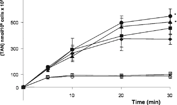
1. Cell energy charge without and with treatment with oligomycin 59
2. Effect of glucose and nucleosides on energy repletion after treatment
with oligomycin 59
3. Activity of enzymes involved in the catabolism of nucleosides 67
4. Determination of purine and pyrimidine salvage in ADF cells 69
5. Activity of enzymes involved in the purine and pyrimidine salvage 75
6 Effect of CTP on the purine and pyrimidine salvage synthesis 75
CHAPTER 4
Discussion 79
CHAPTER 5
INTRODUCTION
1 Astrocyte cells: morphology, physiology and metabolism
1.1 Cells types in the central nervous system
The central nervous system (CNS) is composed of a variety of different cell types, which depend on each other for survival and functional integrity, and have the capacity for intercellular communication.
The mammalian CNS consists of two main cell types, neurons and glia. Neurons are directly involved in electrical transmission and information processing, and glia carry out many indispensable functions, both in development and during the normal function of the mature CNS.
Glial cells are numerically the dominant cell type in the central and peripheral nervous system. They are intermixed with the nerve cells and are found in intimate contacts with neuronal cell bodies, dendrites, axons and synaptic contacts. Like neurons, glial cells are a heterogeneous population of cells that differ in developmental origin, molecular composition, structure and activity. When these cells were first discovered some 150 years ago, they were viewed as a type of connective tissue support for nerve cells. They were considered to assume a passive role of structural support for neurons and to provide a preferential, but metabolically inert, route for the passage of glucose from the circulation to neurons. More recent reports, however suggest that they have much more important and dynamic functions in the brain and other parts of the nervous system: they respond to synaptic activity and they play an important role in the communication network within the central nervous system (Voutsinos-Porche et al., 2003; Jessen et al., 2006).
Glial cells in the CNS are represented by three main types: macroglia (astroglia, oligodendroglia) and microglia. The macroglial cells are of ectodermal origin, whereas microglia stem from the mesoderm; the functions of oligodendrocytes and microglial cells are rather well defined: the oligodendrocytes are responsible for myelination and metabolic support of axons, whereas the microglial cells are involved in brain immune reactivity and defence. In particular,microglia phagocytize foreign particles and cellular debris, and produce many immune factors.
1.2 Astrocyte cells: morphology
Astrocytes (Fig.1) are the most abundant type of glial cell, and in contrast with microglia, are much more intimately involved in the formation of CNS cellular circuits and in information processing in the brain (Verkhratsky et al., 2006).
There are generally two types of astrocytes, protoplasmic and fibrous, similar in function but distinct in morphology and distribution. Protoplasmic astrocytes have short, thick, highly branched processes and are typically found in grey matter.
Fibrous astrocytes have long, thin, less branched processes and are more commonly found in white matter.
Fig.1 Culture of astrocyte cells
Astrocytes participate in numerous functions including structural and metabolic support for neurons, homeostatic and signalling functions in the brain including synthesis of the citric acid cycle (TCA) intermediates, regulation of extracellular glutamate and cation levels, energy storage of glycogen and regulation of blood flow (Mulligan et al., 2004).
Neuronal networks are physically discontinuous, with neurons being separate entities. The integration and communication within these neuronal networks is provided by specialised structures, the synapses, which are considered the substrates of chemical neurotransmission (Verkhratsky et al., 2006).
Because of their capacity to detect and respond to neuronal activity, glial cells, and in particular astrocytes, may be an integral part of the communication network within the central nervous system. Among their new emerging roles, glial cells have been shown to enhance synapse number and spontaneous synaptic activity between developing neurons in culture and to play a role in the stabilization and maintenance of synapses (Voutsinos-Porche et al., 2003).The modulation of synaptic transmission by glial cells has been also described at the neuromuscular junction, in hippocampal cultures, and in the retina (Voutsinos-Porche et al., 2003).
Astroglia divide the grey matter into distinct compartments, and within each of these compartments, a single astrocyte forms contacts with all neuronal membranes and synapses residing within its confines. These contacts are created by fine astroglial processes, called filopodia and lamellipodia. These are highly dynamic structures: lamellipodia being able to glide along neuronal surfaces, whereas filopodia are rapidly extended from the astroglial processes and surround synapses (Verkhratsky et al., 2006). Mitochondria are too large to fit within fine processes, and the filopodia are, therefore, likely to be highly dependent on glycolytic metabolism of glucose and glycogen as local fuel sources. Thus, glycolysis is expected to predominate in astrocytic peripheral fine processes that are too small for mitochondria, whereas oxidative metabolism would be carried out in the soma, larger processes and end-feet (Dienel et al., 2005).
Astrocyte cells not only form interactions with neurons but they also provide a link between neurons and blood capillaries. In fact the end-feet of several astrocytes cover the capillary wall in order to form a glial-vascular interface which is a part of the blood-brain barrier (BBB) (Verkhratsky et al., 2006). These processes express glucose transporters suggesting that astrocytes may constitute a privileged site of glucose uptake as it penetrates within the brain parenchyma (Morgello et al., 1995).
Some astrocyte processes contact blood–brain barrier, also in order to protect the CNS from being exposed to endogenous neurotoxins present in the general circulation (Alberdi et al., 2005).
1.3 Astrocytoma cells
Astrocytomas are primary intracranial tumours derived from astrocyte cells of the brain. They may arise in the cerebral hemispheres, in the posterior fossa, in the optic nerve, and rarely, the spinal cord (Mangiardi et al., 1990). Well-differentiated astrocytomas constitute about 25 to 30% of cerebral gliomas. They have a predilection for the cerebrum, cerebellum, hypothalamus, pons, and optic nerve and chiasm.
Astrocytic tumours are the most common type of gliomas. They can occur in most parts of the brain and occasionally in the spinal cord. However, they most commonly grow in the main part of the brain, the cerebrum (Mangiardi et al., 1990). People of any age can develop an astrocytic tumour, but those in the cerebrum are more common in adults, and the chance of developing one increases as people get older. Astrocytic tumours in the cerebellum are more common in children or young people (Mangiardi et al., 1990). Regional effects of astrocytomas include compression, invasion, and destruction of brain parenchyma. Arterial and venous hypoxia, competition for nutrients, release of metabolic end products (eg, free radicals, altered electrolytes, neurotransmitters), and release and recruitment of cellular mediators (eg, cytokines) disrupt normal parenchymal function (Mangiardi et al., 1990). The astrocytoma cells prefer to produce ATP through glycolysis rather than oxidative phosphorylation and the cells continue with the lactate production also when oxygen is present (Mangiardi et al., 1990). In fact in these cells the Krebs cycle and the oxidative phosphorylation seem to be partially blocked; moreover in the astrocytoma cells there is a decrease of the mitochondrial number in comparison with normal cells (Mangiardi et al., 1990). On the other hand in these cells the pentose phosphate pathway is more active compared to normal cells; therefore these cells are able to produce a great amount of ribose P which is absolutely necessary for 5-phosphoribosyl-1-pyrohosphate (PRPP) production which is a common substrate for the purine and pyrimidine synthesis (Mangiardi et al.,1990). Astrocytoma cells show a relatively low activity of purine metabolism when compared with normal brain and non-glial brain neoplasms (Bardot et al., 1994).
2. Energetic metabolism in the central nervous system
2.1 Glucose
Glucose (Glc), a monosaccharide, is the most important carbohydrate in biology. The cell uses it as a source of energy and metabolic intermediates. Glucose is one of the main products of photosynthesis and starts cellular respiration in both prokaryotes and eukaryotes. Glucose is considered the major if not exclusive energy source for mammalian brain and a continuous supply of this substrate is essential to maintain normal cerebral function. This indicates that oxidative phosphorylation is the most important process used to generate from glucose the energy necessary to sustain the brain functions (Ioudina et al., 2004). Glucose crosses the blood-brain barrier and enters neurons or glial cells via specific glucose transporters and is metabolized via glycolysis and the Krebs cycle in order to produce ATP. The brain is able to maintain cerebral energy metabolism due, in part, to the relatively high glucose concentration in the plasma and the presence of glucose transporters in the capillary endothelial cell, glial cells and neurons. In the rat brain, extracellular glucose concentration ranges from 0.7 to 2.5 mM, corresponding to plasma glucose levels. The total glucose deprivation leads to neuronal degeneration while the effects of a moderate deprivation of glucose are not well understood (Ioudina et al., 2004). In the cells grown in a low glucose media there is a decrease in the cellular ATP and cell viability and an increase of caspase-3 activity which leads to apoptosis and therefore the glucose deprivation could be a serious risk factor that potentiates the pathophysiological consequences of certain neurodegenerative diseases (Ioudina et
al., 2004).
During the physiologic brain activity, glucose crosses the blood-brain barrier via facilitative transporters named glucose transporter 1 (GLUT1). Once within the brain parenchyma, it is taken up by both neurons and astrocytes via glucose transporter 3 (GLUT3) and GLUT1, respectively. In particular, about half of the glucose leaving the capillaries crosses the extracellular space and directly enters neurons; the other half is taken up by astrocytes (Nehlig et al., 2007). GLUT 1 is expressed in all cell types, and it is the only glucose transporter identified in the brain. In the brain it is present at a high concentration at the blood-brain barrier as well as in parenchymal cells, most likely in astrocytes. In particular, GLUT 1 is detected in astrocytic end-feet around blood vessels, and in astrocytic cell bodies and processes in both grey and white matter. GLUT 3, in the brain, is expressed in neurons; in particular is located primarily in pre- and postsynaptic nerve endings and in small neuronal processes (Maher et al., 1994).
In each cell type, glucose is metabolized via glycolysis into pyruvate, leading to the formation of 2 ATP. Then, pyruvate is further metabolized via the TCA cycle, and in conjunction with oxidative phosphorylation using oxygen provided by the blood circulation, it generates 30 ATP (Maher et al., 1994). However calculations suggest that neurons consume more energy than do astrocytes, implying that astrocytes transfer an intermediate substrate to neurons.
Experimental approaches in vitro on the honeybee drone retina and on the isolated vagus nerve also point to a continuous transfer of brain intermediate metabolites in
vivo and in vitro as is well documented in the ANLSH theory (Nehlig et al., 2007).
2.2 Astrocyte-neuron-lactate shuttle theory (ANLSH)
There is controversy at present concerning the metabolic changes that accompany neuronal activation and the energy substrate used by the activated neurons. The traditional theory considers the glucose as the exclusive compound involved in the production of energy necessary for neuronal and astrocyte survival. As already described, glucose is utilized by both neurons and astrocytes in order to produce pyruvate which can enters the TCA cycle and oxidative phosphorylation for ATP production.
Another theory concerning the glucose utilization by neuronal and astrocyte cells is the astrocyte-neuron-lactate shuttle (ANLSH), revealed for the first time by Pellerin and Magistretti (1999). The ANLSH model postulates that activated neurons use lactate which is provided by astrocytes (Fig.2). It is well known that the major amount of energy consumption (90-95%) can be attributed to neurons whereas glial cells contribute only in a small fraction (5-10%) under normal level of activity; on the other hand, several studies reveal that 80% of glucose utilization is taking place in glial cells. And so, to fulfil the greater energy needs of neurons, glial cells must release a metabolic intermediate from glucose that will be eventually taken up and oxidized by neurons (Pellerin and Magistretti, 1999, 2004).
Fig.2 Schematic illustration of the astrocyte-neuron-lactate shuttle (ANLSH) (Image taken from www.nature.com)
It has been known that glial cells and in particular astrocytes are able to produce a large amount of lactate in the presence of normal oxygen levels; this lactate production is enhanced during a neuronal synaptic activity at glutamatergic synapses: after this stimulation there is a significant increase of the extracellular neurotransmitter glutamate which, via glutamate transporters (GLAST or GLT1), enters astrocyte cells and causes an increase in both glucose utilization and lactate production. In addition, glutamate causes a rapid stimulation of glucose transport in astrocytes and it is considered the fastest glucose transport response ever recorded in any other cell type. Lines of evidence of this mechanism have been provided in vivo: in knockout mice for either GLAST or GLT1 transporters, a significant reduction of the glucose utilization during a neuronal stimulation has been observed (Pellerin and Magistretti, 1999, 2004).
Glutamate transporters operate by coupling glutamate transport with the Na+ gradient. For each glutamate molecule transported within the cell, three Na+ (or two Na+ and one H+) ions are cotransported together with one proton while one K+ ion and one OH- is extruded. As a consequence, a significant Na+ influx occurs in astrocytes, with an accumulation of Na+ inside the cell accompanied by an intracellular acidification and extracellular alkalinization (Danbolt et al., 2001).
Changes in the intracellular sodium concentration modulate the activity of the α2
isoform of the Na+, K+-ATPase pump and this correlation is demonstrated in several cell types and also in the astrocyte cells (Pellerin and Magistretti, 1999, 2004). The Na+ influx not only activates the Na+-K+ ATPase but also the enzyme glutamine synthase which leads to the production of glutamine with a concomitant increase of ADP, Pi and AMP levels and a decrease of levels of ATP. As a consequence, an activation of anaerobic glycolysis, which generates lactate from glucose by LDH-5 (lactate dehydrogenase-5) and restores the ATP levels, is observed (Pellerin and Magistretti, 1999, 2004).
The lactate is transported across the glial membrane and leaves the astrocytes by a specific monocarboxylate transporter (MCT1); once in the extracellular space, the increased concentration gradient leads to its net transport into neurons by its own specific monocarboxylate transporter MCT2. The increased lactate in the neurons is converted to pyruvate via LDH-1 (lactate dehydrogenase-1), which enters the TCA cycle and oxidative phosphorylation with the final ATP production (Pellerin and Magistretti, 1999, 2004). The generation of ATP serves also for the production of the neurotransmitter glutamate. Therefore, in this theory there is a metabolic coupling between astrocytes and neurons in brain tissue and the neurons should prefer lactate to glucose as substrate, particularly during periods of intense neuronal activity (Pellerin and Magistretti, 1999, 2004).
2.3 Glycogen
Glycogen is a polysaccharide that is the principal storage form of glucose in animal cells. Glycogen energy stores in the brain are made exclusively by astrocytes which express the gluconeogenesis and glycogenolysis enzymes while neurons do not have these enzymes (Maher et al., 1994). Glycogen is a fuel in the normal astrocytic functions during brain activation, in fact glycogen is located mainly in astrocytes and widely distributed throughout brain, and glycogen phosphorylase is present in the soma, end-feet and fine processes (Dienel et al., 2006). Glycogen levels change during stress conditions: during physiological stimulation the rate of glycogenolysis in brain rises 50-fold above that in the resting state, whereas it increases more than 200-fold during an energy crisis when oxygen or glucose supply is inadequate or eliminated (Magistretti, 1993).
Neurons are more sensitive to ATP depletion than astrocytes. In fact astrocytes, but not neurons, are capable of storing glycogen and glycogenolysis is another mechanism by which astrocytes can resist ischemia-like conditions (Parkinson and Xiong, 2004). Glycogenolysis produces glucose-6-P but the metabolic fate of this compound derived from glycogen in vivo is not well known, moreover the activity glucose-6-phosphatase which produces glucose from glucose-6-P in brain and cultured astrocytes is very low and therefore, the glycogen metabolism generates lactate, which is released to the tissue culture medium (Dienel et al., 2006). On the contrary, the plasma lactate cannot fully substitute for glucose as a metabolic substrate for brain because of its limited permeability across the blood-brain barrier (Dienel et al., 2006).
In response to an acute hypoxic challenge, astrocytes respond to hypoxia by mobilizing their glycogen stores and by increasing their glucose utilization and lactate release for their own survival as well as for the neighbouring cells. In fact in the astrocyte cells, during hypoxia, there is an enhancement of the expression of the sodium-dependent and independent glucose transporters and also of the astrocytic monocarboxylate transporters isoform MCT1 (Vega et al., 2006; Brown et al., 2007). Astrocyte glycogen, therefore, offers some protection against hypoglycemic neural injury and ensures that neurons and axons can maintain their function during very intense periods of activation. These emerging principles about the roles of astrocyte glycogen contradict the long held belief that this metabolic pool has little or no functional significance (Brown et al., 2007). After a prolonged hypoxia exposure, astrocytes decrease their glucose uptake and the lactate release, conserving lactate to ensure their own survival to the detriment of surrounding cells that could use the released lactate as a source of energy (Vega et al., 2006). These findings could explain the higher susceptibility of neurons to hypoxia and their earlier cell death compared with astrocytes when exposed to long-term hypoxic challenges (Vega et
2.4 Lactate
Lactic acid is a chemical compound that plays a role in several biochemical processes. In animals, L-lactate is constantly produced from pyruvate via the enzyme lactate dehydrogenase (LDH) in a process of fermentation during normal metabolism and exercise. A number of recent observations have challenged the canonical view of glucose as the sole energy substrate delivered directly to neurons via the blood supply to sustain their activity: many lines of evidence reveal that monocarboxylates, and in particular lactate, might play useful roles in the adult central nervous system (Parkinson and Xiong, 2004).
Neurons, astrocytes, and oligodendrocytes use lactate as a preferential substrate for both energy purposes and as precursor of lipids (Parkinson and Xiong, 2004). Astrocytes use lactate and other metabolic substrates for the synthesis of oleic acid a fatty acid which is able to increase the fluidity of the neurite bases suggesting that increased fluidity is required at the sites of newly emerging axons and dendrites. Oligodendrocytes mainly use lactate as precursor of lipids, presumably those used to synthesize myelin. Neurons use lactate as a source of energy and as precursor of lipids (Smith et al., 2003).
Lactate produced by muscle during strenuous exercise could be used as a fuel source by the brain (Smith D. et al., 2003). During the perinatal period, neurons may use blood lactate directly to meet the need for the energy and carbon skeletons required for proliferation and differentiation (Medina and Tabernero, 2005). Lactate is a hydrophilic compound which requires transporters to cross cellular membranes. A family of proton-linked monocarboxylate transporters has been described. It contains 14 members sharing sequence homologies and identified as MCT1 to 9 and MCT11 to 14 as well as another member known as TAT1.
A distribution of specific MCTs between different cell types is consistent with a role in mediating a transfer of energy substrates from astrocytes to neurons. It is also correlated with a differential distribution of lactate dehydrogenase (LDH) isoforms: LDH1 appears to be the predominant isoform in neurons, whereas LDH5 is found predominantly in astrocytes (Laughton et al., 2000).
Lactate dehydrogenase is the enzyme catalysing the interconversion of pyruvate and lactate, and is thus essential for both production and utilization of lactate. It is a tetrameric enzyme which can consist of three types of subunit arising from three distinct genes: LDH-A, LDH-B and LDH-C The product of the LDH-C gene is found exclusively in the testis, where it forms a homogeneous tetramer (C4). The LDH-A and LDH-B gene products are more widely expressed, and both are found in the CNS (Laughton et al., 2000; Medina and Tabernero, 2005).
Hypoxia is the oxygen deprivation which provokes a mitochondrial dysfunction and a diminution in the levels of the intracellular energy metabolites which leads to a drastic reduction of ATP production with a concomitant neurodegeneration through both apoptotic and necrotic mechanisms (Cater et al., 2003). In terms of energy metabolism, during hypoxia, there is a rapid switch from oxidative phosphorylation to anaerobic glycolysis, in order to maintain the cellular energy charge. Under normal physiological conditions the NAD+ molecules necessary to drive the glycolytic reactions are regenerated via the TCA cycle.
During anaerobic conditions, the TCA cycle cannot operate in the absence of oxygen so an alternative source of NAD+ is provided through the conversion of pyruvate, derived from glucose, to lactate (Cater et al., 2003).
Therefore, during oxygen deprivation in nervous tissue, a significant rise of the lactate concentrations is observed which could lead to acidosis and for this reason, lactate for a long period was considered the major factor in the resultant neuronal degeneration (Cater et al., 2003). The amount of acidosis differs between animal models, yet intracellular pH typically acidifies to the range of pH 6.5–6.7 (Cater et
al., 2003).
A research in vitro suggests that the role of lactate during neuronal stress may, on the contrary, be beneficial under certain circumstances (Cater et al., 2003). There are also some lines of evidence in vivo that lactate is neuroprotective in a number of ischemic/excitotoxic models as well as following severe insulin-induced hypoglycemia (Schurr et al., 2001).
3 Biochemistry of the purine and pyrimidine nucleotide compounds
3.1 General aspects
A nucleotide is a chemical compound that consists of a heterocyclic base, a sugar, and one or more phosphate groups. In the most common nucleotides the base is a derivative of purine or pyrimidine, and the sugar is the pentose (five-carbon sugar) deoxyribose or ribose. All types of cells (mammalian, bacterial and plant) contain a wide variety of nucleotides and their derivatives. In normal cells the total concentrations of the nucleotides are fixed within rather narrow limits, although the concentration of the individual components can vary. The total concentration of adenine nucleotides (AMP, ADP and ATP) is constant, although there is a variation in the ratio of ATP to AMP+ADP, depending on the energy state of the cells.
Deoxyribonucleotides are formed by the direct reduction of the 2’ position of the corresponding ribonucleotides. This reaction occurs at the level of the nucleoside diphosphates and is catalyzed by nucleoside diphosphate reductase. It is generally accepted that only liver and kidney maintain the de novo pyrimidine and purine synthesis and supply other tissues and organs, including brain, with preformed pyrimidine nucleosides and purine nucleosides and bases.
In the central nervous system, most of the enzymes involved in the purine nucleotide metabolism (except for the de novo pathway and of AMP-5'-nucleotidase), exhibits a higher activity in the astroglia, in comparison to the neurons. Thus, as for other metabolic pathways, the glia appears to be more active than the neurons (Zoref-Shani, et al., 1995).
The functions exerted by nucleotides are:
1. role in energy metabolism: ATP is the main form of chemical energy available to the cell and is generated by oxidative phosphorylation and substrate-level phosphorylation. The ATP is utilized to drive metabolic reactions as a phosphorylating agent and is involved in several processes as muscle contraction, active transport and maintenance of cell membrane integrity. ATP donates the phosphate also for the generation of the other nucleoside 5’-triphosphates (GTP, UTP and CTP). GTP is considered an important non-protein organic cofactor and enzyme regulator present in all cells. GTP produces energy for the assemblage of ribosome, kinesins and myosines.
2. monomeric units of nucleic acids DNA and RNA.
3. mediators of key metabolic processes. (es. cyclicAMP, involved in the molecular signal evoked by hormones)
4. components of coenzymes (NAD, NADP, FAD and CoA) which are involved in many metabolic pathways.
5. allosteric effectors; in fact, many of the regulated steps of the metabolic pathways are controlled by the intracellular concentrations of nucleotides.
3.2 Metabolic pathways of purines and pyrimidines
3.2.1 De novo purine synthesis
Purine nucleotides may be formed de novo, from small molecules, or by salvage, from preformed purines. The former pathway is associated mainly with cell proliferation, requiring formation of new nucleic acids, whereas the latter pathway is associated with maintenance of cellular purine pool size (Zoref-Shani et al., 1995). The de novo synthesis of purine nucleotides in which these precursors are incorporated into the purine ring, proceeds by a 10 step pathway to the final point intermediate IMP, the nucleotide of the base hypoxanthine and AMP and GMP are subsequently synthesized from this intermediate via separate, two step each, pathways. Thus purine moieties are initially formed as part of the ribonucleotides rather than as free bases. The basal precursor for the de novo synthesis of purine (and pyrimidine) is 5-phosphoribosyl-1-pyrohosphate (PRPP). PRPP is formed by ribose 5-phosphate and ATP by a reaction catalyzed by PRPP synthetase.
The purine ring atoms is built using a variety of different precursors: N1 of purines arises from the amine group of aspartate; C2 and C8 originate from N10-formyltetrafolate; N3 and N9 are contributed by the amide group of glutamine; C4, C5 and N7 are derived from glycine; C6 comes from HCO3- (CO2). The de novo
synthesis requires directly four ATP molecules.
In the whole brain, there is a maturation-dependent decrease in the capacity to produce purines de novo, and this particular trend reflects the trend observed in the astroglia, rather than that in the neurons (Zoref-Shani et al., 1995).
3.2.2 Purine catabolism
The end product of purine catabolism in man is uric acid. Other mammals have the enzyme urate oxidase and excrete the more soluble allantoin as the end product. Man does not have this enzyme so urate is the end product. Uric acid is formed primarily in the liver and excreted by the kidney into the urine (Fig.3).
Fig.3 Schematic illustration of purine catabolism (Image taken from web.virginia.edu)
Nucleotides are hydrolyzed to nucleosides by the enzyme family of 5’-nucleotidase (5NT) which have a different subcellular location and a remarkable low sequence homology; 5’-nucleotidases hydrolyze the phosphate esterified at the 5’ position of purine and pyrimidine nucleoside monophosphates (Allegrini et al., 1997, 2004; Pesi et al., 2004).
ATP degradation proceeds via AMP, which can be either dephosphorylated to adenosine, catalyzed by AMP-5'-nucleotidase, or deaminated to IMP, catalyzed by the enzyme adenylate (AMP) deaminase. The reaction catalyzed by AMP deaminase is the rate-limiting step in the operation of the purine nucleotide cycle (AMP →IMP → adenylosuccinic acid →AMP). This cycle has a major role in the maintenance of the normal energy charge in the cell, as well as in the preservation of cellular purine nucleotide content. Guanine nucleotides are hydrolyzed to the nucleoside guanosine which undergoes phosphorolysis to guanine and ribose 1-P.
In the catabolism of purine nucleotides, IMP is further degraded by hydrolysis with nucleotidase to inosine and then phosphorolysis to hypoxanthine in a reaction catalyzed by purine nucleoside phosphorylase (PNP), which acts also on guanosine and xanthosine in order to produce guanine and xanthine and Rib1-P. Based on their structural properties, nucleoside phosphorylases have been classified into two families: NP-I and NP-II. The NP-I family includes homotrimeric and homohexameric enzymes from both prokaryotes and eukaryotes acting on inosine, guanosine, adenosine and uridine. The NP-II family includes homodimeric proteins structurally unrelated to the NP-I family, such as bacterial pyrimidine phosphorylases and eukaryotic thymidine phosphorylase (Tozzi et al., 2006). It has been shown that the activity of purified PNP from bovine spleen may be inhibited by the accumulation of its metabolic products (hypoxanthine and guanine), but it is also inhibited by a negative control exerted by the respective substrates, inosine and guanosine, especially when their concentrations are elevated, to the micromolar range (Tozzi et al., 2006).
The equilibrium of PNP-catalysed reactions is thermodynamically in favour of nucleoside synthesis. Nevertheless, in vivo the equilibrium of the PNP reaction is shifted towards Rib-1-P accumulation because the intracellular concentration of Pi is higher than that of nucleosides inosine and guanosine and also because hypoxanthine and guanine are consumed by hypoxanthine-guanine phosphoribosyltransferase (HPRT) and, in certain tissues, by xanthine oxidase or guanase, respectively (Tozzi
et al., 2006). The phosphorolysis of inosine and guanosine is favoured also because mammals lack the kinase acting on inosine and guanosine (Tozzi et al., 2006). PNP is generally considered as an intracellular enzyme. In fact, the presence of this enzyme is not demonstrated extracellularly (Tozzi et al., 2006).
Adenosine is deaminated to inosine by an adenosine deaminase. Both adenine and guanine nucleotides converge at the common intermediate xanthine. Hypoxanthine is oxidized to xanthine by the enzyme xanthine oxidase. Guanine is deaminated, with the amino group released as ammonia, to xanthine. Xanthine, like hypoxanthine, is oxidized by oxygen and xanthine oxidase with the production of hydrogen peroxide. In man, the urate is excreted and the hydrogen peroxide is degraded by catalase. Xanthine oxidase is present in significant concentration only in liver and intestine. The pathway to the nucleosides, possibly to the free bases, is present in many tissues. The key enzyme for the catabolism of the pentose moiety of deoxyribonucleosides is deoxyriboaldolase, which cleaves deoxyRib-5-P into acetaldehyde and glyceraldehyde 3-P.
3.2.3 Purine salvage
Whereas some organs such as kidney and liver or some cell types such as hepatocytes can synthesize purine and pyrimidine nucleotides de novo, others, in particular protozoan parasites and cells in the brain and bone marrow, rely on salvage pathways for purine and pyrimidine nucleotide synthesis (King et al., 2006). In the cultured astroglia, de novo purine synthesis is found to decrease with aging, but the activity of the salvage enzyme HGPRT, is found to increase (Zoref-Shani et al., 1995).
In contrast to the glia, in the cultured neurons, the rate of de novo purine synthesis is found to increase with maturation (Zoref-Shani et al., 1995). The finding of the age-related increase in the rate of de novo purine synthesis in the non-dividing neurons, may be interpreted to indicate that the functioning mature neurons, more than the glia, have to cope with loss of purines from the cells (Zoref-Shani et al., 1995).
The de novo synthesis of purine and pyrimidine requires a relatively high input of energy in the form of ATP. To compensate for this, most cells have developed a very efficient salvage pathway, by which the preformed purine and pyrimidine bases can be reutilized and so, the cell obtains a considerable energy saving for itself. The synthesis of nucleotides from the purine bases and purine nucleosides takes place in a series of steps known as the salvage pathways. The free purine bases, adenine, guanine, and hypoxanthine, can be reconverted to their corresponding nucleotides by phosphoribosylation. Two key transferase enzymes are involved in the salvage of purines: adenosine phosphoribosyltransferase (APRT), which catalyzes the following reaction:
adenine + PRPP ↔AMP + PPi
and hypoxanthine-guanine phosphoribosyltransferase (HGPRT), which catalyzes the following reactions:
hypoxanthine + PRPP ↔IMP + PPi and guanine + PRPP ↔ GMP + PPi;
these reactions are important not only because they conserve energy, but also because they permit cell such as erythrocytes, neurons and astrocytes (which do not possess the de novo purine synthesis) to form nucleotides from the bases. AMP is an inhibitor of APRT and IMP and GMP regulates the HGPRT activity.
PRPP may be considered as a high-energy sugar phosphate, with a high potential of 5-phosphoribosyl transfer; PRPP is also the common precursor of both de novo and salvage synthesis of nucleotides. During physiological cell conditions, the PRPP pool is maintained at low level, to avoid excessive and unbalanced nucleotide synthesis. PRPP level is regulated by PRPP synthetase and by phosphoribosyl transferases which are the major factors maintaining the low intracellular level of PRPP. PRPP level is also regulated by the inosine nucleoside cycle (INC) constituted by HGPRT (which forms IMP from hypoxanthine), PNP (which forms inosine from hypoxanthine and vice versa) and cN-II (which forms inosine from IMP). Some experiments exerted in rat brain extracts reveal that when PRPP is present, the intermediates of the INC are continuously recycled but as soon as PRPP disappears, HGPRT becomes inactive and the cycle is interrupted causing IMP degradation by cN-II, and hypoxanthine accumulation. The [Pi]/[ATP] ratio regulates the inosine cell cycle and the PRPP levels: in fact, the allosteric activators of cN-II are 2,3-bisphosphoglycerate (BPG),and ATP while Pi is an inhibitor of the enzyme and at the normal low [Pi]/[ATP] ratio, as found in the well-oxygenated cells, cN-II is fully active and the velocity of the cycle is maximal and the PRPP pool is maintained at a low level (Barsotti et al., 2002). On the other hand, during ischemia the [Pi]/[ATP] ratio raises drastically and cN-II is inhibited while PRPP synthetase is activated: as consequence the PRPP level increases and PRPP is made available for purine nucleotide salvage synthesis (Barsotti et al., 2002).
3.2.4 De novo pyrimidine synthesis
The pyrimidine ring is synthesized de novo in mammalian cells utilizing amino acids as carbon and nitrogen donors and CO2 as a carbon donor. In particular the N1,
C4 and C6 of the pyrimidine ring derive from aspartate, while the N3 from glutamine and C2 from CO2. As is true with purine nucleotides, the sugar phosphate portion of
the molecule is supplied by PRPP. Pyrimidine synthesis begins with carbamoyl phosphate II (CPII) synthesized in the cytosol of those tissues capable of making pyrimidines (highest in spleen, thymus and testes). Carbamoyl phosphate condenses with aspartate in the presence of aspartate carbamoyl transferase to yield N-carbamylaspartate which is then converted to dihydroorotate by dihydroorotase and subsequently, oxidated into orotate by dihydroorotate dehydrogenase. Orotate is converted to its nucleotide, orotidine 5’-monophosphate (OMP) with PRPP, which is the ribose-5-phosphate donor. This reaction is catalyzed by orotate phosphoribosyltransferase. In the last step pathway, OMP is decarboxylated into UMP by OMP-decarboxylase. After conversion of UMP to the triphosphate, the amide of glutamine is added, at the expense of ATP, to form CTP by CTP synthetase. The de novo synthesis of purine differs by the de novo synthesis of pyrimidine in two major respects. First, in purine nucleotide synthesis, the N-glycosidic bond is formed in the first committed step of the pathway. In the pyrimidine pathway, the first step is the formation of the ring and then the phosphate is added. Second, all the enzymes of the purine nucleotide pathway are in the cytosol, while in the case of the pyrimidine synthesis, the enzyme dihydroorotate dehydrogenase is localized in the mitochondria.
3.2.5 Pyrimidine catabolism
In contrast to purines, pyrimidines undergo ring cleavage and the usual end products of catabolism are β amino acids plus ammonia and carbon dioxide. Pyrimidines from nucleic acids or the energy pool are acted upon by nucleotidases and pyrimidine nucleoside phosphorylase to generate the free bases. Cytidine and deoxycytidine are deaminated to uridine and deoxyuridine, by the nucleoside deaminase. Uridine phosphorylase catalyzes the phosphorolysis of uridine, deoxyuridine and deoxythimidine. As already described, the equilibrium of the reaction catalyzed by PNP is shifted towards Rib-1-P accumulation even if the reaction is thermodynamically in favour of nucleoside synthesis. On the other hand, some in vitro experiments reveal that uridine phosphorylase may catalyse the Rib-1-P-mediated rybosilation of 5-fluorouracil and uracil, even in the presence of excess of Pi (Mascia et al., 1999, 2001). Uridine phosphorylase plays an important role in the homeostatic regulation of uridine concentration in plasma and tissues (Pizzorno
3.2.6 Pyrimidine salvage
Pyrimidine nucleotide salvage is important for the synthesis of RNA and DNA. Moreover, salvage pyrimidine synthesis is used also for phosphatidylcholine and phosphatidylinositol synthesis in lymphoid cells. In particular, in CNS the continued biosynthesis of phospholipids needs specific activated pyrimidine nucleotides. Even the conversion of uracil to L-alanine is important in neuronal cells since L-alanine, which is produced and exported by liver, enters the CNS and may act as a neurotransmitter, as well as a building block for various dipeptides (Mascia et al., 1999).
A crucial difference between purine and pyrimidine metabolism is that purines are recycled from their bases while pyrimidines are salvaged from their nucleosides, particularly uridine. In fact, in patients with deficient pyrimidine biosynthesis, only uridine is able to overcome this pathological manifestation but uracil is not (Pizzorno
et al., 2002).
Uracil can be salvaged to form UMP through the concerted action of uridine phosphorylase and uridine kinase, as indicated:
uracil + ribose-1-phosphate ↔ uridine + Pi
uridine + ATP → UMP + ADP
uridine phosphorylase type 1 (UPase-1) activity has been reported in the homogenates of many human tissues and from tumours. However, several studies reveal that that functional UPase-1 is confined predominantly to the liver in humans (Loffler et al., 2005). A second minor human uridine phosphorylase, UPase-2 is identified recently, and is expressed predominantly in the kidney (Loffler et al., 2005). Deoxyuridine is also a substrate for uridine phosphorylase. The salvage of dTMP requires thymine phosphorylase and the previously encountered thymidine kinase:
thymine + deoxyribose-1-phosphate ↔ thymidine + Pi
thymidine + ATP → dTMP + ADP
The salvage of deoxycytidine is catalyzed by deoxycytidine kinase deoxycytidine + ATP ↔ dCMP + ADP
Intracellularly, mitochondrial and cytosolic nucleoside kinases participate in the recycling of uridine, deoxycytidine and deoxythymidine arising from cell turnover or diet, thereby restricting the requirement for the energetically expensive de novo synthesis. Although deoxynucleotides are considered to be used exclusively in DNA replication and repair, deoxycytidine is also known to be salvaged as a precursor for phospholipid synthesis (Loffler et al., 2005).
3.3 Metabolism of the pentose phosphate
Pentose phosphates are heterocyclic, five-membered, oxygen-containing phosphorylated ring structures, with ribose-5-phosphate and 2-deoxyribose-5-phosphate being basal structures of ribonucleotides and deoxyribonucleotides, respectively, and 5-phosphoribosyl-1-pyrophosphate (PRPP). There are two pathways of the pentose phosphate biosynthesis (Fig.4): the pentose phosphate pathway where Rib-5-P is generated from glucose-6-phosphate and the phosphorylase-mediated pathway where Rib-1-P and deoxRib-1-P are supplied by various nucleoside phosphorylases, such as thymidine phosphorylase, uridine phosphorylase and purine nucleoside phosphorylase. Rib-5-P can be also formed from free ribose by the action of ribokinase an enzyme well characterized in bacteria but less investigated in mammals. Phosphopentomutase catalyzes the reversible reaction between Rib-1-P and Rib-5-P and between deoxyRib-1-P and deoxyRib-5-P. This enzyme has been extensively studied in bacteria but has been purified also in human cell lines and tissues. The key enzyme for the catabolism of the pentose moiety of deoxyribonucleosides is deoxyriboaldolase, which cleaves deoxyRib-5-P into acetaldehyde and glyceraldehydes 3-P.
Fig.4 Pathways of the pentose phosphate biosynthesis.
Some experiments performed in rat brain extracts show that the activated ribose, originating from inosine and guanosine phosphorolysis as ribose 1- phosphate, can be transferred to uracil to synthesize uridine by uridine phosphorylase and the nucleoside formed is phosphorylated to UMP by uridine kinase, and hence to UDP and UTP (and possibly to CTP). The transfer of the ribose moiety occurs even in the presence of excess Pi, suggesting that at least in brain, uridine phosphorylase might function in vivo as an anabolic enzyme (Mascia et al., 1999). CTP and UTP, the final products of the pyrimidine salvage, inhibit their production and this inhibition occurs at the level of uridine kinase, the enzyme which regulates the flux (Mascia et al., 1999). The absence of any PRPP involvement in this metabolic pathway, is supported by the lack of uracil phosphoribosyltransferase activity.
The rate of this pyrimidine salvage pathway is about threefold higher in brain than in liver, possibly reflecting the need of CNS to synthesize pyrimidine nucleoside diphosphate sugars for myelin biosynthesis (Mascia et al., 1999). The transfer of the ribose moiety stemming from the purine nucleoside to the uracil via uridine phosphorylase in order to produce uridine and then UMP, UDP and UTP, is also demonstrated in liver and PC12 cells (Mascia et al., 2001). In addition, the ribose 1-P stemming from the purine nucleoside is also able to enter the purine salvage pathway and then produce the purine nucleotide compounds: the pentose phosphate is isomerized into ribose 5-P and then converted to PRPP by PRPP synthetase and PRPP can react with adenine, guanine and hypoxanthine in order to form AMP, GMP and IMP respectively through the action of APRT and HGPRT (Tozzi et al., 2006). On the other hand, deoxyRib-5-P is not a substrate of PRPP synthetase and then the pentose phosphate stemming from the purine deoxynucleoside is not able to enter the purine salvage pathway. Thus the ribose 1-phosphate stemming from the purine nucleoside is able to enter also the pyrimidine salvage pathway in order to produce the nucleotide compounds establishing a metabolic link between purine and pyrimidine salvage (Mascia et al., 1999, 2001; Tozzi et al., 2006).
Ribose 1-phosphate formed from guanosine through the action of purine nucleoside phosphorylase acts as ribose donor also in the synthesis of xanthosine catalyzed by the same enzyme (Giorgelli et al., 1997).
3.4 Adenosine metabolism in the central nervous system
Adenosine is an endogenous nucleoside consisting of the purine base, adenine, in glycosidic linkage with the sugar ribose. Adenosine is generated in the cells and in the extracellular space during normal metabolic activity and can exert its biological activity by ligating specific receptors linked to a variety of signalling systems (Hasko
et al., 2005). In the cells, adenosine is generated from AMP which is dephosphorylated by 5’-nucleotidase and from S-adenosylhomocysteine which is hydrolyzed into adenosine and homocysteine by S-adenosylhomocysteine hydrolase (SAH); the first reaction predominates during metabolically stressful conditions while the second predominates in the physiological conditions (Tozzi et al., 2006).
There are two sources of extracellular adenosine: release of adenosine from the intracellular space via specialized bi-directional equilibrative transporters and extracellular conversion of released adenine nucleotides (ATP, ADP and AMP) by a cascade of ectoenzymes of the ecto-nucleoside triphosphatase (E-NTPase) family (Tozzi et al., 2006). E-NTPases include the ecto-ATPase that preferentially transforms ATP into ADP; the ecto-ATP diphosphohydrolase, also named ectoapyrase, that hydrolyses either ATP or ADP, and the ecto-5’-nucleotidase, GPI-anchored plasma membrane protein, that catalyses the hydrolysis of nucleoside 5’-monophosphate to the nucleoside (Tozzi et al., 2006).
The ectonucleotidases reduce the amount of ATP available for the activation of P2 receptors, limiting the excitatory effects of the nucleotide, and, at the same time, increase the extracellular levels of adenosine, which is also released per se from neurons and glial cells, and may activate A1 and/or A2 receptors (Rathbone et al., 1999). Therefore ectonucleotidases could play an important role in regulating the extent and also the duration of the intracellular signal cascade pathway mediated by P1 and P2 receptors (Rathbone et al., 1999).
The levels of extracellular adenosine also depend on the control of the cytoplasmic adenosine concentrations; intracellular levels of adenosine are kept low principally by its conversion to AMP by the salvage enzyme adenosine kinase, but adenosine may also be degraded to inosine by adenosine deaminase (ADA) (Hasko
et al., 2005). These enzymes are able to maintain adequate levels of adenosine available for nucleic acid and ATP synthesis. The reactions of phosphorylation predominate when adenosine occurs at a low physiological concentration (< 1 µM), whereas adenosine deaminase is activated at higher concentrations of the substrate (> 10 µM) during ATP depletion (Borowiec et al., 2006). ADA is mainly a cytosolic enzyme, thought to maintain a strict control of adenosine levels, preventing cytotoxic actions of adenosine and deoxyadenosine in peripheral tissues and CNS.
In the central nervous system, ADA activity is most prominent in glia, where its activity is at least five times that in peripheral ciliary ganglion neurons and nine-fold that in central neurons (Rathbone et al.,1999). ADA is also expressed in the external surface of the cells in the CNS. In particular, ADA has been found in the synaptic vesicles which contains also nucleotidase and the combined effect of ecto-5’-nucleotidase, nucleoside transport and ADA may serve to regulate the effective local concentration of adenosine and therefore the extent of adenosine receptor activation (Rathbone et al., 1999).
In the central nervous system adenosine is present at low concentrations in the extracellular space and its levels are greatly increased under metabolically stressful conditions (Hasko et al., 2005). Under these conditions, the extracellular concentration of adenosine rapidly rises from nanomolar to micromolar levels (van Calker and Biber, 2005). When adenosine deriving from the intracellular degradation of ATP to adenosine by the metabolic enzyme 5’-nucleotidase, reaches high concentrations inside the neuronal cell, it is expelled into the extracellular space by bidirectional, equilibrative, nucleoside transporters, meaning that the net transport of adenosine either into or out of the cell depends upon the adenosine concentration gradient in both sides of the membrane. Inhibition of adenosine transport can, therefore, inhibits either adenosine release or adenosine uptake, depending upon the intra- and extracellular levels of adenosine (Hasko et al., 2005). On the other hand, under ATP-depleting conditions, cultured astrocytes first release adenine nucleotides which are subsequently hydrolyzed to adenosine extracellularly by ecto-nucleotidases (Parkinson and Xiong, 2004).
3.5 Inosine and guanosine-based purine metabolism in the central
nervous system
Also in CNS, inosine which derives from the deamination of adenosine, is phosphorolytically cleaved by a family of enzymes, the purine nucleoside phosphorylases (PNPs), in order to produce the base, hypoxanthine and the phosphorylated sugar. The base can be reconverted to IMP by phosphoribosylation and the activity of this nucleotide cycle, considered as the main salvage pathway of nucleoside, is demonstrated in the brain and dominates in neurons (Tozzi et al., 2006).
The pathway for the degradation of IMP proceeds through inosine, hypoxanthine and xanthine to uric acid. These reactions are catalyzed by IMP-5'-nucleotidase, PNP and xanthine oxidase, respectively. Xanthine oxidase activity could not be detected in the cultured astroglia. Absence of xanthine oxidase activity is an advantage for the reutilization of hypoxanthine for IMP synthesis (Zoref-Shani et al., 1995).
Inosine is degraded further to the stable end-product uric acid, which has anti-inflammatory properties and, as such, is a potential candidate agent for the treatment of multiple sclerosis (Mattle H.P. et al., 2004).
Guanine in the brain is formed by degradation of guanine nucleotides through the pathway: GMP → guanosine → guanine.
Guanine can be reconverted into GMP in the reaction catalyzed by HGPRT, or converted to xanthine in the reaction catalyzed by guanase; thus, the fate of guanine is determined by the relative activities of these enzymes, since the affinity of both enzymes for guanine is apparently similar (Zoref-Shani et al., 1995). In the 45-day-old rat astroglia, the activity of guanase is four to five-f45-day-old that of HGPRT. This ratio indicates increased advantage for guanine degradation in the form of xanthine, over its reutilization for nucleotide synthesis. A similar situation occurs also in the neurons (Zoref-Shani et al., 1995). However, in cultured astrocytes the activity of phosphoribosyltransferases and guanase increased markedly with age; thus, cultured astroglia are able to metabolise guanine more intensively than are neurons. Guanase is considered one of the major responsible for ammonia production in the brain (Tozzi et al., 2006). Cells, including astrocytes, cannot accumulate deoxy purines, as they are toxic (Tozzi et al., 2006).
3.6 Pyrimidine metabolism in the central nervous system
The circulating pyrimidines uridine and cytidine, besides being incorporated into nucleic acids, can serve as substrates for the salvage pathway of pyrimidine nucleotide synthesis (Cansev et al., 2006).
Uridine and cytidine are also considered as precursors of the cytidine triphosphate (CTP) needed in the phosphatidylcholine (PC) and phosphatidylethanolamine (PE) biosynthetic pathway which are the two major membrane phospholipids and they are precursors for the UDP and UTP that activate brain P2Y receptors and that promote brain glycogen synthesis via UDPglucose (Cansev et al., 2006). Some experiments reveal that an increase in neuronal cytidine and uridine levels augments CTP levels both in vitro and in vivo. In humans, the predominant circulating pyrimidine is uridine whereas in rats, it is cytidine. These variations probably reflect the species differences in cytidine deaminase, the enzyme that converts cytidine to uridine in the body (Cansev et al., 2006).
Except for erythrocytes, liver and kidney, which maintain de novo pyrimidine biosynthesis and supply other tissues with uridine or cytidine for salvage, in humans most normal tissues in adults rely on the salvage of uridine and cytidine (Cao et al., 2005). The homeostasis of uridine, which regulates several physiological and pathological processes, is maintained by the relative activities of two enzymes: the UTP-CTP inhibited uridine kinase and uridine phosphorylase. The first produces the uridilic nucleotide compounds by the nucleoside uridine whereas the second catalyzes the Rib-1-P-mediated ribosylation of uracil in order to produce the nucleoside (Tozzi et al., 2006).
As already described, the reaction catalyzed by uridine phosphorylase acts anabolically and at least in rat brain, the anabolism of uridine mediated by the uridine phosphorylase enzyme, is favoured because the degradation of uracil to β-alanine, which would drive uridine phosphorolysis, is absent and also because multiple consecutive phosphorylations of uridine by the ubiquitous uridine kinase and nucleoside mono and diphosphokinases drive the phosphoribosylation of uracil catalysed by uridine phosphorylase. Moreover, the absence of uracil phosphoribosyltransferase (catalyzing the formation of UMP by uracil and PRPP) in mammals, further leads to the uracil phosphoribosylation (Tozzi et al., 2006).
In rat brain, de novo pyrimidine synthesis, although at lower rates than those of liver, has been described and brain pyrimidine levels depend on de novo synthesis even if circulating cytidine and uridine are essential for maintaining various brain functions. Thus, pyrimidines have to be taken up from the circulation in order to maintain all brain functions. Hence, their plasma concentrations, as well as their transport proteins, will be important in modulating brain levels of cytidine and uridine (Cansev et al., 2006).
The liver has been proposed as the major tissue for regulating plasma pyrimidine levels even if, in humans, the liver may not be the major catabolic tissue for pyrimidines; there may be multiple sites of uptake and degradation such as in erythrocytes (Cansev et al., 2006).