UNIVERSITY OF NAPLES FEDERICO II
DOCTORATEMOLECULAR MEDICINE AND MEDICAL BIOTECHNOLOGY XXXI CICLO
Innovative strategies for the generation of novel human
therapeutic anti-tumor and immunomodulatory antibodies
Tutor Candidate
Prof. Claudia De Lorenzo Margherita Passariello
COORDINATOR
ABSTRACT
Immunotherapy, based on the use of novel human mAbs endowed with antitumor or immunomodulatory activity, is an increasingly important strategy for cancer therapy. Monoclonal antibodies can be directed against Tumor Associated Antigens (TAAs), to inhibit their oncogenic function, or against Immune Checkpoints (IC), to modulate specific T cell responses against cancer. Furthermore, ongoing clinical trials in oncology are currently testing combinatorial treatments of anti-TAA and immunomodulatory antibodies.
In our laboratory novel human antitumor immunoagents have been successfully produced against the TAA ErbB2, which is a Tyrosine Kinase Receptor, overexpressed in breast cancer and several other carcinomas. In particular, a fully human single chain antibody fragment (scFv), named Erbicin, able to bind to an epitope of ErbB2 different from those recognized by the clinically validated mAbs Trastuzumab and Pertuzumab, was isolated by phage display selection on live cells. A human anti-ErbB2 “compact antibody” (100kDa) was also generated by the fusion of Erbicin with a human IgG1 Fc, which was found to efficiently inhibit ErbB2-positive tumor growth both in vitro and in vivo.
As a further progress, a trispecific antibody derivative, named Tribody, targeting three noncompeting epitopes on the extracellular domain of ErbB2 was obtained by fusing 3 binding moieties derived from Erb-hcAb, Trastuzumab and Pertuzumab, respectively. The triparatopic Tribody significantly downregulates ErbB2 and inhibits the cell growth of tumor cells, including those resistant to Trastuzumab. We show here that the multiparatope tribody combines and potentiates the therapeutic effects of the 3 different antibodies in 1 single antibody construct, thus allowing for the reduction of costs of antibody production and overcoming the limits related to monotherapy associated drug resistance.
On the other hand, we performed a massive parallel screening of phage antibody library to obtain a large repertoire of fully human
immunomodulatory antibodies against several-immune regulatory checkpoints to be used in monotherapy or in combinatorial treatments for cancer therapy. We used for the first time an innovative selection strategy on human activated lymphocytes to generate a large collection of scFvs against 10 different IC, called “Immunome Library”, from which scFvs specifically recognizing a given receptor could be pulled out by subsequent affinity selection cycles on recombinant purified proteins used as baits. By Next Generation Sequencing and bioinformatic analysis we ranked individual scFvs in each collection and identified those with the highest level of enrichment. Human IgGs from three of these collections (i.e. PD-1, PD-L1 and LAG-3) were generated and tested for their binding and biological activity. In particular, they were found to specifically bind to their targets with high affinity, to efficiently activate T cell proliferation, induce cytokine secretion and inhibit in vivo tumor growth. Interestingly, the novel isolated mAbs have comparable or even better binding affinity and biological activity than the clinically validated anti-PD-1 mAb Nivolumab.
SOMMARIO
L’immunoterapia, basata sull’impiego di nuovi anticorpi umani dotati di attività antitumorale o immunomodulatoria, rappresenta un’importante strategia per la terapia del cancro. Gli anticorpi monoclonali possono essere diretti contro Antigeni Associati a Tumore (TAA), allo scopo di inibire la loro funzione oncogenica, oppure contro proteine di superficie di cellule del sistema immunitario (Immune Checkpoints, IC), per modulare specifiche risposte dei linfociti T contro il cancro. Peraltro, sperimentazioni cliniche in corso nel campo dell’oncologia stanno attualmente valutando l’efficacia di trattamenti combinatoriali che includono anticorpi contro TAA e anticorpi con attività di modulazione del sistema immunitario.
Nel nostro laboratorio sono stati prodotti con successo nuovi immunoagenti umani con attività antitumorale diretti contro il Recettore Tirosina-Chinasico ErbB2, un noto Antigene Associato a Tumore, iper-espresso nel carcinoma mammario ed in diversi altri tipi di tumore. In particolare, è stato isolato, mediante la tecnologia “phage display” applicata su cellule tumorali vive, un frammento anticorpale interamente umano, chiamato Erbicin, capace di legare un epitopo del recettore ErbB2 diverso rispetto a quello riconosciuto dagli anticorpi Trastuzumab e Pertuzumab, attualmente in uso clinico. Mediante la fusione di Erbicin con il dominio Fc di un anticorpo umano con isotipo IgG1 è stato anche generato un “anticorpo compatto” umano (ErB-hcAb, 100kDa) contro il recettore ErbB2, che si è mostrato capace di inibire efficientemente, sia in vitro che in vivo, la crescita di tumori ErbB2-positivi. Come ulteriore progresso, la fusione dei 3 domini di legame derivati da Erb-hcAb, Trastuzumab e Pertuzumab, ha consentito di ottenere un derivato trispecifico, chiamato Tribody, che riconosce 3 epitopi non competitivi sul dominio extracellulare del recettore ErbB2. Il Tribody triparatopico riduce significativamente i livelli di ErbB2 ed inibisce la crescita delle cellule tumorali, incluse quelle resistenti al trattamento con Trastuzumab. In questo lavoro di tesi mostriamo che il Tribody multiparatopico combina e potenzia gli effetti terapeutici dei tre differenti anticorpi in un solo costrutto, avendo pertanto il vantaggio
di consentire la riduzione dei costi di produzione rispetto a quelli di 3 diversi anticorpi e di superare i problemi relativi alla resistenza ai farmaci usati in monoterapia.
Parallelamente, in questo progetto di tesi abbiamo effettuato uno screening massivo di un repertorio di fagi esprimenti frammenti anticorpali umani allo scopo di ottenere una collezione di anticorpi interamente umani con attività immunomodulatoria diretti contro una vasta gamma di molecole chiave regolatrici del sistema immunitario, utili per la terapia del cancro in monoterapia o in trattamenti combinatoriali. A tale scopo, abbiamo usato per la prima volta una strategia di selezione su linfociti umani attivati allo scopo di generare un’ampia collezione di frammenti anticorpali a singola catena (scFvs) diretti contro 10 differenti IC, chiamata “Immunoma”, dalla quale attraverso cicli successivi di selezione su proteine ricombinanti purificate fosse possibile ottenere scFvs capaci di riconoscere specificamente ciascun recettore. Tecniche di sequenziamento di ultima generazione e analisi di bioinformatica ci hanno consentito di identificare le sequenze degli scFvs più arricchite per ciascuna selezione. Sono stati generati anticorpi umani a partire da tre di queste collezioni (i.e. PD-1, PD-L1 e LAG-3), che sono stati analizzati per la loro capacità di legame ed attività biologica. In particolare, essi sono risultati capaci di riconoscere specificamente i propri bersagli e legarli con alta affinità, di attivare efficientemente la proliferazione dei linfociti T, di indurre la secrezione delle citochine ed inibire la crescita tumorale in vivo. E’ interessante notare che i nuovi anticorpi isolati hanno un’affinità di legame ed un’attività biologica paragonabile o anche migliore di Nivolumab, l’anticorpo in uso clinico specifico per PD-1.
INDEX
INTRODUCTION...1
1. Immunotherapy of cancer...1
1.1 Antibody-based immunotherapy against Tumor Associated Antigens...4
1.2 ErbB2 and EGFR receptors as validated TAAs for breast cancer therapy...6
1.3 Immunotherapy targeting immune regulatory checkpoints....10
1.4 Generation of a human repertoire of mAbs for cancer therapy by Phage Display Technology...14
1.5 Programmed cell death receptor-1 (PD-1); Programmed Death Ligand-1 (PD-L1); Lymphocyte Activation Gene-3 (LAG-3) and their relative antibodies in use or clinical development...17
1.6 AIMS...21
MATERIALS AND METHODS...22
Antibodies and human recombinant proteins...22
Aptamers... 23
Bacterial Culture Media...23
Bacterial Strains...23
Antibiotics...23
Cell cultures...23
Production and Purification of Tribodies...24
Western Blotting analyses for the detection of ErbB2 levels on tumor cells...24
Cell Viability Assays to test the effects of the multiparatope Tribody on tumor cells...25
Isolation of human Peripheral Blood Mononuclear Cells...25
FACS analysis of expression levels of immune checkpoints on hPBMCs...26
DNA fragment preparation and high-throughput sequencing...28
scFv recovery... 29
Antibody production and purification...30
Enzyme-Linked Immunosorbent Assay (ELISA)...30
Competitive ELISA assays...32
Lymphocyte proliferation assays by FACS analysis...33
Effects of novel antibodies on the production of cytokines by stimulated hPBMCs...33
Lymphocyte proliferation ELISA assay based on the measurement of BrdU incorporation after co-culture with PD-L1 positive tumor cells...34
In vivo studies...36
Statistical analyses...36
RESULTS...37
1. Immunotherapy based on novel human immunoagents specific for TAAs...37
1.1 In Vitro Effects of the Triparatopic Targeting Tribody on Tumor Cell Growth...37
1.2 Receptor Downregulation is Enhanced by Triparatopic Tribody...39
2. Immunotherapy based on immunomodulatory mAbs...45
2.1 Selection of scFvs on activated human PBMCs...45
2.2 Identification of enriched scFv sequences by next-generation sequencing and conversion into mAbs...48
2.3 Human IgGs generated from selected binders show high binding affinity and receptor/ligand competitive activity...49
2.4 High affinity IgGs against immune checkpoint molecules display T cell immunostimulatory activity and effectors function...52
2.5 Effects of the novel high affinity IgGs against immune checkpoint molecules on secretion of cytokines...57
2.6 In vivo antitumor activity...59 3.
DISCUSSION AND CONCLUSIONS...76
1. Immunotherapy based on novel human immunoagents specific for TAAs...76
2. Immunotherapy based on immunomodulatory mAbs...78
3. REFERENCES...83
LIST OF PUBBLICATIONS...95
List of Figures and Tables Figure 1 - Classification of active and passive cancer immunotherapy...3
Figure 2 - The synergy between different cancer treatments...4
Figure 3 - EGF receptor family...7
Figure 4 - Erb-hcAb...8
Figure 5 - CL4 aptamer...10
Figure 6 - Potential immunomodulatory targets for monoclonal antibody-based therapy...13
Figure 7 - Schematic representation of scFv expressed as a fusion product with the pIII protein of the phage coat…...15
Figure 8 - Strategy of scFvs selection by phage display against
immune checkpoints...16 Figure 9 - Schematic representation of PD-1/PD-L1 interaction and
downstream effects...17 Figure 10 - Schematic representation of LAG-3 and its
interactions...19 Figure 11 - Triparatopic Tribody specific for ErbB2...38 Figure 12 - Effects of the tribodies on ErbB2 downregulation on
tumor cells...40 Figure 13 - Comparison of the effects of the trispecific Tribody with
respect to the combination of the 3 parental antibodies on ErbB2 downregulation and cell viability...42 Figure 14 - Effects of the tribodies on ErbB2 downregulation on
tumor cell lines...44 Figure 15 - FACS analysis of expression of immunomodulators on
human lymphocytes untreated or stimulated for different time intervals...48 Figure 16 - Binding of the selected antibodies to the purified
recombinant proteins and tumor cells...51 Figure 17 - Binding affinity of the selected antibodies for
lymphocytes...52 Figure 18 - Effects of the novel antibodies on lymphocyte
proliferation...54 Figure 19 - Proliferation of mouse PBMCs after stimulation with
PHA at 2,5 μg/ml in the absence or in the presence of the immunomodulatory antibodies...55 Figure 20 - Competitive binding assays...56
Figure 21 - Competitive ELISA assay of PD-1_1 or PD-1_2 mAbs with biotinylated Nivolumab (Nivolumab-B)...57 Figure 22 - Effects of the novel immunomodulatory antibodies on
secretion of cytokines by stimulated T cells...58 Figure 23 - In vivo antitumor activities of PD-1_1 and PD-L1_1
antibodies...60
Table 1 - Anticancer immunotherpeutics approved by FDA and currently in clinical use...2 Table 2 - Expression profile (% of the positive cells) of each target on
Introduction
INTRODUCTION
1. Immunotherapy of cancer
Cancer immunotherapy is aimed at stimulating immune system against cancer and exploiting the properties of its components to specifically recognize the Tumor Associated Antigens (TAAs) and selectively attack cancer cells. Once immune cells are specifically stimulated against tumors, they provide a constant surveillance and protection against tumor relapse, due to induction of specific and long-lasting memory. Nevertheless, tumors escape from immunosurveillance by acquisition of hallmark capabilities to develop several different mechanisms, such as reduction of the expression of Major Histocompatibility Complex I (MHC I) and co-stimulatory ligands, and secretion of immunosuppressive factors (Koebel et al. 2007; Papaioannou et al. 2016; Hanahan and Weinberg 2011). Even though some crucial discoveries started in the 70s relative to dendritic cells, followed by the development of the first chimeric antigen receptors in 1989, the cloning of the first tumor antigen in 1991 and the successive identification of the first checkpoint molecule, named cytotoxic T lymphocyte-associated protein 4 (CTLA 4) in 1995,(Steinman and Cohn 1973; Gross et al. 1989; Leach et al. 1996), cancer immunotherapy has particularly evolved after 2000.
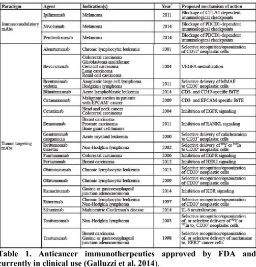
Many immunotherapeutic treatments are already in clinical practice as they have been approved by the Food and Drug Administration (FDA) for treating cancer patients (see Table 1) (Galluzzi et al. 2014). New classes of targeted drugs are emerging and many of them are already in clinical trials.
Table 1. Anticancer immunotherpeutics approved by FDA and currently in clinical use (Galluzzi et al. 2014).
To date, the acquired knowledge on the mechanisms of anti-tumor immune responses and the novel technological platforms suggest possible innovative means to induce more efficient and durable responses in cancer patients.
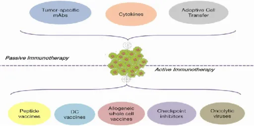
Cancer immunotherapy is based on two main approaches: passive and active treatments (Figure 1) (Papaioannou et al. 2016).
Introduction
Figure 1. Classification of active and passive cancer immunotherapy.
The passive cancer immunotherapy approach includes the tumor specific mAbs, cytokines and adoptive cell transfer. The active immunotherapy approach includes vaccines, checkpoint inhibitors and oncolytic viruses
(Papaioannou et al. 2016).
The active immunotherapy approach involves anti-cancer vaccines (peptide, dendritic cell-based and allogeneic whole cell vaccines), as well as immune checkpoint inhibitors and oncolytic viruses. On the other hand, the best example of passive immunotherapy is represented by the treatment with monoclonal antibodies (mAbs) that have been initially used in targeted therapy against Tumor Associated Antigens (Papaioannou et al. 2016). Recently, many other antibodies targeting immune checkpoints have been approved by FDA for clinical use of melanoma and other cancer types (Galluzzi et al. 2014), due to their immunomodulatory functions, thus representing a novel approach for immunotherapy.
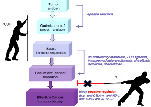
Furthermore, the combinations of different immunotherapy interventions are also under investigation. The combinatorial approach should be elegantly orchestrated by boosting anti-cancer responses (PUSH), on one hand, and by neutralizing negative immune regulators (PULL), on the other hand (Figure 2). The combination of different cancer treatment modalities (e.g., chemotherapy and checkpoint inhibitors) and the expansion of targeted drug repertoire through the discovery of new targets, new drugs and new molecules
targeting multiple molecular pathways (e.g., pan-tyrosine kinase inhibitors) are the new challenges for several research groups (Papaioannou et al. 2016).
Figure 2. The synergy between different cancer treatments. The use of
different immunotherapeutic approaches to combine the induction of immune anti-cancer responses (PUSH) with the inhibition of negative immune regulators (PULL) (Papaioannou et al. 2016).
1.1. Antibody-based immunotherapy against Tumor Associated Antigens
The conventional anti-cancer therapy includes surgery, radiotherapy, and chemotherapy; however, the main limits of the latter two conventional treatments are represented by the absence of selectivity for tumor cells and by non-specific side toxic effects. The immunotherapy targeting cancer cells, by the use of monoclonal antibodies specific for TAAs overexpressed on tumor cells, represent
Introduction
an alternative strategy to overcome these limits. For this reason, the immunologists have worked with the aim of inducing specific immune responses against cancer, by developing drugs for targeted therapies (Wurz et al. 2016; Kirkwood et al. 2012; Galluzzi et al. 2014).
The targets of these therapies can be classified into Tumor Specific or cancer-testis Antigens (TSAs), expressed only on tumor cells, and Tumor Associated Antigens, overexpressed by tumors but present also at low levels on normal differentiated cells from which the tumors arise(Wurz et al. 2016; Savage et al. 2014).
The use of mAbs as magic bullets against these targets has rapidly increased over the last 40 years starting with the production of the first generation of therapeutic mAbs of mouse origin.
Unfortunately, the induction by human body of human anti-mouse antibody (HAMA) response represented an obstacle to the success of this approach and induced further research progresses to overcome these problems (Thorpe et al. 2003). Recombinant DNA technology allowed to replace the mouse costant domains, recognized by the human immune system, and responsible for HAMA response, with the human correspondent sequences, thus producing chimeric mAbs with reduced immunogenicity and improved pharmacokinetic and therapeutic efficacy (LoBuglio et al. 1989). Further improvements in recombinant DNA and cloning techniques led to humanized mAbs (Carter et al. 1992), obtained by inserting only the mouse hypervariable loops in the human V-region frameworks, thus further reducing the immunogenicity of the humanized antibodies.
More recently, new strategies, such as phage display technology, led to the generation of human antibody libraries, to be used for the in vitro selection of fully human antibodies specifically targeting tumor cells (Frenzel et al. 2016).
Some of the therapeutic mAbs approved by FDA for clinical use in cancer therapy are directed against either growth factors, such as vascular endothelial growth factor A, or growth factor receptors, such as human epidermal growth factor receptors EGFR and HER2/ErbB2. In addition to the specific and direct inhibitory effects on the signal transduction of these receptors expressed on tumor cells, therapeutic mAbs can also activate Antibody Dependent Cellular Cytotoxicity (ADCC) and Complement Dependent Cytotoxicity (CDC), or deliver towards tumor cells conjugated therapeutics, such as toxins or
radioisotopes. More recently, bispecific antibodies able to recognize two antigens have been created to retarget cytotoxic T lymphocytes to any cell of choice (Wurz et al. 2016).
1.2. ErbB2 and EGFR receptors as validated TAAs for breast cancer therapy
Worldwide, approximately 10 million people are diagnosed with cancer annually and more than 6 million die of this disease every year (Stuart and Kleiheus 2006). In particular breast cancer affects one in eight women during their lives. It is the most common cancer in women worldwide, with nearly 1,8 million new cases (second most common cancer overall) diagnosed in 2013 (Fitzmaurice et al. 2015). A good candidate as TAA and an attractive target for immunotherapy is ErbB2, a Tyrosine Kinase Receptor (RTK), overexpressed on many carcinoma cells, with a key role in the development of malignancies (Slamon et al. 1989; Tagliabue et al. 1991; Lohrisch and Piccart 2001; Gravalos and Jimeno 2008).
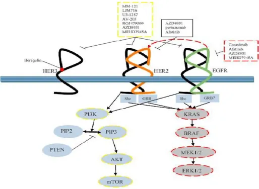
ErbB2 is a receptor belonging to the ErbB family of RTKs. Unlike the other members of the family, ErbB2 lacks natural ligands and acts as the preferential heterodimerization partner for the other members of the family. ErbB2 overexpression on tumor cells leads to the activation of intracellular tyrosine kinase domain and downstream signalling cascades that mediate cell growth, differentiation, and survival (Yarden and Sliwkowski 2001; Klapper et al. 2000; Busse et al. 2000; Baselga et al. 2009), such as those of mitogen-activated protein kinase (MAPK) and phosphatidylinositol-3-kinase (PI3K) pathways (Figure 3), with a consequent dysregulated proliferation.
Introduction
Figure 3. EGF receptor family. Schematic representation of the Tyrosine
Kinase Receptors EGFR, ErbB2/HER2 and HER3, their downstream pathways and inhibitors (Baselga J et al. 2009).
Antitumor strategies have been well established for ErbB2-positive tumors and indicative examples include Trastuzumab, the first humanized antibody approved by the FDA in 1998 for breast cancer therapy, and Pertuzumab approved by FDA more recently for combinatorial treatments with Trastuzumab and Docetaxel (Sabatier and Gonçalves 2014; Stebbing et al. 2000; Romond et al. 2005; Baselga and Swain 2010). They have different mechanisms of action as they bind to different epitopes in the domain II and the domain IV of ErbB2, respectively. Despite the success of these antibodies for breast cancer their clinical efficacy is still limited by resistance and cardiotoxicity issues. Indeed, a high fraction of patients show resistance to their treatment and develop cardiac dysfunction, frequently leading to heart failure in particular in the presence of other
risk factors (Slamon et al. 2001; Natha et al. 2006; Seidman et al. 2002; De Lorenzo et al. 2018).
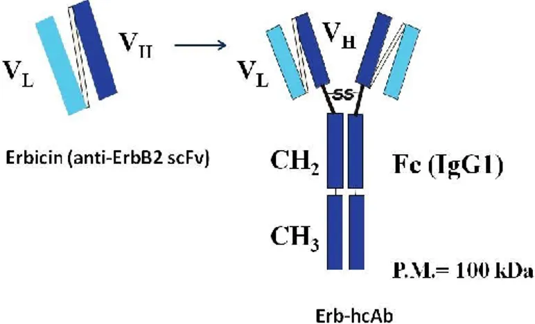
To overcome the limits of Trastuzumab, novel human antitumor immunoagents have been recently engineered in our laboratory by isolation of human single-chain variable fragments (scFvs) through phage display technology. In particular, a human anti-ErbB2 scFv, named Erbicin, was found to be able to selectively bind and inhibit ErbB2-positive tumor cells, and to recognize an epitope of ErbB2 different from that of Pertuzumab and Trastuzumab (Figure 4) (De Lorenzo et al. 2002; Troise et al. 2011).
The following fusion of Erbicin with the Fc region of a human IgG1 led to a new immunoagent construct, called Erb-hcAb (human compact antibody) for its compact size (100 kDa) if compared with the full size (155 kDa) of a natural IgG (Figure 4). This construct acquired the immune effector functions of the Fc domain while retaining the ability to diffuse more easily in the tumor masses due to its reduced molecular size with respect to a full size antibody. Erb-hcAb was found to selectively bind to breast tumor cells expressing ErbB2 and to inhibit their growth in vitro and in vivo, by inducing also Antibody Dependent Cellular Cytotoxicity (ADCC) and Complement-Dependent Cytotoxicity (CDC) (De Lorenzo et al. 2004). Interestingly, Erb-hcAb, differently from Trastuzumab and Pertuzumab, lacks cardiotoxic effects on human cardiomyocytes in in vitro and in vivo models (Riccio et al. 2009; Fedele et al. 2012).
Figure 4. Erb-hcAb. Schematic representation of Erbicin and its derived
Introduction
Unfortunately, a high fraction of breast tumors does not express ErbB2. For instance, Triple-Negative Breast Cancer (TNBC), accounting for ~14% of all breast cancers, is characterized by the absence of Estrogen Receptor (ER), Progesterone Receptor (PR) and ErbB2, excluding the possibility of using efficacious targeted therapies developed against these proteins.
On the contrary, overexpression of EGFR has been reported in ~60% of TNBC and correlates with poor outcome. EGFR is the other member of the EGF tyrosine kinase receptor family, which also regulates the PI3K/AKT and Ras/MAPK signaling upon binding its ligand EGF and receptor dimerization. Increased EGFR expression may influence multiple aspects of tumor biology including cell proliferation, motility and invasiveness, and resistance to treatment (Figure 3) (Stommel et al. 2007; Greenall et al. 2015; Haynes et al. 2014).
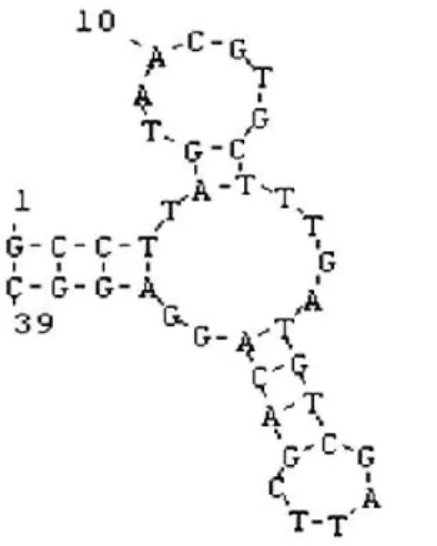
Although more recently, also aptamers have raised increasing attention for cancer therapy due to their low molecular weight, lack of immunogenicity and ready availability. A RNA-aptamer, named CL4 (Figure 5), able to recognize and inhibit EGFR has been recently generated in the laboratory of our collaborators, by an in vitro selection procedure: SELEX (Systematic Evolution of Ligands by Exponential Enrichment) (Tuerk and Gold 1990; Camorani et al. 2018; Camorani and Cerchia 2015). The CL4 oligonucleotide aptamer was found able to bind and inhibit not only the human EGFRwt, but also the EGFRvIII mutant, expressed on the surface of Glioblastoma Multiforme (GBM) cancer cells, lacking most of the extracellular domain (Esposito et al. 2011; Camorani et al. 2015). Indeed, it interacts with domain IV of the receptor, which is still present in the EGFRvIII variant, and inhibits the autophosphorylation of the receptor and its downstream ERK1/2 and STAT3 pathways, thus affecting migration and invasion of cancer cells (Camorani et al. 2015). CL4 was also found able to inhibit the vasculogenic mimicry (VM) and tumor growth in TNBCs resistant to both Erlotinib and Cetuximab (EGFR-TK inhibitors), by preventing the EGFR-integrin αvβ3 interaction (Camorani et al. 2017). Thus, its use could allow for overcoming the tumor resistance to EGFR Tyrosine Kinase Inhibitors (TKIs).
Figure 5. CL4 aptamer. Structure of the anti-EGFR CL4 aptamer (Esposito et al. 2011).
1.3. Immunotherapy targeting immune regulatory checkpoints The other antibody-based approach in oncology exploits the activation of immune system, such as the stimulation of T cells mediated by immunomodulatory antibodies (Peggs et al. 2009).
Activation of immune cells involved in anti-tumor responses is regulated by multiple stimulatory and inhibitory pathways on effector T cells that can be targeted by monoclonal agonistic or antagonistic antibodies. The primary targets for such interventions were envisaged initially to be the effector T cells, but it is becoming clear that, also Natural Killer (NK) cells and regulatory T (Treg) cell populations might be important targets.
Understanding the complexity of immune regulation and tumor microenvironment has been critical to optimize the efficacy of the therapeutic approaches, and to develop new strategies for immune-based cancer therapy. The T cells have high specificity for their own antigen since they bear T cell receptors (TCRs) that specifically recognize cell-surface major histocompatibility complex
Introduction
molecules associated with peptides derived from either endogenous or foreigner proteins.
Moreover, T cells are characterized by memory, because primary T cell responses are generally followed by the production of long-lived memory T cells with accelerated kinetics of response to a second presentation of the antigen (Davis and Bjorkman 1988). In order to induce the activation of effector T cells, the single engagement of the MHC by the recognition of associated antigenic peptides, such as those derived from TAAs, by the TCR is not sufficient for the full activation of T cells and may render them anergic. A series of various stimuli generated by co-regulatory signals are also required to fully activate T cells and to regulate their immune responses. These are exerted by a series of stimulatory or inhibitory receptor-ligand pairs and can determine the fate of the T cell response, such as their activation with the subsequent differentiation into effector T cells, their deletion or anergy (Chen and Flies 2013).
Translation of this concept into the clinic has led the researchers to focuse their efforts on the identification of antibodies agonistic for co-stimulatory receptors or antagonistic for inhibitory signals, in order to use Immune Checkpoints (ICs) to amplify the antigen-specific T cell responses against cancer (Pardoll 2012).
To date, human or humanized mAbs targeting the immunosuppressive receptors CTLA-4 (Ipilimumab), PD-1 (Nivolumab and Pembrolizumab), and PD-L1 (Atezolizumab, Durvalumab and Avelumab) have been approved for the treatment of several tumors, including melanoma, non-small cell lung cancer, renal cell carcinoma, head and neck squamous cell carcinoma, Hodgkin lymphoma, urothelial carcinoma, liver carcinoma, microsatellite instable (MI) colorectal cancer and Merkel-cell carcinoma (Brahmer et al. 2012; Patel et al 2017; Shultz 2017; Callahan et al. 2016).
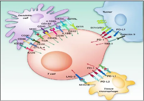
Despite their success, the currently approved antibodies against immune checkpoints (hence collectively named checkpoint inhibitors; CIs) are effective in only about 20-30% of the patients (Lee et al. 2013; Buqué et al. 2015; Pardoll 2012). As shown in Figure 6, new targets are emerging as co-inhibitory receptors, such as lymphocyte activation gene 3 (LAG-3), an immunosuppressive receptor expressed on activated T lymphocytes and T-regulatory
lymphocytes, T cell immunoglobulin and mucin-3 (TIM3), T cell immunoglobulin and ITIM domain (TIGIT), which are expressed on exhausted CD8+ T cells in tumors (Anderson et al. 2016; Goldberg and Drake 2011; Huang et al. 2004). B- and T-lymphocyte attenuator (BTLA) is another co-inhibitory receptor whose expression is induced during activation of T cells, leading to inhibition of human CD8+ cancer specific T cells (Watanabe et al. 2003). Similarly, some agonistic antibodies recognizing co-stimulatory receptors have reached the clinical stage such as those specific for OX40 (CD134), a secondary co-stimulatory immune checkpoint molecule that prevents premature death of activated lymphocytes, and 4-1BB (CD137) expressed on activated CD4+ and CD8+ T lymphocytes whose crosslinking enhances T cell proliferation, IL-2 secretion, survival and cytolytic activity. Other co-stimulatory proteins that are considered good targets for antibody-mediated immunotherapy are inducible T cell costimulator (ICOS), which is an immune checkpoint protein belonging to the CD28-superfamily that is expressed on activated T cells, and CD27, a member of the tumor necrosis factor receptor superfamily, which is required for generation and long-term maintenance of T cell immunity (Chen and Flies 2013; Sanmamed et al. 2015; Wen et al. 2002; Sasso et al. 2018).
Introduction
Figure 6. Potential immunomodulatory targets for monoclonal antibody-based therapy. The interaction between inhibitory receptors, such
as CTLA-4, PD-1, BTLA, LAG-3, TIGIT, TIM-3 with their ligands leads to an inhibition of the T cell activation (red arrows). Co-stimulatory receptors such as CD28, ICOS, 4-1BB, OX40, CD27, on the contrary, enhance the effector function of T cells (green arrows).
Antibodies against different immune checkpoint receptors can also be combined to achieve additive or synergistic activity, potentially translating into a better efficacy. Proof of concept for this approach was provided by the finding of increased efficacy of the Ipilimumab and Nivolumab combination versus monotherapy in the treatment of metastatic melanoma (Mahoney et al. 2015; Larkin et al. 2015). A numerous clinical trials with approved or novel antibodies against immune checkpoints used in monotherapy or in combination with other biologics or small molecules are being carried out worldwide.
A new goal is represented by the generation of a complete repertoire of fully human antibodies specific for all these T cell
checkpoint modulators, so that they can be tested in all the possible combinatorial treatments (Sasso et al. 2018).
1.4. Generation of a human repertoire of mAbs for cancer therapy by Phage Display Technology
To obtain a repertoire of fully human immunomodulatory mAbs against several targets to be used in monotherapy or in combinatorial treatments with other anti-tumor drugs for cancer therapy, innovative strategies based on the well-known phage display technology have been considered in our laboratory.
Phage display is a precious technology that enabled to overcome the disadvantages of humanized mAbs, such as immunogenicity and low efficiency to penetrate tumor masses due to their large size (155 kDa), by producing fully human single chain variable fragments of antibodies and easily selecting them by affinity procedures.
The scFv is the smallest portion of the immunoglobulin, which retains the antigen-binding ability. Indeed, an antibody in the scFv format consists of variable regions of heavy (VH) and light (VL) chains, joined together by a flexible peptide linker, with a molecular weight of 27 kDa. Thanks to their low molecular weight and their retained property to bind to the antigen, the scFvs can be exploited in phage display technology to be expressed on the phage as fusion proteins with a protein of the phage coat, and to be selected for their binding to the targets (Ahmad et al. 2012).
Indeed, phage display is a strategy based on the expression of foreigner (poly)peptides on the surface of filamentous bacteriophages. This strategy was introduced in 1985, by George P. Smith, who demonstrated that the fragment of a protein could be expressed in a native conformation on the phage in order to be immunologically recognized by antibodies, with no significant effects on life cycle and infectivity of the phages (Bazan et al. 2012).
The gene encoding each scFv is packed inside the virion as a single-stranded DNA and the corresponding encoded protein is expressed as a fusion product with the pIII protein of the phage coat (Figure 7).
Introduction
Figure 7. Schematic representation of scFv expressed as a fusion product with the pIII protein of the phage coat.
The phage display technology is based on the generation of libraries containing up to 1010 different variants, that can be used in a screening process to select antibodies against almost unlimited array of biological (including human self-antigens) and non-biological targets (such as toxic molecules). In order to identify the scFvs which specifically bind to the target, this technology allows for panning either on targets immobilized on solid support or expressed on the cell surface, by removing from the whole repertoire the non-binder clones by using affinity selection techniques.
In our laboratory a large number of tumor or other anti-receptors scFvs have been successfully isolated by panning human scFv libraries either on purified proteins or on live cells expressing the targets on their surface (De Lorenzo et al. 2002; Palmieri et al. 2015; Paciello et al. 2016).
In the present project we tailored phage display to the generation of a complete repertoire of fully human antibodies
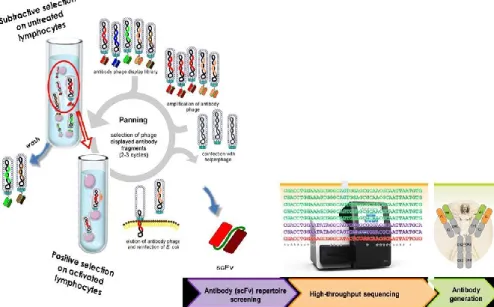
recognizing several different immune checkpoints. As shown in Figure 8, we applied for the first time a novel untested strategy for rapid, parallel screening of phage displayed antibody libraries by directly panning for the first time on activated human lymphocytes. This novel approach allows for the selection of antibodies recognizing the receptors in their native conformation, as that presented on the cell membrane, and to obtain a productive selection of several phage clones against multiple targets in one single panning.
As a proof of concept of the quality/potency of the binders identified by this approach, we generated fully IgGs from three of these collections i.e. PD-1, PD-L1 and LAG-3 (Sasso et al. 2018). These three targets were chosen, as antibodies useful for biological assays and specific for them have been already developed by demonstrating therapeutic benefits, as described in the following paragraph.
Figure 8. Strategy of scFvs selection by phage display against immune checkpoints. The phages were incubated with activated lymphocytes after a
subtractive step on untreated lymphocytes in order to select specific phages for receptors expressed upon activation. The screening of enriched clones was performed by Next Generation Sequencing, and the identified scFvs were then converted into IgG4.
Introduction
1.5. Programmed cell death receptor-1 (PD-1); Programmed Death Ligand-1 (PD-L1); Lymphocyte Activation Gene-3 (LAG-3) and their relative antibodies in use or clinical development
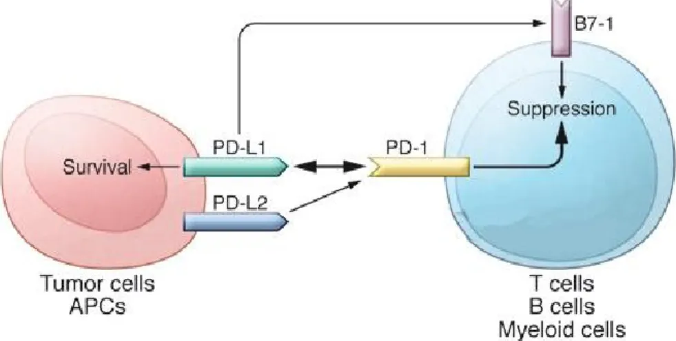
PD-1 is an immune checkpoint firstly identified in lymphoid cell lines undergoing programmed cell death. PD-1 is expressed on activated T-cells, B-cells, dendritic cells (DCs) and monocytes (Figure 9). The role of PD-1 on cells is to limit the activity of T-cells in peripheral tissues during inflammatory responses and to inhibit autoimmune responses. Its expression is induced when T cells become activated; therefore, when engaged by one of its ligands, such as PD-L1, PD-1 inhibits kinases that are involved in T cell activation (Pardoll 2012). This inhibition mechanism mediated by PD-1 can be exploited by the tumor cells expressing its ligand to neutralize the antitumor response of T cells (Haile et al. 2013).
Figure 9. Schematic representation of PD-1/PD-L1 interaction and downstream effects. The interaction of PD-L1 or PD-L2 with PD-1 may
induce T cell suppression.
Since, the interaction of PD-1, expressed on the surface of activated T cells, and PD-L1 on the surface of tumor cells results in immunosuppression, the interference mediated by an antibody specific for PD-1 could be a suitable strategy to reduce the
suppression of effector T cells. Two monoclonal antibodies targeting PD-1 have been already approved by the FDA: Pembrolizumab (Lambrolizumab) a humanized antibody firstly approved in 2014 to treat patients with unresectable or metastatic melanoma after treatment with Ipilimumab, and then approved in 2017 to treat also locally advanced or metastatic urothelial carcinoma (Robert et al. 2014; Barnhart 2015; Shultz 2017), and Nivolumab, a fully human mAb currently in clinical use for the therapy of Hodgkin lymphoma, unresectable or metastatic melanoma, metastatic renal cell carcinoma and non-small cell lung cancer (NSCLC) (Raedler 2015; Kazandjian et al. 2016; Janice 2017).
PD-L1 also acts as an inhibitor of human T cell responses, as the interaction of PD-L1 with PD-1 on activated T cells results in immunosuppression and tumor immune escape (Assal et al. 2015). PD-L1 is expressed on the surface of tumor cells (Figure 9) and its level was found increased in patients with cancer, likely because its expression is inducible by interferon γ (IFN-γ).
A number of antibodies targeting PD-L1 are in clinical development for metastatic melanoma, Non-Small Cell Lung Cancer (NSCLC), Renal Cell Cancer (RCC) and bladder cancer. For instance, Atezolizumab is a humanized anti-PD-L1 IgG1, approved by the FDA for non-small cell lung cancer in October 2016 and currently undergoing evaluation in clinical trials for patients with other types of cancer, including breast cancer, renal cell carcinoma, bladder cancer, and small-cell lung cancer (Janice 2017).
LAG-3, also known as CD223, is another negative regulator of T cell activation and homeostasis that was found to be specifically associated with the CD3–TCR complex following TCR engagement, and is therefore considered as a negative co-receptor (Figure 10).
Introduction
Figure 10. Schematic representation of LAG-3 and its interactions.
LAG-3 is a transmembrane protein with structural homology to CD4, and four extracellular IgG domains. In addition to the membrane-bound form of LAG-3, it can be cleaved at the connecting peptide (CP) by metalloproteases to generate a soluble protein that functions as an immune adjuvant detectable in the serum.
LAG-3 is expressed on activated T cells, NK cells and B cells, and binds with high affinity to major histocompatibility complex (MHC) class II molecules (Goldberg et al. 2011). It has also been found on TReg cells, where it plays a role in amplifying the
immunosuppressive activity of TReg cells, and in decreasing the pool
of memory CD4+ and CD8+ T cells (Huang et al. 2004).
Furthermore, a monoclonal antibody targeting LAG-3, known as BMS-986016, is in phase 1/2a of clinical trials in monotherapy for advanced solid tumors (ClinicalTrials.gov Identifier: NCT02966548. 2018), and in combination with Nivolumab BMS-936558, in relapsed or refractory B-cell malignancies (clinicaltrials.gov identifier: NCT02061761. 2018).
Materials and Methods
AIMS
My PhD project was aimed at developing novel therapeutic approaches for cancer therapy by generating novel human antibodies.
An innovative trispecific multiparatope Tribody, targeting 3 nonoverlapping ErbB2 epitopes, was obtained in our laboratory in collaboration with a laboratory based in Lisbon. The first aim was the characterization of this novel Tribody by testing its ability to bind to tumor ErbB2-positive cells and to inhibit their growth.
A second aim was represented by the generation of a novel repertoire of fully human antibodies recognizing several different immune checkpoints in their native conformation. In this regard, the phases of the research activity were the following:
- set up of an innovative strategy based on phage display by performing a selection on hPBMCs;
- identification of the most enriched scFvs clones by NGS and bioinformatics;
- conversion of the selected scFvs into fully human IgG4; - purification and characterization of the converted
antibodies by: testing the binding affinity of each antibody by ELISA assays on lymphocytes and on purified proteins; analysing their ability to activate T cells, and evaluating their antitumor effects in vivo.
MATERIALS AND METHODS
Antibodies and human recombinant proteins
The following antibodies were used: horseradish peroxidase (HRP)-conjugated anti-His mouse monoclonal antibody (Qiagen); HRP-conjugated anti-M13 mouse monoclonal antibody (GE Healthcare); anti-human LAG-3 mouse monoclonal antibody (R&D Systems); anti-human PD-1 human monoclonal antibody Nivolumab (Opdivo®); anti-human PD-L1 human monoclonal antibody (G&P Biosciences); conjugated anti-human IgG (Promega); HRP-conjugated anti-human IgG (Fab’)2 goat monoclonal antibody (Abcam); HRP-conjugated anti-rabbit immunoglobulin from goat antiserum (Thermo Fisher Scientific). Trastuzumab (Roche), Pertuzumab (Roche) Erb-hcAb (Biotecnol) (De Lorenzo et al. 2004). Anti-ErbB2 rabbit monoclonal antibody and anti-pErbB2 rabbit polyclonal antibody were from Cell signaling Technology.
The following antibodies used for FACS analyses: PE/anti-human CD2 mouse monoclonal antibody (BioLegend), APC/anti-human CD3 mouse antibody, PE/anti-APC/anti-human CD4 mouse monoclonal antibody, PerCP/anti-human CD8 mouse monoclonal antibody were all from BD Biosciences.
APC/antihuman IgG Fc mouse antibody, APC/Cy7 anti-human CD366 (TIM3) mouse antibody, APC/anti-human CD272 (BTLA) mouse antibody, APC/anti-human CD137 (4-1BB) mouse antibody, APC/anti-human CD134 (OX40) mouse antibody, Brilliant Violet 510™/anti-mouse/rat/human CD27 hamster antibody, APC/antihuman/mouse/rat CD278 (ICOS) hamster antibody were all from BioLegend. FITC/anti-human TIGIT mouse antibody (Thermo Fisher Scientific).
The following recombinant chimeric proteins were used: human LAG-3/Fc; human PD-1/Fc; human PD-L1/Fc; TIM3/Fc; BTLA/Fc; TIGIT/Fc; OX40/Fc; 4-1BB/Fc; CD27/Fc and ICOS/Fc; human IgG1-Fc (all from R&D Systems) (Sasso et al. 2018).
Materials and Methods
Aptamers
2’F-Py RNA aptamers (CL4 and CL4Sc), used for treatments in cell viability assays, were synthesized by TriLink Biotechonologies. CL4Sc represents the scrambled form of CL4 and was used as a negative control. Before each treatment, the aptamers were subjected to a short denaturation-renaturation step (85°C for 5 min, on ice for 2 min, 37°C for 10 min).
Bacterial Culture Media
The bacterial growth was carried out in 2xTY and LB (Luria-Bertani) culture media.
Bacterial Strains
TG1 bacterial strain was used for the production of the scFvs expressed as fusion proteins with pIII protein of the phage; E.Coli SF110 bacterial strain was used for expression of the scFvs as soluble proteins; E. coli DH5α (Invitrogen, Life Technologies, Paisley, UK) and BL21 DE3 strains (Novagen-Merck Millipore, Darmstadt, Germany) were used for cDNA amplification.
Antibiotics
The antibiotics used for bacterial growth and selection were Ampicillin (100 μg/ml) and Kanamicin (25 μg/ml).
Cell cultures
MDA-MB-231 and JIMT-1 cells were cultured in Dulbecco’s Modified Eagle’s Medium (GibcoTM DMEM, Thermo Fisher Scientific). MCF-7 cells were cultured in Modified Eagle’s Medium (GibcoTM MEM, Thermo Fisher Scientific). SKBR3 cells (from ATCC, Rockville, MD) were cultured in RPMI 1640 (Gibco BRL,
Life Technologies, Paisley, UK). Media were supplemented with 10% (vol/vol) heat-inactivated fetal bovine serum (FBS, Sigma-Aldrich), 50 UI mL-1 penicillin, 50 μg mL-1 streptomycin (50 μg mL-1 streptomycin), 2 nM L-glutamine all from GibcoTM, Thermo Fisher Scientific. MCF7 cells trasfected with the cDNA encoding the variant of ErbB2 lacking exon16 (Δ16Her2) were grown in minimum essential medium (MEM) medium (Gibco BRL) supplemented with 10% heat-inactivated fetal bovine serum, penicillin (100 UI/mL), streptomycin (100 mg/mL), 2mM glutamine, 1mM sodium pyruvate, MEM aminoacids, and MEM nonessential aminoacids.
Production and Purification of Tribodies
Tribodies were produced by Biotecnol using its proprietary Trisoma platform, which includes proprietary expression and purification technology. This process recovers the Tribody L/Fd chain monomer while limiting impurities such as product variants and endotoxins. Tribodies were sterile filtered, aliquoted, and stored at 70°C. The purified tribodies were analyzed and confirmed to be >95% L/Fd chain Tribody monomer. Low levels of endotoxins were confirmed by a LAL-based method. Binding of the tribodies to their target was confirmed in an enzymelinked immunosorbent assays (ELISA) with a recombinant ErbB2 extracellular domain (Riccio et al. 2017).
Western Blotting analyses for the detection of ErbB2 levels on tumor cells
The cells were plated (6.105 cells/well) in 6-well plates and incubated at 37°C for 16 hours. Multiparatope and monoparatope tribodies (concentration of 10 nM) or each antibody (concentration of 10 nM) were added to the cells and incubated for 30 hours at 37°C. The treated cells were scraped and centrifuged at 1200 rpm for 7 minutes; the cell pellets were lysed in a buffer containing 10mM Tris-HCl (pH 7.4), 0.5% Nonidet-P-40, and 150 mM NaCl in the presence of protease inhibitors (Roche). After incubation on ice for 20 minutes,
Materials and Methods
the extracts were clarified by centrifugation at 12000 rpm for 15 minutes at 4°C. Protein concentration was determined by the Bradford colorimetric assay (Sigma-Aldrich) and Western Blotting analyses were performed by incubating the membranes with a commercial anti-ErbB2 antibody, followed by the HRP-conjugated secondary antibody, indicated above (Riccio et al. 2017).
Cell Viability Assays to test the effects of the multiparatope Tribody on tumor cells
To test the effects of the multiparatope Tribody, or the combination of the 3 parental antibodies (Erb-hcAb, Trastuzumab and Pertuzumab) on tumor cell growth, SKBR3 cells were plated in 96-well flat-bottom at a density of 1.5.104/well and were incubated at 37°C for 72 hours with immunoagents (5–100 nM) in the culture medium. Cell counts were determined in triplicate by using the trypan blue exclusion test. Cell survival was expressed as the percentage of viable cells in the presence of the tested proteins, with respect to untreated control cultures (Riccio et al. 2017).
Isolation of human Peripheral Blood Mononuclear Cells
Human PBMCs were isolated from blood of healthy donors by using Greiner Leucosep® tube (Sigma-Aldrich) following the manufacturer’s instructions, and frozen in a solution containing 90% FBS and 10% dimethyl sulfoxide (DMSO) until use. Cryopreserved cell vials were gently thawed out by using RPMI 1640 medium (GibcoTM, Thermo Fisher Scientific) supplemented with 1% L-glutamine, 1% CTL-Wash™ (Cellular Technology Limited), and 100 U/mL Benzonase (Merck Millipore). The collected hPBMCs were then washed by centrifugation, plated and incubated overnight at 37°C in R10 medium consisting of RPMI 1640 supplemented with 10% inactivated FBS, 1% L-glutamine, 50 U mL-1 penicillin, 50 μg mL-1 streptomycin and 1% HEPES (GibcoTM, Thermo Fisher Scientific). After an overnight resting, the hPBMCs were collected in phosphate-buffered saline (PBS), counted by using the Muse® Cell Analyzer
(Merck Millipore), resuspended at a density of 1·106 cells/mL (Sasso
et al. 2018).
FACS analysis of expression levels of immune checkpoints on hPBMCs
Human PBMCs were isolated as described above. After an overnight incubation they were activated with Dynabeads® Human T-Activator CD3/CD28 at a concentration of 1∙103 beads/mL (GibcoTM, Thermo Fisher Scientific). After 24-96 hours of activation, the cells were seeded in a round-bottom 96-well plate (1·106 cells/well) and then centrifuged at 1200 rpm for 5 minutes to remove the supernatant. Unlabelled anti-LAG-3, anti-PD-1 (Nivolumab) or anti-PD-L1 primary antibodies were added to each well at a concentration of 10 μg/mL and incubated for 90 minutes at room temperature by gently shaking. After extensive washes, the cells were stained with 100 μL of APC/anti-human IgG Fc antibody in FACS buffer (PBS, 1% FBS), and with 10 μg/mL of PE/anti-human CD2 antibody (BioLegend, San Diego) for 45 minutes at room temperature by gently shaking. The labeled antibodies APC/Cy7 human CD366 (TIM3), APC/ human CD272 (BTLA), APC/ anti-human CD137 (4-1BB), PE/ anti-anti-human CD134 (OX40), Brilliant Violet 510™/ anti-mouse/rat/human CD27, APC/ anti-human/mouse/rat CD278 (ICOS), FITC/ antihuman TIGIT antibodies were added to each well at a concentration of 10 μg/mL and incubated for 90 minutes at room temperature by gently shaking. After two washes with FACS buffer, the cells were resuspended in PBS and analyzed on CytoFLEX Flow Cytometer (Beckman Coulter). Lymphocytes were identified by analyzing the forward (FSC) versus side (SSC) scatter and gated to exclude debris. Healthy and apoptotic cell populations were identified by using viability staining (LIVE/DEAD solution, Thermo Fisher Scientific). The live cells only were considered to measure the percentage of positive cells corresponding to the fraction of cell populations double positive for both the expression of CD2 (the T-Lymphocytes lineage marker) and for the specific target of interest (LAG-3, PD-L1, PD-1, TIM3, BTLA, TIGIT, OX40, 4-1BB, CD27 or ICOS). Unstained cells or
Materials and Methods
cells treated with the appropriate isotype control were used as background control. Values were reported as the mean of at least three determinations (standard deviations ≤10%) (Sasso et al. 2018).
Selection of scFv-phage clones
Phagemid particles were recovered from the library by using the M13-K07 helper phage (Invitrogen, Thermo Fisher Scientific), as previously described (De Lorenzo et al. 2002). For each round of selection, phages (1013 cfu) were blocked by incubating them with 5% (wt/vol) Skim Milk Powder (Fluka Analytical, Sigma-Aldrich) for 30 mins in PBS before their use. For the first round of selection, blocked phages (1013 cfu) were firstly submitted to one round of negative selection by incubation with untreated lymphocytes (5.106) in 2.5% (wt/vol) Skim Milk Powder (Fluka Analytical, Sigma-Aldrich, USA) in PBS for 2 hours at 4°C by gently rotation.
The unbound phages were collected in the supernatant by centrifugation at 1200 rpm for 10 minutes and then used for the positive selection, performed by incubating them with activated lymphocytes (1.106) overnight by gently rotation at 4°C in the presence of 2.5% skim milk powder. For the selection of phages binding to CD27, BTLA and TIGIT the negative panning was not carried out to avoid the loss of some specific phages in the subtractive selection. After extensive washes with PBS, the bound phages were eluted from activated lymphocytes with 76 mM citric acid (pH 2.5) in PBS for 5 minutes, and then neutralized with 1M Tris-HCl (pH 8.0). The recovered phages were amplified by infecting E. coli TG1 cells to prepare phages for the next rounds of selection on the purified chimeric protein. To this aim, NuncTM polypropylene tubes (Fisher Scientific, Thermo Fisher Scientific) were coated with the selected recombinant chimeric proteins at a concentration of 20 μg/mL in a 0.05 M NaHCO3 solution, for 72 hours at 4°C. Blocked phages were submitted to two following rounds of negative selection by incubating them in the tubes coated with rhIgG1-Fc protein for 2 hours at 4°C in rotation.
Unbound phages, recovered in the supernatant, were then incubated in the coated tubes, prepared as described above for the
positive selection, overnight at 4°C in rotation, and eluted as described above. Alternatively, since a trypsin site is present between the scFv and the pIII protein of the phage coat, trypsin was used for a more efficient and selective elution of the binders. Briefly, after extensive washes with PBS, the bound phages were incubated with 50 mM Tris-HCl (pH 8.0) buffer containing 1 mM CaCl2 for 1 hour at
4°C in rotation, and then eluted from the chimeric proteins with trypsin (1μg/mL), incubated for further 15 minutes at 25°C by gently shaking. The reaction was then blocked by using protease inhibitors (Complete™ EDTA-free Protease Inhibitor Cocktail, Sigma-Aldrich). Phages were then collected and stored at 4°C until use (Sasso et al. 2018).
DNA fragment preparation and high-throughput sequencing
For each sublibrary, obtained from each selection cycle, the phagemid double-strand DNAs containing the scFvs were purified from cultures of superinfected E. coli TG1 cells using Endo free Plasmid Maxi Kit (Qiagen). The full-length scFvs were excised with restriction enzymes BamHI and HindIII, all from New England Biolabs, and purified with Wizard® SV Gel and PCR Clean-Up System (Promega) from 1.2% agarose gel (Sasso et al. 2015). A second enzymatic excision was performed with NcoI and XhoI, all from New England Biolabs, to isolate the VHs from the previously purified material, and then extracted from a 1,4% agarose gel. Library preparations for sequencing and preliminary bioinformatic analysis of the data were performed at the Center for Translational Genomics and Bioinformatics, Hospital San Raffaele, Milano, Italy. The VHs extracted from sub-libraries were bar-coded by TruSeq ChIP sample prep kit (Illumina). A complementary scheme for bar-coding was implemented to obtain a deep and suitable sequencing of VH mixtures of several subcycles. Subcycles 2 and 3 for each target were mixed in a dedicated run. The first universal cycle 1 was sequenced separately to cover the larger complexity. The bar-coded samples were diluted to a final concentration of 10 pM and sequenced with 2.300 nt SBS kit v3 on an Illumina MiSeq apparatus (Sasso et al. 2018).
Materials and Methods
scFv recovery
The clones of interest were isolated from the sublibrary at cycle 3 of the corresponding target. For high-ranking clones (relative representativeness ≥1% within cycle 3), the QuickChange II XL Site-Directed Mutagenesis Kit (Agilent Technologies) was used to perform copies of clones with overlapping primers, designed within the corresponding HCDR3 regions, according to the procedure previously described (Sasso et al. 2015). Briefly, the extension reactions were assembled as follows: 50-250 ng of template; 2/5 μL QuickSolution reagent; 1 μL Pfu Ultra High-Fidelity DNA polymerase (2.5 U/μL); 5 μL 10x reaction buffer; 1 μL dNTPmix; 125 ng forward primer; 125 ng reverse primer; H2O to a final volume of 50 μL. The template DNA was removed by restriction with 1 μL of DpnI enzyme, as suggested by the kit provider. An appropriate amount of reaction was used to transform XL10-GOLD ultracompetent cells (Agilent Technologies) and then plated on LB/agar containing 100 μg/mL ampicillin. Some colonies were picked and evaluated by double digestion and sequencing. For low ranking clones (relative representativeness <1% within cycle 3), the DNA samples were isolated from cycle 3 by overlapping PCR. Briefly, Phusion High-Fidelity DNA Polymerase (Thermo Fisher Scientific) was used to perform two PCR reactions to obtain separately VH and VL fragments, using primers designed within the corresponding HCDR3 regions and in constant region of plasmid upstream and downstream of VH and VL.
In a second step, the PCR fragments corresponding to each clone were mixed and extended to get the full scFvs. The reactions were assembled as follows: 150 ng of template for VH and VL fragments amplification and 10 ng of template (VH and VL fragments) for full scFv amplification; 0.5μL Phusion DNA Polymerase (0.02 U/μl); 10 μL 5X Phusion HF Buffer; 1 μL dNTP mix; 0.5 μM forward primer; 0.5 μM reverse primer; 1.5 μL DMSO; H2O to a final volume of 50 μL (Sasso et al. 2018).
Antibody production and purification
The scFvs of interest were converted into whole IgG4 antibodies by cloning the corresponding VH and VL cDNAs in the pEU vectors 8.2VH and 4.2VL, expressing respectively, the constant antibody heavy and light chains (Paciello et al. 2016). Briefly, the VHs and VLs were amplified by CloneAmp HiFi PCR Premix in standard conditions with specific primers and purified by Wizard® SV Gel and PCR Clean-Up System (Promega) from 1.3% agarose gel. In-Fusion HD cloning kit (Clontech Laboratories) was used to clone the VHs in BamHI and BssHII (New England Biolabs), linearized pEU8.2VH vector, and the VLs in ApaLI and AvrII (New England Biolabs), linearized pEU4.2VL vector. Stellar Competent Cells (Clontech Laboratories) were transformed with the obtained vectors, and the colonies were screened by digestion and sequence analysis. The vectors containing the correct DNA, prepared with an endotoxin-free system (EndoFree Plasmid Maxi Kit, Qiagen), were co-transfected in HEK293-EBNA by using Lipofectamine Transfection Reagent (Life Technologies, Inc.) and grown up for about 10 days at 37°C in chemical defined CD CHO medium (Gibco, Life Technologies, Inc.) supplemented with 5 mL of L-glutamine 200 mM (Gibco, Life Technologies), 5 mL of Penicillin (100 UI/mL) and Streptomicyn (100 mg/mL), from Sigma-Aldirch, in 6-well plates or in 150 mm Corning® tissue-culture treated culture dishes.
The conditioned media were collected, and the antibodies were purified by using Protein A HP SpinTrap30 (GE Healthcare Life Sciences). The purity of the final products was evaluated by SDSPAGE NuPAGE™ 4-12% Bis-Tris Protein Gels, 1.0 mm, 10-well (Thermo Fisher Scientific) followed by the staining with Coomassie blue solution (Biorad) for 20 minutes and de-stained with 7% CH3COOH and 20% Et-OH. All the antibody preparations were filtered with sterile 0.22 μm durapore hydrophilic filters (Millipore) before their use (Sasso et al. 2018).
Enzyme-Linked Immunosorbent Assays (ELISA)
To detect the surface ErbB2 levels on ErbB2-positive cells, the cells were harvested in nonenzymatic dissociation solution
(Sigma-Materials and Methods
Aldrich), washed and plated in 96-well round-bottom microtiter plates (2.105 cells/well). Then, the cells were incubated in the absence or presence of increasing concentrations of each Tribody (3–100 nM) or other anti-ErbB2 antibodies (in ELISA buffer PBS/BSA 3%) at room temperature for 2 hours (Riccio et al. 2017).
To confirm the binding specificity of the purified immunomodulatory mAbs, ELISA assays were performed on chimeric proteins (coated at 5 μg/mL on microplates), tumor cells (PD-L1-positive breast cancer MDA-MB-231 or PD-L1-negative breast cancer MCF7 cells), and untreated or activated hPBMCs.
The ELISA assays on coated chimeric protein were performed by coating NuncTM flat bottom 96-well plates (Thermo Fisher Scientific) with 5 μg/mL rhPD-L1, rhPD-1 and rhLAG-3 recombinant proteins in a solution of 0,05 M NaHCO3 for 72 hours at 37°C. After blocking of the coated 96-well plates with 5% nonfat dry milk in PBS for 1 hour at 37°C, the purified mAbs were added at increasing concentrations (10-200 nM) to the plates in 2.5% nonfat dry milk in PBS and incubated for 2 hours at room temperature by gently shaking.
Cell ELISA assays were performed by plating the cells in round-bottom 96-well plates (2·105 cells or 2·105 lymphocytes for
each well) and incubating them with increasing concentrations (0.5-200 nM) of mAbs in 2.5% nonfat dry milk for 2 hours at room temperature with gentle agitation (Sasso et al. 2018).
After the incubation with the primary antibodies extensive washes were carried out with PBS, then the plates were incubated with an appropriate HRP-conjugated antibody for 1 hour at room temperature, washed again and incubated with 3,3’,5,5’-tetramethylbenzidine (Sigma-Aldrich) reagent for 10 min before quenching with an equal volume of 1 N HCl. Absorbance at 450 nm was measured by the Envision plate reader (Perkin Elmer, 2102). Binding values were reported as the mean of at least three determinations obtained in three independent experiments.
The Kd values were calculated by elaboration of ELISA binding curve analyses by Prism (Graphpad) tool according to the following model: Y=Bmax*X/(Kd+X) + NS*X + Background. Bmax is the maximum specific binding in the same units as Y; Kd is the equilibrium binding constant, in the same units as X, and it is the ligand concentration needed to achieve a half-maximum binding at
equilibrium; NS is the slope of non-specific binding in Y units divided by X units; background is the amount of nonspecific binding with no added ligand.
Competitive ELISA assays
In order to investigate the ability of the selected PD-L1_1 and PD-L1_2 antibodies to compete in the PD-L1/PD-1 or PD-L1/B7.1 binding, competitive ELISA assays were performed by testing the binding of each biotinylated chimeric protein (PD-1/Fc or B7.1/Fc) to PD-L1 in the absence or in the presence of the unlabelled competitive antibodies. To this aim, a NuncTM flat-bottom 96-well plate was coated with 200ng/ml of PD-L1 recombinant protein in 0.005M NaHCO3 solution for 72 hours at 4°C. Then, the PD-L1 coated plate
was pre-incubated with the competitor PD-L1_1 and PDL1_2 antibodies (at 10:1 M/M excess ratio), and then further treated with the biotinylated PD-1 or B7.1 chimeric proteins, which were added to the plate at the same concentrations of the competitive antibodies (2μg/ml corresponding to 2.4.1012 molecules/well). For the detection of bound biotinylated proteins, HRP-conjugated Streptavidin (Biorad) was added to the plate, whereas an anti-human antibody was used in parallel assays for the detection of bound anti-PD-L1 antibodies.
To determine whether the novel anti-PD-1 mAbs recognize different epitopes from that recognized by Nivolumab, competitive ELISA assays were performed on plates coated with PD-1/Fc chimeric protein (5 μg/mL). After blocking with 5% Nonfat Dry Milk in PBS, the coated 96-well plate was pre-incubated for 2 hours with saturating concentrations of each unlabelled mAb (400 nM) in 2.5% Nonfat Dry Milk in PBS in agitation at room temperature. After extensive washes with PBS, increasing concentrations of biotinylated Nivolumab mAb were added. For the detection of the binding, the plate was incubated with HRP-conjugated Streptavidin for 30 minutes in agitation at room temperature, washed again and analyzed as described above (Sasso et al. 2018).