EUROPEAN COMMISSION E C
Structural Funds
MINISTERO DELL’UNIVERSITÀ’ E DELLA RICERCA SCIENTIFICA
E TECNOLOGICA
Università degli Studi di Catania
DOTTORATO DI RICERCA IN
DOTTORATO DI RICERCA IN
DOTTORATO DI RICERCA IN
DOTTORATO DI RICERCA IN
METODOLOGIE SPERIMENTALI ED APPLICAZIONI
METODOLOGIE SPERIMENTALI ED APPLICAZIONI
METODOLOGIE SPERIMENTALI ED APPLICAZIONI
METODOLOGIE SPERIMENTALI ED APPLICAZIONI
TECNOLOGICHE IN CHIRURGIA
TECNOLOGICHE IN CHIRURGIA
TECNOLOGICHE IN CHIRURGIA
TECNOLOGICHE IN CHIRURGIA
Ciclo XX
Ciclo XX
Ciclo XX
Ciclo XXII
II
II
II
Facoltà di Medicina e Chirurgia
Facoltà di Medicina e Chirurgia
Facoltà di Medicina e Chirurgia
Facoltà di Medicina e Chirurgia
UNIVERSIT
UNIVERSIT
UNIVERSIT
UNIVERSITA
A
A
A’’’’ DEGLI STUDI DI CATANIA
DEGLI STUDI DI CATANIA
DEGLI STUDI DI CATANIA
DEGLI STUDI DI CATANIA
_________________________________________
_________________________________________
_________________________________________
_________________________________________
Dott.
Dott.
Dott.
Dott. Floriana Gona
Floriana Gona
Floriana Gona
Floriana Gona
BACTERIAL
BACTERIAL
BACTERIAL
BACTERIAL AND FUNGAL
AND FUNGAL
AND FUNGAL
AND FUNGAL INFECTIONS
INFECTIONS
INFECTIONS
INFECTIONS
IN RENAL ALLOGRAFT RECIPIENTS
IN RENAL ALLOGRAFT RECIPIENTS
IN RENAL ALLOGRAFT RECIPIENTS
IN RENAL ALLOGRAFT RECIPIENTS
_______________________
_______________________
_______________________
_______________________
TESI DI DOTTORATO
TESI DI DOTTORATO
TESI DI DOTTORATO
TESI DI DOTTORATO
_______________________
Coordinatore:
Tutor:
Prof. Pierfrancesco Veroux
Prof. Stefania Stefani
_______________________
_______________________
_______________________
_____________________________________________________
______________________________
______________________________
______________________________
QUADRI
QUADRI
QUADRI
2
INDEX
RIASSUNTO Pag. 3 ABSTRACT Pag. 6 INTRODUCTION Pag. 10 CLASSIFICATION Pag. 12 Living donor Cadaver donors Recipient evaluationTRANSPLANT IMMUNO-BIOLOGY Pag. 20
INFECTIVE COMPLICATIONS
Risk factors
Temporal succession of infections Donor Screening
Recipient Screening
Posted transplantation prevention
Pag. 25
TYPES OF INFECTION Citomegalovirus (CMV) BKV and JCV Infections
Urinary Tract Infections (UTIs) Infections by Candida spp Other fungal infections
Pag. 37
THE AIMS OF THIS STUDY Pag. 41
MATERIALS AND METHODS Pag. 42
RESULTS AND CONCLUSIONS Pag. 50
FUTURE PROSPECTIVE Pag. 56
REFERENCES Pag. 57
3 RIASSUNTO
Con l'aumento della sopravvivenza a breve termine un sempre maggior numero di pazienti ha goduto di ottimi risultati anche a lungo termine, portando ad un graduale ma progressivo spostamento dell' attenzione dei clinici su differenti problematiche come la qualità della vita dei pazienti trapiantati e l'aumentata incidenza di complicanze infettive e neoplasie legate allo stato di immunosoppressione indotto dalla terapia antirigetto. Al fine di poter valutare in modo completo ed esauriente il tema delle complicanze infettive nel paziente sottoposto a trapianto di organo solido, è necessario comprendere a fondo la complessità del problema-trapianto e soffermarsi brevemente sui principali concetti generali di trapiantologia. Inizierò ad esaminare in breve la terminologia, i vari modi in cui un trapianto può essere classificato ed i principi che guidano scelta e valutazione del donatore, infine la breve valutazione di problematiche quali le reazioni di rigetto e la terapia immunosoppressiva mi permetteranno di introdurre, trattare ed approfondire il complesso panorama delle complicanze infettive del paziente trapiantato. Infatti, sebbene l’aumento e il successo dell’ attività di trapianto d’organo sono il risultato di importanti progressi ottenuti in chirurgia, nella diagnosi e nel trattamento, la gestione delle complicanze infettive sottolinea la necessità di sorveglianza, prevenzione ed adeguate profilassi nella pratica clinica. La sorveglianza e la prevenzione sono divenute il fine principale del follow-up dei pazienti trapiantati che prende inizio nel periodo pre-trapianto per protrarsi poi nei tempi successivi all’intervento. Lo scopo di tale studio è stato valutare lo stato infettivologico dei pazienti sottoposti a trapianto di rene e a trapianto combinato rene – pancreas presso il Centro Trapianti del Policlinico di Catania in due fasi: una prima fase in cui ho valutato lo stato infettivologico dei pazienti, sia donatori che riceventi, prima del trapianto al fine di evidenziare processi infettivi acuti e cronici e l’eventuale rischio di attivazione di infezioni latenti; e una seconda fase, nel periodo post-trapianto, lo stato infettivologico è stato monitorato inizialmente ogni mese per i primi tre mesi successivi al trapianto e poi con cadenza trimestrale. Questo monitoraggio microbiologico ha permesso di individuare, prevenire e trattare precocemente le infezioni nei pazienti sottoposti a trapianto presso il nostro Centro, al fine di salvaguardare la vitalità del graft e del paziente trapiantato. Inoltre considerando le difficoltà relative, ad esempio a una
4 accurata diagnosi di Infezioni fungine invasive, è risultato di fondamentale importanza riuscire ad evidenziare markers di laboratorio che servano a differenziare i soggetti più a rischio per avviare, qualora le condizioni cliniche lo richiedano, un eventuale trattamento di pre-emptive therapy o profilassi. Tale monitoraggio è stato eseguito dal 1 Novembre 2007 al 30 giugno 2010 in una popolazione di 101 pazienti (84 da cadavere e 17 da vivente, età media 44 anni) sottoposti a trapianto di rene singolo (n=94), doppio (n=4) o combinato rene/pancreas (n=3). Le ricerche microbiologiche e virologiche sono state eseguite mediante vari esami: esami colturali, esami sierologici, esami in PCR. In particolare i campioni biologici esaminati sono stati: tamponi faringei, tamponi nasali e analisi del liquido di trasporto dell’organo, studio della colonizzazione mediante tamponi e indagini sierologiche di base relative alla ricerca di anticorpi anti-Candida (fase miceliale) rispettivamente in batteriologia e micologia. Un dato significativo è stato ottenuto dalla raccolta del liquido di trasporto poiché dagli 84 campioni esaminati in 46 (54%) vi era la presenza di almeno un microrganismo. In tutti i riceventi sottoposti a terapia antibiotica nessuno ha sviluppato batteriemie attribuibili alla contaminazione del liquido di trasporto. Questi risultati confermano che, sebbene un alto rischio di perdita dell’organo e morte dei pazienti sono stati riportati nei precedenti studi, soprattutto con microrganismi gram-negativi quali
Escherichia coli, la contaminazione del donatore non è una controindicazione al trapianto se una terapia antibiotica di profilassi è iniziata prima del trapianto. Al contrario l’incidenza di contaminazione fungina è stata dal 2 al 10%, e non essendoci linee guida per la prevenzione vascolare o per le complicazioni di infezioni funginee dal donatore o della contaminazione del liquido di trasporto, la raccolta del liquido di trasporto al pre-trapianto è utile per l’identificazione di riceventi ad alto rischio di insorgenza di infezioni e può essere utilizzata come una terapia pre-emptive. L’analisi batteriologica sia dei tamponi nasali che di quelli faringei, non ha dato risultati significativi dal momento che nella maggior parte dei casi sono stati isolati batteri commensali mentre risultati significativi sono stati mostrati per le infezioni del tratto urinario. In particolare ancora una volta è emerso che Escherichia coli e Klebsiella pneumoniae risultano le specie batteriche che ancora oggi rappresentano gli agenti eziologici principali delle infezioni delle vie urinarie sia in ambito nosocomiale che comunitario, inoltre per quel che riguarda la
5 sensibilità antimicrobica, gli antibiotici risultati più attivi sia verso E. coli che K.
pneumoniae sono state amikacina ed imipenem che hanno inibito la totalità dei ceppi saggiati. E’ da rilevare che da Marzo a Settembre 2009, all’interno del reparto del Centro Trapianti c’è stata la diffusione di due cloni multi-resistenti di K.
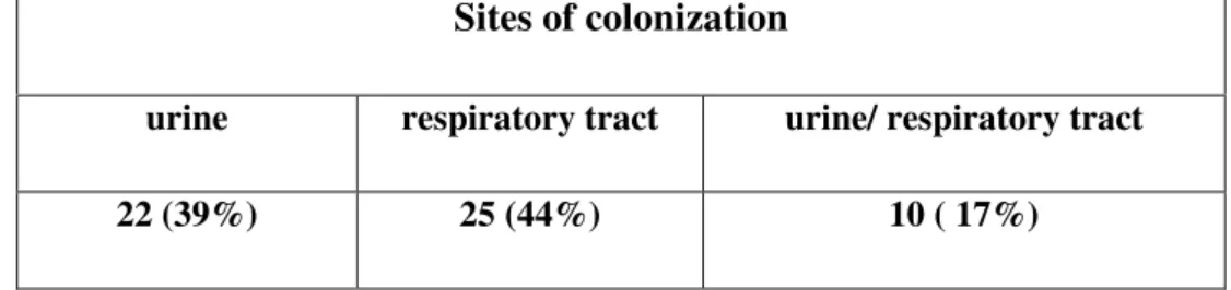
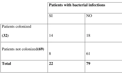
pneumoniae produttrici di ESBLs. Questi cloni non erano mai stati isolati prima e furono responsabili per la prima volta di gravi infezioni urinarie con difficile risoluzione per alcuni pazienti. In conclusione quindi le infezioni del tratto urinario rimangono le più frequenti tra i riceventi il trapianto renale, ma misure di controllo, adeguati programmi di educazione e un attenta sorveglianza sono importanti per contenere la rapida diffusione di tali ceppi resistenti. Il monitoraggio micologico su 101 pazienti ha mostrato una colonizzazione in almeno uno dei siti indagati del 56%; in tutti i siti indagati le specie trovate sono state 123 con una incidenza del 52% nel tratto respiratorio (tampone oro-faringeo/espettorato) e del 48% nelle urine. In particolare, sia nel tratto respiratorio che nelle urine, la specie più frequentemente isolata è stata Candida albicans ma pur essendo la specie maggiormente presente quello che è apparso evidente è che nel tratto respiratorio le specie appartenenti al genere Candida si ritrovavano più spesso rispetto alle urine. Analizzando poi la distribuzione dei pazienti con infezioni batteriche del tratto urinario rispetto alla colonizzazione fungina nel medesimo distretto si evince un’ associazione estremamente significativa (χ2 = 13.267, P<0.001) tra colonizzazione fungina e infezioni batteriche ed inoltre, nel 64,3% dei casi, la colonizzazione fungina precedeva il primo episodio di infezione batterica, lasciando ipotizzare che la colonizzazione fungina possa rappresentare un fattore predisponente all’insorgenza di una infezione batterica. Infine la ricerca delle IgG dirette verso la fase miceliale di C. albicans sembra essere utile, in associazione allo studio della colonizzazione, a selezionare i pazienti sui quali iniziare un eventuale trattamento antifugino la cui precocità è determinante per la riduzione della mortalità dei pazienti.
6 ABSTRACT
With the increase of short-term survival, a greater number of patients have had optimal results also in the long term, which is leading to a gradual but progressive change in focus of the clinicians to different problems such as the quality of life of transplant patients, and the increased incidence of infective and neoplastic complications linked to the state of immunosuppression induced by antirejection therapy. With the aim of evaluating infective complications in patients undergoing solid-organ transplantation it is necessary to completely understand the complexity of the transplantation problem and to briefly look at the general principles of transplantology. I will begin by briefly examining the terminology, the various ways in which transplantation can be classified, and the principles that guide the choice and evaluation of the donor. Finally there will be a brief evaluation of transplantation problems such as rejection reactions and immunosuppressive therapy. I will introduce and explain in the following chapters the complex panorama of the infective complications of transplant patients. In fact even if the increase in and the success of organ transplantation are the result of progress obtained in the fields of surgery, diagnosis and treatment, the management of infective complications underlines the necessity for monitoring, prevention and adequate prophylaxis in clinical practice. Monitoring and prevention have become the principal aims of follow-up in transplant recipients and begin in the pre-transplantation period to continue well after the pre-transplantation. The aim of this study was to evaluate the infective state of patients undergoing renal transplantation and combined renal-pancreas transplantations at the transplantation centre of the University Hospital of Catania subdivided in two phases : first I evaluated the infective state of the patients, both donors and recipients, before transplantation in order to detect acute and chronic infective processes and the eventual risk of activation of latent infections. Then, in the post-transplantation period, the infective state of the patients was monitored initially every month for the first three months after transplantation and then once every three months. This microbiological monitoring identified, prevented and rapidly treated infections in the patients undergoing transplantation at our centre, with the aim of safeguarding both the graft and the transplanted patient. Moreover, considering the relative difficulty, for example an accurate diagnosis of invasive fungal infections (IFI), it
7 was fundamentally important to be able to find laboratory markers that could differentiate the subject most at risk and initiate, based on clinical conditions, an eventual pre-emptive therapy or prophylaxis. This monitoring was performed by 1 November 2007 to 30 June 2010 the incidence and time to appearance of infective complications were evaluated in a population of 101 organ transplantation recipients (84 from cadavers and 17 from live donors, average age 44 years) having undergone transplantation for a single kidney, double kidney or kidney/pancreas combination. Microbiological and virological investigations were carried out: by means of various examinations culture exams, serum exams, PCR. In particular the biological samples examined were: pharyngeal swab, nasal swabs and analysis of the organ transport liquid, colonisation studies by means of swabs (oropharyngeal, cutaneous, right and left nasal) and basic serum investigations for the identification of anti-Candida (mycelial phase) antibodies respectively in bacteriology and mycology. A significative result was obtained from the examination of the transport liquid from the 84 samples examined; in 46 (54%) there was the presence of at least one microorganism. In all the recipients undergoing antibiotic therapy bacteremia did not develop that could be attributed to the transport liquid. These results confirm that, even if a high risk of organ loss and death of patients was reported in previous studies, above all by gram-negative microorganisms such as
Escherichia coli, the contamination of the donor was not a contraindication for transplantation if prophylactic antibiotic therapy was initiated before transplantation. On the other hand, the incidence of fungal contamination is from 2 to 10%. There are no guidelines for vascular prevention or for complications from fungal infections of the donor or of contamination of the transport liquid. Various studies have underlined the need for nephrectomy of the organ as prophylaxis when the transport liquid is found to be contaminated. In conclusion the analysis of the transport liquid before transplantation is useful for the identification of recipients at high risk of infections and can be used as pre-emptive therapy. The bacteriological analysis of both nasal and pharyngeal swabs did not give significative results as most cases isolated were commensal bacteria while results significative were shown for urinary infections. In particular from this study it can be seen that
Escherichia coli and Klebsiella pneumoniae were the bacterial species that are still the principal etiological agent of infections, both nosocomial and community of the
8 urinary tract. The antibiotics that were the most active against E. coli and K.
pneumoniae were amikacin and imipenem that inhibited all the sampled strains. Particular attention was paid to 10 patients from March to September 2009: three patients developed bacteraemia, seven patients developed symptomatic UTIs, due to the presence of fever, urgency, frequency, dysuria, and supra-pubic tenderness, caused by K. pneumoniae that showed a pattern of resistance to multiple antibiotics. These microorganisms were characterised to determine the mechanisms responsible for antibiotic resistance by means of specific phenotypic tests (double disc for ESBLs) and molecular techniques for the amplification of resistance genes (DNA extraction, PCR). Moreover, to evaluate the type of diffusion inside the hospital these strains underwent molecular typing by means of Pulsed Field Gel Electroforesis (PFGE). The ten strains belonged to two different clones, A and B, moreover, clone A was an MDR clone resistant to all β-lactams and amikacin, piperacillin/tazobactam and ciprofloxacin. Clone B was susceptible to amikacin and ciprofloxacin. Subtle differences in the bla gene content were observed between both clones, demonstrating that lateral gene transfer drives the diffusion of many antibiotic resistance genes. These clones had never been isolated before and were responsible, for the first time, for severe upper UTIs with difficult resolution: one failure and one relapse. Finally, UTIs remain the most frequent infections among renal transplant recipients. Until recently infections were sustained by susceptible microrganisms in which complete resolution of infections was easily obtained; the increase of potentially life-threatening multi-resistant strains now emerging in hospital settings for renal transplant recipients has changed the severity of infections and the corresponding outcome. The implementation of control measures, focusing on hand hygiene and appropriate urinary catheter manipulation through educational programs and contact isolation procedures were able to limit the spread of resistance clones in the renal transplantation unit. Therefore it is mandatory to continue epidemiological surveillance of transplantation units in order to tailor a correct therapy to maintain antibioticspotent, such as carbapenem, which are losing their potency. Of the 101 patients monitored 57(56%) were colonized in at least one of the investigated sites, generally, in all the sites investigated, 123 species were found: 64(52%) in the respiratory tract(oral-pharyngeal swab/sputum) and 59(48%) in urine. In particular
9 in both sites the most frequently isolated species was C. albicans. Therefore, even if C.albicans was the most frequently isolated species it appears that in the respiratory tract the species have a greater distribution with respect to urine. Analyzing the distribution of the patients with bacterial infections of the urinary tract with respect to fungal colonization of the same tract, it can be seen that there is an extremely significant association (χ2 = 13.267, P<0.001) between fungal colonization and bacterial infection. Moreover, in 64.3% of the cases, fungal colonization caused the first episode of bacterial infection, suggesting that fungal colonization can be a predisposing factor for the appearance of bacterial infection. Finally the identification of IgG stowards the mycelial phase of C. albicans seems to be useful, in association with the study of colonization, to select those patients who should receive eventual antifungal treatment that when started early can lead to a reduction in mortality.
10 INTRODUCTION
The first solid-organ transplantation was carried out by Joseph Murray in 1954, Nobel Prize winner for medicine in 1990: the recipient was a 23 year old patient affected by chronic glomerular nephritis and the donor was the homozygous twin. Research carried out on twins showed that genetic identity was the principal factor for the success of this type of operation. Even with this information, and the success of this and other transplantations between homozygous twins, problems of transplantation between patients who are not genetically identical still need to be overcome. Over the last 20 years, the constant and intensive research activity, the development of potent antirejection drugs with immunosuppressive activity, the possibility of immunologically typing tissue and the notable improvement in surgical techniques has allowed organ transplantation to leave the experimental field and become a reality for treating diseases that were once fatal.
There are situations in which an organ transplantation is the only solution to resolve a serious and invalidating dysfunction: kidney transplantation is the only alternative to dialysis patients suffering from renal failure, and pancreas transplantation is an optimal method of treatment for insulin independent diabetes mellitus sufferers, as it allows for the repair of a system of insulin secretion that is self-regulated based on the blood glucose levels, completely resolving this problem. Furthermore, thanks to a great experience, and to improvements in surgical techniques for the preservation of tissue and post-transplant therapy, the percentage of successes is constantly increasing. In 1988 one-year survival after transplantation from a cadaver donor was 76%, while in 2007 this increased to 90%. The results from kidney transplantation from live donors have improved from 89% in 1988 to 93% in 2008, for one-year survival [1]. With the increase of short-term survival, a greater number of patients have had optimal results also in the long term, which is leading to a gradual but progressive change in focus of the clinicians to different problems such as the quality of life of transplant patients, and the increased incidence of infective and neoplastic complications linked to the state of immunosuppression induced by antirejection therapy. With the aim of evaluating infective complications in patients undergoing solid-organ transplantation it is necessary to completely understand the complexity of the transplantation problem
11 and to briefly look at the general principles of transplantology before entering into the principal subject of this thesis. I will begin by briefly examining the terminology, the various ways in which transplantation can be classified, and the principles that guide the choice and evaluation of the donor. Finally there will be a brief evaluation of transplantation problems such as rejection reactions and immunosuppressive therapy. I will introduce and explain in the following chapters the complex panorama of the infective complications of transplant patients.
12 CLASSIFICATION
Transplantology is a medical science that is constantly changing, and increasing: the techniques that have been developed have opened various paths increasing the possibilities for potential recipients. Other than solid-organ transplantation other types of transplantations exist that are useful for the repair of structures that have been damaged by trauma or pathologies: these are tissue and cell transplantation.
Tissue transplantation
Tissue transplantation is carried out to improve the quality of life of the recipient rather than as a life-saving technique as is the case of organ transplantation, or the transplantation of stem cells.
When we speak about the substitution of tissue it would be better to speak about grafting, rather than transplantation. Tissues that can be used for grafting are: cornea, skin, artery, vain, cardiac valve, bone, muscle and tendon.
Cell transplantation
Cell transplantation or infusion is one of the areas of study and experimentation at the cutting edge. Stem cells, present in every organism with the function of regenerating and producing new tissue are, in this field, the most important. Unlike organ and tissue donation, which take place more often from cadaver donors, those of stem cells come from living donors. For transplantation haemopoietic stem cells are preferred, progenitors of all the hepatic cell lines, removed from bone marrow, or from the blood present in the umbilical cord.
Bone marrow transplantation has come of age over the last 20 years as a possible therapy, even if sometimes not definitive, for neoplasias (leukemia and lymphomas), as well as non-neoplastic diseases (thalassemia, congenital immunodeficiency).
Solid-organ transplantation
This is a surgical procedure, divided into two phases: the removal of the organ from a donor and the successive implantation of this organ in the recipient, with the eventual removal of the non-functioning organ from the latter. The law 91/99, “regulations for the removal and transplantation of organs and tissues” does not
13 permit the removal of gonads, brain and the genetic manipulation of embryos also with the aim of transplanting organs. Theoretically, any organ or tissue can be removed from a donor and transplanted into a recipient. The solid-organs that are currently used for transplantation are the following:
• kidney; • liver;
• heart;
• lung;
• pancreas, and intestine.
The removal of organs for transplantation is generally carried out from cadaver donors whose vital functions are artificially maintained as long as possible to preserve the quality and vitality of the organs to be transplanted. Notwithstanding this, the possibility of removing organs from living donors is a possibility, as long as this does not put the donor's life at risk, of overcoming the gap that exists between the request and the availability of organs and to reduce waiting list time.[2] The type of donor directly affects both the survival of the recipient and that of the organ; the best results have been obtained for patients who received a transplantation from a homozygous twin: recent data show that for kidney transplantation there is a survival rate of 100% for organ and patient at five years from transplantation; a patient who receives a kidney from a living donor has a five-year survival of 90% and a graft half-life of more than 30 years. In the case of kidney transplantation from a cadaver donor, recipient survival at five years is 81%, with a graft half-life of about 14 years.[3]
Kidney transplantation
Kidney transplantation is the substitution therapy of choice for patients with terminal renal failure. In fact, for the same age patient having similar risk factors, the survival of patients on dialysis is always less than a transplanted patient. The transplanted kidney is normally positioned at the back of the peritoneum in the fossa iliac. First, the renal vein is anastomosed, generally with the external vena iliac, then the renal artery, generally with the external iliac artery. Finally, the ureter is implanted in the bladder by means of a sub-mucosal tunnel, similar to the
14 natural one, with the aim of preventing reflux. The return of urination can happen during the first few hours after the operation, but there can also be a delay of days or even weeks. This delay depends on factors linked to the “procurement” of the organ such as duration of ischemia (that is the time from removal to transplantation), factors linked to the donor (shock, hypoperfusion, age and quality of the kidney), and finally factors linked to the recipient, as regards kidney transplantation, results from a living donor are superior to those from a cadaver.
Living donor
”Acts of disposition of one’s own body are forbidden when they cause a permanent physical decrease, or when they are otherwise agaist the law, public order or good custom.” (Italy, Civil Code, Book I, art. 5: Acts of disposition of one’s own body
parts). From a technical/scientific point of view, the most recent acquisitions in the medical/surgical field, make it possible to remove various organs from living donors: kidney, lung, body-tail of the pancreas, intestine, and parts of the liver are all examples of structures that can be removed from living subjects without significantly compromising quality and duration of life. However, Italian law (in particular, the above-mentioned article 5 of the civil code) does not allow the voluntary donation of organs by a living individual due to the “ permanent decrease
15 of physical integrity” that removal would cause. There have been dispensations to article 5 of the civil code in Italy, but so far there have been two laws that allow the removal of kidneys and part of the liver from living donors. Law n. 458 of 26 June 1967 allows the gratuitous removal of a kidney for transplantation between living people; the act of deposition and destination of the organ in favour of a certain patient is under the jurisdiction of a magistrate, who will evaluate the donor as regards his or her mental state, understanding of the limits of transplant therapy, and the personal consequences that the donation could have and that this act must be free, spontaneous and gratuitous.[4] As has already been mentioned transplantation from a living donor has notable advantages with respect to a transplantation from a cadaver: the operation can be carried out at an established time, and, in the case of a kidney, can be carried out according to a protocol that is called pre-emptive, that is before the recipient begins dialysis. Moreover, the screening of the living donor can be carried out over a longer period of time as there are no restrictions such as those during a transplantation from a cadaver donor, and can that be more accurate; often waiting time is also reduced with positive repercussions on the psychophysical state of the recipient; finally, in the case of a transplantation between relations the rejection reaction is often less intense, thanks to a better HLA compatibility. A person who donates an organ is, however, exposed to some risks. Transplantation from a living donor is the only case in medicine in which a healthy subject undergoes major surgery [5]: the surgical operation in itself, notwithstanding the great experience of the specialised centres, is not immune from complications regarding anestesia, haemorrhage and infections. These risks are not identical for every organ removed: in fact, the removal of a kidney is much safer with respect to the removal of part of the liver. The risk of death following donation is, however, minimum; International studies show a donor mortality of 0.03% after kidney donation, [6] and 0.2% after donation of part of the liver [7]. A further risk for the donor is the reduced capacity of the organ, or part of the organ that remains, to compensate for eventual functional deficits, traumas or pathologies that can arise following the transplantation.
Paradoxically, in some recent studies it has been observed that living donors for kidneys have a life expectancy higher than that of the normal population: this could
16 be explained by the fact that donors are healthy individuals, generally young, and undergo medical checkups over a long period of time in order to diagnose early and treat rapidly any eventual pathological condition.
Diseased donors
The possibility to carry out transplantation from cadaver donors has led to the medical/legal need for a definitive definition of the concept of death and of the criteria to verify it, an indispensable presupposition to allow the removal of organs and tissues for therapeutic purposes. To declare the decease of an individual, cerebral death is a criterion that has become accepted almost unanimously. Even if death is identified by the irreversible ceasing of all brain functions, it is necessary to distinguish between: death due to cardiac arrest, in which the time between the ceasing of respiratory and cardio circulartory functions lead to the irreversible loss of all cerebral functions (still-heart cadaver), and death by brain pathologies with the maintenance of recipe respiratory and cardio circulatory functions by means of artificial external devices (beating-heart cadaver) [8]. Even if the removal of many organs and tissues is possible from both types of cadaver, the beating-heart cadaver donors are, for most transplantation centres, the principal source from which to remove kidneys, lungs, liver and pancreas, and are the only source as regards the heart. Transplantation in Italy has imposed a continuous updating of the law; after the Zanardelli code that in 1889 ratified the respect and the inviolability of the cadaver, only in 1957 was a law approved for the first time that determined the lawfulness of the removal of part of a cadaver for therapeutic purposes; from that moment on there have been numerous laws, among which dispensations and revisions that have focused, above all, on two particularly delicate points of transplantology, the identification of criteria to ascertain death and deregulation of the activity of removal and transplantation of organs and tissues.
Preoperative evaluation and biological safety
The cadaver donor identified by the coordination of the transplantation system must be correctly evaluated to exclude the presence of pathological conditions that potentially threaten the life of the eventual recipient or reduce the vitality and
17 efficiency of the organs to be transplanted, and are thus contraindications for transplantation.
It is clear that from an ethical point of view it is desirable to only use healthy and functional organs coming from younger donors, however, in a context characterised by a permanent scarcity of organs, over the last few years there has also been organ removal and transplantation using older people, often with reduced function, and sometimes affected by pathologies that are able to damage the graft, for example organs coming from a donor affected by diabetes.
In in this case, we can speak about organs coming from these so-called “ marginal donors” or “ sub-optimal”. The transplantation of a sub-optimal organ leads to a life expectancy of the graft and the recipient that is less with respect to an organ coming from a “ optimum” donor. Here arises the ethical question of which type of patient could receive organs that are not perfectly functional: the Eurotransplant Association has created an example of a programme of preferential attribution of sub-optimal kidneys for older patients, and those that have been on the waiting list for a long time. Other than systemic or organ pathologies and an advanced age it is important to also evaluate the biological safety of the donor. The principal transmissible diseases from donor to recipient are infections and neoplasias, whose presence plays an important role in the decision to use or not a certain donor: among the absolute contraindications for transplantation there are, in fact, active uncontrollable infections, presence of anti-HIV antibodies and a positive work up for existing infection and neoplasias with the exception of: tumours of the CNS with low possibility for metastases (based on the WHO guidelines); in situ carcinomas at the level of any organ; carcinomas with a particularly low metastatic potential.
Recipient evaluation
The typical patient candidate for organ transplantation is an individual affected by a pathology that has caused the irreversible dysfunction of one or more transplantable organs.
For each organ there exist specific criteria that allow the inclusion or exclusion of a given patient in a waiting list; based on the results of the analyses, and of the evaluations carried out the medical team of the transplant centre decide if
18 transplantation is the only therapy possible for the patient bearing in mind: possibility of success, eventual complications, medical consequences, social and psychological consequences, compliance of the patient for immunosuppressive therapy, life expectancy, as well as clinical, hemato-biochemical and instrumental checkups at established intervals.
Once the patient has been included in the list, the data necessary for attribution (type of graft, blood group, HLA typing, age, height, weight, etc.) are memorised by means of SIT, which accelerates the management of the waiting lists. As the organs removed in each region or interregional aggregation are principally assigned to patients on the waiting list of the areas that are served and as not all the areas have the same percentage of donations to be able to guarantee equality, each patient is offered the possibility to be on the waiting list of a transplant centre in the region in which he or she is resident and also a different transplant centre in Italy of the patient's choice.
Based on the donor-recipient characteristics there are the following transplant modalities:
• autologous transplant (self-transplantation): cells, tissues or organs removed from a patient are transplanted in the same patient (for example, skin graft); this is a type of surgery that eliminates the risk of rejection and thus there is no need for immunosuppressive therapy.
• allogeneic therapy (Allotransplatation): therapy of cells, tissues or organs between two individuals from the same species.
• Singeneic therapy (Isotransplatation): type of allotransplatation in which the donor and the recipient are genetically identical (homozygous twins); the immune system of the transplanted patient recognises as “self” the received organ and there is no immune response.
• Trapianto xenogeneic (Xenotransplant): transplanted cells, tissues or organs between individuals not belonging to the same species. The possibility of transplanting in man organs removed from other animal species is today considered one of the possible solutions to the always dramatic discrepancy between number of available donors and number of required donors. The principal obstacle that has until now stopped the development in this sector is the violent and rapid immune reaction that leads to the loss of the organ immediately after transplantation
19 (hyperacute rejection). The possibility of hyperacute rejection in xenotransplatation is linked to various factors: natural preformed antibodies, complement system and proteins that regulate its activation.[5]
Moreover, based on the implantation site transplants can be:
• orthopic transplant: the organ is implanted in the normal anatomical site after the elimination of the diseased organ (liver transplantation).
• heterotopic transplant: the non-functioning organ is left in situ and the transplanted organ is situated in a different site.
20 TRANSPLANT IMMUNO-BIOLOGY
Rejection is the consequence of a normal defence activity by the immune system of an individual against an antigen that is not self, this is carried out by the mechanisms of humeral immunity, mediated by antibodies produced by B-lymphocytes, and by those of cellular immunity sustained by T-lymphocytes. While cell mediated immunity plays a principal role in rejection of allogeneic organs, humeral immunity plays a principal role in xenogeneic transplantation. Finally, in the same way as nearly all immune responses, also rejection has memory: the transplantation between two individuals belonging to the same species, but genetically different (allotransplantation), is rejected by the recipient within 7-10 days (primary rejection), while a second transplantation between the same two individuals is rejected within 2-3 days (secondary rejection). The signs and symptoms that should lead to the suspicion of a rejection reaction are fever, influenza-like symptomatology, arterial hypertension, edema or the increase in body weight, tachycardia and tachypnoea. There can also be alterations in the specific function indexes of the transplanted organ: creatinine for the kidney, ALT, AST, ALP for the liver and amilase for the pancreas. The increase in the serum of these enzymes is often a late marker as it only takes place after inflammatory cell infiltration in the parenchyma and organ damage has already taken place, however, it is an indication for graft biopsy that is the only procedure that can diagnose rejection.[1,9] The time between transplantation and rejection, the immunological mechanism involved and, above all, the characteristics of the anatomical-pathological picture, are the criteria used to classify rejection in : hyperacute, acute and chronic.
Hyperacute rejection
This takes place within minutes of the revascularisation of the transplanted organ and consists of the rapid occlusion of the vascular system of the organ. From the physio-pathological point of view hyperacute rejection is produced by preformed antibodies towards the endothelium that are able to activate the complement cascade with the successive activation of the hemocoagulative system and consequent thrombosis and ischaemia leading to the necrosis of the transplanted organ. The preformed or natural antibodies are probably the response to
21 polysaccharide antigens present on the surface of the bacteria that colonise the digestive apparatus and are generally against the antigens of the ABO blood group or the major histocompatibility complex MHC. The problem of hyperacute rejection by anti-AB0 antibodies has been resolved by selecting a recipient compatible with the donor; as regards the MHC cross-matching identifies these antibodies and thus avoids the risk of hyperacute rejection.
Acute rejection
This is the most common form of rejection and generally takes place in the first six months after transplantation. Based on the anatomical-pathological picture we can distinguish: acute vascular rejection and acute cellular rejection.
Vascular
Acute vascular rejection is mediated by the antibodies of the IgG class against the allo-antigens of the endothelial cells and is characterised by fibrinoid necrosis of the arteries and the arterioles in the presence of a modest infiltration of mono-nucleated elements, in particular T-lymphocytes and macrophages.
Cellular
Acute cellular rejection is characterised by parenchymal necrosis and the presence of sustained lymphocyte and macrophage cellular infiltration of the parenchyma of the transplanted organ.
Chronic rejection
Chronic rejection can follow an episode of acute rejection, but in some cases can arise on its own. Even if chronic rejection takes place most frequently after some years from the transplantation it can also happen during the first 6-12 months after transplantation.[1] From the anatomical-pathological point of view chronic rejection is characterised by progressive fibrosis with the loss of the normal structure of the transplanted organ.
Fibrosis could be the result of repair phenomenon following cellular necrosis caused by acute rejection, or it could derive from the release of growth factors for the mesenchymal cells by activated macrophages, and, finally, it could be the result of chronic ischaemia caused by circulatory alterations of the vascular system around the graft.
22 Rejection therapy
Due to the elevated polymorphism of the alleles of the MHC system, the possible combinations of the HLA antigens is infinite and the possibility of finding two individuals genetically identical, with the exception for homozygous twins, is practically impossible. The gold standard for survival in allotransplantation is thus linked to the possibility of preventing or reducing the natural reaction of the immune system against antigens that are recognised as foreign.
Multiple strategies able to interfere with the immune response of the recipient have been experimented to date. The objective is to obtain a permanent acceptance of the transplantation without the necessity for chronic treatment with immunosuppressive drugs: therefore the so-called “immunological tolerance” has been followed. Immuno manipulation is a field of research that is extremely interesting and concerns the induction of a permanent immunitary tolerance by means of the transfer of APC cells from the donor to the recipient. APC cells in the recipient proliferate creating a mixed donor-recipient lymphocyte population (chimerism) that seems to be able to trigger a series of mechanisms that would lead to the permanent acceptance of the graft, eliminating the necessity for immunosuppressive treatment.[2,10] Even if immuno manipulation is today an extremely promising area of research, drug immunosuppression is still the most common procedure used to prevent rejection in transplanted patients. There are no fixed therapy regimens and every transplant centre uses its own experience trying to personalise treatment for each patient, following international guidelines, by evaluating toxicity and the co-presence of risk factors in order to reduce to the minimum the complications of immunosuppressive therapy.[2,11] The general principles when programming immunosuppressive therapy include planning therapy into phases. The first phase is called induction, carried out during the first two weeks after transplantation with the administration of high doses of steroids, cyclosporin or tacrolimus and azathioprine. The second phase, called maintenance, is aimed at avoiding episodes of eventual future acute rejection by using drugs in different combinations.[2] There are many drugs that are available today for the prevention and treatment of rejection and they can be distinguished based on their different mechanisms of action. The first class of drugs is the corticosteroids that allow an “aspecific” immunosuppression. The side effects of prolonged steroid
23 therapy include: cushing’s syndrome, corticosurrenalic suppression, myopathy with muscular hypotrophy, glucidic intolerance and diabetes mellitus, osteoporosis, ulcer and gastrointestinal haemorrhage, wound healing delay, liquid retention and an increased incidence in bacterial, viral and fungal infections. The principal drug used in the protocols of immunosuppression is ciclosporin A whose side-effects are: nephrotoxicity, neurotoxicity, arterial hypertension, hyperglycaemia, hyperlipidemy, transitory hepatic dysfunction, mialgie. The combination of cyclosporin with agents of recent synthesis is proving to be extremely effective in clinical and experimental protocols in which a better and less toxic immunosuppression is called for. Tacrolimus is an alternative to cyclosporin and is from 10 to 100 times more potent in inhibiting immune response, even if recent multicentric studies carried out in Europe and the United States have not indicated a significant difference between the two drugs in terms of survival both of the graft and of patients.[12] The two most important side-effects are nephrotoxicity and a reduced therapeutic timeframe that necessitate continuous monitoring of hematic levels; other undesirable effects have also been described such as neurotoxicity, hyperglycaemia and alterations of the gastrointestinal apparatus.[2]
Two important new generation drugs are: sirolimus and everolimus. Sirolimus is a drug derived from Streptomyces hygroscopicus discovered in a soil sample coming from the island of Rapa Nui: hence its name rapamicin. Everolimus, a derivative of rapamicin, in association with reduced doses of cyclosporin, obtains an immuno suppression characterised by low rates of acute rejection and rare side effects such as: arterial hypertension, iperlipidemia and alterations of the gastrointestinal apparatus.[2,12,13] Finally, azathioprine and mycophenolate mofetil are used in association with cyclosporin or FK-506 to improve their activity. The most important toxic effect of azathioprine is, without a doubt, myeloinhibition, which generally manifests as leukopenia, even if anaemia and thrombocytopenia are also possible. The toxic effects of mycophenolate mofetil are principally at the level of the gastrointestinal apparatus.[2,12] Even if the use of ever more powerful drugs has dramatically reduced the incidence of rejection, over the last three decades there has been an increase in neoplasias and infections in patients undergoing solid-organ transplantation. Among the causes of death in
24 transplanted patients infective complications is third after tumours and cardiovascular complications.
Cardiovascular complications: these are the most frequent cause of death due to cardiac and vascular alterations caused by a previous state of uraemia, dislipidemia, hypertension and metabolic syndrome (associated with the use of steroids, cyclosporin) and underlying disease (diabetes mellitus, glomerulopathy, etc). Neoplastic Complications: the prevalence of neoplasias in patients undergoing transplantation of any organ is from 3 to 5 times more with respect to the not transplanted population. The principal risk factors for the development of malignant neoplasias include: factors shared with the general population (genetic factors, advanced age, male, cigarette smoking, exposure to the sun, etc.); immuno suppressive therapy (high doses, drug combinations) and infections by oncogenic viruses, 60% of tumours are neoplasias of the skin, neck of the uterus, but above all lymphomas (Kaposi’s sarcoma, non-Hodgkin lymphoma etc.)[2,14].
25 INFECTIVE COMPLICATIONS
Infective complications are an important limit for the complete success of a transplantation representing the most important cause of morbidity and mortality such that a large part of recent literature has been dedicated to the analysis of these complications.[15] The incidence of infections in transplanted patients in relation to the type of transplantation varies from transplantation centre to transplantation centre, even if an estimate is about 80% of patients undergoing an immunosuppressive regimen develop at least one infective episode after transplantation: 52% bacterial infections, 33% viral and 15% fungal infections [2]. Generally it is believed that the direct consequences of microbial invasion are those of the classic bacterial, viral, fungal or parasitic diseases, and indirect consequences those that always accompany microbial invasion: accentuation of the state of immuno depression of the host that can open the way for further opportunistic agents, acute or chronic damage of the graft and the oncogenic effect of some pathogens, particularly viral.[16] Patients undergoing solid-organ transplantation are at risk of infections not only by pathogens capable of infecting and immunocompetent host, but also from pathogens considered opportunistic that use the particular state of immunodepression of the host. Microorganisms that are considered contaminants, commensal or saprophytic when isolated from immunocompetent hosts must be considered potential pathogens when isolated from one or more sites of a transplanted host. Any microbial infection that develops in the immunodepressed host can be more difficult to recognise and diagnose as typical signs and symptoms of the presence of an infective pathology, for example leucopsychosis and fever, can be less clearly visible in an immunocompromised host.[16,17] Furthermore, pathogens responsible for banal infections in immunocompetent hosts can cause serious and fatal diseases in transplanted subjects.
Risk factors
Any type of infection has two subjects: susceptible host and pathogenic microrganisms; the susceptibility of transplanted subjects is not the same for all pathogenic agents, and, furthermore, susceptibility for a given pathogen is a necessary criterion but not sufficient to begin an infective episode.[18,19] Within
26 the first 30 days after transplantation technical and anatomical factors relative to the surgical procedure and the management of the patient undergoing solid-organ transplantation are the principal risk factors for infection, among which there are also factors correlated to the intra-operatory procedures (presence of body fluids and dead tissue, contamination of the operating area) and factors correlated to intensive post- transplantation therapy (use of multiple devices, drainage, endotracheal intubation).[20, 21-26] Moreover, also rejection or GVHD, the excessive duration of the surgical procedure and eventual re-transplantation are factors able to make the graft a locus minoris resistentiae encouraging infections.[27, 28] After the first month the role of primum movens is, instead, taken by the interaction of factors able to increase the individual susceptibility of the patient undergoing solid-organ transplantation: net state of immunosuppression, and factors able to increase exposure of the subject to potentially pathogenic agents: epidemiological exposure.[29, 30, 31]
The net state of immunosuppression is a complex function deriving from numerous factors of which the most obvious and probably the most important is immunosuppression whose net effect is determined not only by the introduction of a state of quantitative and qualitative immunodeficiency, but is also directly linked to the various posologies used, to the times in which different drugs are used alone or in association and the temporal sequence with which these therapeutic regimens are introduced; for example patients that receive a graft from a cadaver require a more substantial immunosuppressive regime with respect to those who receive an organ from a relation-donor and are thus at greater risk of infective complications: in particular subjects at the highest risk are those that receive anti-lymphocyte antibodies. Infections by immune-modulating viruses such as CMV, EBV, HIV, HBV, HCV and probably also HHV-6 and HHV-7 seem to be associated with more than 90% of opportunistic mycotic infections.
From the epidemiological exposure point of view transplanted patients are considered as “litmus paper” able to reveal the presence of microrganisms in the environment: an excessive environmental colonisation is invariably reflected in the appearance of clinically relevant infections in these subjects. Infections following environmental exposure can derive from the common environment by means of recent or remote contact with pathogenic agents and, in particular, with naturally
27 endemic microrganisms present in the community; if the infection takes place in a hospital these are known as nosocomial infections often caused by antibiotic resistant microrganisms (Enterococcus faecium vancomicin-resistent,
Staphylococcus aureus methicilin-resistent, Clostridium difficile, Gram-negative bacilli with multiple antibiotic-resistence and Candida spp. azolo-resistent).[30, 32, 33] In the pathogenic bacteria that cause infections the contemporary presence of various resistance determinants responsible for therapeutic failure is often evident. The emergence of resistance is the natural response of the microbes to the presence of antibiotics and it is widely accepted that the use, and, often, the abuse of these substances is the primary cause of the diffusion of resistance genes. Therefore, if on the one hand significant improvements have been made in the fight against infections thanks to the use of valid antibiotics, on the other the abuse or the misuse of these substances has created problems of various types without benefiting human health. Many factors can contribute to the wrong use of drugs and therefore to the diffusion of resistant bacteria, in particular it is in clinical therapy and thus in hospitals that one sees the greatest diffusion of resistance genes. One example are the β-lattam antibiotics which are among the molecules most frequently used in the world for various reasons among which their activity, specificity and complete absence of secondary effects in higher organisms. Bacterial resistance against β-lattam antibiotics has become a real problem, above all over the last 20 years, following the introduction of therapies using new molecules such as extended spectrum cefalosporin (cefotaxime, ceftazidime), monobactams (aztreonam), carbapenems (imipenem and meropenem) and the combinations of β-lattams with other β-lattam inhibitors (amoxicillin-clavulanic, piperacillin-tazobactam). There are four resistance mechanisms towards β-lattam antibiotics and of particular interest is the production by microrganisms of β-lattams: enzymes localised in the periplasmatic space that inactivate the antibiotic destroying the lattam ring. β-lattams are the primary cause of resistance and currently we know of more than 250 types, the genes that code for these enzymes can be localised on chromosomes, plasmids and transposons. The finding of these genes on mobile genetic elements has allowed a rapid diffusion and dissemination in the microbial population. Extended spectrum β-lattams (ESBLs), so called for the capacity to hydrolyse a larger range of substrates have been described in almost all Enterobacteriaceae,
28 and in non-fermenting Gram-negatives, even if with different frequencies, depending on the species: they are more diffused in K. pneumoniae while in other species of Enterobacteriaceae these enzymes are found with notably lower frequency. The greatest diffusion of ESBLS for K. pneumoniae derives, at least in part, from the fact that these microrganisms can survive longer than others on the skin or on other surfaces, thus facilitating the diffusion from one patient to another. Most of the ESBLS are an evolution of the classic narrow spectrum β-lattams that carry out their activity against first-generation cefalosporins, while they are not able to hydrolyse those of the third generation. The diffusion of botanic strains producing ESBLS is closely linked to the introduction in therapeutic practice of wide spectrum β-lattam antibiotics; the use of these drugs, in fact, if on the one hand has helped in the treatment of serious infections caused by Enterobacteriaceae, on the other it has led to the emergence of resistant clones. DESβLS are divided into various groups among which the most diffused are the derivatives of the enzymes of TEM, SHV, CTX-M type and TEM and non-SHV enzymes. These enzymes have been principally found in E. coli and K.
pneumoniae, but also in Proteus spp., Providencia spp., and other genera of
Enterobacteriaceae. Finally, the presence of infections in the donor or the recipient at the moment of transplantation is a risk factor of notable impact due to the reactivation or worsening of this pathologic process in the post-transplant period; this observation underlines the necessity of an accurate pre-transplant microbiological screening to carefully evaluate the biological risk relative to the infective condition of both donor and recipient.[34]
Temporal succession of infections
The risk of infection in patients undergoing solid-organ transplantation varies over time: from the first studies carried out on these patients it was observed how the different infective pathologies follow a stereotypic temporal line and are often predictable. Approximately from 50% to 75% of transplant patients develop an infective complication in the first year after transplantation, and the most feared infections tend to take place in the first 3-4 months after transplantation. This is the period in which all of the risk factors for infection can be present at the same time: the underlying disease of the patient is still able to have some effect, surgery and
29 the period the patient stays in intensive therapy after surgery are relevant factors, immunosuppressive therapy has its maximum expression in this period and, finally, acute rejection can take place. After this period the incident, morbidity and mortality from infective complications tend to reduce; however, infective risk is always present and, as is well known, often different infective aetiologies can occur in various periods. The infections that take place in the first 30 days after transplantation (early period) are, in 95% of cases, the classic post-operative infections associated with the sight of surgery caused by microrganisms present both in the recipient and in the transplanted organ. These infections are commonly caused by bacteria and Candida spp. and include: infections of the surgical wound, pneumonias, infections of the urinary tract, septicaemia from infections from
devices, and intra-abdominal infections secondary to anastomotic complications. Many of the post-operative infections are localised at the graft site: it is well-known that patients after kidney transplantation frequently develop infections of the urinary tract; while patients following liver, pancreas and intestinal transplantation more frequently have intra-abdominal abscesses; and finally, heart and lung transplant recipients develop bronchitis or pneumonias. In the remaining 5% of cases, however, infections are caused by microrganisms present in the donor or in the recipient before transplantation and include: reactivation of latent viruses; bacteraemia or miss diagnosed fungemie of the donor and or recipient that can lead to the colonisation of the graft, in particular at the level of the vascular anastomoses, causing the formation of mycotic aneurysms and eventually the opening of the anastomoses. In the period from 1 to 6 months after transplantation (intermediate period) there is a greater frequency of the classic opportunistic infections; however, introduction of efficacious prophylaxis has modified the classic pattern preventing the appearance of infections from herpes viruses, infections of the urinary tract and other opportunistic infections (Listeria
monocytogenes, Toxoplasma gondii, Nocardia spp. sulfamethoxazole -sensitive), in favour of infections from immune-modulating viruses often acquired by the donor or latent in the recipient whose incubation period is generally that of the early period. Among the infections occurring most frequently during the intermediate period there are endemic mycosis, aspergillosis, criptococcosis, strongiloidiasis and
30 infections from the so-called emerging viruses such as polyomavirus (BK and JC virus) and adenovirus. [34]
In 80% of the patients undergoing solid-organ transplantation the infective risk tends to diminish after the first six months from surgery (late period) as in subjects with a good graft function immunosuppressive therapy is tapered off. [29, 30, 20, 35, 36] What is seen most frequently in these patients is the decrease in opportunistic infections, while the risk of developing community infections from inhaling bacteria and viruses remains higher with respect to the general population. In about 10-15% of patients chronic viral infections caused by HBV, HCV, CMV or HPV can damage the graft, complications due to other organs (retinitis from CMV) and the increased incidence of malignant neoplasias (HCC, PTLD). Finally, about 5-10% of transplant patients belong to the category of patients in which chronic rejection or repeated episodes of acute rejection lead to a reduced graft function and to the necessity of maintaining the levels of immunosuppressive therapy high. These patients remain at high risk for opportunistic infections often sustained by unusual microrganisms (Listeria monocytogenes, Nocardia spp.,
Rhodococcus spp., zigomiceti). The chronic increase in infective risk and the diminished graft function necessitate accurate monitoring and the maintenance of a long-term prophylactic regimen with cotrimoxazole and eventually fluconazol in this subgroup of patients. Over the years this “timeline” has been modified due to the introduction of new immunosuppressive drugs, efficacious routine antimicrobial prophylaxis in all patients undergoing transplantation, and the recognition of clinical syndromes that were previously unknown (nephropathy by
polyomavirus BKV), and to the numerous innovative microbiological tests that have been developed in the field of molecular diagnosis and finally to the progressive increase of the survival of the graft over time. The temporal succession of the infections for each single patient tends to modify itself with respect to the general pattern in relation to the type of graft, to episodes of rejection and to the variations in immunosuppressive therapy. The presence of a well-defined “timetable” for the therapeutic-diagnostic course of infective complications of patients undergoing solid-organ transplantation functions with the following aims:
• To guide differential diagnosis between different infective syndromes within a precise post-transplantation time interval;
31 • To make epidemiological observations: verifying infections that are “exceptions” to the “classic” temporal succession suggest an environmental exposure.
• To make a base on which to impose checkup and therapeutic strategies of infective complications of the transplanted patient.[37, 34, 38, 29, 28]
Donor Screening
On the basis of direct clinical effects, but above all the indirect effects, of infections occurring in patients undergoing solid-organ transplantation surveillance and prevention rather than the treatment of the already clinically manifest disease have become the principal aim of follow-up in transplant patients that begins in the pre-transplantation period and continuing for an indefinite period after transplantation. [17, 39] In order for transplantation to be successful it is essential to accurately check for possible infections present in the donor that must be diagnosed before transplantation: an insufficient number of donors, the risk-benefit ratio expected and the different susceptibility to cold ischaemia of the organs
32 condition the modality and timing of the evaluation of biological suitability of potential donors. In transplantology, respecting the guidelines for the evaluation of donor suitability, a certain degree of biological risk is always present making an unequivocal definition of the level of acceptable/not acceptable risk for the use of organs for transplantation necessary. These are:
1. Unacceptable risk: patients are affected by HIV 1-2, HBV/HDV co-infection or super-infection, malignant neoplasia (with some exceptions), uncontrollable systemic infections.
2. Increased, but acceptable, risk: cases in which, notwithstanding the identification of infection in the donor, the use of the organ is justified by the precarious conditions of health of the recipient, subject to informed consent.
3. Calculated risk: cases in which, even in the presence of transmissible pathologies, transplantation is permitted in patients affected by the same pathology as the donor or with an immunological state that is considered protective. Included here are also donors affected by meningitis or bacteraemia undergoing controlled antibiotic therapy of at least 24 hours.
4. Unassessable risk: cases in which screening does not permit an adequate evaluation and stratification of the risk due to the lack of one or more evaluation elements. In these cases the use of organs is not excluded a priori, but must be evaluated case-by-case based on the available information about the donor, the urgency for transplantation and the conditions of the recipient.
5. Standard risk: cases in which from the evaluation process there are no risk factors for transmissible diseases. [40, 41] The infections linked to the donor and reactivated in the recipient represent one of the most important aspects of infective complications in patients undergoing solid-organ transplantation.
Donor screening, with the aim of minimising the risk of transmission of these infections, is limited to the available technology and a brief time period during which it is possible to evaluate the infective state of the cadaver. At the moment of routine evaluation emphasis is placed on the epidemiological history of the patient with particular attention on: vaccinations, infections pregresse and specific exposure (travelling, contact with animals, drug use, risky sexual behaviour, imprisonment), on the identification of specific antibodies against the most common microrganisms by serum tests, and finally on instrumental