E
E
X
X
P
P
L
L
O
O
R
R
I
I
N
N
G
G
H
H
O
O
N
N
E
E
Y
Y
B
B
E
E
E
E
-
-
M
M
I
I
T
T
E
E
I
I
M
M
M
M
U
U
N
N
E
E
I
I
N
N
T
T
E
E
R
R
A
A
C
C
T
T
I
I
O
O
N
N
S
S
F
F
O
O
R
R
D
D
E
E
V
V
E
E
L
L
O
O
P
P
I
I
N
N
G
G
R
R
N
N
A
A
I
I
B
B
A
A
S
S
E
E
D
D
C
C
O
O
N
N
T
T
R
R
O
O
L
L
T
T
E
E
C
C
H
H
N
N
O
O
L
L
O
O
G
G
I
I
E
E
S
S
O
O
F
F
V
V
A
A
R
R
R
R
O
O
A
A
D
D
E
E
S
S
T
T
R
R
U
U
C
C
T
T
O
O
R
R
Andrea Becchimanzi
Dottorato in Biotecnologie – XXXI ciclo
Dottorato in Biotecnologie – XXXI ciclo Università di Napoli Federico II
E
E
X
X
P
P
L
L
O
O
R
R
I
I
N
N
G
G
H
H
O
O
N
N
E
E
Y
Y
B
B
E
E
E
E
-
-
M
M
I
I
T
T
E
E
I
I
M
M
M
M
U
U
N
N
E
E
I
I
N
N
T
T
E
E
R
R
A
A
C
C
T
T
I
I
O
O
N
N
S
S
F
F
O
O
R
R
D
D
E
E
V
V
E
E
L
L
O
O
P
P
I
I
N
N
G
G
R
R
N
N
A
A
I
I
B
B
A
A
S
S
E
E
D
D
C
C
O
O
N
N
T
T
R
R
O
O
L
L
T
T
E
E
C
C
H
H
N
N
O
O
L
L
O
O
G
G
I
I
E
E
S
S
O
O
F
F
V
V
A
A
R
R
R
R
O
O
A
A
D
D
E
E
S
S
T
T
R
R
U
U
C
C
T
T
O
O
R
R
Andrea Becchimanzi
Dottorando:
Andrea Becchimanzi
Relatore:
Prof. Francesco Pennacchio
A tutta quella gente che soffre
e combatte quotidianamente
per la liberta’
INDICE
RIASSUNTO ... 1
SUMMARY ... 6
INTRODUCTION ... 7
1.1 Economic and ecological importance of honeybees... 7
1.2 Biology of the western honeybee Apis mellifera ... 8
1.3 Honeybee colony losses: the role of Varroa destructor ... 9
1.4 Biology of Varroa destructor ... 11
1.4.1 Morphology ... 11
1.4.2 Life-cycle ... 13
1.4.3 Feeding ... 14
1.5 Pathology of Varroa destructor parasitization ... 16
1.5.1 Varroa destructor as a vector ... 17
1.5.2 Honeybee immunity and viruses: a matter of stress ... 18
1.6 Varroa mite control ... 19
AIM OF THE WORK AND EXPERIMENTAL APPROACH ... 20
MATERIALS AND METHODS ... 21
2.1 Secretome prediction of Varroa destructor ... 21
2.2 Biological material ... 21
2.3 Salivary glands dissection ... 22
2.4 RNA isolation ... 22
2.5 Quantitative real time PCR (qPCR) analysis ... 22
2.6. Digoxigenin labeled probe synthesis ... 23
2.7 In situ hybridization ... 24
2.8 Differential expression of a salivary Chitinase in reproductive and phoretic mites ... 25
2.9 Vd-CHI sequence analysis and phylogenetic tree ... 25
2.10 dsRNA production and administration ... 26
2.11 Artificial infestation ... 26
2.12 Effectiveness and time course of Vd-CHI knockdown ... 27
2.12 Survival of V. destructor after Vd-CHI gene knockdown ... 27
2.13 Expression profile of honeybee immune response to Vd-CHI dsRNA treated mite exposure ... 28
2.14 Statistical analysis ... 28
RESULTS ... 30
3.1 In silico predicted Varroa destructor secretome ... 30
3.2 Differential expression study of putative salivary glands transcripts ... 34
3.3 Localization of selected transcripts in V. destructor sections ... 34
3.4 Differential expression of Vd-CHI in reproductive and phoretic mites ... 36
3.5 Sequence analysis and phylogenetic reconstruction of Vd-CHI ... 36
3.6 Effectiveness and time course of Vd-CHI knockdown in Varroa mites ... 40
3.7 Effects of Vd-CHI knockdown on mites’ survival ... 40
3.8 Effects of Vd-CHI knockdown on A. mellifera immune response ... 41
DISCUSSION ... 44
CONCLUSIONS AND FUTURE PERSPECTIVES ... 50
REFERENCES ... 51
1
RIASSUNTO
Il valore economico del settore apistico, considerando non solo i vari prodotti dell’alveare ma anche il valore dell’impollinazione, è difficile da determinare. Secondo la Federazione Italiana Apicoltori (FAI), in Italia questo valore è stimato intorno a 1,5 miliardi di euro l’anno, generati da circa un milione di alveari. Negli USA si presenta uno scenario diverso, in cui la stragrande maggioranza degli apicoltori gestisce più di 500 alveari, per un totale di circa 2,6 milioni di alveari totali, utilizzati soprattutto per erogare servizi d’impollinazione, e un giro di affari che supera i 14 miliardi di dollari. L’impollinazione non favorisce solo l’aumento della produzione, ma anche quello della qualità, della shelf-life e, quindi, del valore commerciale delle colture impollinate. Questo aumento di valore influisce positivamente sui ricavi degli agricoltori e promuove lo sviluppo economico del settore apistico. Allo scopo di comprendere il reale valore dell’impollinazione, è necessario considerare che più del 75% delle principali specie vegetali coltivate sfrutta l’impollinazione entomofila, la quale è responsabile di circa un terzo della produzione mondiale di frutta, ortaggi, noci e semi. Le colture che beneficiano dell’impollinazione entomofila sono tra le più ricche in micronutrienti e, quindi, un improvviso declino degli insetti impollinatori potrebbe causare carenze nutrizionali, così come altri problemi per la salute umana. É chiaro, quindi, che al di là degli aspetti economici ed ecologici su scala locale, l’impollinazione ad opera degli insetti è un servizio d’importanza globale minacciato dal progressivo cambio d’uso del suolo e dall’agricoltura intensiva. L’ape mellifera (Apis mellifera) è la specie impollinatrice per eccellenza e la sua popolazione mondiale è aumentata negli ultimi decenni. Ciononostante, dati della FAO mostrano che la crescita della popolazione mondiale di A. mellifera durante l’ultimo mezzo secolo (∼45%) non ha assecondato il rapido aumento (>300%) della diffusione di colture dipendenti dall’impollinazione entomofila. Questo disequilibrio tra domanda e offerta di servizi d’impollinazione suggerisce che esiste un urgente bisogno di sviluppare un’agricoltura più sostenibile, in grado di ottimizzare impollinazione e produzione, salvaguardando la biodiversità e la salute delle api.
Sebbene le colonie d’ape mellifera siano aumentate su scala globale, significative morie sono state riportate in alcune aree negli ultimi anni. In particolare, durante l’inverno 2006/2007, negli USA è stata registrata una mortalità delle colonie inspiegabilmente elevata (∼30%). Una parte di queste perdite è stata caratterizzata da una serie di particolari sintomi (la rapida perdita di api operaie adulte unita all’assenza di api morte all’interno o nei dintorni dell’alveare) che, in seguito, hanno portato a definire questa sindrome come Sindrome dello Spopolamento degli Alveari (il cui acronimo in inglese è CCD, Colony Collapse Disroder). Allo stesso tempo, in Europa, la 4a Conferenza COLOSS (Prevention of honeybee COny LOSSes) riportava una mortalità media di circa il 20% delle colonie, con variazioni dal 1.8 al 53% a seconda del paese considerato. La dimensione drammatica di queste recenti perdite ha attratto l’attenzione dell’opinione pubblica e il crescente interesse della comunità scientifica. Dopo anni d’intense ricerche, una causa specifica di questo fenomeno non è stata individuata, bensì è stata proposta un’origine multifattoriale per la CCD. Oggi è largamente condivisa l’opinione che il degrado ambientale e l’utilizzo di pesticidi siano concause più o meno dirette di questa sindrome. Ad ogni modo, numerosi studi di monitoraggio hanno evidenziato che la perdita delle colonie è costantemente associata ad una forte presenza di parassiti e patogeni e, in particolar modo, alla compresenza dell’acaro Varroa destructor e del Virus delle Ali Deformi (Deformed Wing Virus, DWV).
2 L’acaro V. destructor è un ectoparassita obbligato dell’ape che si alimenta creando una ferita nella cuticola, attraverso la quale si nutre dell’emolinfa e del corpo grasso dell’ospite. Questo parassita indebolisce l’ape e agisce da vettore di numerosi virus, tra i quali il più studiato è il DWV. Quest’ultimo, così come gli altri virus, era considerato un problema minore per la salute delle api prima della diffusione, relativamente recente, dell’acaro. In colonie severamente infestate da V. destructor, il DWV, che normalmente è presente a livelli subletali e asintomatici, si manifesta con infezioni potenzialmente distruttive, attraverso la replicazione incontrollata mediata dall’attività trofica dell’acaro e dalla conseguente immunosoppressione dell’ape. In seguito all’arrivo di V. destructor negli anni ‘70-80, in Europa e Nord America le perdite di colonie di ape mellifera sono quasi triplicate. In assenza di misure efficaci di controllo del parassita, una colonia infestata non può sopravvivere più di 2-3 anni. Nonostante i numerosi studi circa l’immunodepressione causata dal virus, gli effetti dell’attività trofica di V. destructor, mediati dall’azione delle secrezioni salivari, sono ancora poco noti. Come riportato in un recente articolo sul sialoma di V. destructor, le proteine salivari dell’acaro sono probabilmente coinvolte nella manipolazione e nello sfruttamento delle risorse nutrizionali offerte dall’ospite, sebbene esistano poche evidenze sperimentali a riguardo. Dai pochi studi effettuati, emerge che la saliva dell’acaro è in grado di favorire la replicazione del virus nell’ospite e di interferire con la capacità degli emociti (le cellule immunitarie degli insetti) di formare aggregati. L’approfondimento delle conoscenze circa l’attività biologica del sialoma di V.
destructor rappresenta un passo importante per comprendere i meccanismi
molecolari alla base delle interazioni ospite-parassita e per sviluppare nuove strategie di controllo.
Nel presente lavoro di tesi è descritta una pipeline di genomica funzionale impiegata per caratterizzare il secretoma di V. destructor e generare una lista di probabili proteine salivari. All’interno di questa lista sono stati selezionati alcuni geni, successivamente studiati tramite qPCR e in situ hybridization, allo scopo di valutarne l’espressione nelle ghiandole salivari dell’acaro. Questo approccio ha portato all’identificazione di una chitinasi (Vd-CHI) specificamente espressa nelle ghiandole salivari di V. destructor. La successiva caratterizzazione funzionale mediante interferenza dell’RNA, ha dimostrato che Vd-CHI è in grado di favorire la sopravvivenza dell’acaro e di alterare la risposta immunitaria dell’ape.
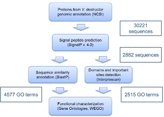
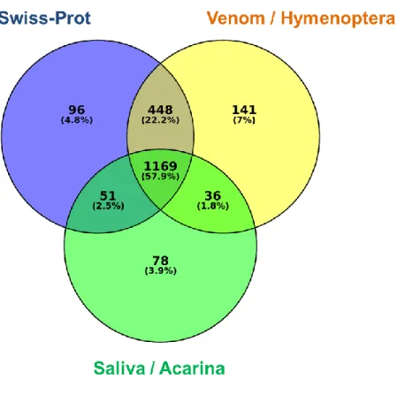
Per la caratterizzazione del secretoma sono state utilizzate le 30221 proteine predette in seguito alla più recente annotazione del genoma di V. destructor (Ottobre, 2017). Queste sono state filtrate per la presenza del peptide segnale, allo scopo di considerare quelle più probabilmente secrete. L’annotazione delle 2882 proteine con peptide segnale, oltre a consentire una preliminare caratterizzazione funzionale del secretoma, ha evidenziato una notevole presenza di omologie tra le componenti della saliva di acari e zecche e quelle del veleno degli Imenotteri. In particolare, circa il 60 % delle proteine del secretoma di V. destructor annotate è risultato condiviso da questi due taxa. Ciò è in accordo con quanto osservato da alcuni autori circa la convergenza evolutiva delle secrezioni nel regno Animale. Infatti, vari studi riportano che zecche e Imenotteri parassitoidi, così come altri animali velenosi, utilizzano le stesse famiglie di proteine, da veicolare attraverso l’iniezione di saliva e veleno, allo scopo di alterare la fisiologia dell’ospite e sfruttarlo da un punto di vista nutrizionale. Esempi di questa convergenza sono rappresentati da fosfolipasi A2, inibitori delle serin proteasi e chitinasi, proteine reclutate tra quelle partecipanti a funzioni biochimiche fondamentali, altamente conservate nel regno Animale. Queste ultime
3 esibiscono funzioni simili a quelle endogene quando vengono iniettate nell’ospite, rappresentando un particolare esempio di elementi attivi a livello intergenomico. Al contrario, altre proteine coinvolte nei fenomeni parassitari possono essere molto specifiche ed esibire una bassa identità di sequenza con proteine di specie evolutivamente vicine. La nostra analisi ha riportato la presenza di 863 proteine che non mostrano alcuna similarità di sequenza con i database presi in esame. Non è da escludere che ci possano essere fattori di parassitizzazione tra queste proteine non annotate, che, ovviamente, risultano di difficile caratterizzazione funzionale. Abbiamo, quindi, deciso di prendere in considerazione per il presente lavoro unicamente candidati dotati di una funzione putativa nota.
Dalla lista dei candidati identificati in seguito all’annotazione del secretoma, in accordo con dati di letteratura, abbiamo selezionato cinque geni, per valutarne l’espressione nelle ghiandole salivari dell’acaro, mediante qPCR. Tra i geni analizzati, solo Vd-CHI è risultato sovraespresso nelle ghiandole salivari. Sebbene gli altri geni valutati (α-macroglobulina, protease aspartica, q-carboxypeptidase, serine protease 42) sono probabilmente espressi anche in altri tessuti, non è da escludere che la loro espressione nelle ghiandole salivari possa avere un ruolo nella regolazione dell’ospite.
Vd-CHI è stato, quindi, selezionato per un successivo esperimento di ibridazione in
situ, che ne ha rilevato l’espressione specifica nelle ghiandole salivari. Questo
risultato ha contribuito a indirizzare i successivi biosaggi funzionali verso la caratterizzazione di questo putativo fattore di regolazione dell’ospite.
Le chitinasi sono glicosil idrolasi responsabili della degradazione della chitina, il secondo più abbondante biopolimero sul pianeta, che è una componente strutturale di matrici biologiche protettive come, ad esempio, l’esoscheletro degli artropodi o la parete cellulare dei funghi. Negli artropodi le chitinasi hanno un ruolo fondamentale come enzimi degradativi durante la muta e un ruolo difensivo contro funghi e nematodi parassiti. La presenza di chitinasi enzimaticamente attive, ma anche di tutti gli enzimi necessari alla completa degradazione della chitina, nelle femmine adulte di
V. destructor è già stata descritta in precedenza. Gli autori degli studi in questione
hanno ipotizzato che questi enzimi possano avere un ruolo nell’idrolisi della chitina dell’ape e nel mantenimento del sito di alimentazione, attraverso il quale la femmina fondatrice e la progenie di V. destructor si nutrono.
Allo scopo di valutare un eventuale ruolo nel favorire l’alimentazione dell’acaro, abbiamo effettuato delle infestazioni artificiali utilizzando acari in cui è stato silenziato il gene Vd-CHI. La sopravvivenza dell’acaro è risultata ridotta del 50% in seguito al silenziamento di Vd-CHI, a 72 ore dal trattamento con dsRNA. Considerando che la maggior riduzione della sopravvivenza avviene intorno alle 24 ore dall’inizio del silenziamento (48 ore dal trattamento) e che V. destructor non può sopravvivere più di 24-36 ore senza alimentarsi, è ragionevole assumere che il silenziamento di Vd-CHI determina una riduzione della capacità dell’acaro di nutrirsi.
Sebbene l’apertura del sito di alimentazione sia principalmente dovuta alla rottura meccanica della cuticola da parte dell’acaro, è possibile che questo processo possa essere facilitato dalla presenza di Vd-CHI. Questa ipotesi è anche supportata dal fatto che questa chitinasi, specificamente espressa nelle ghiandole salivari di V.
destructor, è stata rilevata da recenti studi di proteomica solo nelle femmine adulte
dell’acaro, le uniche in grado di creare e mantenere aperto il sito di alimentazione. Comunque il meccanismo molecolare alla base dell’effetto positivo di Vd-CHI sull’alimentazione dell’acaro resta poco chiaro. La sequenza aminoacidica di Vd-CHI presenta i motivi conservati delle chitinasi GH18 (DXXDXDXE, dove D = acido
4 aspartico, E = acido glutammico, e X = qualsiasi aminoacido), che includono il sito attivo dell’enzima. É interessante notare che Vd-CHI manca del domino putativo di legame con la chitina, nonostante quest’ultimo sembri non essenziale per l’attività chitinolitica delle chitinasi degli artropodi. Comunque, l’attività chitinolitica di Vd-CHI dovrebbe essere valutata attraverso appropriati saggi enzimatici utilizzando estratti delle ghiandole salivari e/o la proteina ricombinante.
Allo scopo di indagare circa un probabile coinvolgimento nella formazione del sito di alimentazione, l’espressione di Vd-CHI è stata misurata in due diverse fasi del ciclo biologico di V. destructor. Abbiamo ipotizzato che femmine foretiche e riproduttive, alimentandosi principalmente attraverso le morbide membrane intersegmentali e la parte centrale del secondo sternite addominale, rispettivamente, potessero avere diverse esigenze in termini di un fattore parassitario in grado di agevolare la perforazione della cuticola.
Vd-CHI è risultato più espresso nelle femmine riproduttive estratte da cellette opercolate, supportando l’ipotesi dell’importanza di questo fattore per la formazione e il mantenimento dei siti di alimentazione, processi che possono influenzarne il livello di espressione durante il ciclo biologico di V. destructor.
Allo scopo di studiare l’impatto di Vd-CHI sulla presenza, la dimensione e il numero delle ferite di alimentazione su pupe artificialmente infestate, abbiamo effettuato delle prove di colorazione con Trypan Blue, che colora selettivamente le cellule morte. Queste prove hanno però dato esito negativo nelle nostre condizioni sperimentali, che prevedevano solo 24 ore di infestazione artificiale delle pupe (occhi marroni), all’interno di capsule di gelatina.
Abbiamo quindi valutato l’effetto di Vd-CHI sull’ospite determinando il profilo di espressione di alcuni geni immunitari dell’ape.
La presenza di Vd-CHI ha determinato la sovraespressione di acidic-mammalian
chitinase e la sottoespressione di sgabd8, due geni che non sono differenzialmente
espressi in pupe infestate con acari silenziati rispetto alle pupe-controllo non infestate. Questo risultato è in linea con recenti studi di proteomica quantitativa in cui un’altra chitinasi (XP_623995.1) e la stessa proteina sgabd8 sono, rispettivamente, sovra- e sottorappresentate in pupe parassitizzate.
Utilizzando un approccio simile a quello descritto nel presente lavoro (silenziamento del fattore di parassitizzazione seguito da RNA-seq dell’ospite parassitizzato), è stata studiata la funzione di una chitinasi GH19 identificata nel veleno di Nasonia
vitripennis, Imenottero parassitoide. La presenza di questa proteina nel veleno
dell’imenottero parassitoide causa la sovraregolazione di una chitinasi nell’ospite
Sarcophaga bullata. Le chitinasi endogene degli insetti sono coinvolte nella risposta
immunitaria all’invasione di funghi patogeni. Sembra quindi ragionevole ipotizzare che la sovraregolazione del gene acidic-mammalian chitinase nelle pupe di A.
mellifera infestate da V. destructor possa essere indotta dall’attività chitinolitica di
Vd-CHI, in grado di stimolare una risposta immunitaria anti-fungina allo scopo di prevenire eventuali infezioni della ferita di alimentazione.
Questa ipotesi è anche supportata dal fatto che Vd-CHI determina la sovraregolazione di spaetzle, che attiva il pathway di Toll in Drosophila e la successiva produzione di peptidi antimicrobici, come osservato anche per la chitinasi GH19 di N. vitripennis.
L’unico trascritto risultato sottoregolato codifica per una glicoproteina strutturale dell’endocuticola dell’ape, sgabd8, associata alla resistenza all’acaro Varroa e probabile indicatore una compromessa capacità della cuticola danneggiata di curarsi in presenza di Vd-CHI.
5 Inoltre, la sovraregolazione di β-1,3-glucan binding avviene solo in pupe infestate con acari silenziati per la Vd-CHI. Questa proteina è coinvolta nel rilevamento delle infezioni nell’ospite e stimola la melanizazzione, processo alla base della cura delle ferite e dell’incapsulamento negli artropodi. É quindi ragionevole considerare la sovraregolazione di questo trascritto come un indicatore di infezioni batteriche causate dall’assenza di Vd-CHI a livello delle ferite di alimentazione. Infatti, Vd-CHI potrebbe essere coinvolta nella prevenzione della proliferazione di batteri attraverso (1) la produzione di chito-oligosaccaridi antimicrobici derivanti dalla degradazione della chitina dell’ospite e (2) l’attività anti-biofilm, come osservato per altre chitinasi. Inoltre, a supporto dell’ipotesi di infezioni batteriche in atto, il peptide antimicrobico
hymenoptaecin risulta sovraregolato in pupe parassitizzate da acari privi di Vd-CHI,
così come già osservato in seguito a iniezione di buffer o di omogenato di V.
destructor.
Nel presente lavoro viene riportata la sovraregolazione del peptide antimicrobico
defensin-1in pupe infestate con acari silenziati e non silenziati, rispetto alle pupe
controllo non infestate. Questo risultato conferma quanto già riportato in altri lavori, ovvero che la sovraespressione di defensin-1viene indotta dalla parassitizzazione. Questo approccio sperimentale soffre della mancanza di informazioni circa il titolo virale delle pupe e degli acari impiegati negli esperimenti. L’effetto del virus DWV sul sistema immunitario dell’ape potrebbe, infatti, mascherare l’impatto della sola infestazione da Varroa. Inoltre, come già menzionato in precedenza, è improbabile che l’infestazione artificiale all’interno di capsule di gelatina possa riprodurre la stessa comunità microbica e l’effetto dei composti antimicrobici caratteristici della cera naturale (in sinergia con propoli e miele) che costituisce i favi dell’alveare. Nonostante questi limiti, il presente studio di espressione differenziale dei geni immunitari suggerisce che le proteine salivari di V. destructor sono in grado di stimolare la risposta immunitaria dell’ape, probabilmente allo scopo di limitare le infezioni a carico delle ferite di alimentazione.
Prolungate infestazioni artificiali di pupe di A. mellifera con acari silenziati, seguite da RNA-seq degli ospiti, potranno rivelare i pathway biologici sollecitati dall’azione di Vd-CHI, contribuendo alla caratterizzazione funzionale di questa proteina salivare. Insieme, questi risultati fanno luce sull’interazione Varroa-ape, confermando che il sialoma dell’acaro riveste un ruolo importante nello sfruttamento delle risorse alimentari offerte dall’ospite. In particolare, la capacità di alimentarsi di V. destructor è mediata dall’azione di Vd-CHI, che probabilmente facilita l’apertura e la pervietà delle ferite di alimentazione sulla cuticola dell’ape. É ragionevole ipotizzare che questa chitinasi possa interferire con il normale processo di cura della cuticola danneggiata, garantendone l’apertura e al contempo limitandone l’invasione da parte di possibili patogeni.
La presenza di funghi e batteri a livello dell’interfaccia ospite-parassita rende questa interazione anche più complessa, suggerendo di adottare un approccio ologenomico per simili studi. L’utilizzo di particolari tecniche di microscopia, esperimenti di RNA-seq e studi di metagenomica delle ferite di alimentazione contribuiranno a caratterizzare i patogeni opportunisti e l’impatto che essi hanno sui processi curativi della cuticola, allo scopo di chiarire il ruolo di Vd-CHI nell’intricata interazione tra parassita, ospite e microrganismi.
Il presente lavoro contribuisce ad una conoscenza più profonda circa il ruolo delle proteine salivari di V. destructor e dimostra, inoltre, che l’utilizzo di dsRNA allo scopo di silenziare geni espressi nelle ghiandole salivari costituisce un’interessante
6 strumento per il controllo dell’acaro, sebbene sia necessario lo sviluppo di nuovi metodi di rilascio nell’ambiente di tali molecole.
SUMMARY
The economic impact of beekeeping, including crop pollination, is very difficult to assess, but, according to the Italian Federation of Beekeepers (FAI), it can be estimated around 1,500 millions of euros annually, generated by about one million of hives. Honeybee colony losses have been a major problem since the beginning of modern apiculture; however, in 2006, the dramatic dimension of this phenomenon attracted the attention of the public opinion and the increasing interest of the scientific community. After a few years of intense investigation, a specific causal agent for the widespread colony losses was not found, but rather a multifactorial origin was proposed for this syndrome. It is now largely accepted that both landscape deterioration and agrochemicals can be directly or indirectly responsible for colony losses. However, several monitoring programs indicate that the large majority of losses are associated with the co-presence of the mite Varroa destructor and the Deformed Wing Virus (DWV). V. destructor is an ectoparasitic mite that creates a wound in the host’s cuticle through which it feeds on haemolymph and fat body, representing an important stress factor that weakens honeybee colonies and promotes the spreading of viral diseases. In order to facilitate feeding, V. destructor delivers a complex of factors, including proteins, through its salivary secretions. The scarcity of information about the sialome of the mite limits the functional analysis of the host regulation process and, thus, the opportunity to impair the mite’s fitness using biotechnological approaches. Here, we have used a functional genomics pipeline to identify V. destructor candidate salivary proteins, along with in situ hybridization detection to assess their expression in salivary glands. Using this approach, we identified a chitinase (Vd-CHI), specifically expressed in salivary glands, which affects mite survival and causes alterations of honeybee immune response. In particular, gene knockdown experiments revealed that Vd-CHI deficient mites tend to show a reduced survival as a likely consequence of reduced feeding capacity on honeybee pupae, due to the lack of chitinoloytic activity that favours the patency of the feeding wound. The importance of Vd-CHI for the feeding success of
Varroa destructor is also supported by the upregulation of this gene during the
reproductive phase of the mite, when the adult female creates large communal feeding site for her and the offspring, usually in the middle of the 2nd abdominal sternite of honeybee pupae. Vd-CHI has also an impact on honeybee’s immune response, determining the upregulation of an endogenous chitinase and the downregulation of an endocuticle structural glycoprotein, associated with wound healing and Varroa-resistance. On the other hand, infestation by Vd-CHI deficient mites led to upregulation of β-1,3-glucan binding protein and hymenoptaecin, as a likely consequence of bacterial infections, suggesting that Vd-CHI could also prevent detrimental bacterial proliferation.
Collectively, these results shed light on Varroa-honeybee interaction, confirming that
V. destructor sialome plays an important role in host exploitation. In particular, V. destructor feeding success on A. mellifera is mediated by the action of Vd-CHI, which
7 probably facilitates the opening and patency of the feeding wound in the honeybee’s cuticle. Indeed, this chitinase is likely involved both in maintaining the feeding site open, by interfering with the regular healing process of cuticle, and in limiting opportunistic infections, by priming antimicrobial defenses. The present work contributes to a better knowledge of salivary repertoire of V. destructor and also demonstrates that dsRNA targeting of genes expressed in salivary glands can offer a promising new tool for controlling the mite.
INTRODUCTION
1.1 Economic and ecological importance of honeybees
The interaction between humans and honeybees had its origins with honey-hunting: the opportunistic stealing of honey from wild bees. The first Neolithic evidence of this activity comes from beeswax residues in Anatolian pottery nearly 9000 years old (Kritsky, 2017). However, the origins of true beekeeping, where bees are provided with artificial cavities in which the comb can be built, can be found in the ancient Egypt and date back to 2450 BC (Kritsky, 2015). In the ancient Egypt honey and the other products of apiculture were important commodities for trade, food, medicinal ingredients, cosmetics and religious rituals (Cilliers and Retief, 2008).
Nowadays the economic impact of beekeeping, including crop pollination, is very difficult to assess. In Italy, according to the Italian Federation of Beekeepers (FAI), this value can be estimated around 1.5 billion € annually, generated by about one million of hives. In the US, a different scenario can be found, where professional beekeepers manages more than 500 hives, for a rough total of 2.6 million honey bee colonies, largely used for crop pollination, and a value of over 14 billion $ (Ellis, 2012). Indeed, crop pollination increases not only yield but also quality, shelf life and commercial value of crops (Klatt et al., 2014) and, then, by significantly influencing farmers’ income, promotes the economic development of the beekeeping industry (Geslin et al., 2017).
More than 75% of all globally important crops depend to some degree on animal pollination, that contributes to about one-third of global crop production (Klein et al., 2007). Assuming complete removal of pollinators, global fruit supplies could be reduced of 22.9%, vegetables by 16.3% and nuts and seed by 22.1% (Smith et al., 2015). Furthermore, animal-pollinated crops are among the richest in micronutrients (Daily and Karp, 2015) and pollinator declines could thus cause micronutrient deficiencies as well as other human health concerns (Chaplin-Kramer et al., 2014; Ellis et al., 2015). It thus become clear that, beyond the ecologic-economic aspects at a local scale, pollination is a service of global and strategical importance threatened by land-use change and agricultural intensification (Lautenbach et al., 2012). Wild bee species, central to the agro-ecosystem service of pollination (Garibaldi et al., 2011, 2013), have been declining in many parts of the world
(Goulson et al., 2015; Potts et al., 2010a). Moreover, the western honeybee, Apis
mellifera, represents the most important species for crop pollination, showing a rapid
8 González-Varo, 2018). However, available data from FAO reveal that during the last half century the growth of the global population of managed honey-bee hives (∼45%) is not matching the rapid (>300%) increase in the fraction of agriculture that depends on animal pollination (Aizen and Harder, 2009). This mismatch between demand and supply of pollination services (Breeze et al., 2014; Schulp et al., 2014) suggests that there is an urgent need to develop a more sustainable agriculture by optimizing pollination and agricultural production, while conserving biodiversity (Garibaldi et al., 2014; Geslin et al., 2017) and health of wild and managed bees.
The biology of the western honeybee A. mellifera will be briefly described in the next section, with special emphasis on the aspects that are relevant to the present work.
1.2 Biology of the western honeybee Apis mellifera
The honeybee Apis mellifera (Hymenoptera: Apidae) is an eusocial insect that lives in colonies where a caste division occurs and only some individuals are capable of reproducing (Winston, 1991). A typical A. mellifera colony consists of a reproductive queen (a fertile female), 20,000-60,000 adult workers (non-reproductive females), 10,000-30,000 worker brood (eggs, larvae and pupae) and several hundred drones (reproductive males) (Bailey and Ball, 1991). The comb, made of beeswax produced by worker bees, is the substrate for all colony interactions and consists of cells with different dimension: smaller cells that are used for brood rearing and food storage, larger cells used to rear drones and queen cells, acorn-shaped, built along the lower edge of the combs (Contessi, 2016). The three castes can be easily distinguished due to some morphological differences (Fig.1).
Fig.1 – Honeybee castes
a) queen; b) worker; c) drone
9 Sex-determination in honey bees is controlled by haplodiploidy: fertilized diploid eggs develop into females, while unfertilized haploid eggs develop into males (Rinderer, 2013). The drones are present only during the early summer in Europe and their sole function is to mate with queens. The queen only leaves the colony to mate, during swarming (colony division) or when a colony deserts a nest site. Her main task is to lay eggs (Denholm, 1999). The queen can control egg fertilization that occurs during oviposition and depends by the type of cell. A fertilized egg has the potential to develop in a fertile queen or a sterile worker, two castes genetically identical, differing only for the type and amount of food received after the first three days of larval development (Contessi, 2016). Nurse workers feed larvae with a protein-rich food secreted by the hypopharyngeal and mandibular glands: the royal jelly (Snodgrass, 1984). If a larva receives a large amount of this brood food throughout the developmental stages it will become a queen; if the same larva receives smaller quantity of royal jelly plus pollen and honey during the latter part of the development it will become a worker (Contessi, 2016). All individuals show the same four developmental stages: egg, larvae, pupae and adult. Eggs are laid by the queen at the bottom of the brood’s cells. After three days from oviposition, a small larva emerges and worker bees start immediately to take care of it. The larva develops very quickly and undergoes five molts; during the last larval stage, workers cap the cell with wax and the larva starts spinning a silk cocoon. Into the sealed cell, the larva develops into a pupa, starts the metamorphosis and, 12 days after sealing, it emerges from the cell. The total developmental time from egg to adult is 16 days for queens, 21 for workers and 24 for drones. Life expectation is very different among the different castes: usually a worker lives 4-6 weeks during the summer but can lives 4-5 months during the winter, the drones survive for 3-5 weeks while the queen typically lives 1-3 years (Contessi, 2016).
Among the workers, there is an age-correlated behavioural division of labour, referred to as temporal polyethism (Winston, 1991). Younger workers perform brood-nest associated tasks such as brood-cell cleaning; when they are 4 day old, they become nurses and start feeding older larvae; then later, according to the hypopharyngeal glands’ development, they feed the younger larvae. Middle-aged bees typically perform food processing, nest construction, and guarding. Finally, from about 20 days of age, older bees progress to foraging outside the nest for nectar and pollen (Johnson, 2008; Seeley, 1982; Seeley and Kolmes, 1991; Winston, 1991). This polyethism schedule is not perfectly rigid, as bees can engage in other duties, if necessary, according to the colony’s needs (Johnson, 2010). Juvenile hormones, food and pheromones act in concert to ensure the reproductive dominance of the queen, the workers’ behavioural maturation, the coordination of colony reproduction through swarming and many other functions (Bortolotti and Costa, 2014; Grozinger et al., 2003; Hoover et al., 2003; Moritz and Southwick, 2012; Robinson, 1985).
1.3 Honeybee colony losses: the role of Varroa destructor
Although global stocks of managed honeybee colonies appear to be increasing (Aizen and Harder, 2009), substantial regional losses have been documented in the last decade (Pettis and Delaplane, 2010; Potts et al., 2010b; Stokstad, 2007). In particular, during the 2006/2007 winter an apparently unexplainable high colony
10 mortality (∼30%) affected managed honeybees in the United States (van Engelsdorp et al., 2007). A portion of those losses were characterized by a common set of specific symptoms (mainly the rapid loss of adult worker bees and the lack of dead workers within or surrounding the hives) that subsequently led to define this syndrome as Colony Collapse Disorder (CCD) (van Engelsdorp et al., 2009). Meanwhile the proceedings of the 4th COLOSS Conference (Zagreb, Croatia, 3-4 March 2009) reported a colony mortality of about 20% in Europe, ranging from 1.8 to 53% among countries (Williams et al., 2010). Extensive colony losses are not unusual and have occurred repeatedly over many centuries and locations during the long-lasting history of apiculture (van Engelsdorp and Meixner, 2010; van Engelsdorp et al., 2008; Oldroyd, 2007; Underwood and van Engelsdorp, 2007), but the dramatic dimension of these recent declines attracted the attention of the public opinion and the increasing interest of the scientific community (Stokstad, 2007). After a few years of intense investigation, a specific causal agent for the widespread colony losses was not found, but rather a multifactorial origin was proposed for this syndrome (Neumann and Carreck, 2010).
It is now largely accepted that both landscape deterioration and agrochemicals can be directly or indirectly responsible for colony losses (Goulson et al., 2015). However, several monitoring programs (van der Zee et al., 2012, 2015) indicate that high loads of parasites and pathogens are constantly associated with this problem (Neumann and Carreck, 2010), and, in particular, the symbiotic association between Varroa
destructor and Deformed Wing Virus (DWV) (Di Prisco et al., 2016) account for the
large majority of losses (Kielmanowicz et al., 2015). The mite V. destructor is an obligate ectoparasite of bees which feeds on host’s haemolymph and fat body (Ramsey and van Engelsdorp, 2017; Ramsey et al., 2018; Rosenkranz et al., 2010). This parasite has a severe impact on honeybee physiology, causing the reduction of weight at the emergence and shortening the life span of the host (Annoscia et al., 2012; Daly et al., 1988; Jong et al., 1982). Furthermore, V. destructor acts as vector of viral pathogens such as deformed wing virus (DWV), sacbrood virus (SBV), chronic bee paralysis virus (CBPV), acute bee paralysis virus (ABPV), Israeli acute paralysis virus (IAPV), black queen cell virus (BQCV) and Kashmir bee virus (KBV) (Tentcheva et al., 2004). The best known is DWV infection, which causes the symptoms of crippled wings and shortened abdomens in individuals from heavily infested colonies (Boecking and Genersch, 2008). DWV, as the other viruses, has been considered a minor problem to honeybee health before the occurrence of
Varroa mites (Bowen-walker et al., 1999). The vectoring activity and the immune
stress caused by the mite Varroa destructor, a relatively novel parasite for Apis
mellifera, promoted the spread and the uncontrolled replication of this virus,
triggering the transition of common harmless covert infections to devastating overt infections (Nazzi and Le Conte, 2016; Nazzi et al., 2012; Wilfert et al., 2016). After the arrival of Varroa in the 1970s and 1980s, the rate of colony loss in Europe and North America nearly tripled (Ellis et al., 2010). In absence of effective control measures of this parasite, an infested colony cannot survive longer than 2-3 years (Fries et al., 2006; Rademacher and Harz, 2006).
In order to have a better understanding of the mechanisms underlying this dramatic effect on honeybee colonies, in the next two sections the biology of the mite V.
destructor (Section 1.4) and the immunity of honeybees (Section 1.5) will be
11
1.4 Biology of Varroa destructor
The mite responsible for the abovementioned clinical symptoms in A.
mellifera belongs to the species V. destructor (Acari: Varroidae), which was
erroneously considered as Varroa jacobsoni until the year 2000 (Anderson and Trueman, 2000). Therefore, every Varroa publications of the last century refer to V.
jacobsoni although in nearly all cases the research subject was V. destructor, the
only mite of economic importance (Rosenkranz et al., 2010). V. destructor successfully shifted from its original host, the Asiatic honeybee Apis cerana, to the western honeybee A. mellifera when this latter were transported to the Far East in the first half of the past century (Oldroyd, 1999). Rapidly spread by beekeeping activities, V. destructor were found on A. mellifera in the eastern coastal region of the USSR (1952), in Pakistan (1955), Japan (1958), China (1959), Bulgaria and ex Jugoslavia (1967), South-America (Paraguay, 1971), Germany (1977), Italy (1981), USA (1987), UK (1992), New Zealand (2000), Hawaii (2007) (Fontana, 2017; Rosenkranz et al., 2010). Today the mite is cosmopolitan, except for Australia, where on some occasions has been detected in hives on docking ships, but without consequences (Fontana, 2017; Rooth, 2018). In this section morphology, life-cycle and behaviour of V. destructor will be discussed, emphasizing the aspects that are relevant to mite feeding and the linked effects on honeybees.
1.4.1 Morphology
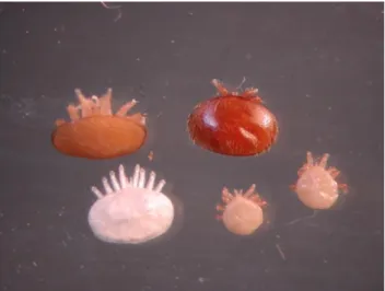
V. destructor displays a marked sexual dimorphism (Ifantidis, 1983): females are
flattened, oval-shaped (1.6 x 1 mm); males are pear shaped and show only weak sclerotisation (Fig.2).
Males are clearly smaller than females in all developmental stages (Rosenkranz et al., 2010). In the adult phase they are easily distinguishable by colour: males are light-yellow while females have a reddish-brown colouration.
Fig. 2 – Varroa destructor mature and immature stages
Clockwise from top left: mature daughter mite, mother mite, two mature males and an immature (deutonymph) daughter (Huang, 2012).
12 The body of Varroa mites, like in other species belonging to the Acari group, is subdivided in two well defined parts: a complex of mouthparts called gnathosoma (Greek: gnathos = jaws, soma = body) and the unsegmented idiosoma (Greek: idio = belonging to one’s self). The gnathosoma (Fig. 3) consists of:
- the hypostome, which ventrally delimits the gnathosoma and dorsally forms a furrow for food and saliva;
- the pair of chelicerae, situated above the hypostome, enclosed in a protrusion of the exoskeleton from which they can be protruded or retracted;
- the labrum, between the two chelicerae;
- two sensory pedipalps (Bautz and Coggins, 1992; Lucius et al., 2017).
The distal part of the chelicera is the movable digit (digitus mobilis), straight and spearshaped, provided with a sharp ventral edge and two thin dorsal hook-like teeth. Together the movable digits constitute a well-adapted saw-like structure to pierce the honeybee integument (Griffiths et al., 1988; de Lillo et al., 2001).
Fig. 3 – Anterior gnathosoma
The gnathosoma consists of a pair of lateral pedipalps (P) and a pair of modified chelicerae (C). Note the pointed tip of the hypostome legs (arrow). The first pair of legs (L) are visible. Scale bar 150 µm (Bautz and Coggins, 1992).
13 The idiosoma comprises one dorsal shield and different ventral shields (Lucius et al., 2017). Thin and flexible membranes between the shields enable the mite to dilate during feeding and egg formation (Rosenkranz et al., 2010). Four pair of legs, short and strong, are attached to the idiosoma and show specialized structures that facilitate the adherence to the host, the apoteles (De Ruijter and Kaas, 1983). The body and extremities, legs included, have an enormous range of bristles, the setae, which act as mechano- or chemoreceptors that help the mite to be oriented in the darkness of the hive (Lucius et al., 2017; Rosenkranz et al., 2010).
1.4.2 Life-cycle
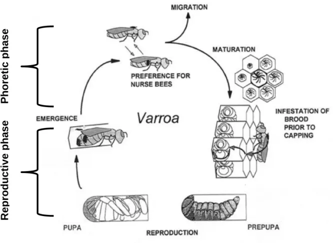
The life cycle of Varroa mites involves two distinct stages: the phoretic phase, spent on the adult bee and the reproductive phase, spent inside a bee brood cell (Fig. 4). Carried by nurse bees and guided by chemical cues, Varroa mated female enters a brood cell containing a 5th instar bee larva 15 to 20 h before worker brood cells are sealed and 40 to 50 h before drone brood cells are sealed (Boot et al., 1992; Nazzi and Le Conte, 2016). The mite uses bee brood pheromone as a kairomonal cue to choose the cell to invade, showing a strong preference for drone brood (Le Conte et al., 1990; Nazzi and Le Conte, 2016; Zuk and Kolluru, 1998). The invasion of the brood cell represents the beginning of the reproductive phase. After entering the cell, the mite reaches the bottom, where becomes stuck within the larval food, probably to avoid detection and removal by hygienic bees (Ifantidis, 1988; Rosenkranz et al., 2010). When, about 5 hours after cell capping, the honeybee larva has consumed all the food and starts to spin the cocoon against the cell wall, then the Varroa female climbs on the host and feeds itself for the first time (Donzé and Guerin, 1994; Rosenkranz et al., 2010). Oogenesis begins rapidly and the first egg is laid on the surface of the cell wall about 70 hours after the capping (Ifantidis, 1983; Steiner et al., 1994). This first egg is normally unfertilized and, due to the haplo-diploid sex determination system, it develops into a haploid male, while subsequent eggs are fertilized and laid every 30 hours to give rise female progeny (Martin, 1994; Rehm and Ritter, 1989). Reproductive phase ends when the adult bee hatches, thus usually a Varroa female can lay up to five eggs in worker brood (12 days of capping) and up to six eggs in drone brood (14 days of capping) (Garrido and Rosenkranz, 2003). In the reproductive phase, a “foundress” mite (the mite invading the cell) will produce only one male and an average of 1.30–1.45 mature females when infesting worker cells, but close to double this amount when infesting drone cells (Martin, 1994). When the prepupa moults into a pupa a major reduction in the free space available for Varroa occurs. Immediately after pupation the foundress engages in “leg-pushing”, resulting in the displacement of the pupa’s third legs, to enlarge the space around the so called “fecal accumulation site” (where the mites aggregate in the cell and defecate) (Donze et al., 1998). Then, near this site, the mother mite creates a hole in the cuticle of the pupa, generally on the 2nd sternite, for her offspring to feed trough (Donzé and Guerin, 1994).
Preimaginal development of Varroa mites consists of egg, proto- and deutonymph stages (Donzé and Guerin, 1994; Rehm and Ritter, 1989). Varroa mites become sexually mature immediately after the last molt, about 6 days after oviposition (Donzé et al., 1996). The offspring mate with each other within the sealed brood cell, at the fecal accumulation site. The foundress and the mated daughters leave the cell when
14 the newly adult bee emerges (Nazzi and Le Conte, 2016). They then transfer onto a nurse bee, where they spend the phoretic phase, before entering a brood cell to reproduce again (Le Conte and Arnold, 1987). Male mites cannot survive outside the cell, and die. Female mites can go through two or three cycles over the course of their life span (Fries and Rosenkranz, 1996).
1.4.3 Feeding
Feeding behaviour is difficult to observe and is an aspect of the mite’s biology which is still relatively poorly understood. Despite a lack of experimental evidence, for the past 5 decades the conclusion that Varroa feed exclusively on the haemolymph (the “blood”) of host bees has been the accepted view among researchers and beekeepers. Recent work, however, has shown that Varroa is not consuming haemolymph, but damages host adult bees by consuming fat body (Ramsey and van Engelsdorp, 2017; Ramsey et al., 2018). The fat body is analogous to mammalian liver and adipose tissue, and is abundant in insects’ abdomen where it is localized subjacent to the epidermis (Keeley, 1985). During larval and pupal stage of
Ph
o
reti
c
p
h
ase
Re
p
ro
d
u
cti
ve
p
h
a
se
Fig. 4 – Varroa life cycle
After a phoretic phase on the adult bee, mite adult females invade the brood cells immediately before capping, start sucking haemolymph from the larva and reproduce (Donzè et al., 1998).
15 honeybee fat cells are detached to each other and are floating free in the haemolymph (Snodgrass, 1984). In the mature worker fat body consists of a thin layer of cells spread against the dorsal and ventral body wall, more developed in nest bees compared to foragers (Chan et al., 2011; Snodgrass, 1984). Fat body is the central storage depot of nutrients and energy reserves (Arrese and Soulages, 2010). This tissue is integral to proper immune function, pesticide detoxification, overwinter survival and several other essential processes in healthy adult and immature bees (Lemaitre and Hoffmann, 2007).
During the phoretic phase, mites are frequently found hiding between the abdominal sternites of the bee in a position that is difficult for other bees to reach (Nazzi and Le Conte, 2016). From this favourable position the phoretic mite is able to feed herself piercing the membrane between the overlapping abdominal plates (Ramsey et al., 2018). The importance of these meals for the mite is probably negligible for the reproductive success, but has relevant consequences on the viral level of the mite. Indeed, V. destructor induces more wing deformity in emerging honeybees when it has experienced a longer phoretic phase, because of the Deformed Wing Virus transmission (Piou et al., 2016).
During the host's pupal stage, adult females spend nearly 60 minutes to perforate the integument of bee pupae in such a way that they and their progeny can feed (Donzé and Guerin, 1994; Rosenkranz et al., 2010). The mother restricts her feeding to one site that is usually localized on the 2nd abdominal segment and never on head or torax, thus avoiding possible damage to the developing appendages of the host bee, essential for the cell opening at the emergence (Donzé and Guerin, 1994; Kanbar and Engels, 2004a). Following penetration of the intersegmental membrane by the bidentate chelicerae, the contact chemosensillae, chemosensitive receptors located distally on the movable digits, are immersed into the wound, providing a sensory feedback on the quality of the food before and during extra-oral digestion (de Lillo et al., 2001). Strick & Madel (1988) state that Varroa uses its chelicerae to 'rasp' a wound, first by inserting the tip of the dentate chelicerae into the membrane and then by making 'alternate sawing movements of the chelicerae (Strick and Madel, 1988). Then the mite can insert its hypostome to inject saliva and aspirate liquid tissues of the pupa (Bautz and Coggins, 1992; Nuzzaci and de Lillo, 1995), with a mechanism similar to blood sucker arthropods (Griffiths et al., 1988). However, Varroa is phylogenetically more closely related to predatory mites (i.e. Gamasiphis,
Macrocheles, Lasioseius) (Klompen et al., 2007) that feed through a different
feeding mechanism: the extra-oral digestion. Recent findings by Ramsey et al. (2018) on parasitized adult bees show degradation of fat body cells beneath the pierced intersegmental membrane, likely due to the extra-oral digestion of the mite at the feeding site.
Very little is known about Varroa mite saliva, but, due to the abovementioned findings, mite’s salivary secretions are likely involved into host manipulation and exploitation. In the first study on V. destructor saliva bioactivity, Richards et al. reported that mite’s salivary secretions damage haemocytes (invertebrate immune system cells) from the caterpillar Lacanobia oleracea and disrupt their ability to form aggregates (Richards et al., 2011). A recent work identified a Varroa toxic protein (VTP), in the saliva of the mite, which exhibits toxic activity toward A. cerana preimaginal stages and elevates DWV titer in A. mellifera (Zhang and Han, 2018a). Investigating the poorly known feeding behaviour and the role of salivary effectors represents an important step towards understanding the molecular
16 mechanisms of the host-parasite interaction and for developing new effective control strategies.
1.5 Pathology of Varroa destructor parasitization
Feeding activity of the mite has profound implications for the honeybee, both at individual and colony scale. At the individual level, honeybee is damaged by Varroa
destructor in a variety of ways, ranging from physiological to behavioural alterations.
Parasitization affects longevity of honeybees (Dainat et al., 2011; Yang and Cox-Foster, 2007). De Jong reported that uninfested control bees live an average of 27.6 days, while infested ones only 13.6. The number of mites per bee is significantly negatively correlated with both longevity and weight at emergence (De Jong et al., 1983).
The loss of haemolymph and fat body during ontogenetic development decreases the weight of worker at the emergence of 7% (Jong et al., 1982). Drones are even more susceptible, varying from 11% to 19% of weight loss, depending on the infestation rate (Duay et al., 2003). The weight loss seems to be related, not just to direct feeding of the mites, but also to the alteration of cuticular hydrocarbons and the consequent water loss through cuticular transpiration (Annoscia et al., 2012; Salvy et al., 2001).
Varroa parasitization impairs the over-wintering capacity of honeyebees, through the
reduction in protein, mainly vitellogenin, and carbohydrate content of haemolymph (Amdam et al., 2004; Bowen‐Walker and Gunn, 2001). The yolk protein vitellogenin is produced in the fat body as a storage protein and have evolved alternative functions in honeybees, regulating immunity and longevity (Amdam et al., 2003; Tufail and Takeda, 2008). Varroa affects also juvenile hormone titre, hypopharingeal glands development, learning ability and attraction to brood (Zanni et al., 2018). Together these alterations contribute to premature transition to foraging in worker bees parasitized during development (Alphen et al., 1996; Amdam et al., 2004; Jong et al., 1982). When honeybees switch from the hive bee to the forager stage, their cellular defence machinery is down-regulated by a dramatic reduction in the number of functioning haemocytes (Amdam et al., 2003). But in emerging parasitized bees a stronger reduction of the functioning haemocyte proportion in haemolymph is observed (Amdam et al., 2004).
Furthermore, several authors reported alterations of behaviour in Varroa infested honeybees. Parasitized foragers show a decreased capability of non-associated learning, prolonged absences from the colony and a lower rate of return to the colony (Kralj and Fuchs, 2006; Kralj et al., 2007).
Considering the colony as a “superorganism”, V. destructor impacts the infested honeybee fitness by lowering the drones chance to mate (Duay et al., 2002) and reducing the capacity of parasitized colonies to produce swarms (Fries et al., 2003; Villa et al., 2008).
The final breakdown of a honey bee colony is associated with the typical “parasitic mite syndrome” (see Section 2) such as scattered brood, crippled bees and unexplainable reduction of the adult bee population (Rosenkranz et al., 2010). It is thus clear that V. destructor has a dramatic impact on physiology, immunity and behaviour of honeybees. However, it is difficult to distinguish between the direct effects of Varroa parasitization and indirect effects related to other parasites, mainly viral pathogens, vectored through feeding.
17
1.5.1 Varroa destructor as a vector
The feeding activity of Varroa mites on honeybess not only subtracts nutrients but also promotes proliferation and transmission of several pathogens. Observing ultrastructural features of the feeding hole made by the foundress on honeybee pupa, Kanbar and Engels (2003) reported that the repeated and communal use of the wound, especially in multiple infested cells, is often associated with delayed healing. The remaining opening allow pathogens to grow at the feeding site and enter the host’s body. Large colonies of Melissococcus plutons, the European foulbrood agent, were found in wounds on worker and drone pupae (Kanbar and Engels, 2003; Kanbar et al., 2004). Furthermore, manipulative experiments have demonstrated that Varroa can transmits bacteria, leading to septicemia in infested larvae (Gliński and Jarosz, 1992). Honeybee’s haemocele invasion and subsequent septicemia it happens only for some harmful bacteria, but represents just one of the possible deleterious effects of bacteria vectored by the mite (Hubert et al., 2016). Indeed, Hubert et al. (2017) reported that regular microbial community, integral of many nutritional and immune functions, is altered in parasitized bees. The author suggests that this disruption of immune homeostasis in the gut is triggered by the immune response of the host toward bacteria vectored by Varroa (Hubert et al., 2017). Different routes of bacterial transmission have been proposed, such as external contamination of mouthparts (dirty needle) and acquisition through infected host fluids ingurgitation (brush). However, the mode of transport of bacteria via mites seems to be passive, because there is no evidence of bacterial replication inside the body of the mite, which is the criterion for active transport and vectoring (Hubert et al., 2016).
Varroa can also be responsible of fungal spores (conidia) dispersion throughout the
bee colony. Soil saprophytes are normally associated with bees, their combs and provisions. The most abundant species found in this context are (in order of abundance): Aspergillus, Penicillium, Fusarium, Trichoderma, Alternaria, Rhizopus and Mucor (Benoit et al., 2004). Some fungi have been shown to be
beneficial to the colony by preserving stored pollen and bee bread, whereas others are pathogenic and contribute to colony losses (Gilliam, 1997; Morse and Nowogrodzki, 1990). Aspergillus flavus, the agent of stonebrood disease in A.
mellifera larvae, known to attack and damage the brood by producing a neuropathic
aflatoxin (Batra et al., 1973; Glinski and Buczek, 2003), has been isolated from the external surfaces of V. destructor (Benoit et al., 2004). Although stonebrood is a rare disease considered to be of minor importance (Morse and Nowogrodzki, 1990), the immune stress caused by the facultative parasite A. flavus has been poorly investigated.
The aforementioned pathogens are passively transported by Varroa destructor and became harmful only in some cases. Indeed, transmission routes, dynamics of infection and effects at colony level are considerably different for viruses, in particular for DWV, which can replicate within the mite’s body (Di Prisco et al., 2011; Gisder et al., 2009; Sumpter and Martin, 2004; Yue and Genersch, 2005).
Honeybees can be infected by 15-20 different viruses and many of them can be vectored by Varroa mites (Chen and Siede, 2007), including Acute Paralysis Virus (Ball, 1985), Slow Bee Paralysis Virus (SBPV) (Denholm, 1999) and Deformed Wing Virus (Bowen-walker et al., 1999). Much of the pathology and mortality observed in severely mite-infested bee colonies is linked to the mite-mediated transmission of viruses (Martin, 1998). Transmission occurs during feeding, which consists in a continuous interchange of fluids between the Varroa mite and the haemocele of the