POLITECNICO DI MILANO DEPARTMENT OFPHYSICS DOCTORAL PROGRAMME INPHYSICS
C
ALCIUM
I
MAGING BY
L
IGHT
S
HEET
F
LUORESCENCE
M
ICROSCOPY
Doctoral Dissertation of:
Alessia Candeo
Supervisor:
Prof. Cosimo D’Andrea
Tutor:
Prof. Paola Taroni
The Chair of the Doctoral Program:
Prof. Paola Taroni
The eye of a human being is a microscope, which makes the world seem bigger than it really is. Khalil Gibran
Acknowledgment
I
WOULD LIKE TO THANKwho gave me the possibility to do this PhD. Cosimo D’Andrea, for being my supervisor and especially teaching me a lot about time resolved spectroscopy, and Paola Taroni for being my tutor and organizing the PhD program. I am strongly in debt with Andrea Bassi for introducing me to the light sheet microscopy, for picking me up randomly one day and working with me from then on, always promoting me. With Gianluca Valentini, for always being helpful and for supporting me, with his patience, kindness and exaggerate knowledge. You both have my deep gratitude.I am grateful to all the external people I have collaborated with during these years. Alex Costa is for sure the best collaborator ever, he thought me every-thing I know about plant biology without him even realizing it. Thanks also to Gandolfo, who is now working with the light sheet setup. A special thank goes to German Sumbre, who welcomed me from the first day, and all his team in Paris which made me discover a whole new field. I want to especially thank Adrien Jouary, who worked together with me, thought me and was my mentor in the Parisian lab, Jonathan Boulanger-Weill and Selma Mehyaoui who helped with the softwares. I hope to work and hang out with you guys again.
I would like to thank the people that spent these 3 years with me in the depart-ment. Firstly, my office mates, Luca, Rebecca, Lucia, Valentina and Edoardo. Thanks for all the advice, the discussions, the fights, the laughs. Edo and Vale, a special thank you for the amazing lunches! And of course the people of the office next door, Laura, Sara, Sara, Sanathana and all the others. Thanks also to the people from the administration and the technicians, who somehow helped in this PhD.
Of course, I want to thank all the people that supported me in a way or another in this adventure, my friends, among which Petra stands, and all my family, mom and dad in particular, that always stayed by my side.
Milano, March 2016 Alessia Candeo
Abstract
O
VER THE LAST DECADES there has been an impressive progression in optical microscopy to explore fundamental issues in life science. The understanding of complex phenomena like the mechanisms of calcium signalling requires a multiscalar method, to be applied from cells to organs in physiological condition, with minimum damage for the sample. An inno-vative approach for the field was provided by Light Sheet Fluorescence Mi-croscopy (LSFM), a technique that introduces optical sectioning in wide-field microscopy.In the following work, an overview of the main calcium pathways is presented, along with a review of fluorescence probes for calcium imaging. This is fol-lowed by the description of the theoretical basis and the design features of three LSFM systems: a Selective Plane Illumination Microscope (SPIM), a Digital Scanned laser Light sheet based fluorescence Microscope (DSLM) and a two-photon DSLM.
The first system was employed in the study of Arabidopsis plant roots in the context of improving the growth of crops and their tolerance to stresses. Finger-prints of calcium signalling related to the application of specific stimuli were observed across the whole plant root, for extended periods of time, with single cell resolution and low photodamage. Spontaneous calcium oscillations in root hairs were correlated to their growth by tree-dimensional screenings and vali-dated by the analysis of knockout mutants showing growth issues.
The DSLM was used for neuroimaging in the study of spontaneous activity in the brain of zebrafish larva. Spontaneous activity could be recorded at high frame rate and for many hours, over different brain areas, with neuronal res-olution. This provided insights on the activity patterns of neurons and on the possibility of predicting behavioural effects of the fish. Two-photon excitation was instead implemented to observe visually evoked responses and understand how visuomotor behaviours are processed.
Contents
Introduction 1
1 The Ion Calcium: from the Primordial Soup to Signals 5
1.1 Introduction . . . 6
1.2 Calcium stores and sources . . . 8
1.3 The signalling toolkit . . . 9
1.4 General Ca2+ encoding . . . . 12
1.5 Calcium channels in plants . . . 13
1.6 Calcium channels in neurons . . . 15
1.7 Seeing the calcium . . . 16
1.7.1 Calcium probes . . . 18
1.7.2 Strategy of calcium evaluation . . . 19
1.7.3 Families of indicators . . . 20
2 Light Sheet Fluorescence Microscopy 25 2.1 Fluorescence microscopy for calcium imaging . . . 26
2.2 A novel approach to microscopy . . . 29
2.2.1 Light sheet fluorescence microscopy . . . 29
2.2.2 The long story of LSFM . . . 33
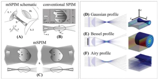
2.2.3 Different illumination techniques . . . 35
Contents
3 Calcium Dynamics in Plant Root 47
3.1 Introduction . . . 48
3.1.1 From the improvement of crops to the imaging of roots . 48 3.1.2 State of the art of calcium imaging in plant biology . . . 49
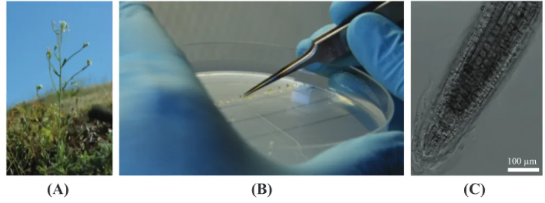
3.1.3 Plant description . . . 50
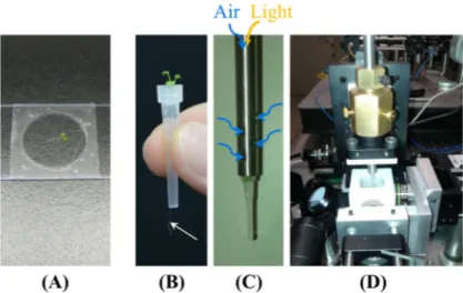
3.2 Material and methods . . . 51
3.2.1 Plant preparation . . . 51
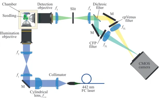
3.2.2 Experimental setup . . . 52
3.2.3 Seedling mounting . . . 54
3.2.4 Plant root treatments . . . 56
3.3 Results and discussion . . . 56
3.3.1 Image quality . . . 56
3.3.2 Ca2+dynamics observation through SPIM-FRET . . . . 59
3.3.3 Calcium oscillations in root hair of Arabidopsis thaliana 67 3.4 Conclusions . . . 74
4 Calcium Imaging in the Brain of Zebrafish Larva 77 4.1 Introduction . . . 78
4.1.1 Recording the brain activity . . . 80
4.1.2 Zebrafish as a model organism . . . 81
4.2 Experimental setup . . . 83
4.2.1 Zebrafish preparation . . . 83
4.2.2 The Digital Scanned laser Light sheet based fluores-cence Microscope . . . 84
4.2.3 Sample mounting . . . 88
4.2.4 Imaging performances . . . 89
4.2.5 Preprocessing and signal extraction . . . 94
4.3 Experimental results . . . 97
4.3.1 High speed recording of neuronal pattern in the optic tectum . . . 97
4.3.2 Long-term recording for the study of neuronal patterns predictive of spontaneous behaviours . . . 105
4.3.3 Two-photon excitation for visual stimulation analysis . . 108
4.4 Conclusions . . . 114
Bibliography 117
List of Figures
0 Statistics of the Method of the year 2014 . . . 2
1.1 The Ca2+signalling toolkit . . . . 10
1.2 Schematic evolution of an action potential . . . 16
1.3 Ca2+ pathways in neurons . . . . 17
1.4 FRET-based calcium indicators . . . 20
1.5 Families of indicators . . . 22
2.1 Standard microscopes . . . 27
2.2 Light sheet fluorescence microscopy principle . . . 30
2.3 Gaussian beam profile . . . 31
2.4 Advantages of LSFM . . . 33
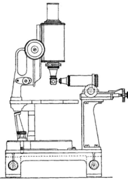
2.5 The ultramicroscope . . . 34
2.6 Different types of light sheet . . . 36
2.7 Principle of DSLM . . . 38
2.8 Further illumination techniques . . . 41
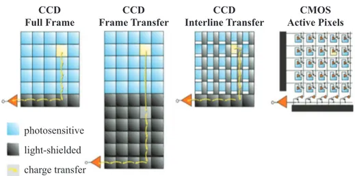
2.9 Detectors . . . 44
3.1 Arabidopsis thaliana plants . . . 51
3.2 SPIM setup . . . 53
3.3 Seedling mounting . . . 55
3.4 Image quality of the SPIM-FRET setup for plant imaging . . . 57
List of Figures
3.6 Single-plane FRET response to L-Glu of plants expressing the
nuclear targeted cameleon . . . 60
3.7 Single-plane FRET response to eATP of plants expressing the nuclear targeted cameleon . . . 61
3.8 Statistical analysis of the response amplitude and duration . . . 62
3.9 Single-plane FRET response to L-Glu and eATP of plants ex-pressing the cytosolic targeted cameleon . . . 63
3.10 Four-dimensional imaging in seedlings expressing the nuclear targeted cameleon during eATP stimulus . . . 65
3.11 Four-dimensional imaging in seedlings expressing the cytosolic targeted cameleon during eATP stimulus . . . 66
3.12 Root hairs growing steps . . . 68
3.13 Autocorrelation of Ca2+oscillations . . . . 70
3.14 Correlation between growth rate and Ca2+ oscillations . . . . . 70
3.15 Knock-out mutant shows a clear phenotype in the root hairs . . 71
3.16 Comparison of Ca2+ oscillations in root hairs wild-type and mutant seedlings . . . 72
3.17 Fourier analysis of Ca2+oscillations . . . . 73
3.18 Three-dimensional CICR . . . 75
4.1 Animal models in neuroscience . . . 79
4.2 Adult zebrafishes of different transgenic lines . . . 82
4.3 Coarse brain anatomy of a 6 dpf zebrafish larva . . . 82
4.4 Top view of the excitation path of the DSLM setup . . . 85
4.5 Side view of the detection path of the DSLM setup . . . 87
4.6 Zebrafish larva embedded in agarose . . . 89
4.7 One and two-photon 2D images of zebrafish brain . . . 91
4.8 3D imaging of zebrafish brain . . . 92
4.9 Axial light sheet profile . . . 93
4.10 Inference of fluorescence events . . . 96
4.11 Retinotopic organization of the optic tectum . . . 99
4.12 PCA-promax functional assemblies in the optic tectum . . . 100
4.13 Raster plot and threshold from null model of spontaneous ac-tivity in the optic tectum . . . 101
4.14 Single events maps . . . 102
4.15 Wave-like patterns in the optic tectum . . . 103
4.16 Repeated events of coactivation . . . 103 4.17 Analysis of the lag of neuronal firing during activation events . 104
List of Figures
4.18 Increase in activity prior to spontaneous movement . . . 106
4.19 Specificity for the directionality of routine turns . . . 107
4.20 Raster plot of visually evoked activity . . . 109
4.21 Orientation selectivity map . . . 110
4.22 Orientation selectivity graphs . . . 111
4.23 Polar plots of orientation selectivity . . . 112
Introduction
E
VERY TIME WE LOOK AT LIFE ON EARTH, we remain fascinated by how incredibly perfect, yet complex, it is. From our childhood to oldness, we all remain amazed by the majesty of life. Even simple actions, like taking a picture of a flower, imply a series of complicated events, taking place both in the macroscopic and microscopic scale, happening in extremely short time but that can have long term effect and are moulded by experience. We need to see images of the flower with our eye, while the eye muscles keep the field of view centred on the flower, with the retinas sending images of the flower to the brain; this latter, at the same time decides when to take a picture, communicating with the muscle of our arm and our finger to press the shutter button. Then, we press in a fast, controlled way, converting energy to mechan-ical motion. And the image remains impressed also in our memory.From the primordial soup, an ubiquitous element involved in all these pro-cesses is the calcium ion. It is so effective that it became necessary, early in the evolution of life, to create mechanisms to reduce calcium ions concentration from the cell cytosol. The existence of large concentration gradients across cell membranes created the perfect conditions to enable calcium signalling, spatiotemporal calcium patterns that encode a variety of process taking place in an organism.
These patterns can be explored thanks to increasingly refined techniques, which ideally should allow the measure of calcium concentration in a single cell surrounded by a large number of other cells, in order to understand its
func-Introduction
Figure 0: Light sheet microscopy statistics from Web of Knowledge. The number of the published and especially cited papers about light sheet fluorescence microscopy has increased exponentially in the last decade.
tional role. Among these techniques, fluorescence microscopy has always had a prominent role. Lots of progresses have been made in calcium imaging tech-niques in the last decades, leading even to the formulation of new questions about decoding and improving life conditions on earth.
This thesis describes a novel approach to study calcium dynamics in living samples, called light sheet fluorescence microscopy. This technique puts the sample at the very centre of the microscope, approaching it with a multiscalar scheme, under different points of view. It allows one to observe a wide field of view with single cell resolution and sectioning capability, with the possibility to record at high speed, for long time an organism in non-damaging conditions. This method comes from a century-old idea, though the first biologically rele-vant application only arrived in 1993 and the scientific community discovered it in 2004. But, after that, the number of publications (more than 500) and of citations (11.7 per paper in average) increased exponentially, till it was named the Method of the year 2014 by Nature (Figure 0).
This tomographic device, running in the visible or near-infrared ranges, of-fers the possibility to investigate a variety of living biological samples, mainly small (in the order of one millimetre) translucent specimens. The two research topics that I faced during my PhD, deal with plant roots of Arabidopsis thaliana and zebrafish (Denio rerio) brain.
In Chapter 1, the main motivations about the importance of the ion calcium are highlighted, and a review of the main calcium channels is presented, with emphasis to plant roots and neurons. This is followed by a report on calcium indicators, the tools that allow one to actually see calcium dynamics.
Chapter 2 explains the principle of light sheet fluorescence imaging with
Introduction parison to standard techniques. Three main implementations of light sheet microscopy are discussed to lay the theoretical bases of the systems that I have implemented: static light sheet, digitally scanned light sheet and two-photon digitally scanned light sheet.
The third and fourth chapters discuss the two main subjects that I had dealt with. Chapter 3 concerns the improvement of crop yield and their tolerance to diverse stresses. Arabidopsis roots have been used as a model specimen and have been studied by static light sheet microscopy. The variety of benefits of using such a technique for the characterization of the response to the applica-tion of different stimuli are highlighted. Calcium signalling in the root cells across the whole plant organ has been observed, revealing specific fingerprints related to the application of different stimuli. The possibility to observe also spontaneous activations in single cells, allowed us to correlate root hair growth and spontaneous calcium oscillations in the root hair apex by tree-dimensional screenings. The deregulation of these calcium variations observed in knockout mutants was indeed linked to altered plant health.
Chapter 4 deals with a field born two decades ago and strongly emerged since then: neuroimaging. The brain is an intricate network, whose understanding cannot disregard its interaction with the external world, as well as its spon-taneous intrinsic dynamic. Decoding the brain would open incredible new opportunities both in medical research and artificial intelligence studies. A key model for optical neuroimaging is the zebrafish larva, a small transparent behaving vertebrate. Neuronal activity from different brain regions of larvae was acquired with neuronal resolution and minimal photodamage by digital scanned laser light sheet based fluorescence microscopy. Spontaneous activ-ity was recorded with one-photon illumination at high frame rate and in the long term, providing insights on the structure of large neuronal circuits and on the possibility of predicting behavioural outcomes. Two-photon excitation was instead implemented to observe visually evoked responses and to understand how orientation selectivity is encoded to process visuomotor behaviours. All the experiments were run with specially designed light sheet microscopes, giving results that could have been hardly obtained with standard techniques. The achieved results shed new light on both the fields of investigation, opening new perspectives for further studies.
These projects are the outcome of a synergy with the Dipartimento di Bio-scienze of Università degli Studi in Milano for the plant biology project and with the Institut de Biologie of the École Normale Supérieure in Paris for the neuroimaging part.
CHAPTER
1
The Ion Calcium: from the Primordial Soup to
Signals
I
N THE LAST DECADES, there has been a great emphasis in life science on the study of calcium dynamics. This ubiquitous ion is involved in all the basic processes that can take place in an organism from cells to organs. The changes in its cellular concentration occur with refined spatial and/or tem-poral patterns, essential for most functional processes.The following chapter gives an overview of the main calcium pathways to un-derstand this signalling toolkit, with specific reference to plant roots and neu-rons. Since the interest is on calcium imaging, a review of the principal calcium indicators is given.
Chapter 1. The Ion Calcium: from the Primordial Soup to Signals
1.1
IntroductionLife science investigates the diversity, complexity, and interconnectedness of life on earth. The study of a cell, tissue, organ or entire organism requires a multiscale approach from microscopy to macroscopic observation of the sam-ple under test. Understanding the interaction among cells as well as the one with the external environment requires the capability to comprehend how cells process information. The gathering and processing of this information takes place through the modulation of membrane potentials. These are electric po-tential differences between the sides of cellular membranes, created by the different concentration of ions between the inside environment and the outside one. The migrations of ions are regulated by the opening of pore-forming pro-teins, or channels, positioned not only on the plasma membrane, but also in the ones of internal compartments, which selectively change their conforma-tion according to the electrochemical gradient. At the same time, the opening of a channel influences the membrane potential by locally varying the ion con-centration. This complex interaction between channels opening and membrane potential variation creates an intricate feedback pattern, which can lead to dif-ferent physiological outcomes, like adaption, growth, or even apoptosis of the cells.
Knowledge of the intracellular concentration of ions is thus fundamental. Mono-valent ions (Na+, K+, H+, and Cl ) generally provide for electrical excitability and energy production, while divalent cations (Ca2+, Mg2+, Zn2+) are crucial for cellular biochemistry and regulation. In this whole mechanism of cellular signal processing, the calcium Ca2+ ion plays a central and fundamental role. The unique biophysical properties of Ca2+ has been known from more than a century, and, since their discovery, millions of experiments aided in building up the fame of intracellular Ca2+ as a “universal signalling molecule” [95]. For this reason, in the last decades the study of the dynamics of intracellu-lar calcium concentration [Ca2+] has been a major focus in life science, and the knowledge of calcium signalling and of its corresponding physiological outcome has grown significantly. As a matter of fact, Ca2+ ions are used as transductory molecules, or second messengers, by almost all cell types, from prokaryotic to eukaryotic ones. Many endogenous stimuli and stress signals of both biotic and abiotic nature lead to transient variations in the intracellular calcium concentration [Ca2+], which in turn activate specific downstream sig-nalling cascades, triggering or mediating a wide variety of cellular processes. From an evolutionary point of view, why the Ca2+ has been chosen to be so
1.1. Introduction unique among all the possible ions is still not completely understood, but its universality and effectiveness has an important evolutionary implication. As a matter of fact, specificities of ATP-based energetics of primordial cells re-quired tight control over [Ca2+]. An excess of Ca2+inside the cell would have been toxic: high [Ca2+] would induce aggregation of proteins and nucleic acids affecting the integrity of lipid membranes, and would initiate precipitation of phosphates [15]. As a result, cells developed a highly sophisticated system to maintain the equilibrium. First of all, the activatory actions of Ca2+ ions are usually accomplished not directly by the ions alone, but through their binding to a number of high-affinity binding proteins (CaBPs), like calmodulin (CaM), troponin C, etc. These proteins are negatively charged at physiological pH and can be highly Ca2+-selective. Moreover, Ca2+is favoured as a regulatory molecule thanks to its double positive charge, which results in a higher affinity when binding to negatively charged proteins than monovalent cations such as Na+ or K+. At the same time, Ca2+ has a lower affinity compared to larger divalent cations such as Cu2+, Zn2+ or Mn2+, but also a lower dissociation constant. Finally, Ca2+ has a higher and more flexible coordination chemistry than that of Mg2+for example. Concentration of free Ca2+ ions in the cytosol of all cells is maintained at approximately 50 to 150 nM, around 20000 times lower than that in the extracellular space, even lower when compared to that of intracellular domains or organelles. Considering that for a good signalling sys-tem the intrinsic signal-to-noise ratio is essential, it is possible to understand the importance of Ca2+ in this context. The existence of a large concentration gradient across membranes created the ideal condition for calcium to be the signalling molecule par excellence. Indeed, because of this large electrochem-ical Ca2+ gradient always directed towards the cytosol, even very short-lived changes in the Ca2+permeability of the membranes will rapidly generate con-siderable signals. Obviously, to allow cells to be always prompt for the acti-vation, an effective mechanism of cytosolic Ca2+removal has been developed during the evolution process, permitting the return of [Ca2+] to the low basal level for the system to be primed again. The combination of Ca2+ movements between intracellular compartments and the cell and extracellular environment produce spatiotemporally organized fluctuations that are generally referred to as “Ca2+ signals”.
Chapter 1. The Ion Calcium: from the Primordial Soup to Signals
1.2
Calcium stores and sourcesThe stores of Ca2+ during these ion movements include not only the extracel-lular environment, but also some intracelextracel-lular compartments such as the mito-chondria, the nucleus, the endoplasmic reticulum (ER) and sarcoplasmic reti-culum (SR). Mitochondria can actually both accumulate excess of Ca2+during periods of intense cellular activity and release it after stimulation, sustaining calcium signals. While the nucleus and the mitochondria are just stores of Ca2+, the ER and SR can be seen as the main intracellular source for initiating Ca2+ signalling, and in fact the majority of the eukaryotic cells prefers using these intracellular Ca2+ stores as their primary source for Ca2+ signal genera-tion.
Cells that exclusively use these stores are generally known as non-excitable cells and, since their membrane depolarization is not followed by a subsequent regenerative depolarization, they cannot generate or propagate electrical sig-nals. In this case, the general mechanisms for Ca2+ signal generation involves activation of plasma membrane (PM) receptors and the synthesis of an inter-mediate signalling molecule, which then acts on specific Ca2+release channels situated on the ER membranes. This first signal can then be followed by a sec-ondary release of intracellular Ca2+mediated by other channels sensitive to the cytosolic [Ca2+], which acts like a signal amplifier.
On the other hand, also the extracellular Ca2+alone can initiate signalling. For example neurons, considered as the ultimate excitable cells, primarily exploit extracellular Ca2+ for their signal generation, though the possibility of contri-bution of intracellular Ca2+ stores is still being debated. In functional terms, it makes sense that neurons, which require a rapid speed of processing, have developed a system of using preferentially the extracellular Ca2+: in this way, the activation and full Ca2+ response is completed within milliseconds, while intracellular Ca2+ can still participate in regulation and excessive Ca2+ buffer-ing.
Nonetheless, in some cells the shaping of the Ca2+ signal involves extracel-lular and intracelextracel-lular Ca2+ at the same time, for example by means of store-operated Ca2+channels or SOCC, (as discussed below) in the PM, sensitive to the exhaustion of the finite amount of Ca2+ in the ER. Anyway, every type of cell has its specific mechanism of interplay between intracellular and extracel-lular Ca2+ sources.
1.3. The signalling toolkit
1.3
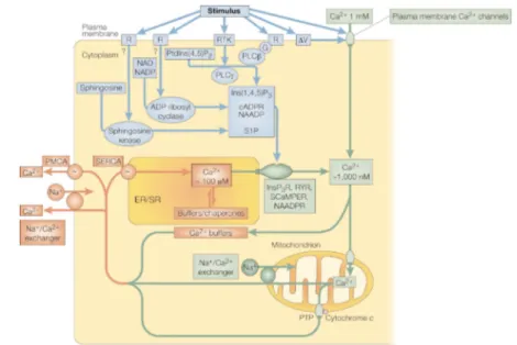
The signalling toolkitBy analysing the temporal behaviour of these processes, we can distinguish between immediate and delayed responses activated by Ca2+signals. The im-mediate response is the one taking place in the milliseconds-to-seconds range, with a short delay between signalling and execution and, as a consequence, in short spatial ranges and primed pathways. It is the case for example of mus-cle contraction, exocytosis and neurotransmitters. The delayed responses are more varied and less defined, since the lag time between the signalling and the final response could vary from seconds to even months. This response could include regulation of intracellular enzymes, gene expression, cellular develop-ment, growth and death. Since in the same cell multiple different responses can take place at the same time to provide a functional continuum, every Ca2+ signal triggering specific responses must be finely encoded, to be in the end de-coded by the system. In this sense, the versatility of Ca2+ arises from the use of an extensive molecular repertoire of signalling components, which creates a “toolkit” of signals whose simultaneous interplay produces a large variety of different spatial and temporal output profiles. This toolkit basically relies on “ON” mechanisms of increase of the [Ca2+] in the cytoplasm and “OFF” mech-anisms that remove Ca2+ to restore the basal state. A graphical representation of the toolkit is given in Figure 1.1.
The “ON” mechanisms
The “ON” mechanisms depend on Ca2+ channels that are built up by a hetero-geneous family of proteins, providing Ca2+ entry from the extracellular envi-ronment and/or release of ions from intracellular organelles. In the PM it can be found the best known typology of channels, the voltage-operated calcium channels (VOCCs), which are activated by the depolarization of the membrane. The VOCCs are highly Ca2+permeable and consist of a set of protein subunits, where usually one acts as the real channel while the others regulate the gating, but a wide variety of isoforms of these protein and, of course, of combinations, have been detected. The binding of a specific agonist (e.g. ATP, glutamate, serotonin etc.) to the extracellular domain instead activates ligand-gated or receptor-operated Ca2+ channels (ROCCs). These have a lower permeability for Ca2+ and include an assortment of structurally and functionally various channels. Many cell types show mechanically activated Ca2+channels, which respond to stress/shape changes that the cell is undergoing. Finally, on the PM we can find the already mentioned store-operated Ca2+channels (SOCCs),
ac-Chapter 1. The Ion Calcium: from the Primordial Soup to Signals
Figure 1.1: Elements of the Ca2+ signalling toolkit. Ca2+-mobilizing signals (blue)
are generated by stimuli acting through a variety of cell-surface receptors (R), including G-protein (G)-linked receptors and receptor tyrosine kinases (RTK). The signals generated include: inositol-1,4,5-trisphosphate (Ins(1,4,5)P3), gen-erated by the hydrolysis of phosphatidylinositol-4,5-bisphosphate (PtdIns(4,5)P2) by a family of phospholipase C enzymes (PLC , PLC ); cyclic ADP ribose (cADPR) and nicotinic acid dinucleotide phosphate (NAADP), both generated from nicotinamide-adenine dinucleotide (NAD) and its phosphorylated derivative NADP by ADP ribosyl cyclase; and sphingosine 1-phosphate (S1P), generated from sph-ingosine by a sphsph-ingosine kinase. ON mechanisms (green) include plasma mem-brane Ca2+ channels, responsive to transmitters or to membrane depolarization,
and intracellular Ca2+ channels like the Ins(1,4,5)P3 receptor (InsP3R),
ryan-odine receptor (RYR), NAADP receptor and sphingolipid Ca2+ release-mediating
protein of the ER (SCaMPER). The Ca2+ released into the cytoplasm activates
different Ca2+ sensors and processes, depending on cell type and context. OFF
mechanisms (red) pump Ca2+ out of the cytoplasm: the Na+/Ca2+exchanger and
the plasma membrane Ca2+ ATPase (PMCA) pumps Ca2+out of the cell and the
sarco-endoplasmic reticulum Ca2+ATPase (SERCA) pumps it back into the ER/SR.
(CAM, calmodulin; PTP, permeability transition pore). Adapted from Berridge and coworkers [9].
1.3. The signalling toolkit tivated in response to depletion of the intracellular Ca2+ store, whose effect is to sustain and shape the signal. How the exhaustion of the organelles is per-ceived is still unknown.
Other sets of Ca2+ channels are expressed in intracellular stores membrane. Their Ca2+ release is mediated by several types of intermediate messengers: the binding of many hormones or growth factors to specific receptors on the PM leads to the production of the intracellular messengers inositol 1,4,5-tris-phosphate (InsP3) and diacylglycerol. InsP3 moves in the cytoplasm where it encounters specific receptors (InsP3Rs) on the ER/SR, which change their conformation, opening and releasing Ca2+. The InsP3Rs activation is however modulated by the [Ca2+] itself: an increase in [Ca2+] within a specific thresh-old enhances the channel opening, whereas a higher [Ca2+] inhibits it. A struc-turally and functionally analogues family of ER/SR receptors are the Ryan-odine (RyRs) or Ca2+-gated Ca2+ channels. Also these channels are sensitive to cytosolic Ca2+concentrations, although in their case the Ca2+concentration must be higher, and they are actually the main responsible of the phenomenon known as Ca2+-induced Ca2+release (CICR). The capability of RyRs to sense Ca2+ variations is mediated by cyclic ADP ribose (cADPR), while a related messenger, nicotinic acid dinucleotide phosphate (NAADP), acts on a separate channel still under study. The RyRs can also act like signal amplifiers, since their activation can follow the Ca2+ release from InsP3Rs. Finally, also mito-chondria can participate in CICR, though they have to accumulate Ca2+ before it. A further channel is the sphingolipid Ca2+-release mediating protein of en-doplasmic reticulum (SCaMPER), whose physiologic function is under strong research.
The “OFF” mechanisms
The second step in the signalling toolkit is the removal of the Ca2+ from the cytoplasm by various pumps and exchangers, the “OFF” mechanism. Firstly, in the cytosol we can find various Ca2+ binding proteins, some fixed and some mobile, buffering the Ca2+and participating in the spatial coding mecha-nism. The recovery of the basal state is mainly mediated by high-affinity ATP-dependent Ca2+ pumps present in membranes: the plasma membrane Ca2+ -ATPase (PMCA) pumps and Na+/Ca2+exchangers return Ca2+to the extracel-lular environment, while the sarco-endoplasmic reticulum ATPase (SERCA) pumps segregate Ca2+ in the internal stores. Once the cytosolic Ca2+ has re-turned to its resting level, a mitochondrial Na+/Ca2+ exchanger can pump the
Chapter 1. The Ion Calcium: from the Primordial Soup to Signals
large load of Ca2+ back into the cytoplasm, from which it is either returned to the ER or removed from the cell. Lastly, a permeability transition pore (PTP) can form on mitochondria, activated by the build-up of Ca2+ within the mi-tochondrial matrix and hence participating in CICR. Two functional states of PTP are known: a reversible low conductance state, allowing mitochondria to become excitable, and an irreversible high conductance state, which can make the mitochondrion collapse and lead to apoptosis.
1.4
General Ca2+ encodingOn a coarse level, further versatility of the signalling toolkit is achieved through both a spatial and temporal encoding. The spatial properties are due to an am-plitude modulation of the Ca2+ signals, each prompting a different specific effect, determined by the affinity with the intracellular Ca2+ sensors. More-over, variations in the Ca2+ signal are determined not only by the changes in the total amount of cytosolic Ca2+, but also by the type and distribution of the various Ca2+ buffers and sensors, which are not homogenous in the cells and indeed create local Ca2+domains or “Ca2+ gradients”. This heterogeneity accounts for the directionality in the intracellular propagation of the Ca2+ sig-nal, which follows specific pathways producing a sort of vectorial code. The propagation of the signalling appears as a cytosolic “Ca2+wave”, with specific spatial and temporal properties. For waves to occur, most of the InsP3Rs and the RyRs respond to each other through the process of CICR: Ca2+ emitted from one receptor diffuses to the neighbouring one exciting further release, therefore setting up a regenerative process. For example, since InsP3Rs and/or RyRs have different degrees of excitability depending on the levels of the ap-propriate Ca2+-mobilizing messenger, their opening depends on how small, but higher than a threshold, the level of calcium is. These single-channel events of InsP3Rs and RyRs have been known as quarks or blips, respectively, and may be considered the building blocks of more complex Ca2+ signals. At higher [Ca2+], coordinated opening of clusters of InsP3Rs or RyRs, known as puffs or sparks, respectively, can be reached, and the amplitude of the signal can be linked to the number of opened channel involved. Also, increasing sparks frequency can result in a cascade and become regenerative, setting up two- or three-dimensional waves of changes in [Ca2+] that propagate within cells. From a temporal point of view, this repeated activation of the Ca2+-mobilizing receptors can evoke oscillatory patterns of [Ca2+l. This temporal coding can be analyzed at three main levels: i) the shape of these oscillations; ii) the
1.5. Calcium channels in plants pendency of the frequency of Ca2+oscillations on stimuli; iii) the specificity of the cytosolic oscillation of an individual cell compared to that of a population. Variations of the basic pattern from cell to cell is a phenomenon called “Ca2+ fingerprint”.
When looking at a whole tissue or organism, it becomes of more interest the mechanism of intercellular communication. In fact, intracellular waves can spread to neighbouring cells, coordinating the activity of many cells. How the waves traverse the gap junction is a matter of considerable debate.
Cell-to-cell signalling can occur thanks to the presence of gap junction chan-nels, which circumvent cellular Ca2+ autonomy. For some cells types, the messenger seems instead to be InsP3 or even ATP. Intercellular signals can also be prompted by VOCCs, which rapidly rise periplasmic [Ca2+] triggering protein fusion and enabling vesicles containing transmitter molecules to fuse to the plasma membrane. Whatever the mechanism, the molecules released in the external environment can gate channels and receptors on adjacent cell membranes. These Ca2+-mediated events are of great interest in neuroscience, where VOCCs create a local pulse of Ca2+to trigger vesicle release in the pro-cess of exocytosis at synaptic endings, but corollaries have been formulated in intercellular communication in almost all cell types.
1.5
Calcium channels in plantsIn resting plant cells, the cytosolic [Ca2+] is maintained around 100 nM by the activity of Ca2+-ATPases and Ca2+/H+ antiporters in cell membrane. Ca2+ channels have been characterized in the PM, the SR, the vacuole, nuclear and plastid membranes. In the PM, the channels can be divided into depo-larisation-activated, hyperpolarization-activated and voltage-insensitive chan-nels. Depolarisation-activated Ca2+-permeable channels (DACC) have been found in the PM of all plant root, leaf mesophyll and suspension-cultured cells, in which a characteristic Ca2+ current has been identified. This current is activated by membrane depolarisation to voltages more positive than about 140mV, whereas at extreme negative voltages it shows a slow and reversible inactivation. In general, these channels are thought to regulate stress-related signals like environmental, developmental and pathological stimuli, generating a global signal. As an example, the distribution and activation of proteins in the PM can be coordinated by the cytoskeleton, which can regulate the direction of growth of root hairs.
proto-Chapter 1. The Ion Calcium: from the Primordial Soup to Signals
plasts from various cell types. In the PM of onion epidermal cells they act as mechanoreceptors activated by stretch. In guard cells they appear to trigger PM depolarization, coordinating the loss of solutes and initiating stomatal closure. In root endodermal cells they are supposed to take part in the mineral nutrition process. Mechanosensitive ion channels may serve a role in the regulation of turgor as a response to gravity, touch or flexure stimulation.
An interesting case is the elongation of root hairs [100] and pollen tubes, stud-ied by analogy with the tip-growth of algal rhizoids and fungal hyphae. The proposed mechanism of growth for polarized cell is that the PM stretches as a consequence of cell wall yielding and local turgor-driven evagination; this opens locally stretch-activated Ca2+ channels, which makes the extracellular Ca2+ enters into the cytosol generating a Ca2+ gradient in the tip. Finally, movements of secretory-vesicles and exocytosis reduces the elevated [Ca2+]. However, the actual Ca2+ channels involved in these processes have not yet been elucidated. In Arabidosis thaliana root, depolarization-activated Ca2+ -current was observed in all cell types, while hyperpolarization was observed in a subgroup of cells in the elongation zone, including endodermal cells. It has indeed been suggested that the ubiquitous depolarization-activated chan-nels are involved in cell signalling, while the hyperpolarization one may have a role in mineral nutrition.
Two elicitor-activated, plasma-membrane Ca2+-permeable channels have been documented, triggering the Ca2+ influx as an early event in the initiation of defence responses to pathogens. The first type activates at voltages more nega-tive than 120 mV, while the activity of the second, termed large-conductance elicitor-activated channel (LEAC), does not appear to depend on the membrane potential (between 30 and 150 mV).
The vacuole may occupy more than 90% of the volume of a plant cell and con-stitutes a major source of Ca2+. Both the resting membrane potential ( 20 mV to 50 mV) and the difference between cytosolic (100 to 500 nM) and vacuo-lar [Ca2+] (micromolar to millimolar) favour the entering of Ca2+in the cyto-plasm. This Ca2+ efflux from the vacuole could sustain large and prolonged elevations in the cytosolic [Ca2+] by propagating and amplifying an initial sig-nal. When the initiation comes from a cytosolic [Ca2+] rise, a CICR appears, but the role of the different channels in this event is still of great debate. Also in the case of vacuole membrane, there are several classes of channels like de-polarization activated Ca2+ channels, comprehending the slow vacuolar chan-nels with still an unclear physiological role, and the hyperpolarization activated Ca2+ channels, which catalyse Ca2+ influx to the cytoplasm. These channels
1.6. Calcium channels in neurons may serve a role in cell signalling. Several classes of second-messenger acti-vated Ca2+ channels have also been found, like those activated by the InsP3 and the cADPR, highly selective for divalent cations.
Calcium channels have also been characterized in the SR and the calcium efflux from them has been shown to be implicated in cell signalling. For example, it has been shown that in Brassica oleracea Ca2+ can be released by the applica-tion of cADPR, NAADP and possibly InsP3, suggesting the presence of Ca2+ channels dependent on these molecules.
Finally, a channel permeable to Ca2+, Mg2+and K+has been found in swollen thylakoids from spinach chloroplasts and one permeable to K+, Na+, Cs+and Ca2+ in the nuclear envelope of nuclei from red beet. For a wide variety of Ca2+ channels, an electrical characterization have been done. However, the physiological role as well as the stimuli activating many of them is still under study.
1.6
Calcium channels in neuronsNeurons are the excitable cell par excellence. They present numerous Ca2+ channels with different localizations and different functions. As in all cells, the different concentration of ions in the intracellular and extracellular environ-ment, creates an electric potential difference between the sides of the cellular membrane, leading, for most neurons, at a resting state of about 70 mV. All neuron types are able to generate a stereotyped event, called an “action poten-tial” (Figure 1.2).
An action potential occurs in the cell when the membrane potential increases up to a threshold, generally around 55 mV, at which VOCCs for Na+ open. Since the concentration of sodium is much higher outside the cell than inside it, this causes a rapid increase in the membrane potential, up to positive val-ues, where VOCCs for K+ open and Na+ channels close. The concentration of K+ is higher inside the cell, so that the opening of the channels abruptly lowers the membrane potential towards negative values. The action potential propagates along the axon at high speeds (up to 100 m/s) through the serial opening of VOCCs for Na+equally spaced along the axon. At its termination the axon ramifies, and contacts other neurons by synapses. Here local VOCCs open, with the influx of Ca2+ triggering exocytosis of a neurotransmitter in the synaptic space, passing information between neurons. ROCCs on the adjacent neuron can open and receive the neurotransmitter, altering the membrane po-tential. The described event results in a fast spike in membrane potential, which
Chapter 1. The Ion Calcium: from the Primordial Soup to Signals Time 1 ms -80 mV -70 mV -55 mV 40 mV M em bra ne pot ent ia l 1 2 3 4 5 6
Figure 1.2: Schematic evolution of an action potential. 1. Resting state. 2. Sodium channels opening threshold. 3. Potassium channels open. 4. Sodium channels close. 5. Refractary period. 6. Resting state.
lasts, in most vertebrate organisms, humans included, around one millisecond. Neurons also have internal Ca2+stores. The store function is usually attributed to the ER, where the InsP3Rs and of RyRs have been found. Internal Ca2+ release have been documented in various types of neurons at different devel-opmental stages. While InsP3-mediated calcium release is mostly triggered by some neurotransmitters, RyRs can be activated by elevations of the cytosolic [Ca2+], which by CICR amplifies the neuron activation or firing. Both InsP3Rs and RyRs are regulated by calcium itself along with other intracellular factors. In the case of InsP3Rs, calcium influx plays an essential role in generating Ca2+ waves in neocortical and other types of neurons. In hippocampal neurons, for example, electrical activity resulting in Ca2+ entry through VOCCs acts to-gether with InsP3 produced by metabotropic glutamate receptors (mGluR1) to produce a synergistic release of internal Ca2+ (see Figure 1.3).
Communication between neurons is actually an intricate subject not only from the point of view of the channels that can be found expressed on them, but also for the wire of communication they can establish. The study of how and why these connections form, is becoming more and more of interest and is making neuroscience an incredibly wide interdisciplinary field.
1.7
Seeing the calciumThe understanding of the biological complexity of calcium signalling has been dependent on the methodologies of measuring the calcium concentration [Ca2+] in living cells. Nowadays the ideal technique for Ca2+ evaluation should allow
1.7. Seeing the calcium
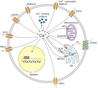
Figure 1.3: Ca2+ pathways in neurons. Sources of Ca2+ influx are
calcium-permeable ↵-amino-3-hydroxy-5-methyl-4-isoxazolepropionic acid (AMPA) and N-methyl-D-aspartate (NMDA) glutamate-type receptors, voltage-gated calcium channels (VGCC), nicotinic acetylcholine receptors (nAChR), and transient recep-tor potential type C (TRPC) channels. Calcium release from internal srecep-tores is medi-ated by inositol trisphosphate receptors (InsP3Rs) and ryanodine receptors (RyRs). InsP3 can be generated by metabotropic glutamate receptors (mGluR). Calcium efflux is mediated by the plasma membrane calcium ATPase (PMCA), the sodium-calcium exchanger (NCX), and the sarco-endoplasmic reticulum sodium-calcium ATPase (SERCA). Also the mitochondria are important for neuronal calcium homeostasis. Reproduced from Grienberger and coworkers, [32].
one to measure [Ca2+] in a single cell (microscopic level), as well as in a large number of cells surrounding it, to understand the effect of calcium signalling (macroscopic level). The two main techniques to study the functional output of Ca2+signals over a population of cells are electrophysiology and fluorescence imaging. The first one involves the use of microelectrodes inside the cells to provide direct measurements of the voltage changes in the channels. The dis-advantage however is its complexity and invasiveness, as well as its intrinsic incapability to record signals from many cells. Although single-cell patch-clamp techniques can nowadays be parallelized to enable recordings from even 200 cells using multi-electrode arrays [13], a powerful alternative for
record-Chapter 1. The Ion Calcium: from the Primordial Soup to Signals
ing the activity of hundreds of cells at the same time is calcium imaging. This strategy is based on fluorescence microscopy and has a lower temporal res-olution compared to electrophysiology, but balances this with the advantage of being non-invasive and providing precise spatial localization of each of the cells being monitored. In the last decades, the advances of optical methods and sensors increased tremendously the attention on fluorescent Ca2+ probes and microscopy development.
1.7.1 Calcium probes
Ca2+probes (also known as indicators, reporters or sensors) are molecules that can selectively and reversibly bind with Ca2+ ions. These indicators estimate the amount of free and bound ions. The concentration of free Ca2+ is indi-rectly calculated taking into account the effective dissociation constant (Kd) of the probe for Ca2+in the environment where it is located. Inevitably, since all indicators act as Ca2+buffers, their use leads to an increase in the Ca2+ buffer-ing capacity.
The first measurements of this type were done with organic fluorescent dyes like murexide, azo dyes and chlortetracycline, whose spectral properties changes when binding to Ca2+, but had several limitations. For example, azo dyes have complex and variable Ca2+-dye stoichiometries, small signal-to-noise ratio and had to be introduced into cells by technically tough methods (microinjection or intracellular perfusion). Chlortetracycline shows instead large increases in fluorescence on Ca2+binding but is mainly sensitive to [Ca2+] changes only in membrane as well as to several other parameters ([Mg2+], membrane potential, pH). In 1980 Roger Tsien presented the first fluorescent probe for intracellular use specifically designed for Ca2+imaging, called BAPTA [94]. These fluores-cent polycarboxylate dyes are based on modifications of the best known selec-tive Ca2+-chelator available, ethylene glycol tetraacetic acid (EGTA), properly revised to change its excitation and/or emission characteristics when binding to Ca2+. By the way, BAPTA itself is not satisfactory as an indicator since its ab-sorption spectrum is peaked in far ultraviolet range, but its improved derivatives became soon popular probes. This led to the synthesis of their acetoxymethyl esters [93], which allowed the trapping of these indicators in living cells in a technically simple way in fundamentally all cell types with minimal side ef-fects.
The field experienced an even bigger burst with the investigation of the phe-nomenon of bioluminescence in the medusa Aequora Victoria, which brought
1.7. Seeing the calcium to the identification of aequorin (AEQ) [87] and green fluorescent protein (GFP) [59] by Shimomura, Nobel Prize in Chemistry in 2008, as well as altered ver-sion of these to produce variants with different excitation and emisver-sion spectra. The big improvement introduced by these proteins is that they can be selec-tively targeted to most subcellular compartments by genetically encoding them (GECIs, Genetically Encoded Calcium Indicators). While AEQ has a small flu-orescence, affecting space and time imaging resolution, encoded GFPs main-tains their strong signal, which explains their great diffusion. Indeed, starting from the 1997, three main families of Ca2+ sensors were developed based on GFPs: the camelons [56], the camgaroos [6] and the pericams [62]. These probes are all linked to a Ca2+-binding protein, calmodulin, which undergoes a conformation change in presence of Ca2+. This variation in the molecular structure of the sensor alters the fluorescence characteristics of the GFP, and the quantification of this variation gives an indirect estimation of the Ca2+ con-centration around the indicator.
1.7.2 Strategy of calcium evaluation
We can distinguish two main classes of Ca2+ sensors, the non-ratiometric and the ratiometric one. Non-ratiometric probes can be excited with a single wave-length and the [Ca2+] is determined by looking at relative variations in the fluorescence intensity as follows
⇥
Ca2+⇤ = Kd⇥ F Fmin
Fmax F (1.1)
where Kdis the dissociation constant; F is the fluorescence intensity; Fmaxis F at saturating [Ca2+]; Fminis F at zero [Ca2+].
Ratiometric dyes experience instead a substantial change in the excitation (emis-sion) spectrum when the [Ca2+] varies, for example a spectral shift. The [Ca2+] is therefore evaluated as the ratio between two different fluorescence intensity values, taken at two different wavelengths of the altered excitation (emission) spectrum. In this case, [Ca2+] is calculated as
⇥ Ca2+⇤= Kd⇥ R Rmin Rmax R ⇥ Sb2 Sf 2 (1.2)
where R is the ratio between the fluorescence of the acceptor and that of the donor; Rmax is R at saturating [Ca2+]; Rmin is R at zero [Ca2+]; Sb2 and Sf 2 are the values of the fluorescence at saturating [Ca2+] and at zero [Ca2+] at the
Chapter 1. The Ion Calcium: from the Primordial Soup to Signals
Figure 1.4: Emission spectrum of FRET-based ratiometric dyes, here the cameleon YC3.6. An increase in the [Ca2+] shifts the emission to higher wavelengths,
chang-ing the ratio between the emission at two wavelengths 1 and 2.
wavelength 2, which is the one used in the denominator. The advantage of this last class of indicators is that they are intrinsically correct, for example, for unequal dye loading, bleaching and focal-plane shifts, as the ratio does not depend on the absolute intensity of the two signals, allowing to calculate an exact value of the [Ca2+]. Despite of that, non-ratiometric sensors usually have a stronger fluorescence change when binding to Ca2+, allowing faster recordings, and since just one wavelength is acquired, they require simpler instrumentation and analysis. Moreover, it must be considered that usually only relative [Ca2+] variations are of interest.
1.7.3 Families of indicators
The first two probes based on GFP that has been developed belong to the fam-ily of cameleons (Figure 1.5(A)) and are ratiometric: they both rely on the phenomenon of Forster Resonant Energy Transfer (FRET) between two differ-ent GFP mutants, whose reciprocal distance changes with the [Ca2+]. FRET occurs between an excited donor molecule and an acceptor molecule in the ground state. Depending on the distance between the donor and acceptor, as well as other parameters like the extent of overlap of the emission spectrum of the donor with the absorption spectrum of the acceptor, the quantum yield of the donor and the relative orientation of the donor and acceptor transition dipole, there can be an energy transfer between the two molecules. It is worth noting that FRET relies on a long range dipole-dipole interactions between the
1.7. Seeing the calcium donor and acceptor and consequently the energy transfer by definition takes place without the presence of a photon [49]. In the first cameleon, designed by Tsien and coworkers [56], a blue (or cyan) and a green (or yellow) vari-ants of GFP are expressed at the termini of a construct made of the fusion of the CaM-binding peptide M13 and CaM. An increase in [Ca2+] makes the CaM wrap around the M13 domain, like the tongue of a chameleon, reduc-ing the distance between the two fluorophores and therefore changreduc-ing colour, thus increasing the efficiency of FRET (Figure 1.4). In FIP-CBSM developed by Persechini [79], the two fluorophores are linked only by the M13 peptide, which elongates when binding to CaM in presence of high [Ca2+], reducing the FRET phenomenon.
All the experiments on plants described in the following Chapters were per-formed taking advantage of the Yellow Cameleon YC3.6, which is the most popular for plant biology [63], whose spectrum is depicted in Figure 1.4. It has been specifically designed to improve the brightness and energy transfer between the FRET couple, CFP and cpVenus. In fact, with respect to other yellow cameleons, the YC3.6 uses as an acceptor cpVenus, which is a circu-larly permuted variant of YFP. This chromophore proved to be more resistant to the cellular environment and to have a higher fluorescence expression. More prominently, the relative orientation of CFP and cpVenus transition dipoles is improved, increasing the dynamic range of the indicator. The YC3.6 probe properties that fit well the needs of plant biology can be indeed resumes in: i) high signal-to-noise ratio; ii) high dynamic range; iii) pH stability in the physiological range; iv) an in vitro Kdfor Ca2+ of 250 nM. In this ratiometric indicator, the two fluorescent proteins sandwich the calmodulin and the M13. When there is an increase in the [Ca2+] around it, the M13 domain binds the calmodulin peptide reducing the distance between the two fluorophores and, therefore, increasing the efficiency of FRET.
However, in general one of the main disadvantages of cameleons is their small changes in signal when binding to Ca2+. Besides, their large size and molec-ular complexity in some cases might weaken their targeting efficiency, though on the contrary the associated low diffusion rate can help when high spatial accuracy is pursued, for example, to localize Ca2+puffs or sparks.
As an answer to these issues, non-ratiometric probes based on a single GFP variant were designed. The general mechanism is the alteration of the pKa (the pH at which 50% of the probe is protonated) of the fluorescent protein, which follows the binding of Ca2+ to CaM, that is in one way or another fused to the GFP. This results in a consistent fluorescence variation in
pres-Chapter 1. The Ion Calcium: from the Primordial Soup to Signals
Figure 1.5: Families of GFP-based indicators: in the cameleon (A), a Ca2+-induced
interaction between CaM and the CaM-binding peptide M13 increases the rate of FRET leading to a decrease in the fluorescence of the CFP and an increase in the fluorescence of the YFP; in the camgaroo probe(B), the Ca2+-induced
conforma-tional change in CaM increases the fluorescence of YFP; the Ca2+-induced
inter-action between CaM and M13 of pericam(C) leads to changes in the fluorescence characteristics of the cpYFP. Reproduced from Rudolf and coworkers [80].
ence of even subtle changes in the protein structure, which however depends to the compartment pH. Moreover, thanks to the discovery that the termini of a yellow-emitting GFP (YFP) can be rearranged without negatively affecting the fluorescence properties of the protein, CaM started to be inserted within GFP-based proteins themselves. In camgaroos [6] CaM is interleaved in a site in between YFP, which makes its fluorescence “bounce” high when interact-ing with Ca2+ (Figure 1.5(B)). In pericams [62] a variant of YFP is circularly permuted, meaning that a peptide linker is inserted between the termini of the protein and that a new terminal pair is created elsewhere in the sequence, and sandwiched between CaM and M13 (Figure 1.5(C)). There are three main vari-ants of pericam, one of which, known as ratiometric pericam, has an excitation spectrum that shifts on Ca2+ binding, thereby allowing ratiometric measure-ment. The issue with ratiometric pericams is that they must be excited at two
1.7. Seeing the calcium different wavelengths, which makes them not adequate for fast imaging. To the family of pericams also belong GCaMP indicators, single wavelength non-ratiometric sensors based on circularly permutated GFP instead of YFP, where again the termini are concatenated to M13 and CaM [64]. These indicators have improved rapidly, and as an example, the GCaMP5G variant has an im-proved sensitivity compared to its predecessors, with a fluorescence increase by a factor relative to the baseline up to 1000, with a decay and rise half-time of 667 ± 43 ms and 166 ± 20 ms respectively. At the time of redacting this manuscript, an even more sensitive and faster probe, the GCaMP6, is already available.
So far, most research in Ca2+ signalling has been carried out in cultured cells or ex vivo tissues. However, the final goal of modern biology is to investigate life on earth, ultimately researching highly complex systems and whole living organisms. This is probably one of the reasons why the genetically encoded probes are rapidly becoming popular, given that they can be exploited in cul-tured cells and be ready for the following in vivo measurements. By the way, improving [Ca2+] imaging means not only to develop new and better probes, but also instruments that can benefit from the innovative probes and that, at the same time, are powerful and flexible enough to permit the study of a whole living specimen.
CHAPTER
2
Light Sheet Fluorescence Microscopy
T
HE20TH CENTURYhas seen major advances in optical microscopy. The main struggle was to be able to see the smallest details of biological samples. While huge advances in the knowledge of how things are made were achieved, new quests for functional investigation has emerged. Over the past decades, light sheet fluorescence microscopy showed its unique ability to image large biological specimens with single cell resolution and minimum damage, for long time periods, shedding new light on biological functions. In the following chapter, the principles underlying this technique will be dis-cussed with comparison to standard microscopy for life science, and a detailed description of three implementations of light sheet microscopy i.e. static, digi-tally scanned and two photon light sheet microscopy will be discussed.Chapter 2. Light Sheet Fluorescence Microscopy
2.1
Fluorescence microscopy for calcium imagingThe easiest and most used technique for general-purpose fluorescence imag-ing is of course epifluorescence microscopy. An epifluorescence microscope is based on a standard microscope, with an incoherent light source reflected by a dichroic mirror making it pass through the objective. At the focal plane of the objective, where the sample is placed, the fluorescence signal is excited and collected by the same objective, which sends it through the dichroic mirror and the tube lens which finally projects it on a pixelated detector. The main issue with this approach is the contribution of out of focus fluorescence, which limits its usage only to thin samples, like cultured cells or tissue slices. Looking at thick samples leads to blurred images, with no possibility of recovering the lost information. Therefore, making an image of a volume requires the elimination of the unwanted photons from out-of-focus regions, operating an optical sec-tioning of the sample. The secsec-tioning capability provides greater contrast and permits three-dimensional (3D) reconstructions by computationally combining images from different planes of a sample.
The confocal microscope [85] is the most diffused microscope able to do so, which brought a first huge contribution to calcium imaging. Basically, the light from a point source, usually coherent, is focalized on a single point in the focal plane of the objective and a pinhole is placed on an image plane in front of a single-point detector, such as a photomultiplier. The pinhole let only the light coming from the very on-focus plane to reach the detector, while blocking all the rest of the light, as depicted in Figure 2.1(A). The illuminated spot is then scanned across the whole field of view and the complete image is generated by a computer. The scanning can be achieved in two main ways: by using a couple of galvanometric mirrors to actually move the beam in a raster way, or by means of the so-called spinning disk. In this last approach, a rotating disk with multiple pinholes is placed in a position conjugated with both the focal plane of the objective and a pixelated detector, creating an array of excitation spots and of pinholes at the same time. Three-dimensional images can be cre-ated by moving the objective in the axial direction. Nevertheless, confocal microscopy suffers from few drawbacks. First of all, the time required for the acquisition of an image is proportional to the number of sampling points that compose it. Since it is a point-scanning technique (or array-scanning with the spinning disk) it is intrinsically slow, and, as the image quality also depends on the pinhole size, obtaining one image can take even few seconds. This means that the sample remains exposed to a large dose of excitation light, with a big
2.1. Fluorescence microscopy for calcium imaging
Figure 2.1: Standard point scanning microscopy techniques: confoncal microscope (A) and two-photon microscope (B). Note that the fluorescence in (A) is generated also on the cones below and above the focal plane, being rejected later by the pinhole, while in(B) is generated only in the focal point.
contribution to the bleaching of the specimen. To reduce the acquisition time maintaining a good sampling in an image, one can either reduce the field of view or use an even higher and damaging excitation power, but anyway up to a saturation level for the fluorescent molecules. What is more important, out of focus fluorescence is rejected, but still excited: this means that the sample is being bleached on planes that are not even being recorded. Since the ex-citation light is being absorbed and also scattered by out of focus molecules, the photons that reach the focal plane are only a fraction of the initial ones, further decreasing the total useful signal and the acquisition speed, along with the penetration depth.
A more suitable approach to image thick and scattering media is to avoid the excitation of the out of focus fluorescence from the beginning. This is
Chapter 2. Light Sheet Fluorescence Microscopy
the idea behind two-photon (Figure 2.1(B)) [18] and multiphoton microscopy in general, tools that have become crucial for research in biology in general, and calcium imaging in particular. Two-photon fluorescence excitation con-sists in the simultaneous absorption of two photons by a fluorophore in a single event. The two photons absorbed have roughly twice the wavelength of the one of single photon absorption. The process is nonlinear, as the probability of this phenomenon to happen is quadratically dependent on the excitation power. This means that to increase this probability a high photon density is needed: this is achievable by focusing the excitation light in a tight, diffraction-limited volume at the focal point of a high numerical aperture (NA) objective, together with a high repetition rate femtosecond lasers for the excitation. These pulsed sources, usually tunable Ti:Sapph laser, provide a high peak intensity while keeping the average power relatively low. In this way, only the fluorophores at the focal point are excited, while the rest of the sample does not absorb any photon. Like in confocal microscopy, the focal point is necessarily scanned over the whole field of view to create the full picture. The purpose of em-ploying the two-photon effect is that the axial point spread function is substan-tially lower than for single-photon excitation. As a result, the axial resolution is improved, allowing thin optical sections to be recorded without the use of the pinhole. Moreover, since most fluorescent molecules used in biology have absorption spectra in the visible range, the typical light used in two-photon imaging is in the near infrared range of the spectrum. This leads to a higher penetration depth for this technique, as light scattering is proportional to 1/ 4 and the absorption properties of water, haemoglobin and melanin are minimum in the near infrared region. The weakness in the use of infrared excitation light is the loss in lateral resolution, which is directly proportional to the excitation wavelength.
Although technical improvements (such as the acquisition parallelization [11]) or refinements in measurement strategy [83] (scanning the beam only across area of interest of the field of view) allow to gain in speed while maintaining the sequential scanning, a real jump in the performances could only come from a radical change in the geometry of the microscope.
It is worth mentioning that optical microscopes have an inherent limitation in spatial resolution because of the wave nature of light. The effect of light diffraction limits the optical resolution to approximately half of the wave-length of the light used, meaning that many fine cellular structures cannot be resolved. In 1994 Stefan W. Hell, who received the Nobel Prize in Chemistry in 2014, and Jan Wichmann [33] developed the STimulated Emission Depletion
2.2. A novel approach to microscopy (STED) microscopy, creating super-resolution images by the selective deacti-vation of fluorophores. In 2006, three research groups independently demon-strated super-resolution microscopy using high-precision localization of single fluorophores, named as PhotoActivated Localization Microscopy (PALM) [10] STochastic Optical Reconstruction Microscopy (STORM) [81] and Fluores-cence PhotoActivation Localization Microscopy (FPALM) [35]. These tech-niques allowed to look at cellular structures with a resolution of even 20 nm, but at the cost of the acquisition speed, which is limiting their diffusion for in vivo imaging.
2.2
A novel approach to microscopy2.2.1 Light sheet fluorescence microscopy
In the 80s the confocal configuration led to a breakthrough in microscopy, im-proving resolution and introducing optical sectioning. Confocal imaging re-lies on diffraction-limited scanning spot and spatial filtering, which make the method intrinsically slow and prone to photodamage, due to the high light dose required. As already stated in Chapter 1, life science and in particular calcium imaging require multiscale observation from cell level to systemic level of en-tire organs , which means having a wide Field Of View (FOV) with still at least single cell resolution and sectioning capability. Since we also want to look at dynamic behaviours, the image acquisition must be fast enough to record them, without damaging the sample.
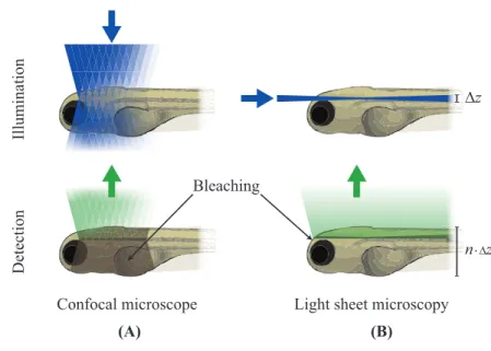
Confocal imaging limitations can be overcome by using a different geometry of the microscope. A promising approach relies on the selective excitation of a thin plane of the sample, matched with the focal plane of a wide field objective. It is then possible to image an entire plane of the sample in one shot using a pixelated detector, still preserving optical sectioning. This is the principle be-hind Light Sheet Fluorescence Microscopy (LSFM), a technique based on two different optical paths for the excitation and the detection paths, as shown in Figure 2.2. In LSFM a thin sheet of laser light illuminates the sample, while the fluorescence is collected orthogonally to the excitation path and recorded by a camera.
This geometry gives an inherent sectioning capability even in its simplest con-figuration, with no need for pinholes or expensive femtosecond lasers. By mov-ing the specimen through the light sheet or the light sheet through the sample, many 2D images at different depth can be acquired, allowing the reconstruction