UNIVERSITÀ DEGLI STUDI DI SASSARI
SCUOLA DI DOTTORATO DI RICERCA IN SCIENZE BIOMEDICHE
Direttore della Scuola: Prof. Andrea Fausto Piana
INDIRIZZO IN ODONTOSTOMATOLOGIA PREVENTIVA
Responsabile di Indirizzo: Prof. Guglielmo Campus
XXVIII CICLO
ORAL DISEASES EPIDEMIOLOGY IN ITALIAN AND NON-ITALIAN
POPULATION. A MULTICENTER CROSS-SECTIONAL STUDY
Direttore:
Prof. Andrea Fausto Piana
Tutor:
Prof. Guglielmo Campus
Tesi di dottorato di: Dott.ssa Cynthia Lara Capi
Cynthia Lara Capi
Oral diseases epidemiology in Italian and non-Italian population. A multicenter cross-sectional study. Tesi di dottorato in Odontostomatologia Preventiva, Università degli Studi di Sassari
ORAL DISEASES EPIDEMIOLOGY IN ITALIAN
AND NON-ITALIAN POPULATION.
A MULTICENTER CROSS-SECTIONAL STUDY
Doctoral Thesis
PhD in Preventive Dentistry
Department of Biomedical Sciences - University of Sassari
PRESENTS:
SUPERVISOR:
Cynthia Lara Capi
Oral diseases epidemiology in Italian and non-Italian population. A multicenter cross-sectional study. Tesi di dottorato in Odontostomatologia Preventiva, Università degli Studi di Sassari
“
Tu naturaleza no ha cambiado sigues siendo un ser alado y hasta el último de mis días pongo mi mano e tu mano”Dedicated to the life of Dr. Mídore Lara Capi
Comenzamos este capítulo de mi vida juntas y hoy que lo concluyo recuerdo cuando hace
cuatro años cumpliste tu sueño de ser doctora. Nunca olvidaré cuando te graduaste, ese
instante en el que me miraste a los ojos y sonreíste con la más pura alegría, como si no
existiera ningún obstáculo cuando se va en busca de la felicidad: entonces supe que eras la
persona mas valiente que he conocido.
Daría todo porque estuvieras sentada junto a mí en este día tan importante, eres mi primer y
mi último pensamiento del día y te extraño con toda el alma… pero se que estás presente y
que te sientes orgullosa de mí. Te dedico este y cada uno de mis logros, a ti, a tu amor por la
vida, a tu fuerza y a tu determinación, a tu bondad y generosidad, eres y serás siempre mi
más grande inspiración, mi mas precioso sentimiento y mi mayor orgullo.
Gracias por todo lo que me enseñaste y por todo lo que me amaste.
Bubu
Cynthia Lara Capi
Oral diseases epidemiology in Italian and non-Italian population. A multicenter cross-sectional study. Tesi di dottorato in Odontostomatologia Preventiva, Università degli Studi di Sassari
Agradecimientos
Le doy gracias a Dios por haberme sostenido en esta prueba, por traerme aquí, a un lugar que se convirtió en mi refugio en un momento en el que necesitaba más que nunca sentirme en casa pero sobretodo le agradezco por mi Madre.
Gracias Mamá: hoy estoy de pie por ti. La vida nos ha puesto de rodillas muchas veces, pero tu nos has levantado entre tus brazos. Me he convertido en quien soy porque he seguido tu reflejo; hoy me miro al espejo y veo a alguien que se parece mucho a ti y que también, cuando da lo mejor de si, se parece a Mido. Nos enseñaste a triunfar ante la adversidad y a luchar por nuestra felicidad… quiero que sepas que lo hicimos, realizamos nuestros sueños sabiendo que estabas allí para sostenernos, y sabes una cosa, junto a ti pasamos los momentos más bellos. Recuerda que el amor verdadero es eterno e invencible y que un día volveremos a encontrarnos.
Cynthia Lara Capi
Oral diseases epidemiology in Italian and non-Italian population. A multicenter cross-sectional study. Tesi di dottorato in Odontostomatologia Preventiva, Università degli Studi di Sassari
Ringraziamenti
Voglio ringraziare a tutte le persone che mi hanno aperto le porte del loro cuore e che sono entrati nel mio. Sono sicura che il destino mi abbia portato proprio qui per incontrare il bene che bisognavo ricevere e dare.
Grazie Flavi sei una benedizione meravigliosa.
Grazie Don Peppino per diventare il padre che ho sempre necessitato, per insegnarmi a pregare con il cuore nel posto giusto, e per tutti i momenti che abbiamo passato insieme. Grazie G per la tua comprensione e per il tuo sostegno, per insegnarmi tutto quanto so ora e per guidarmi nella vita e nella professione, non esistono parole in nessuna lingua per dirti quanto hai cambiato la mia esistenza.
Grazie Zia Farica e Zio Sergio, mi avete accolto come una figlia e vi voglio bene come tale. Grazie Marco e Giovanna per tutto l’affetto, incessante e fiducioso, e per le lacrime trasformate in sorrisi.
Grazie Anna, Giorgio, Valentina, siete diventati parte della mia famiglia e mi avete regalato inaspettati e meravigliosi momenti.
Grazie Antonella e Antonio: la vita ha dei piani che non riusciamo a comprendere fino a che ci troviamo di fronte una risposta che ci ruba un attimo di gioia: grazie per averla replicata. Grazie Alessandra, sei un dono splendido e immeritato.
Grazie Angela, Giuseppe, Roby, Sara, e ai miei amici di San Vincenzo.
Grazie Rosaria e Aracellis: la lontananza non ha impedito che il vostro affetto mi stringa forte.
Grazie all’Università di Sassari, a tutto il Dipartimento di Scienze Biomediche, ai miei colleghi e collaboratori, specialmente a Gianfranco e Roberto, a Maria Teresa e a Riccardo del Dipartimento di Alta Formazione, e a ognuno che ha fatto possibile questa meta.
Cynthia Lara Capi
Oral diseases epidemiology in Italian and non-Italian population. A multicenter cross-sectional study. Tesi di dottorato in Odontostomatologia Preventiva, Università degli Studi di Sassari
Acknowledgements
My heartfelt appreciation to my mentor, Prof. Guglielmo Campus, for the immeasurable dedication, genuine care, indefatigable professionalism and incomparable support he put into guiding me to become a true researcher in science and in life. You made this achievement possible!
I’m forever grateful to the professors that motivated me to grow and learn, especially to Prof. Gail Douglas for the life-changing opportunity and to Prof. Franklin García-Godoy for believing in me.
My sincere gratitude to the collaborators who shared their experience with me, in particular to Dr. Fabio Cocco and Dr. Maria Grazia Cagetti.
Thank you to everyone at the Department of Biomedical Sciences, the Department of Surgery, Microsurgery and Medicine Sciences and the Department of High Education from the University of Sassari.
Cynthia Lara Capi
Oral diseases epidemiology in Italian and non-Italian population. A multicenter cross-sectional study. Tesi di dottorato in Odontostomatologia Preventiva, Università degli Studi di Sassari
PROLOGUE
The doctoral work I am presenting: “Oral diseases epidemiology in Italian and non-Italian population. A multicenter cross-sectional study” has been achieved during a three-year period in the international scenery to promote the cooperation between institutions from different parts of the world.
The thesis is divided into two chapters that contain the main topics of the research: Dental Caries and Dental Erosion (Erosive wear). Under the dental caries chapter, the subjects covered are: Diagnosis, Risk, Early detection technologies, Minimally Invasive Therapy, Behavioral and socio-cultural factors, Taste preference and Prevention.
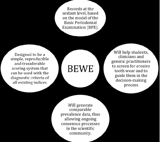
The chapter on erosive wear focuses on the condition and starts from the Tooth wear definition and classification, Differential diagnosis, Measurement of wear and the BEWE model.
A third part of the volume contains a detailed account of the Multicenter study achievements in three different countries: Italy, Mexico and Saudi Arabia.
Each chapter contains the relative scientific evidence published and presented in international journals and congresses.
This dissertation conceives the current oral health status of three different populations, the advanced approaches on its diagnosis and treatment.
Cynthia Lara Capi
Oral diseases epidemiology in Italian and non-Italian population. A multicenter cross-sectional study. Tesi di dottorato in Odontostomatologia Preventiva, Università degli Studi di Sassari
INDEX
ABSTRACT INTRODUCTION
CHAPTER A – DENTAL CARIES I. Diagnosis
II. Risk
III. Early detection
IV. Minimally Invasive Therapy
V. Behavioral and socio-cultural risk factors
VI. Taste Preference
VII. Prevention
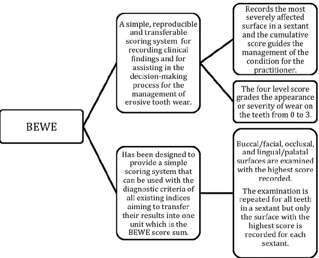
CHAPTER B – EROSIVE WEAR I. Differential diagnosis
II. Measurement of wear
III. BEWE (Basic Erosive Wear Examination)
MULTICENTER STUDY Aims
Stage I. Data Collection Phase Stage II. Analysis Phase Stage III. Informative Phase
Cynthia Lara Capi
Oral diseases epidemiology in Italian and non-Italian population. A multicenter cross-sectional study. Tesi di dottorato in Odontostomatologia Preventiva, Università degli Studi di Sassari
ANNEXES
ANNEX I. Digital Imaging Fiber-optic Transillumination Device versus Radiographic and Clinical Examination in the Detection of Dental Caries.
ANNEX II. Comparison of Carisolv system vs traditional rotating instruments for caries removal in the primary dentition: A systematic review and meta-analysis.
ANNEX III.
Differences on the impact of BMI and behavioural factors on dental caries in
Mexican urban and rural populations: a comparative study.
ANNEX IV. Taste Preference in Relation to Dental Caries in Italian Adolescents. ANNEX V. Taste Perception in Relation to Dental Caries in Saudi Schoolchildren. ANNEX VI. Taste Perception and Dental Caries – A Multicenter Study.
ANNEX VII. The use of polyols in caries prevention: a systematic review and meta-analysis. ANNEX VIII. Erosive Wear In Adolescents from Italy, Mexico And Saudi Arabia: A Multicenter Study.
Cynthia Lara Capi
Oral diseases epidemiology in Italian and non-Italian population. A multicenter cross-sectional study. Tesi di dottorato in Odontostomatologia Preventiva, Università degli Studi di Sassari
Contributors Recognition
Prof Guglielmo Campus
Department of Surgery, Microsurgery and Medicine Sciences,
School of Dentistry, University of Sassari, Italy
WHO Collaborating Centre for
Epidemiology
and Community Dentistry, University of Milan, Italy
Dr. Fabio Cocco
Department of Chemistry, University of Sassari, Italy
WHO Collaborating Centre for
Epidemiology and Community
Dentistry, University of Milan, Italy
Prof. Peter Lingström
Department of Cariology, Institute of Odontology
The Sahlgrenska Academy
University of Gothenburg, Sweden
Dr. Maria Grazia Cagetti
Assistant Professor, University of Milan WHO Collaborating Center of Milan for Epidemiology and
Community Dentistry
San Paolo Hospital, Dental Clinic
Prof. Franklin García-Godoy
Bioscience Research Center, College of Dentistry
University of Tennessee Health
Science Center
Memphis, Tennessee, USA
Prof. Gail Douglas
Chair in Dental Public Health School of Dentistry
University of Leeds Leeds, England
Cynthia Lara Capi
Oral diseases epidemiology in Italian and non-Italian population. A multicenter cross-sectional study. Tesi di dottorato in Odontostomatologia Preventiva, Università degli Studi di Sassari
ABSTRACT
Oral diseases epidemiology in Italian and non-Italian population. A multicenter cross-sectional study
LARA-CAPI C1, LINGSTROM P2, ASHI H2, COCCO F3, CAMPUS G3
1Department of Biomedical Sciences, University of Sassari, Italy
2Department of Cariology, The Shalgrenska Academy, University of
Gothenburg, Sweden
3Department of Surgery, Microsurgery and Medical Sciences, School of
Dentistry, University of Sassari, Sassari, Italy [email protected]
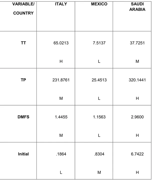
Aims: To assess the prevalence of the major oral diseases and the impact
of the risk factors in adolescents from three different countries representative of Europe, Latin America and Middle East: Italy, Mexico and Saudi Arabia.
Methods: Caries (ICDAS and DMFS), erosion (BEWE), gingival status
(Silness-Löe Index) were recorded. Body Mass Index (BMI) was calculated (WHO guidelines). A questionnaire investigated dietary patterns, oral habits and socio-economic status. Sweet taste evaluation was performed to determine threshold (TT) and preference (TP).
Results: 909 subjects (Italy n= 220; Saudi n=225; Mexico Urban n=224;
Mexico rural n=240). Mean DMFS was 2.96 SD 4.01. The prevalence of erosion was higher in Mexico (18.7%), followed by Italy (12,7%) and Saudi (6.7%) while caries experience was higher in Saudi (3.0±4.0), followed by Italy (1.4±2.3) and Mexico (1.2±1.5)(p<0.001). The frequency of soft drinks intake was a constant variable for erosion (p<0.01). Overweight (BMI) was positively related to caries severity. TP and caries had a significant correlation (p<0.01). Subjects in the rural area presented less caries (p<0.01).
Conclusions: The distribution of oral diseases is related to behavioral,
socio-cultural and geographic situations. Both caries and dental erosion are associated to diet and diet being determinant to BMI is decisive in oral and general health where prevention can make a decisive change in the quality of life.
Cynthia Lara Capi
Oral diseases epidemiology in Italian and non-Italian population. A multicenter cross-sectional study. Tesi di dottorato in Odontostomatologia Preventiva, Università degli Studi di Sassari
Cynthia Lara Capi
Oral diseases epidemiology in Italian and non-Italian population. A multicenter cross-sectional study. Tesi di dottorato in Odontostomatologia Preventiva, Università degli Studi di Sassari
Great improvements have been accomplished in oral health all over the globe, yet in
under-privileged populations, both in the developed world as in developing countries,
this remains to be addressed. The distribution and severity of oral diseases diverge
between different parts of the world and within the same country or region.
Socio-behavioral and environmental factors play a fundamental role in oral disease making
necessary the surveillance of the patterns and the evaluation of the risk factors in
order to develop and implement community preventive measures.
1Oral diseases are a main public health problematic with an outcome on the quality of
life. The diversity between their prevalence, incidence and development requires
operative health programs. The most common oral diseases reported worldwide are:
1.
Dental caries
2.
Periodontal disease
3.
Tooth loss
4.
Oral mucosal lesions
5.
Oropharyngeal cancers
6.
Human immunodeficiency virus/Acquired immunodeficiency syndrome
(HIV/AIDS) -related oral disease
7.
Dental trauma* (although is not a disease, is a highly prevalent linked
condition and influences the quality of life).
Risk factors:
1.
Poverty
2.
Oral hygiene
3.
Tobacco
4.
Alcohol
Another conditions that affect oral
health:
1.
NOMA (a form of orofacial
gangrene).
2.
Developmental disorders.
3.
Down syndrome, cerebral
palsy and genetic defects.
Cynthia Lara Capi
Oral diseases epidemiology in Italian and non-Italian population. A multicenter cross-sectional study. Tesi di dottorato in Odontostomatologia Preventiva, Università degli Studi di Sassari
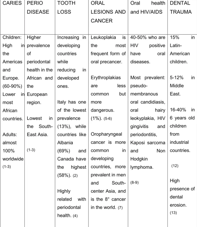
Table 1. Comparison among the characteristics of prevalent oral diseases.
CARIES PERIO
DISEASE
TOOTH
LOSS
ORAL
LESIONS AND
CANCER
Oral
health
and HIV/AIDS
DENTAL
TRAUMA
Children: High in the Americas and Europe. (60-90%) Lower in most African countries. Adults: almost 100% worldwide (1-3) Higher prevalence of periodontal health in the African and the European region. Lowest in the South-East Asia. (1-3) Increasing in developing countries while reducing in developed ones.Italy has one of the lowest prevalence (13%), while countries like Albania (69%) and Canada have the highest (58%).(2) Highly related with periodontal health. (4)
Leukoplakia is the most frequent form of oral precancer. Erythroplakias are less common but more dangerous. (1%).(5-6) Oropharyngeal cancer is more common in developing countries, more prevalent in men and
South-center Asia, and is the 8° cancer in the world. (7) 40-50% who are HIV positive have oral diseases. Most prevalent: pseudo-membranous oral candidiasis, oral hairy leokyplakia, HIV gingivitis and periodontitis, Kaposi sarcoma and Non Hodgkin lymphoma.
(8-9) 15% in Latin-American children. 5-12% in Middle East. 16-40% in 6 years old children from industrial countries. (12) High presence of dental erosion. (13)
Cynthia Lara Capi
Oral diseases epidemiology in Italian and non-Italian population. A multicenter cross-sectional study. Tesi di dottorato in Odontostomatologia Preventiva, Università degli Studi di Sassari
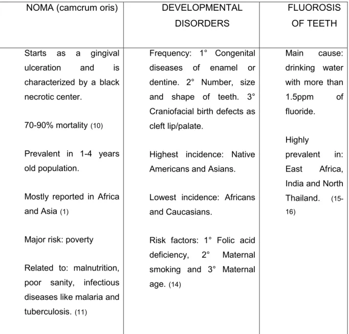
Table 2. Comparison of the most frequently related conditions that affect oral heath.
NOMA (camcrum oris)
DEVELOPMENTAL
DISORDERS
FLUOROSIS
OF TEETH
Starts as a gingival ulceration and is characterized by a black necrotic center. 70-90% mortality (10) Prevalent in 1-4 years old population.Mostly reported in Africa
and Asia (1)
Major risk: poverty
Related to: malnutrition, poor sanity, infectious diseases like malaria and
tuberculosis. (11)
Frequency: 1° Congenital diseases of enamel or dentine. 2° Number, size and shape of teeth. 3° Craniofacial birth defects as cleft lip/palate.
Highest incidence: Native Americans and Asians. Lowest incidence: Africans and Caucasians.
Risk factors: 1° Folic acid
deficiency, 2° Maternal
smoking and 3° Maternal
age.(14)
Main cause:
drinking water with more than
1.5ppm of
fluoride. Highly
prevalent in:
East Africa,
India and North
Thailand.
(15-16)
To acknowledge the way that oral diseases compromise the health status of different
populations is a public health matter that concerns us all. Its identification and
description are important for the education and prevention worldwide. The impacts of
oral disease in the general health condition as well as in the economy are high. Is the
fourth most expensive in the majority of the industrialized countries, and there
difference between developed and developing countries is quite large.
16Its relation
with living conditions, lifestyle and prevention is a valuable information to determinate
the populations at risk and implement effective health programs.
17Cynthia Lara Capi
Oral diseases epidemiology in Italian and non-Italian population. A multicenter cross-sectional study. Tesi di dottorato in Odontostomatologia Preventiva, Università degli Studi di Sassari
CHAPTER A
DENTAL CARIES
Cynthia Lara Capi
Oral diseases epidemiology in Italian and non-Italian population. A multicenter cross-sectional study. Tesi di dottorato in Odontostomatologia Preventiva, Università degli Studi di Sassari
Dental caries is a demineralization process, a disease caused by the action of
bacteria that can result in a lesion (initial caries) that can affect the enamel and
dentinal surface (manifest caries).
The World Health Organisation (WHO) has recorded dental caries globally
through its oral disease surveillance systems. Although preventive programmes
have had a positive effect on the caries figures in developed countries, several
factors may act on the disease. In developing countries on the other hand, the
adoption of a western lifestyle in absence of public prevention programmes
have caused a rapid increase in the develop of the dental disease.
18Caries is a growing oralh health problem that needs to be appraised in:
I. Diagnosis – ICDAS criteria
II. Risk - Caries Risk Assessment and Risk Factors
III. Early detection - DIFOTI technology
IV. Minimally Invasive Therapy - Chemical removal of caries using Carisolv
V. Behavioral and Socio-cultural Risk factors – Diet, BMI and poverty
VI. Taste Preference
Cynthia Lara Capi
Oral diseases epidemiology in Italian and non-Italian population. A multicenter cross-sectional study. Tesi di dottorato in Odontostomatologia Preventiva, Università degli Studi di Sassari
I. DIAGNOSIS
Caries diagnosis is mainly evaluated with the World Health Organization
criteria, this is, the DMFT/DMFS (Decay, Missing or Filled Tooth/Surface) that
registers only obvious caries lesions along with the missing and restored teeth.
A new model, called ICDAS, has been developed to assess both enamel
(non-cavitated) and dentin ((non-cavitated) lesions, and is capable to record appropriate
the pertinent information of the presence of caries and the relationship between
the primary and the permanent dentition.
19,20Characteristics of ICDAS
- Reliable
- Reproducible
- Practical
Evaluates
1. Lesions according to the type of teeth (anterior, posterior)
2. According to the surfaces (occlusal, proximal and free smooth)
Criteria
- Visual examination
- plaque-free surfaces
- wet and dried teeth
21Cynthia Lara Capi
Oral diseases epidemiology in Italian and non-Italian population. A multicenter cross-sectional study. Tesi di dottorato in Odontostomatologia Preventiva, Università degli Studi di Sassari
Training
The ICDAS Committee provides an e-learning program. As well, calibration
programs can improve the reliability of the method for the use of both
epidemiologist and clinicians.
Scoring system
ICDAS evaluates the overall caries experience: tooth status (previous
treatment: Range 0 to 8) and the caries status (current status: Range 0 to 6)
and using one code for each giving as a final result a two-digit code.
22Cynthia Lara Capi
Oral diseases epidemiology in Italian and non-Italian population. A multicenter cross-sectional study. Tesi di dottorato in Odontostomatologia Preventiva, Università degli Studi di Sassari
ICDAS CHART
17 16 15 14 13 12 11 21 22 23 24 25 26 27 O B P D M TOOTH 47 46 45 44 43 42 41 31 32 33 34 35 36 37 O B L D MTooth status (1st number): 0= Sound
1= Sealant, partial 2=Sealant full
3=Tooth coloured restoration 4=Amalgam restoration 5=Stainless steel crown
6=Porcalain or gold PFM crown or veneer 7=Lost or broken restoration
8=Temporary restoration
9=Used for the following conditions:
97=Tooth extraxted because of caries (all tooth surfaces will be coded 97)
98=Tooth extracted for reasons othe rthan caries
99= Unerupted (all tooth surfaces coded)
Caries status (2nd number): 0=Sound
1=First visual change in enamel 2=Distinct visual changes in enamel
3=Localized enamel breakdown due to caries with no visible dentin
4=Non-cavitated surfaces with underlying dark shadow from dentin
5=Distinct cavity with visible dentin
6=Extensive distinct cavity with visible dentin and extensive cavity involves at least half of the tooth surface and possibly reaching the pulp
7=Tooth extraxted bacause of caries (all tooth surfaces will be coded 97)
8=Tooth extracted for reasons othe rthan caries ( all tooth surfaces coded 98)
Cynthia Lara Capi
Oral diseases epidemiology in Italian and non-Italian population. A multicenter cross-sectional study. Tesi di dottorato in Odontostomatologia Preventiva, Università degli Studi di Sassari
II. CARIES RISK ASSESSMENT AND RISK FACTORS
Caries risk assessment is the identification of individuals at risk for future caries
and it is a science that lies within the field of health risk assessment.
Etiology of caries
- The host, mainly represented by tooth resistance, saliva factors and
remineralization capacity.
- The microflora, amount of plaque and type of oral microorganisms
- The substrate, diet, its content and frequency of intakes
- Time needed before a cavity becomes visible.
Table 3. Caries risk factors (Beck 1998).
23Caries risk factors HIGH RISK LOW RISK
Clinical evidence New lesions
Premature extractions
Anterior caries or restorations Multiple restorations
No fissure sealants
Fixed appliance orthodontics Partial dentures
No new lesions
Nil extractions for caries Sound anterior teeth No or few restorations
Restorations inserted years ago
Fissure sealed No appliance
Dietary habits Frequent sugar intake Infrequent sugar intake
Social history Social depreciation
High caries in siblings
Low knowledge of dental disease
Irregular attendance
Ready availability of snacks
Social advantage Low caries siblings Dentally aware
Regular attendance limited availability o f snacks
Cynthia Lara Capi
Oral diseases epidemiology in Italian and non-Italian population. A multicenter cross-sectional study. Tesi di dottorato in Odontostomatologia Preventiva, Università degli Studi di Sassari
Low dental aspirations
Use of fluoride Drinking water not fluoridated
No fluoride supplements No fluoride tooth paste
Drinking water fluoridated Fluoride supplements used Fluoride toothpaste used
Plaque control Infrequent ineffective cleaning
Poor manual control
Frequent, ineffective cleaning Good manual control
Saliva Low flow rate
Low buffering capacity
High S. mutans and
lactobacillus counts
Normal flow rate High buffering capacity
Low S. mutans and
lactobacillus counts
Medical history Medically compromised
Physical disability Xerostomia Long-term cariogenic medicine No medical problems No physical problems Normal salivary flow No long term medication
Moderate Risk
Individuals who do not clearly fit into high or low risk categories are considered
to be at moderate risk.
CARIOGRAM. A tool for the assessment of caries.
A model developed at the Dental University in Goteborg that illustrates the
multifactorial background of dental caries in a simple way. It was
computer-based by the Dental University in Malmo.
Cynthia Lara Capi
Oral diseases epidemiology in Italian and non-Italian population. A multicenter cross-sectional study. Tesi di dottorato in Odontostomatologia Preventiva, Università degli Studi di Sassari
To improve the understanding of the multifactorial aspects of dental caries and
to estimate the caries risk where the patient is seen as a “whole”.
Evaluation
1. Collecting relevant information.
2. Scoring according to a standardized protocol.
3. Entering the scores into the computer program.
Description
Cariogram is a pie-circle diagram divided into colored sectors. There are five
sectors:
1. Green (actual chance to avoid new cavity).
2. Dark blue (diet contents and diet frequency).
3. Red (bacteria). A combination of amount of plaque and mutans streptococci
4. Light blue (susceptibility). Based on a combination of fluoride program, saliva
secretion and saliva buffer capacity.
5. Yellow (circumstances). Comprises the combination of caries experience and
related diseases.
Scoring
For all patients the factor “0” is the best while “3” (or “2” where that is maximum)
the most unfavorable scores.
The assessment of the risk of dental caries has been showed to benefit from
the use of computer-based programs. The caries risk profile in a group of
Sardinian schoolchildren evaluated the caries prevalence, gingival conditions,
diet, oral hygiene and saliva with success, determining that more than a quarter
had less than 40% possibility to avoid dental caries.
24Cynthia Lara Capi
Oral diseases epidemiology in Italian and non-Italian population. A multicenter cross-sectional study. Tesi di dottorato in Odontostomatologia Preventiva, Università degli Studi di Sassari
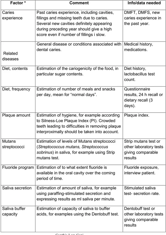
Table 4. Caries related factors and the date needed to create a Cariogram
Factor * Comment Info/data needed
Caries experience
Past caries experience, including cavities, fillings and missing teeth due to caries. Several new cavities definitely appearing during preceding year should give a high score even if number of fillings i slow.
DMFT, DMFS, new caries experience in the past year.
Related diseases
General disease or conditions associated with dental caries.
Medical history, medications.
Diet, contents Estimation of the cariogenicity of the food, in
particular sugar contents.
Diet history, lactobacillus test count.
Diet, frequency Estimation of number of meals and snacks
per day, mean for “normal days”.
Questionnaire
results, 24 h recall or dietary recall (3 days).
Plaque amount Estimation of hygiene, for example according
to Silness-Loe Plaque Index (PI). Crowded teeth leading to difficulties in removing plaque interproximally should be taken into account.
Plaque index.
Mutans streptococci
Estimation of levels of Mutans streptococci (Streptococcus mutans, Streptococcus sobrinus) in saliva, for example using Strip mutans test.
Strip mutans test or other laboratory tests giving comparable results
Fluoride program Estimation of to what extent fluoride is available in the oral cavity over the coming period of time.
Fluoride exposure, interview patient.
Saliva secretion Estimation of amount of saliva, for example using paraffing-stimulated secretion and expressing results as ml saliva per minute.
Stimulated saliva test- secretion rate.
Saliva buffer capacity
Estimation of capacity of saliva to buffer acids, for examples using the Dentobuff test.
Dentobuff test or other laboratory tests giving comparable results
Cynthia Lara Capi
Oral diseases epidemiology in Italian and non-Italian population. A multicenter cross-sectional study. Tesi di dottorato in Odontostomatologia Preventiva, Università degli Studi di Sassari
III. EARLY CARIES DETECTION
Early diagnosis and treatment of caries is necessary to implement strategies.
25The
clinical diagnosis of caries lesions is fundamental, and the use of radiopgraphs has
enhance practitioners. However, although radiographs are highly sensitive specially
for detection of interproximal caries, it has been showed to be limited to a
comprehensive diagnosis.
26DIFOTI
A Digital Imaging Fiber-optic Transillumination Device, also known as DIFOTI, was
designed to support the identification of cavitated and non-cavitated lesions aloft the
gingiva that differs the optical properties of the lesion with the ones from the sound
dental tissue (DIAGNOcam 2170 KaVo). DIFOTI uses digital image processing in
real time that permits quantitative information to monitore lesions in present and in
time.
27,28Transillumination
Transillumination of the teeth with intense fiber-optic light amplifies the change in
scattering and absorption of light photons in the carious tissue and thereby makes
the caries lesion appear as a dark shadow.
Advantages
The main asset of this technology is that, unlike x-rays, is noninvasive. This give the
opportunity to use it as many times necessary and to follow the arrest or progression
of the lesions by capturing an digital image on the computer that can be stored and
take again if needed.
29It has also the potential to detect missing homogeneity in the
tooth structure by accentuating the features of the lesions. This is very important
given the recent reports about
the risk related to dental radiographic exposure and
the suggestion of radiographic selection criteria.
30Cynthia Lara Capi
Oral diseases epidemiology in Italian and non-Italian population. A multicenter cross-sectional study. Tesi di dottorato in Odontostomatologia Preventiva, Università degli Studi di Sassari
DIFOTI in the literature
Clinical studies to measure the lesions and correlate the obainted results with the
ones given by radiographs is limited.
31Although DIFOTI has demonstrated to be
more effecive than radiographs to detect enamel lesions under in vitro conditions,
further analysis is needed to validate the scale of diagnosis and the intra-examiner
and inter-examiner reproducibility of the method.
29This is the reason we developed a study design to evaluate the effectiveness of the
DIFOTI technology in the clinic of the University of Sassari as well as to assess the
reproducibility of the instrument in dental professionals with different backgrounds
belonging to different parts of the country.
Cynthia Lara Capi
Oral diseases epidemiology in Italian and non-Italian population. A multicenter cross-sectional study. Tesi di dottorato in Odontostomatologia Preventiva, Università degli Studi di Sassari
CASE 1. Clinical and radiographical evaluation of an lower left second premolar.
Dx: Premolar No. 35 was classified as caries free, as no color change or
demineralization was observed during the clinical analysis and bitewing radiographic
examination.
Cynthia Lara Capi
Oral diseases epidemiology in Italian and non-Italian population. A multicenter cross-sectional study. Tesi di dottorato in Odontostomatologia Preventiva, Università degli Studi di Sassari
DIFOTI Evaluation
A defined dark area is detected with the DIFOTI device in the mesial and distal
surfaces of the second mandibular premolar. It was corroborated that is not a shadow
produced by the inclination of the instrument. In different shoots, this was confirmed.
Cynthia Lara Capi
Oral diseases epidemiology in Italian and non-Italian population. A multicenter cross-sectional study. Tesi di dottorato in Odontostomatologia Preventiva, Università degli Studi di Sassari
ANNEX I
Digital Imaging Fiber-optic Transillumination Device versus
Radiographic and Clinical Examination in the Detection of Dental
Cynthia Lara Capi
Oral diseases epidemiology in Italian and non-Italian population. A multicenter cross-sectional study. Tesi di dottorato in Odontostomatologia Preventiva, Università degli Studi di Sassari
Cynthia Lara Capi
Oral diseases epidemiology in Italian and non-Italian population. A multicenter cross-sectional study. Tesi di dottorato in Odontostomatologia Preventiva, Università degli Studi di Sassari
Cynthia Lara Capi
Oral diseases epidemiology in Italian and non-Italian population. A multicenter cross-sectional study. Tesi di dottorato in Odontostomatologia Preventiva, Università degli Studi di Sassari
Digital Imaging Fiber-optic Transillumination Device versus Radiographic and Clinical
Examination in the Detection of Dental Caries
Cynthia Lara-Capi
a, Peter Lingström
bc, Gianfranco Lai
a, Maria Grazia Cagetti
cd, Fabio
Cocco
ace, Charlotte Simark-Mattsson
band Guglielmo Campus
aca
Department of Surgery, Microsurgery and Medical Sciences, School of Dentistry,
University of Sassari, Sassari, Italy;
b
Department of Cariology, Institute of Odontology, The Sahlgrenska Academy,
University of Gothenburg, Gothenburg, Sweden
c
WHO Collaboration Centre for Community Dentistry and Epidemiology, University of
Milan, Italy
d
Department of Helth Science, School of Dentistry, University of Milan, Milan, Italy;
eDepartment of Chemistry and Pharmacy, University of Sassari, Sassari, Italy
Running head
Caries detection using DIAGNOcam
Key Words
Caries detection - DIFOTI - ICDAS - Radiographs
Corresponding author
Cynthia Lara Capi
Department of Biomedical Sciences
School of Dentistry, University of Sassari, Italy
Viale San Pietro 43/C, I-07100 Sassari
tel +39-324-9030047
email [email protected]
Cynthia Lara Capi
Oral diseases epidemiology in Italian and non-Italian population. A multicenter cross-sectional study. Tesi di dottorato in Odontostomatologia Preventiva, Università degli Studi di Sassari
Abstract
Aim: To evaluate the reliability of a Digital Imaging Fiber-Optic Transillumination
device (DIFOTI) for the detection of caries lesions and compare it with the results of
clinical or radiographic examinations. In addition, the reliability of DIFOTI method was
evaluated in a group of dental professionals. Methods: 52 selected subjects were
included into the study. Two calibrated dentists evaluated premolars and molars
using DIFOTI (DIAGNOcam) and a clinical examination (CE) for assessing caries
lesions on occlusal surfaces (CAMo), and DIAGNOcam and a radiographic
examination (BW) for caries in approximal surfaces (CAMa). Forty-eight trained
dental professionals evaluated thirty randomly selected surfaces (EVA1) derived from
CAMo/a images analysis. One month later, the same dentists re-evaluated the same
images (EVA2). Cohen’s Kappa was used to evaluate the grade of accordance while
Intra-Class Correlation coefficients (ICC) for the reproducibility for each surface.
Results: The number of detected occlusal caries lesions was similar for CAMo and
CE (Kappa=0.99). DIAGNOcam identified a higher number of approximal lesions
compared to BW (Kappa=0.91). The same number of lesion in dentine (Kappa=1)
was identified by the two detection methods, while in enamel a low agreement was
found with more lesions detected by CAMa (Kappa=0.24). For EVA1, 87.5% of the
participants had high concordance of Cohen’s Kappa compared to DIAGNOcam
images and an higher concordance in EVA2. The intra-examiner reliability was
substantial/almost perfect in 59.4% of the participants. Conclusion: DIAGNOcam
images may be useful for early caries detection on approximal surfaces. The device
seems easy to decode for professionals without experience.
Cynthia Lara Capi
Oral diseases epidemiology in Italian and non-Italian population. A multicenter cross-sectional study. Tesi di dottorato in Odontostomatologia Preventiva, Università degli Studi di Sassari
INTRODUCTION
Caries clinical management is linked to the number of teeth and surfaces affected, as
well as the severity (depth) and the activity (progression or new development) of the
lesions. Caries detection, including assessment of non-cavitated and cavitated
carious lesions, is an important issue in operative dental practice [Pitts, 2004;
Piovesan et al., 2013]. Radiographic examination is a highly sensitive method to
detect carious lesions on surfaces that can not be inspected visually, such as
approximal surfaces. However, limitations in its sensitivity to diagnose early lesions
have been reported [Bader et al., 2002]. In addition, the risk related to radiographic
exposure needs to be taken into consideration [Ludlow et al., 2008].
There is a need for improvement of the current methods for caries detection. As a
complementing aid to visual examination, a Digital Imaging Fiber-Optic
Transillumination Device (DIFOTI) was designed with the task to support clinicians in
the identification of caries lesions in different stages [Keem and Elbaum, 1997,
Schneiderman et al., 1997; Astvaldsdóttir et al., 2012]. Using the specific optical
properties of a carious tissue, transillumination of the teeth with DIFOTI amplifies the
change in scattering and absorption of light photons and thereby, makes the lesion
appear as a dark shadow [Astvaldsdóttir et al., 2012]. DIFOTI was developed to
facilitate in real time the detection, localization and quantitative characterization of
lesions [Schneiderman et al., 1997]. The major advantage of the method is that it is
non-invasive and therefore can be used as frequently as needed, providing an
immediate digital image capture that can be stored and compared with previously
acquired images [Astvaldsdóttir et al., 2012]. Caries lesion activity may be monitored
by quantification of the changes in mineral content of the lesion over time using the
comparison of DIFOTI images acquired at different time points. The detection of early
lesions is extremely relevant from clinical point of view as implies an uplift caries
activity and the need for additional non-invasive intervention [Keem & Elbaum, 1997;
Astvaldsdóttir et al., 2012].
Although it is subjective, the interpretation of the DIFOTI images seems to be
relatively easy to learn. In literature, clinical studies that compare the in situ depth of
carious lesions with DIFOTI versus radiographs are quite limited [Bin-Shuwaish et al.,
2008]. A recent in vitro study used the transillumination device to identify approximal
Cynthia Lara Capi
Oral diseases epidemiology in Italian and non-Italian population. A multicenter cross-sectional study. Tesi di dottorato in Odontostomatologia Preventiva, Università degli Studi di Sassari
carious lesions and compared the diagnostic accuracy/efficacy of the device with
both traditional and digital x-ray examination, finding that DIFOTI identified a higher
number of enamel caries by detecting lesions at an earlier stage than radiographs,
providing more accurate results. In contrast, radiographs showed a better sensitivity
in deeper lesions, this is, DIFOTI identified a higher number of incorrect dentin
lesions. Radiography is able to identify great change in lesion depth although small
changes in the mineral content are not detectable. Moreover, DIFOTI and film
radiography showed a high intra-examiner concordance [Astvaldsdóttir et al., 2012].
The International Caries Detection and Assessment System (ICDAS) is a more a
visual scoring systems than tactile, developed to assess the caries lesions at both
initial and manifest thresholds [International Caries Detection and Assessment
System Coordinating Committee, 2005; Honkala et al., 2011].
Meticulous and reliable data collection is vital for success in all fields of research
[Lesaffre et al, 2004]. The training of the examiners is fundamental, and it can be
defined, according to the Guidance on the Statistical Aspects of Training and
Calibration of Examiners for Surveys of Child Dental Health by British Association for
the Study of Community Dentistry (BASCD) [Pine et al, 1997; Assaf et al., 2006;
Agustsdottir et al., 2010], as teaching the agreed interpretation of the diagnostic
criteria.
The main aim of this study was to evaluate the effectiveness and reliability of the
DIAGNOcam. The null-hypothesis was that the reliability of a DIFOTI device (KaVo
DIAGNOcam 2170) for the detection of caries lesions did not differ from that obtained
through the clinical or radiographic examinations. To validate this hypothesis, an
observational study was designed and evaluated as well in a group of dental
professionals. In the first part of the study, the DIAGNOcam was compared with a
clinical examination appraising the occlusal surfaces and with x-ray bitewings
assessing the approximal surfaces. In the second part, the reproducibility of image
evaluation using DIAGNOcam was determined in a group of dental professionals.
Materials and Methods
The study was approved by the Ethical Committee at the University of Sassari
(authorization number 389/2013) and it was conducted over 6 weeks from June 9th
Cynthia Lara Capi
Oral diseases epidemiology in Italian and non-Italian population. A multicenter cross-sectional study. Tesi di dottorato in Odontostomatologia Preventiva, Università degli Studi di Sassari
to July 15th 2014.
Study design
The study was designed in two parts: the first was a comparison among three
detection methods (DIFOTI, bitewing radiographs and clinical examination), the
second was a reliability study among dental professionals using the DIFOTI imagines
derived from the first part.
Comparison among three detection methods
The new KaVo DIAGNOcam 2170 is a camera system that reads the tooth's
structure to verify occlusal, approximal and secondary caries lesions when the tooth
is transilluminated. A digital video camera records the image and displays it on a
computer screen.
For the radiographic examination, Planmeca intraoral radiographic equipment
(Planmeca, Helsinki, Finland) and Kodak UltraSpeed DF42 films, with settings of 70
kV and 7 mA and an exposure time of 0.25 s, were used for bitewing radiographs.
The radiographs were manually developed via conventional standard conditions and
standard processing times, and examined according to O’Mullane criteria [O’Mullane
et al., 1997].
The clinical examinations were performed under standard conditions. The subjects
were seated in a dental unit and the teeth were examined using a plan mirror
(Hahnenkratt, Königsbach, Germany) and the WHO CPITN ballpoint probe
(Asa-Dental, Milan, Italy) under optimal light.
Calibration of the examiners
Calibration exercises for all the three methods (DIAGNOcam unit visual, clinical
caries diagnostic system (ICDAS) and radiographic examination) were carried out by
two dentists before the start of the study. One of the authors (GCampus) acted as
benchmark, training and calibrating the two examiners. The calibration process was
divided for each diagnostic method in four steps:
• lectures regarding the disease and the method (i.e. DIAGNOcam, ICDAS, x-ray) for
eight hours;
Cynthia Lara Capi
Oral diseases epidemiology in Italian and non-Italian population. A multicenter cross-sectional study. Tesi di dottorato in Odontostomatologia Preventiva, Università degli Studi di Sassari
• first examination, no discussion was allowed between the examiners and the dental
advisors as to the interpretation of the criteria during the calibration sessions;
• re-evaluation by the examiners after 72 hours (clinical examination) and one week
(DIAGNOcam and x-ray)
• evaluation of the agreement or disagreement and statistical analysis.
Fifty volunteers were clinically examined for presence of caries lesions in a dental
chair using the ICDAS criteria and re-examined after 72 hours. Intra- and
inter-examiner reliability was calculated through percent agreement and Cohen’s Kappa
statistics. Good inter-examiner reliability was found with no significant difference from
benchmark values (p=0.15) and a low mean square of error (0.47). The Pearson’s
correlation coefficient between the two examiners was high (r = 0.83, p < 0.01, R2 =
0.71). Intra-examiner reliability was also high, Cohen’s K=0.88.
Forty extracted human teeth (10 premolars and 30 molars), in total 80 approximal
and 40 occlusal surfaces, were selected for the calibration of the DIFOTI device and
the radiographic examination. The teeth were selected from a pool of extracted teeth
from the Department of Oral Surgery at the University of Sassari. The teeth were
cleaned, any remaining soft tissues and calculus were removed, and they were
subsequently frozen at -20° until used. Selection criteria match the line of the first
evaluation. Evaluations were carried out at one-week interval; Kappa values for inter-
and intra-examiner agreement were high for both methods (0.79 for DIFOTI and 0.83
for x-ray). The Pearson’s correlation coefficient for the two examiners was high (r =
0.84, p < 0.01, R2 = 0.74). The clinical examiner did not have the opportunity to look
at DIAGNOcam (CAMo/a) or BW images for the entire period.
Study population
The study population consisted of students of the School of Medicine of the
University of Sassari, Italy. To be suitable for enrolment, subjects had to meet these
inclusion criteria: no missing teeth, no secondary caries and no fillings in premolars
or molars. The exclusion criteria were subjects wearing fixed orthodontic appliances
and subjects unable to be exposed to x-rays for medical/specific reasons. All
students (n=1145) attending the School of Medicine were invited to participate via
email/leaflet where the aim of the study was described in detail. A total of 678
Cynthia Lara Capi
Oral diseases epidemiology in Italian and non-Italian population. A multicenter cross-sectional study. Tesi di dottorato in Odontostomatologia Preventiva, Università degli Studi di Sassari
students accepted and were examined (59.2% acceptance rate) and 52 subjects
(19-23 years, mean age 21.2±1.2) fulfilled the inclusion/exclusion criteria.
Power analysis (G*Power 3 software) was performed to establish the number of
subjects needed to evaluate the estimated difference in caries diagnosis using
DIFOTI and/or clinical evaluation and x-ray. Data [Virajsilp et al., 2005] related to the
reliability of two diagnostic methods were used to calculate the sample size, even if
data used were on primary teeth. The standardized effect was set at 0.39 with a
sample size of 48 subjects and an upper 95% one- sided confidence limit of 0.52. All
subjects (n=52) that fulfilled the inclusion/exclusion criteria were enrolled. Each
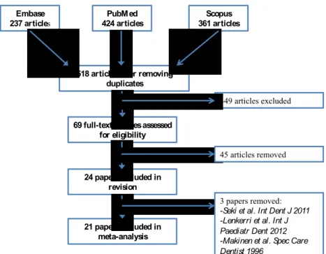
subject was codified with a number in order to protect his/her identity. The flow chart
of the study is displayed in Figure 1.
The DIFOTI device was used to assess caries lesions on occlusal surfaces (CAMo)
and on approximal surfaces (CAMa). In addition, a clinical examination of the
occlusal surfaces (CE) and a radiographic examination (BW) for approximal surfaces
were performed.
Each tooth were cleaned for 30 seconds with a prophylaxis paste (Clinpro™ Prophy
Paste: 3M ESPE Dental Products, USA) and then rinsed by a water spray for 10
seconds. The clinical examination was performed under standardized conditions
describe above after drying teeth for 5 seconds. The students were examined and
analysed during the same day by both examiners, first attending the clinical and
radiographic examination and afterwards they were asked to go to another room
where the DIFOTI device was installed with a computer in a dental chair. The
International Caries Detection and Assessment System (ICDAS) was recorded for
both enamel and dentinal lesions [International Caries Detection and Assessment
System Coordinating Committee, 2005; Ismail et al., 2007; Honkala et al., 2011]. The
radiographs were taken using an 8-inchround cone that was placed in contact with
the ring of the film-holding system (RINN XCP, Dentsply, York), which in turn was
placed in contact with the patient’s cheek during exposure. Not perfectly clear or
overlapping images were taken a second time. Then the DIFOTI device was used
according to the manufacturer’s instructions, placing the mouthpiece over the
occlusal surfaces. The image appeared in real time on the computer monitor, and the
examiner saved it in the electronic patient record.
Cynthia Lara Capi
Oral diseases epidemiology in Italian and non-Italian population. A multicenter cross-sectional study. Tesi di dottorato in Odontostomatologia Preventiva, Università degli Studi di Sassari
The DIAGNOcam was used for the detection of occlusal and approximal caries at
enamel or dentine. When a defined approximal shadow in the enamel was present, it
was scored as 1 and when reaching into the dentine it was scored as 2. Due to the
impossibility to measure the lesion vertically all dark occlusal areas were scored as 1.
The ICDAS scores were performed on the occlusal surface. Radiographs were
examined according to O’Mullane criteria [O’Mullane et al., 1997] and mesial and
distal surfaces were assessed.
Reliability among dental professionals using DIFOTI
Forty-eight Italian dental professionals with no experience of the DIFOTI device were
asked to participate in the second part of the study. Their professional experience
was at least 7 years. On the day of the study they underwent at 60-minute training
session describing the DIFOTI technology and the DIAGNOcam by one of the
authors (CLC). Immediately after the training session, each participant had to
diagnose ten teeth images randomly obtained from the first part of the study,
analysing 10 occlusal, 10 mesial and 10 distal surfaces. Participants were asked to
fill in a form containing two possible answers (1 - presence of caries, 2 - absence of
caries) (EVA1). One month later, participants were contacted via email and were
asked to revaluate the same images with the same criteria (EVA2). These results
were compared with their previous answers.
Statistical Analysis
All data were analysed using STATA 13. For all analysis a p-value <0.05 was
considered statistically significant. The general grade of accordance between the
different detection methods was evaluated using the Cohen’s Kappa [Cohen, 1960],
while the reproducibility for the two methods for each surface (occlusal or
approximal) was assessed using Intra-Class Correlation coefficients (ICC). ICC
values equal to 0 represent agreement equivalent to that expected by chance, while
1 represents full agreement.
The inter-examiner DIFOTI reliability among dental professionals compared to the
results derived from DIAGNOcam analysis was evaluated categorizing the kappa
value of each professional respect to DIAGNOcam following the criteria described by
Landis and Koch [1977], who characterized values <0 as indicating no concordance
and 0-0.20 as slight, 0.21-0.40 as fair, 0.41-0.60 as moderate, 0.61-0.80 as
Cynthia Lara Capi
Oral diseases epidemiology in Italian and non-Italian population. A multicenter cross-sectional study. Tesi di dottorato in Odontostomatologia Preventiva, Università degli Studi di Sassari
substantial, and 0.81-1 as almost perfect concordance. The method by Bland and
Altman [1986] was used to display the variability of the two examinations (EVA1 and
EVA2) by each examiner and the plot of EVA1 respect to the DIAGNOcam results,
the plot of EVA2 respect to DIAGNOcam and the comparison between EVA1 and
EVA2. This method allows to investigate the existence of any systematic difference
between the measurements and to identify possible outliers.
Results
Comparison among the three detection methods
A total of 2496 surfaces (832 mesial, occlusal and distal, respectively) were
analysed. The occlusal surfaces were analysed using DIAGNOcam (CAMo) and
Clinical Examination (CE), while the approximal surfaces were analysed with
DIAGNOcam (CAMa) and Bite-Wing radiographs (BW). The total number of occlusal
caries lesions detected was similar, 149 using CAMo and 152 with CE with a
Cohen’s Kappa of 0.99. The ICC for the occlusal, mesial and distal surfaces of each
tooth is reported in Figure 2. The mean ICC for the occlusal surface was 0.93 with a
lowest value for maxillary right second molar (ICC=0.78), while a perfect agreement
(ICC=1) was observed for several premolars. Approximal caries identified using
CAMa were 83 and 70 using BW (Cohen’s Kappa of 0.91). CAMa and BW identified
the same number (31) of caries in dentine. The Cohen’s Kappa was 0.24 for enamel
lesions with a low agreement, while a complete concordance (Kappa=1) was
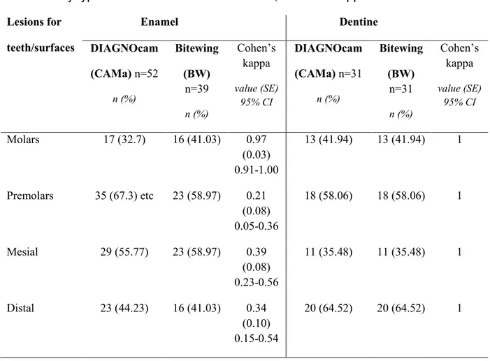
observed for dentinal lesions (Table 1). The mean ICC for approximal surfaces was
0.97 for the distal and 0.95 for the mesial surfaces (Figure 2). Regarding enamel
lesions, 17 lesions in molars were detected with CAMa, while 16 with the BW method
(Cohen’s kappa=0.97); 35 lesions were detected in premolars with CAMa respect to
23 with BW (Cohen’s kappa=0.21). Twenty-nine decayed mesial surfaces were
registered with CAMa respect to 23 with BW (Cohen’s kappa=0.39). For the distal
surfaces, 23 lesions were recorded with CAMa and 16 with BW (Cohen’s
kappa=0.34). A complete concordance was observed for dentinal lesions between
the two methods.
Cynthia Lara Capi
Oral diseases epidemiology in Italian and non-Italian population. A multicenter cross-sectional study. Tesi di dottorato in Odontostomatologia Preventiva, Università degli Studi di Sassari
Reliability among dental professionals using DIFOTI
Forty-eight dental professionals participated in the first evaluation (EVA1) and
thirty-two (drop out rate 33.3%) in the second evaluation (EVA2). The Cohen’s Kappa of
each subject regarding the reliability between the two evaluations was categorized
following the scale proposed by Landis and Koch [1977] (Table 3). Regarding
inter-examiner reliability, in EVA1 the majority of the inter-examiners (87.5%) had either a
substantial (46.9%) or an almost perfect concordance (40.6%) compared to
DIAGNOcam results, while in EVA2 a higher percentage had a substantial
concordance (75.00%) and a lower percentage an almost perfect (18.8%), with a
shift towards substantial concordance grade. Nineteen examiners (59.4%) showed a
substantial/almost perfect agreement, while 13 examiners (40.6%) a fair/moderate
agreement (Figure 3). The Bland-Altman plot showed a good intra-examiner (Figure
3a) and a higher over-rating of the number of the lesions in EVA2 (Figure 3c).
Discussion
The main findings of this study are that the DIFOTI device (DIAGNOcam) proved to
be consistent to clinical examination for the detection of lesions on the occlusal
surface and to bite-wing x-ray for dentinal lesions on approximal surfaces; a higher
number of enamel lesions was detected by DIAGNOcam compared with x-rays,
especially in premolars. In the calibration process, no statistically significant
differences were observed between benchmark and examiners and no systematic
bias between examiners’ scores was noted. The level of concordance among dental
professionals, with respect to the DIAGNOcam analysis result derived from the first
part of the study, was really high in both examinations (EVA1/EVA2). The
intra-examiner reliability of the dental professionals was quite good even if in 40% of the
examiners the level of agreement was moderate or less.
The DIFOTI device used in this study, KaVo DIAGNOcam 2170, is a non-invasive
real-time recording tool that was developed for regular practice use with no exposure
of ionizing radiations to the patient. The device was designed to be useful to identify
lesions at the initial caries stage and the technique allows for more frequent
re-evaluations of these diagnoses than what is feasible using radiographs [American
Dental Association, 2012]. The DIFOTI method has been shown to be more sensitive
Cynthia Lara Capi
Oral diseases epidemiology in Italian and non-Italian population. A multicenter cross-sectional study. Tesi di dottorato in Odontostomatologia Preventiva, Università degli Studi di Sassari