UNIVERSITA’ DEGLI STUDI DI SASSARI
PhD course in Life Sciences and Biotechnologies
Curricula: Biochemistry, Physiology and Molecular
Biology.
Metabolic reprogramming of oestrogen
receptor positive breast cancer
in endocrine therapy resistance
PhD course coordinator:
Prof. Leonardo A. Sechi
Tutor:
Prof. Gianfranco Pintus
Co-Tutor:
Prof. Paola Chiarugi
PhD Thesis:
Dr. Marina Bacci
Academic year 2015/2016
Dr. Marina Bacci
Metabolic reprogramming of oestrogen receptor positive breast cancer in endocrine therapy resistance. PhD Course in Life Sciences and Biotechnologies XXIX cycle - Università degli Studi di Sassari.
2
INDEX
Abstract ... 5
Abbreviations used in thesis ... 6
Introduction ... 11
1- Molecular subtypes of breast cancers ... 14
2- ER positive breast cancer ... 16
2.1 Oestrogen and breast cancer risk ... 16
2.3 Mechanisms of oestrogen action ... 17
3- Endocrine therapy ... 22
3.1 Tamoxifen ... 23
3.2 Aromatase inhibitors ... 24
3.3 Fulvestrant ... 25
4- Endocrine therapy resistance ... 26
4.1 ESR1 mutations ... 27
4.2 Growth factor receptors: PI3K/AKT/mTOR and MAPK pathway activation ... 29
4.3 Cell cycle checkpoint alterations ... 30
4.4 Enhanced autophagy ... 31
4.5 Epigenomic signature ... 31
5- Tumour metabolism ... 32
5.1 Glucose metabolism ... 33
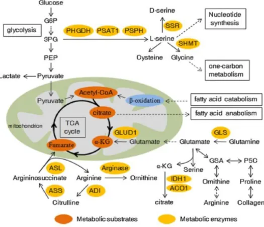
5.2 Amino acids metabolism ... 37
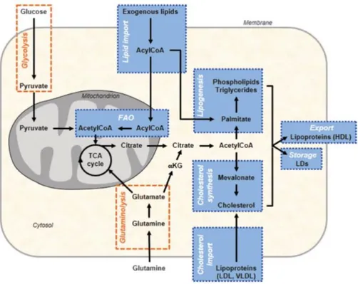
5.3 Lipid metabolism ... 41
5.4 Therapies targeting tumour metabolic reprogramming ... 43
6- Metabolic reprogramming and breast cancer ... 44
6.1 Metabolism and therapy resistance in breast cancer ... 47
6.2 Metabolic targeting in breast cancer ... 48
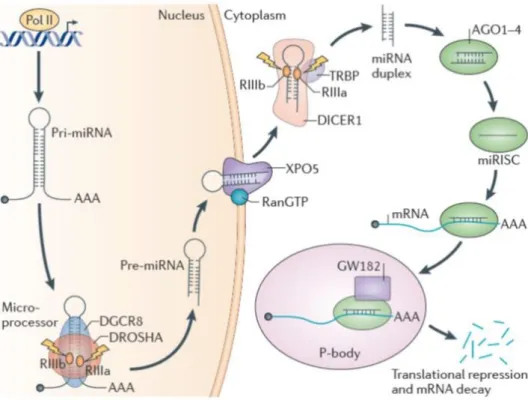
7- MicroRNAs ... 49
7.1 miRNAs and cancer ... 52
7.2 miRNAs and breast cancer ... 54
7.3 miR-155 and cancer ... 56
7.4 miR-23b and cancer ... 58
7.5 miRNAs and tumour metabolism ... 60
8- Autophagy ... 62
8.1 Autophagy and metabolism ... 64
8.2 Autophagy and cancer ... 66
Dr. Marina Bacci
Metabolic reprogramming of oestrogen receptor positive breast cancer in endocrine therapy resistance. PhD Course in Life Sciences and Biotechnologies XXIX cycle - Università degli Studi di Sassari.
3
Materials ... 68
Drugs and Compounds ... 68
Common use solution ... 69
Antibodies: ... 70
Cell lines ... 70
Methods ... 71
General culture conditions ... 71
Frozen storage of cells ... 71
Cell viability ... 71
Protein manipulation ... 72
In vitro Boyden motility and invasion assay ... 74
Three dimensional (3D) tumour spheroid invasion assay ... 75
RhoA or Rac1 activity assay ... 75
Gelatin zimography ... 75
FITC-collagen release assays ... 76
Gene expression and miRNA analysis ... 76
Quantitative real-time RT-PCR (qRT-PCR) ... 76
RNAi transfection ... 77
Glucose, lactate, glutamine and amino acids uptake ... 77
Detection of released CO2 by radioactive glucose or lactate ... 78
3H thymidine incorporation assay ... 78
In vivo experiments ... 78
Immunohistochemistry (IHC) ... 79
Statistical analysis ... 79
Results and Discussion ... 80
Role of central carbon metabolism in response and adaptation to AI ... 81
The combination of letrozole and glycolysis inhibitors synergistically inhibits MCF7-2A cancer cell growth in vitro ... 81
Aerobic glycolysis is enhanced in an in vitro model of AI resistance ... 83
MCF7-LTED cells display high metabolic plasticity following the metabolic targeting ... 85
In addition to metabolic plasticity, MCF7-LTED cells also display high motile plasticity ... 87
ER-dependent miR-155 is responsible for metabolic and motile plasticity of MCF7-LTED cells ... 91
Glycolytic key players and miR-155 levels are decreased in AI-sensitive human breast cancer xenografts following the letrozole treatment ... 93
Glycolytic key players expression correlate with response to AI treatment in vivo ... 96
Dr. Marina Bacci
Metabolic reprogramming of oestrogen receptor positive breast cancer in endocrine therapy resistance. PhD Course in Life Sciences and Biotechnologies XXIX cycle - Università degli Studi di Sassari.
4
miR-155 expression in ER+/HER- breast cancers identifies a subset of
patients that do not respond to AI anastrozole ... 99
Conclusion ... 99
miR-23b-3p regulate amino acids metabolism and influences the response and adaptation to endocrine therapy of ER+ breast cancer ... 101
Global gene expression and miRNAs analysis in parental MCF7 and MCF7-LTED cells show that miR-23/SLC6A14 node is deregulated in MCF7-LTED cells and seems to have a role in endocrine therapy response. ... 101
AI-resistant MCF7-LTED cells have lower SLC6A14 expression when compared to parental cells ... 104
The increase of miR-23b-3p correlates with low SLC6A14 levels and decreases amino acids uptake in AI-resistant MCF7-LTED cells ... 106
Low levels of SLC6A14 and high miR-23b-3p expression correlate with poor prognosis and lower survival in ER+ breast cancer patients ... 106
miR-23b-3p modulates SLC6A14 expression and influences the proliferation of MCF7-LTED cells in oestrogen-deprived conditions ... 108
miR23b-3p/SLC6A14 node is involved in the response and resistance to tamoxifen and fulvestrant treatments. ... 112
Gene expression data reveals that autophagy-related markers are deregulated in MCF7-LTED cells compared to parental MCF7 cells ... 113
AI-resistant MCF7-LTED cells show an autophagic phenotype compared to parental cells ... 117
Conclusion ... 118
Discussion ... 118
Dr. Marina Bacci
Metabolic reprogramming of oestrogen receptor positive breast cancer in endocrine therapy resistance. PhD Course in Life Sciences and Biotechnologies XXIX cycle - Università degli Studi di Sassari.
5
Abstract
The majority of breast tumours express oestrogen receptor (ER) and are dependent on oestrogen (E2) for their growth and survival. Endocrine therapy is the standard of care for this breast cancer subset and acts by targeting ER pathway in different ways: selective ER modulators compete with E2 to bind ER (e.g. tamoxifen), selective ER downregulators promote ER degradation (e.g. fulvestrant) and aromatase inhibitors (AI) block E2 biosynthesis. Despite the efficacy of these endocrine agents, a large proportion of women relapse with endocrine-resistant disease. In this study, we investigated the link between altered breast cancer metabolism and endocrine therapy resistance. We found that AI-resistance cells can adapt to metabolic stress and switch ad hoc between OXPHOS and glycolysis. In particular, we identified the miR-155/hexokinase-2 (HK2) axis as an important regulator of this tumour plasticity. In addition to central carbon metabolism, we found a deregulated node between miR-23b-3p and the amino acid transporter SLC6A14 in endocrine therapy resistant cells, which leads to an impairment of amino acids metabolism in the resistant cells with subsequent activation of autophagy. Furthermore, the miRNA characterised have prognostic (miR-155 and miR-23b-3p) and predictive (miR-155) value in ER positive breast cancer. These results suggest that high metabolic plasticity is involved in acquiring adaptive features that allow breast cancer cell survival even in the presence of endocrine therapy.
Dr. Marina Bacci
Metabolic reprogramming of oestrogen receptor positive breast cancer in endocrine therapy resistance. PhD Course in Life Sciences and Biotechnologies XXIX cycle - Università degli Studi di Sassari.
6
Abbreviations used in thesis
2-DG 2-Deoxy-Glucose
ACC Acetyl-CoA carboxylase ACLY ATP citrate lyase
ACSS2 Acyl-CoA synthetase 2
AF Activation Function
Ago Argonaute protein
AI Aromatase inhibitors
AKT Protein Kinase B, PKB AMPK AMP-activated protein kinase ASS Arginosuccinato synthase ATGs Autophagy-related genes ATP Adenosine 5-triphosphate Bcl-2 B-cell lymphoma 2
BECN1 Beclin-1 gene
BIF-1 BAX-interacting factor 1 BSA Bovine serum albumin CBP CREB-binding protein
CCND1 Cyclin D1
CDK Cyclin Dependent Kinases
CI Combination index
CIC Protein citrate carrier
CK Cytokeratins
CLL Chronic lymphocyte leukemia c-Met Hepatocyte Growth Factor Receptor
CV Cristal Violet
DCC Dextran charcoal treated DCIS Ductal Carcinoma in situ DMSO Dimethyl sulfoxide
Dr. Marina Bacci
Metabolic reprogramming of oestrogen receptor positive breast cancer in endocrine therapy resistance. PhD Course in Life Sciences and Biotechnologies XXIX cycle - Università degli Studi di Sassari.
7
E1 Oestrone
E2 Oestradiol, or 17β-oestradiol
E3 Oestriol
ECL Enhanced chemiluminescence EGFR Epidermal Growth Factor Receptor EMT Epithelial-Mesenchymal Transition
ER Oestrogen Receptor
EREs Oestrogen Response Elements ERK Extracellular-signal-regulated kinase
FA Fatty Acids
FANS Fatty acid synthase FBP Fructose-1-6-biphosphate FBS Foetal bovine serum FDG 18F-fluorodeoxyglucose
FOXO3a Forkhead box O3A
FSH Follicle-Stimulating Hormone GAB3 GRB2 Associated Binding Protein 3 GAPDH Glyceraldeid-3-phosphate dehydrogenase
GDH Glutamate Dehydrogenase
GDP Guanosin-Di-Phosphate
GLS Glutaminase
GLUTs Glucose transporters GSA Glutamic-γ-semi-aldehyde GTP Guanosine Tri-Phosphate HAT Histone Acetyltransferase HCC Hepatocellular carcinoma cell HER2 Human Epidermal Growth Factor 2 HIF1-α Hypoxia-inducible factor 1 α
HK Hexokinase
HRP HorseRadish Peroxidase
Dr. Marina Bacci
Metabolic reprogramming of oestrogen receptor positive breast cancer in endocrine therapy resistance. PhD Course in Life Sciences and Biotechnologies XXIX cycle - Università degli Studi di Sassari.
8
ICI Fulvestrant
LBD Ligand Binding domain LCIS lobular carcinoma in situ LDH Lactate Dehydrogenase LDs Lipid droplets
LH Luteinising Hormone
LKB1 Liver Kinase B1
MAPK Mitogen-Activated Protein Kinase MCTs Monocarboxylate transporters
Met Metformin
miRNA microRNA
MPC Mitochondrial Pyruvate Carrier mTOR Mammalian target of rapamycin
mTORC1 Protein kinase complex mTOR complex 1 NADH Nicotinamide Adenine Dinclueotide
NADPH Nicotinamide Adenine Dinucleotide Phosphate NCOA1 Nuclear-Receptor Co-activator 1
NCOR Nuclear-Co-Repressor
NISCH Nischarin
ORF Open Reading frame
OXPHOS Oxidative phosphorylation P5C α-pyrroline-5-carboxylate PAK2 P21 (RAC1) Activated Kinase 2 PBS Phosphatase Buffered Saline PCAF p300/CBP-associated factor PCR Polymerase Chain Reaction
PDAC Pancreatic Ductal Adenocarcinoma PDCD4 Programmed cell death protein 4 PDGH Phosphoglycerate Dehydrogenase PDH Pyruvate Dehydrogenase complex PDK1 Pyruvate Dehydrogenase Kinase 1
Dr. Marina Bacci
Metabolic reprogramming of oestrogen receptor positive breast cancer in endocrine therapy resistance. PhD Course in Life Sciences and Biotechnologies XXIX cycle - Università degli Studi di Sassari.
9
PE Phopshatidyl ethanolamine
PEP Phosphoenolpyruvate
PET Positron Emission Tomography PFK1 6-phosphofructokinase
PI3K Phosphoinositide 3 Kinase PI3P Phopshatydilinositol-3-phosphate PKM1 Pyruvate Kinase M1
PKM2 Pyruvate Kinase M2
PR Progesterone Receptor
PRODH/POX Proline dehydrogenase (oxidase) PTEN Phosphatase and tensin homolog
qRT-PCR Quantitative real-time reverse transcription PCR RAB6A Ras-Related Protein Rab-6A
Rb Retinoblastoma protein
RhoA Ras homolog family member A SDS Sodium Dodecyl Sulphate SERMs Selective ER Modulators
SHMT Serine Hydroxymethyl Transferase SOCS1 Suppressor of cytokine signalling 1
SREBP-1 Sterol regulatory element-binding protein 1 STAT3 Signal transducer and activator of transcription 3 SWI/SNF Switch/Sucrose Non-Fermenting
TAM Tamoxifen
TCA Tricarboxylic Acid Cycle TIMP3 Metallopeptidase Inhibitor 3
TP53INP1 Tumour protein 53-induced nuclear protein 1
TRAP/DRIP/SMCC Thyroid-Hormone-Receptor-Associated Protein TSC2 Tuberous Sclerosis Complex 2
ULK1 Autophagy activating kinase 1
uPA Urokinase-Type Plasminogen Activator VDAC Voltage Dependent Anion Channel
Dr. Marina Bacci
Metabolic reprogramming of oestrogen receptor positive breast cancer in endocrine therapy resistance. PhD Course in Life Sciences and Biotechnologies XXIX cycle - Università degli Studi di Sassari.
10
WB Western Blotting
α-KG α-ketoglutarate
Dr. Marina Bacci
Metabolic reprogramming of oestrogen receptor positive breast cancer in endocrine therapy resistance. PhD Course in Life Sciences and Biotechnologies XXIX cycle - Università degli Studi di Sassari.
11
Introduction
Breast cancer is the second most common cancer in the world and the most frequent cancer among women with an estimated 1.67 million new cancer cases diagnosed in 2012, representing the 25% of all cancers. Incidence rates are very different across the world regions, with rates ranging from 27 per 100000 people in Middle Africa and Eastern Asia to 96 in Western Europe. In Europe, approximately 464000 new cases were diagnosed in 2012, in particular ~50000 cases in Italy. Breast cancer ranks as the fifth cause of all the cancer related death with 522000 cases/year; it is the leading cause of cancer death in women in less developed regions (324.000 deaths, 14.3% of total) and the second cause of cancer death in more developed region (198000 deaths, 15.4%) after lung cancer. In Europe, breast cancer deaths were ~130000 in 2012 of which 12000 in Italy. In Western Europe and United States, breast cancer mortality rate is lower than incidence rate with respect to undeveloped regions, because of the majority availability of diagnostic technologies and cares in developed regions, which allow an increase of survival and better prognosis of breast cancer patients (1, 2).
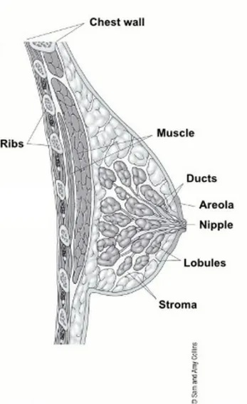
The breasts are composed of fat, connective tissue and gland tissue and are divided into lobes. A network of tubular structures (ducts) originates from the lobes and collectively culminates into the nipple (Figure 1). The breasts composition changes during lifetime: pre-menopausal women have more glandular tissue, whereas in post-menopausal women the glandular tissue is gradually replaced by fat. Breast tissue covers a large area of the chest. It extends from just below the clavicle to the axilla and across to the sternum. The breast is characterised by many blood and lymph vessels. The lymph vessels collect and move lymph fluid away from the breast into the small bean-shaped masses of lymphatic tissue, called lymph nodes, in the area around the breast. Lymph nodes are located all-round the breast tissue, but the axillary lymph nodes are most important and are divided into
Dr. Marina Bacci
Metabolic reprogramming of oestrogen receptor positive breast cancer in endocrine therapy resistance. PhD Course in Life Sciences and Biotechnologies XXIX cycle - Università degli Studi di Sassari.
12
Figure 1. Anatomy of the female breast
three levels according to how close they are to the pectoral muscle. The lymph vessels and lymph nodes are part of the lymphatic system, which has a crucial role during extravasation of cancer cells. Once cancer cells leave the primary site, they can arrive to the axillary lymph nodes through the lymph vessels and from there they can invade to other tissues and metastasize. (Figure 2).
Adenocarcinoma is the most common type of breast cancer and originates from breast glandular tissue. Depending on the site of origin, breast tumours are classified as ductal carcinoma and lobular carcinoma, localized in the breast ducts and lobules, respectively. Ductal carcinoma can be defined in situ (DCIS) when the tumour is localised inside glandular tissue and invasive ductal carcinoma when cancer cells invade the proximal
Dr. Marina Bacci
Metabolic reprogramming of oestrogen receptor positive breast cancer in endocrine therapy resistance. PhD Course in Life Sciences and Biotechnologies XXIX cycle - Università degli Studi di Sassari.
13
lymph nodes and metastasise to other part of the body. In addition, lobular carcinoma can be classified as lobular carcinoma in situ (LCIS or lobular
Figure 2. Breast lymphatic system.
neoplasia) and invasive lobular carcinoma, when the cancer cells have already invaded the surrounding tissue. Around 90% of breast carcinomas diagnosed are ductal carcinomas, and around 10% are invasive lobular carcinoma, which is most common in women between 45 and 55 years old. Furthermore, there are other less common malignant breast tumours, such as inflammatory breast cancers, Paget’s disease of the nipple and phyllodes tumours.
Following breast cancer diagnosis and staging of tumour based on the cancer size and the presence of cancer cells in lymph nodes, breast cancer patients are treated with specific therapy. When possible, the patient undergoes surgery, followed by radiotherapy, chemotherapy (e.g.
Dr. Marina Bacci
Metabolic reprogramming of oestrogen receptor positive breast cancer in endocrine therapy resistance. PhD Course in Life Sciences and Biotechnologies XXIX cycle - Università degli Studi di Sassari.
14
antracycline), biological therapy (antibody against epidermal growth factor receptor HER2, trastuzumab), hormonal therapy (endocrine therapy) or a combination of treatments, depending on the genetic and/or molecular profile of the cancer.
1- Molecular subtypes of breast cancers
Breast cancer is a highly heterogeneous disease. Over the last decades, genomic, transcriptomic and proteomic analyses were applied to identify new molecular markers with prognostic and predictive value to better determine the appropriate therapy.
Two distinct types of epithelial cells compose the human mammary gland: basal (and/or myoepithelial) cells in contact with the basement membrane and luminal epithelial cells that are in continuum with the basal cells and are polarized culminating/facing the lumen. These breast cells are characterised by specific cytokeratins (CK) expression. In particular, luminal epithelial cells are characterised by the expression of CK 8, 18 and 19, while basal cells express CK 5/6, 14 and 17 (3). Expression profile studies showed two main groups of breast cancer based on oestrogen receptor α (ER) expression: ER positive (ER+) breast cancer characterised by high ER expression and ER negative (ER-) breast cancer characterised by low or absence of ER levels (4). Moreover, breast cancer can be classified into four different phenotypes based on the gene expression profile. These subtypes are associated to specific tumour characteristics and clinical outcomes. Accordingly, they are defined as ER+/ Luminal-like, basal like, HER2 positive (HER2+), characterised by overexpression of HER2neu/ ERBB2 oncogene, and Normal like. It is important underling that the clinical designation of ER- breast carcinoma encompasses at least two biologically distinct subtype of tumours, basal like and HER2+ (4). Subsequent studies demonstrated that the ER+ luminal subtype can be divided into additional different subgroups, according to their distinct expression profiles: luminal A, characterised by high expression levels of ER and ER-related genes; luminal B that show, in addition to ER, high expression of a set of genes related to proliferation and the cell cycle; and a new heterogeneous subtype denominated luminal C, which show a more
Dr. Marina Bacci
Metabolic reprogramming of oestrogen receptor positive breast cancer in endocrine therapy resistance. PhD Course in Life Sciences and Biotechnologies XXIX cycle - Università degli Studi di Sassari.
15
aggressive evolution than luminal A or B subtypes (5). Moreover, the biological behaviour of breast cancer is correlated with their gene expression profile, indicating that ER- tumours have a worse prognosis with reduced overall and clinical survival compared to ER+ tumours(5). In the clinical practice, three biomarkers are usually analysed to evaluate the subtype of a given breast cancer, that is, ER, progesterone receptor (PR) and HER2 (6). Taking these markers into consideration, we can correlate ER, PR and HER2 expression to different molecular subtypes. Indeed, luminal A are ER+ and/or PR+, HER2-; luminal B are ER+ and/or PR+, HER2+; HER2 type are ER-, PR- and HER2+; basal like are negative for ER-, PR- and HER2- and are also called triple negative. In addition to ER, PR and HER2, the basal like group can be defined more precisely by antibody staining against to typical basal CK 5/6 and epidermal growth factor receptor (EGFR) (7). These molecular subtypes have specific prognosis and clinical outcome, described in figure 3 (8).
Dr. Marina Bacci
Metabolic reprogramming of oestrogen receptor positive breast cancer in endocrine therapy resistance. PhD Course in Life Sciences and Biotechnologies XXIX cycle - Università degli Studi di Sassari.
16
2- ER positive breast cancer
Oestrogens stimulate the proliferation and the growth of the epithelial cells of normal human breast (9). Approximately 10-15% of luminal epithelial cells of the mammary epithelium express ER at detectable levels. In normal human breast, ER+ cells do not proliferate, although they are often in close proximity to proliferating cells. Interestingly, oestrogens stimulate the proliferation of ER- epithelial cells through the secretion of paracrine factors by surrounding ER+ cells (10, 11). In contrast, in human breast tumours, ER+ cells are proliferating and their proliferation is directly regulated by oestrogens (10). Approximately 75% of breast cancers are ER+ (12) and they are dependent on oestrogens for their survival and proliferation. About two-third of ER+ tumours regress after oestrogen deprivation by endocrine therapy (13).
2.1 Oestrogen and breast cancer risk
Oestrogens have a key role in the aetiology of breast cancer due to their proliferative effects. Exposure to oestrogens is associated with an increased risk of breast cancer (14). Factors that correlate with increased risk include early menarche, late first full-term pregnancy, late menopause and the use of hormone replacement therapy (HRT), all of which likely enhance lifetime breast cancer risk by increase of exposure to oestrogens (15, 16). The molecular aspects underlying increased breast cancer risk due to oestrogen exposure are not fully understood. This prolonged exposure could increase cell proliferation, thus enhancing the errors associated with DNA replication. Additionally, oestrogen metabolites can have a genotoxic effects (17). In addition, prolonged exposure to other hormones involved in the oestrogen signalling, such as prolactin (18), progesterone (14) and testosterone (19) may have a role in the increased breast cancer risk.
2.2 Oestrogens synthesis
Oestrogens are a class of steroid hormones synthesised from cholesterol. Oestrogen physiological effects are mediated by ER, which acts as transcription factor regulating the expression of different genes (20). There are three major forms of physiological oestrogens in females: oestrone
Dr. Marina Bacci
Metabolic reprogramming of oestrogen receptor positive breast cancer in endocrine therapy resistance. PhD Course in Life Sciences and Biotechnologies XXIX cycle - Università degli Studi di Sassari.
17
(E1), oestradiol (E2, or 17β-oestradiol), and oestriol (E3). E2 is the major product from the whole biosynthesis process and is the most potent oestrogen during the premenopausal period in a woman's life. Oestrogens play key role in the development and maintenance of female sexual and reproductive function and regulate physiological process in the cardiovascular, skeletal, immune and central nervous system (21). In addition to these roles, oestrogens are also involved in the development and progression of breast cancer.
In premenopausal women, oestrogens synthesis occurs predominantly in the ovaries and is stimulated by follicle stimulating hormone (FSH) and luteinising hormone (LH), the pituitary gonadotropins (22). Androgen hormones produced by theca cells are transported to the granulosa cells where they are converted into oestrogens, in a reaction catalysed by the aromatase enzyme. Ovarian synthesis of oestrogen ceases at menopause, when the main source of oestrogens is no longer the ovaries. In post menopause the major oestrogens synthesis occurs in distal organs, including bone, adipose tissue, the vascular endothelium, aortic smooth muscle and the brain. This localised production has an important role in tumour progression in post-menopausal women (23). Indeed, in these type of patients, intratumoral concentration of E2 are more than 20-fold higher than those present in the plasma. This is probably because also breast tumour tissue concurs with the other tissues in converting androgens into oestrogens (24, 25).
2.3 Mechanisms of oestrogen action
Oestrogens action is mediated by two oestrogen receptors, respectively receptor α (ERα) and receptor β (ERβ). ERs belong to the nuclear receptor superfamily and act as ligand dependent transcription factors. ERs contains six structural domains, which are defined by the putative functions contained in each region (Figure 4) (26).
Dr. Marina Bacci
Metabolic reprogramming of oestrogen receptor positive breast cancer in endocrine therapy resistance. PhD Course in Life Sciences and Biotechnologies XXIX cycle - Università degli Studi di Sassari.
18
Figure 4. Schematic representation of human ERα and ERβ. Both receptors contain six functional domains (A-F) including the DNA Binding Domain (DBD), the Ligand Binding Domain (LBD) and both Activating Function domains 1 and 2 (AF1, AF2). The percentage amino acid similarity between ERα and ERβ is indicated for ERβ.
The domains are: the highly conserved DNA-binding domain (DBD), which contains two zinc finger motifs that permit ER binding to DNA, and the
ligand binding domain (LBD) which mediates oestrogen binding. Moreover,
there are two additional domains with transcriptional activation functions (AFs), known as AF1 and AF2. The first regulates the ligand-independent transcriptional activation in response to phosphorylation mediated by downstream signalling events orchestrated by growth factors, including Mitogen-Activated Protein Kinase (MAPK) and Protein Kinase B (PKB or AKT). Conversely, AF2 is ligand dependent and regulates the transcriptional activation upon oestrogen binding (26, 27). ERα and ERβ show the 96% of amino acid identity in their DBD and only 53% homology in their LBD, the latter could explain the difference in the response of the two receptors. The characterisation of ERα and ERβ in knockout mice has revealed distinct, non-redundant, role for ERβ. Particularly, ERβ seems to have opposing proliferation related effects when compared to ERα (28-30). In particular, some ERβ splice variants act as dominant-negative effectors of ERα (31). In this context, it is interesting to note that during the proliferative phase of pregnancy in rats, mammary epithelial cells express only one of the two ERs isoforms, whereas up to 60% of epithelial cells co-express the two receptors during the non-proliferative, oestrogen insensitive lactational phase (32). Despite these differences, both ERs seem to have similar affinity for oestrogens and bind to the same DNA response elements (33). Several studies show that ERβ is expressed in
Dr. Marina Bacci
Metabolic reprogramming of oestrogen receptor positive breast cancer in endocrine therapy resistance. PhD Course in Life Sciences and Biotechnologies XXIX cycle - Università degli Studi di Sassari.
19
breast cancer, but its role is highly controversial (34, 35). Since it is well characterised the role of ERα and its mechanism of activation, here I will limit my discussion to ERα, hereafter called ER.
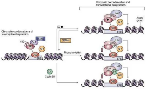
After entering cells, oestrogen binds to and activates ER. Oestrogen binding results in a conformational change that enables oestrogen-regulated genes to be activated. The ER binds as a dimer to small palindromic DNA motifs, known as oestrogen response elements (EREs), in the promoters of specific genes, through the action of two zing fingers (36). Two distinct activation domains, AF1 and AF2, mediate transcription activation. AF2 is integral to the ligand-binding domain (LBD) and its activity requires the binding between LBD and oestrogen, whereas AF1 activity is regulated by phosphorylation (Figure 5) (37, 38). AF1 and AF2 activate the transcription independently and/or synergistically and there is evidence that AF1 and AF2 activities are influenced by the promoter and cell type (39). ER activates gene expression by stimulating recruitment of the general transcription machinery to the transcription start site through the action of its activation domains.
Figure 5. Mechanisms of oestrogen-receptor activation. The oestrogen receptor (ER) has three domains: AF1, which is regulated by phosphorylation; AF2, which is regulated by oestogen binding; and a DNA binding domain (DBD). In the inactivated state, the ER is bound to corepressor (CoR) complexes, which recruit histone deacetylases (HDACs). HDACs maintain histones in a deacetylated state, which favours chromatin condensation. Oestrogen binding results a conformational change in AF2 that facilitates interaction with co-activators (CoA), which bind histone acetyltransferases (HATs). Acetylation of histones by HATs leads to chromatin decondensation, facilitating transcriptional activation. Modulation of
Dr. Marina Bacci
Metabolic reprogramming of oestrogen receptor positive breast cancer in endocrine therapy resistance. PhD Course in Life Sciences and Biotechnologies XXIX cycle - Università degli Studi di Sassari.
20
ER activity by selective ER modulators (SERMs) is likely to be achieved by a balance between coactivator and corepressor complex recruitment to AF2, depending on the conformation induced by the SERM, as well as tissue-specific differences in co-activator/corepressor availability. Other factors, such as cyclin D1 and growth-factor-induced phosphorylation of AF1, might facilitate ligand-independent recruitment of co-activators. ERE, oestrogen response element; H12, helix 12 (taken from 40).
Different studies have identified co-activator complexes that mediate trans-activation by nuclear receptors. These co-activators facilitate recruitment of the general transcription machinery through direct interaction but, importantly, several co-activator complexes also mediate chromatin remodelling (41, 42). These include: the SWI/SNF complexes (switch/sucrose non-fermenting), which facilitate transcription-factor binding to nucleosomal DNA by ATP dependent chromatin remodelling (43, 44); the TRAP/DRIP/SMCC complex, these alternative names stand for thyroid-hormone-receptor-associated protein (TRAP); vitamin-D-receptor-interacting protein (DRIP), and SRB and mediator-protein containing complex (SMCC), which associates with RNA polymerase II63; CREB-binding protein (CBP), and p300/CBP-associated factor (PCAF), which are histone acetyltransferase (HAT) complexes. Hyperacetylation of histones seems to correlate with more actively transcribed regions of the genome than to hypoacetylated regions. Therefore, histone acetylation through recruitment of HATs might be crucial in overcoming the repressive effects of chromatin on transcription (45, 46). Three related co-activators, collectively known as the p160 co-activators, stimulate ER activity following ligand stimulation, through direct interaction with AF2 (41, 42). These three proteins, known as nuclear-receptor co-activator 1 (NCOA1; also known as SRC1) NCOA2 (also known as TIF2 or GRIP1) and NCOA3 (also known as P/CIP, ACTR, AIB-1, RAC3 or TRAM1) associate with the transcription factor CBP to facilitate histone acetylation (47, 48). Furthermore, NCOA1 and NCOA3 can themselves acetylate histones (43, 44, 49). Several nuclear receptors that are not bound to the cognate ligands, such as the thyroid hormone receptor, repress gene expression. This repression is mediated, at least in part, through recruitment of histone deacetylase complexes (HDACs) by interaction of nuclear receptors with nuclear-receptor corepressor 1 (NCOR1) or NCOR2 (also known as SMRT) (50-52). NCOR recruitment, like that of co-activators, is mediated by the
Dr. Marina Bacci
Metabolic reprogramming of oestrogen receptor positive breast cancer in endocrine therapy resistance. PhD Course in Life Sciences and Biotechnologies XXIX cycle - Università degli Studi di Sassari.
21
LBD/AF2, albeit in the absence of ligand. Therefore, ligand binding results in dissociation of co-repressors and recruitment of co-activator complexes. Other histone-modifying proteins, such as arginine methyltransferases, act as co-activators for nuclear receptors, including ER, either through direct interaction with the LBD (53) or through association with co-activators (54, 55). The LBD is encoded by about 300 amino acids. Structural studies have shown that it is a wedge-shaped structure that contains the ligand-binding pocket. Ligand binding results in a remarkable conformational change in the LBD, that induces the exposition of a surface for the recruitment of co-activators. Co-activators recruitment to the LBD is mediated by a short motif, characterised by the amino-acid sequence leucine–X–X–leucine– leucine (where X is any amino acid), which forms an α-helix (56-58). Different studies identified cis-regulatory domains that augment transcription of these ER gene targets. Specifically, ER association with gene targets results from an association with the pioneer factor FoxA1, responsible for recruitment of ER to the genome. Recruitment of ER to the genome does not seem to occur at the promoter proximal regions, but instead involves distal enhancer elements that function together with the ER complex at the promoter of the target genes (59).
Many of the genes regulated by oestrogen signalling promote tumorigenic phenotypes, including cell proliferation, inhibition of apoptosis, invasion and metastasis, and angiogenesis (60). Of particular importance are Cyclin D1 (61, 62) and c-Myc (63, 64), both of which are essential drivers of oestrogen stimulated cellular proliferation and tumorigenesis. Cyclin D1 binds to and activates cyclin dependent kinases (CDK) 4/6, which phosphorylate the retinoblastoma protein (Rb), resulting in release of the E2F transcription factor, and progression through the restriction point within the G1 phase of the cell cycle (65, 66). Inhibition of Cyclin D1, either with antibodies or by expression of its inhibitor, CDK4 p16INK, prevents oestrogen stimulation of cellular proliferation and progression through the G1 checkpoint (67). c-Myc is a proto-oncogene transcription factor that regulates the expression of a large number of target genes that promote cell growth and cell cycle progression (68, 69). Similarly to Cyclin D1, inhibition of c-Myc prevents oestrogen stimulated cell proliferation (70). Conversely, overexpression of Cyclin D1 or c-Myc can mimic the effects of
Dr. Marina Bacci
Metabolic reprogramming of oestrogen receptor positive breast cancer in endocrine therapy resistance. PhD Course in Life Sciences and Biotechnologies XXIX cycle - Università degli Studi di Sassari.
22
oestrogen by reinitiating cell cycle progression in anti-oestrogen arrested cells (71). These results indicate that Cyclin D1 and c-Myc are essential drivers of oestrogen stimulated cellular proliferation.
Since ER positive breast cancers are dependent upon oestrogen for their growth and progression, this type of tumour can be treated with endocrine therapies that deprive cells of ER signalling, resulting in tumour inhibition.
3- Endocrine therapy
ER+ breast cancer cells are dependent to oestrogens for their growth and proliferation. The hypothesis that oestrogen could have a crucial role in the tumour progression dates back to 1936. In that year, Lacassagne demonstrated in mice with high incidence of mammary cancer that ovariectomy or oestrogen replacement prevents or enhance tumorigenesis, respectively (72). However, the mechanism of tumour inhibition was not elucidated until the role for the ovarian hormone oestrogen in stimulating breast cancer growth was discovered (73). The discovery of ER and the development of an assay quantifying ER expression in patients with breast cancer made possible to identify women likely to respond to endocrine therapy (20, 74). Endocrine ablation by ovariectomy in pre-menopausal patients has now been replaced by pharmacological agents, generally called selective ER modulators (SERMs). SERMs are structurally different compounds that interact with intracellular ERs in target organs as oestrogen receptor agonists or antagonists. These drugs have been intensively studied over the past and have been proven to be a highly versatile group for the treatment of different conditions associated with post-menopausal women’s health including osteoporosis and hormone responsive cancer (75, 76). The first SERM developed was MER25, non-steroidal antioestrogen capable of blocking oestrogen action (77). However, MER25 was unsuccessful due to toxicity issue (hallucination) and the first successful oestrogen antagonist to enter the clinic was tamoxifen (ICI 46474) (78). Although SERMs have many benefits, they also have some potentially serious adverse effects, such as thromboembolic disorder and, in the case of tamoxifen, uterine cancer.
Dr. Marina Bacci
Metabolic reprogramming of oestrogen receptor positive breast cancer in endocrine therapy resistance. PhD Course in Life Sciences and Biotechnologies XXIX cycle - Università degli Studi di Sassari.
23
3.1 Tamoxifen
Tamoxifen binds to ER and inhibits ER signalling, limiting cellular proliferation of breast cancer cells (79, 80). The major metabolites of tamoxifen in human are N-desmethyltamoxifen and trans-4-hydroxytamoxifen; the affinity of the latter for ER is equivalent to that E2 (81). The anti-tumour effects of tamoxifen are mediated by competitive inhibition of oestrogen binding to ER (82) and by recruitment transcriptional co-repressor (e.g. NCoR) instead of co-activator (83). As a consequence, tamoxifen inhibits the expression of oestrogen-dependent genes, including growth factor and angiogenic factor secreted by cancer cell that in turn may stimulate tumour growth by autocrine or paracrine mechanism (84). The net result is a block in the G1 phase of cell cycle and subsequent reduction of the cell proliferation rate. Furthermore, it has been shown that tamoxifen may also directly induce apoptosis (85).
Tamoxifen has been the mainstay endocrine therapy in breast cancer for the last 25 years. Compelling data have demonstrated a significant overall survival benefit; in patients with ER+ breast cancer, tamoxifen treatment results in a 51% reduction in recurrence and a 28% reduction in death as well as improved quality of life for patients with metastatic disease (86). Tamoxifen has also been shown to be effective in reducing the incidence of breast cancer in patients at risk for developing the disease (87) and in women with ductal carcinoma in situ (88).
Despite the documented benefits of ER-targeted therapy in breast cancer, it is known that not all patients who have ER expressing tumours respond to endocrine manipulation (de novo resistance) and a substantial number of patients who do respond will develop disease progression or recurrence while on therapy (acquired resistance): all patients with metastatic disease and 40% of early stage breast cancer patients treated with adjuvant tamoxifen, eventually relapse with tamoxifen resistant disease (89).
Tamoxifen is classified as a SERM; although tamoxifen acts as an ER antagonist in the breast, it exerts agonistic effects in some tissues such as the endometrium and the vascular system. This agonistic activity is associated with rare yet life-threatening side effects such as thromboembolic events and uterine cancer (87, 90). Whether tamoxifen
Dr. Marina Bacci
Metabolic reprogramming of oestrogen receptor positive breast cancer in endocrine therapy resistance. PhD Course in Life Sciences and Biotechnologies XXIX cycle - Università degli Studi di Sassari.
24
acts as an antagonist or agonist of ER signalling depends on the cellular context. As discussed previously, ER contains two domains that regulate transcriptional activation, AF1 and AF2 (Figure 5). AF2 acts in a ligand dependent manner, whereas AF1 is largely controlled by phosphorylation and as such tamoxifen only inhibits AF2 activation. Since ER activity in the breast is mainly AF2 driven, tamoxifen acts largely as an antagonist. This is in contrast to other tissues, such as the uterus, where ER activity is also controlled by AF1, resulting in greater agonistic activity of tamoxifen (91).
3.2 Aromatase inhibitors
Aromatase is an enzyme belonging to the cytochrome P-450 superfamily (92) and is highly expressed in the placenta and in the granulosa cells of ovarian follicles, where its expression depends on cyclical gonadotropin stimulation. Aromatase is also present, at lower levels, in several non-glandular tissue, including subcutaneous fat, liver, muscle, brain, normal breast and breast cancer tissue (93, 94). After menopause, oestrogen source derived exclusively from non glandular tissues, in particular from subcutaneous fat. In postmenopausal patients, an alternative to oestrogen antagonists for endocrine therapy are aromatase inhibitors (AI), which block the conversion of androgen to oestrogen by aromatase inhibition. Therefore, treatment of postmenopausal women with AI results in oestrogen deprivation and reduced ER signalling (95). Three generations of AI have been developed. The first- (aminoglutethimide) and second-generation AI (e.g., fadrozole and vorozole) were less selective and in addition to aromatase, they decreased aldosterone and cortisol production. These drugs were poorly tolerated and had limited clinical efficacy (96). Third-generation AI such as letrozole, anastrozole or exemestane, are highly selective for the enzyme aromatase and are well tolerated from the patients. Anastrozole and letrozole are nonsteroidal inhibitors that reversibly bind aromatase, whereas exemestane is a steroidal AI that irreversibly binds aromatase (97). These drugs are effectively challenging tamoxifen for use in postmenopausal patients with ER+ breast cancer (98). The clinical benefits associated with AI include significantly greater disease-free survival, a longer median time to recurrence, and a reduced incidence of contralateral breast cancer. Indeed, AI markedly supress
Dr. Marina Bacci
Metabolic reprogramming of oestrogen receptor positive breast cancer in endocrine therapy resistance. PhD Course in Life Sciences and Biotechnologies XXIX cycle - Università degli Studi di Sassari.
25
plasma oestrogens levels in postmenopausal women and in contrast with tamoxifen, AI have no partial agonist activity reducing incidences of thromboembolic events, vaginal bleeding, and endometrial cancer (99). As a consequence, aromatase inhibitors are increasingly being used for the endocrine treatment of postmenopausal ER+ breast cancer patients. Despite clinical benefits of AI, up to 50% of treated patients develop resistance to AI (100), again limiting effectiveness.
3.3 Fulvestrant
Oestrogen ablation therapy has been intensively used as a mean of treating ER+ breast cancer: antioestrogen (e.g. tamoxifen) and AI are now established as first–line agents in adjuvant endocrine therapy of ER+ breast cancer patients. Although adjuvant endocrine therapy is an effective treatment for breast cancer, most patients with advanced disease will eventually exhibit resistance to individual therapy (101). However, an initial response to endocrine treatment is generally indicative of a positive response to further alternative endocrine agents (102). Consequently, therapeutic options against ER+ breast cancer have expanded tremendously. In contrast to tamoxifen, which exhibits partial antagonist activity, fulvestrant (or ICI 182,780) is a “full” or “pure” anti-oestrogen that has no known oestrogen agonist effects (WO 2001051056 A1, Astrazeneca 2001). Fulvestrant exerts its antitumor activity preventing the oestrogen-ER interaction, thus abrogating the oestrogen–regulated transcription pathway (103, 104). Its binding affinity for ER is higher than tamoxifen (103, 105). Following binding to ER, fulvestrant blocks dimerization of the receptor and limits its nuclear translocation (106-108). Furthermore, fulvestrant-ER complex is instable and more susceptible to degradation by proteasome (109). Fulvestrant also blocks the recruitment of both transcriptional activating factors, AF1 and AF2 (109). As a result, in contrast to tamoxifen, which blocks recruitment of AF-2 only, fulvestrant exhibits full ER antagonist and no agonist effects (109). Fulvestrant has similar efficacy to tamoxifen as a first line therapy in patients with advanced ERα positive breast cancer (110) and has been shown to be as effective as the aromatase inhibitor anastrozole as a second line therapy in patients whose disease has progressed on prior endocrine therapy (110). It has been
Dr. Marina Bacci
Metabolic reprogramming of oestrogen receptor positive breast cancer in endocrine therapy resistance. PhD Course in Life Sciences and Biotechnologies XXIX cycle - Università degli Studi di Sassari.
26
demonstrated that fulvestrant monotherapy may be superior to AI in patients who have not received adjuvant endocrine therapy and in patients that present inoperable locally or advanced cancer and treated in first line setting (111). Fulvestrant monotherapy is associated with less arthralgia, but the combination with AI increase the risk of hot flashes and gastrointestinal disturbance (111). Despite its usefulness, resistance to fulvestrant also occurs frequently (112). These findings have stimulated the search for new mixed SERMs, which are anti-oestrogenic for the breast, but oestrogenic for other tissues, in which the protective actions of oestrogen are desirable (113).
4- Endocrine therapy resistance
Despite the relative safety and significant anti-neoplastic activities of endocrine therapies, the major limitation remains de novo and acquired resistance to endocrine agents. Although clinicians are encouraged from positive effects of second- and third-line endocrine therapy for patients who initially benefited from first line treatment (114), the clinical response rate declines from approximately 70% for first line-therapy fulvestrant or AI to around 30% in the successive lines of treatments (115, 116). Several approaches have been used to elucidate the mechanisms that drive endocrine therapy resistance to discover predictive markers and/or alternative targets that could be investigate for therapeutic approach. However, the molecular mechanisms that underlie resistance are not fully understood and as such, definitive approaches for preventing and overcoming resistance are not yet available. Several mechanism have been associated with endocrine therapy resistance, such as mutations of
ER (ESR1), growth factors driven signalling cross talk, cell cycle alteration,
Dr. Marina Bacci
Metabolic reprogramming of oestrogen receptor positive breast cancer in endocrine therapy resistance. PhD Course in Life Sciences and Biotechnologies XXIX cycle - Università degli Studi di Sassari.
27
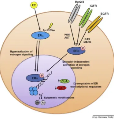
Figure 6. Schematic representation of major endocrine-resistance mechanisms. Several mechanisms have been shown to contribute to endocrine therapy resistance, which encompass hypersensitivity to E2 stimulation, phosphorylation of the ER by several kinase cascades, such as phosphoinositide 3 kinase (PI3K) or mitogen-activated protein kinase (MAPK), which in turn can be activated by tyrosine kinase receptors. Furthermore, changes in the expression of transcriptional regulators of the ER transcriptional complex are responsible for increased expression of ER-responsive genes (taken from 117).
4.1 ESR1 mutations
Although loss of ER expression may be a reasonable explanation for the emergence of endocrine therapy resistance, loss of ER occurs in only 10% of primary and metastatic tumours that show the resistance (118). Therefore, ER remains a potential target in the majority of the endocrine resistant cancers. It has been reported that in solid tumours, the resistance to “oncoreceptor”-targeted therapies, such as tyrosine kinase inhibitors, is frequently driven by the emergence of additional mutations in the target oncogene (119). A similar molecular mechanism might underpin resistance to therapies targeting the ER. Specifically, attention has recently focused on mutations in the gene ESR1. A recent work has revealed that ESR1 mutations are more frequent in patients bearing metastatic breast cancers or that have already received a therapeutic intervention (120). Highly
Dr. Marina Bacci
Metabolic reprogramming of oestrogen receptor positive breast cancer in endocrine therapy resistance. PhD Course in Life Sciences and Biotechnologies XXIX cycle - Università degli Studi di Sassari.
28
recurrent mutations were noted at two residues in the LBD of the receptor: Y357S and D538G (Figure 7) (121-123).
Figure 7. Structural diagram of the ERα protein encoded by the ESR1 gene. Schematic representation of the ER protein and its functional domains; the position and number of cases (n) of the ESR1 (ER) LBD point mutations reported in metastatic ER+ breast cancers are indicated. Black circles indicate each mutation at the specific amino acid residue; numbers in parentheses indicate the total number of samples reported to harbour the specific indicated mutations. Abbreviations: AF-1, activation function-1; AF-2, activation function-2; DBD, DNA-binding domain; ERα, oestrogen receptor α; LBD, ligand-binding domain (taken from reference (124).
These mutations generally are observed in 10-30% of all endocrine-resistant advanced breast cancers and have been linked to enhanced sensitivity to oestrogen as well as to the constitutive activation of transcriptional activity of ER in the absence of an ER agonist (121-123, 125-127). These mutations seem to be more common only after exposure to one or more lines of endocrine treatments (in particular AI) (121, 128), as highlighted by paired analysis of primary tumours and their metastatic therapy-resistant counterparts (128, 129). Furthermore, gene expression of
ESR1-harboring breast cancer cells show dysregulation of both
ER-dependent and ER-inER-dependent genes, suggesting that ESR1 mutants alter the natural landscape of ER interaction network, or arise together with other resistance mechanisms (128). However, since ESR1 mutations could be the indication of the emergence of endocrine therapy resistant clones, monitoring ESR1 mutation status in patients that are undergoing endocrine
Dr. Marina Bacci
Metabolic reprogramming of oestrogen receptor positive breast cancer in endocrine therapy resistance. PhD Course in Life Sciences and Biotechnologies XXIX cycle - Università degli Studi di Sassari.
29
therapy might help clinicians to prevent and combat therapy resistance (128-130).
4.2 Growth factor receptors: PI3K/AKT/mTOR and MAPK
pathway activation
Overexpression and/or amplification of growth factor receptor including FGFR1, HER2, HER3, EGFR, IGF1R and RET are associated with the emergence of endocrine therapy resistance (131-135). The activation of growth factor receptors can induce the phosphorylation of ER and AIB1 through cross talk mechanism, which have been shown to empower oestrogen signalling and induce tamoxifen resistance (136-139). Furthermore, their signalling pathways converge on the Phosphoinositide 3 kinase (PI3K)/AKT/ mammalian target of rapamycin (mTOR) and MAPKs pathway. Several kinases belonging to MAPK family, such as ERK1/2 and ERK3, can phosphorylate ER (e.g. Ser-118), prompting ligand-independent activation of the receptor and altering the response to endocrine therapies (89, 136, 140, 141). PI3K and AKT also have a role in the activation of ER in absence of oestrogen trough of the AF-1 (PI3K) and AF-2 (PI3K and AKT) domains of the receptor ((142). PI3K gene is mutated in ~40% of human breast cancers (143, 144). These mutations promote a PI3K pathway hyper-activation that induces oestrogen-independent ER transcriptional activation (145). mTOR is a key downstream effector of PI3K/AKT pathway involved in important cellular processes, such as protein synthesis and cellular metabolism (146). Therefore, mTOR has become an attractive target for therapies attempting to reverse the endocrine therapy resistance. Accordingly, randomized Phase III BOLER2 trial has shown that the combination of mTOR inhibitor everolimus with the AI exmestane improve progression-free survival in ER+ breast cancer patients previously treated with non-steroidal AI (BOLERO2 clinical trial.gov number NCT00863655, (147). However, it has been reported that combinatorial treatment can promote several toxicity issues in breast cancer patients. Therefore, we should consider the real benefits of the combination in relation to toxicity before that patients undergo to these treatments. Other approaches to revert endocrine therapy resistance are based on combination of endocrine agents with selective PI3K inhibitors
Dr. Marina Bacci
Metabolic reprogramming of oestrogen receptor positive breast cancer in endocrine therapy resistance. PhD Course in Life Sciences and Biotechnologies XXIX cycle - Università degli Studi di Sassari.
30
(https://www.clinicaltrials.gov/ct2/show/NCT02340221, accessed online 18
January 2016) and FGFR, EGFR or IGFR inhibitors
(Https://www.cliniclatrials,gov/ct2/results?term=%22FGFR+AND+%22breas t+cancer%22&recr0Open accessed online 28Febraury 2016;
https://www.clinicaltrials.gov/ct2/show/NCT02115282, accessed online 19 January 2016)
4.3 Cell cycle checkpoint alterations
Normal and cancerous cells receive a plethora of proliferative and antiproliferative signals and the balance of these inputs determines whether a cell will undergo cell division or will enter into quiescent phase (148). The deregulation of the cell cycle progression via alterations of key cell cycle checkpoints can also contribute to endocrine therapy resistance (149). Tumour suppressor Rb is a negative regulator of the cell cycle able to mediate antiproliferative signals. Rb itself is regulated by a complex of cyclin and CDK, a family of serine-threonine kinases (150). Progression through the G1-S phase requires the phosphorylation of Rb by CDK4 in complex with cyclin D1, D2 or D3 (151). Rb hyper-phosphorylation leads to an increase of genes synthesis whose products are essential for DNA replication and mitotic progression (152). Many tumours increase cyclin D-dependent activity and thereby escape senescence via multiple mechanisms such as CDK4 amplification, CDK4 mutations, cyclin D1 translocation, amplification or overexpression (153). Cyclin D1 amplification is a common event in ER+ breast cancer, identified in 58% of luminal B cancers and 29% luminal A cancers (154). Anti-oestrogen induced growth
arrest in ER+ breast cancer cells is accompanied by decreased cyclin D1 expression, whereas there is a persistent cyclin D1 expression and Rb phosphorylation in the case of the endocrine therapy resistance (155, 156). The first drugs developed to target cell cycle progression abnormalities in human cancer were non-selective pan-CDK inhibitors (157, 158). The therapeutic potential of this strategy in breast cancer is increased by development of highly selective inhibitors of CDK. Accordingly, Palbociclib is a small-molecule inhibitor of CDK4 and CDK6, and preclinical data have shown that it was able to inhibit the growth of ER+ breast cancer and reverse endocrine therapy resistance (159). These results have led to a
Dr. Marina Bacci
Metabolic reprogramming of oestrogen receptor positive breast cancer in endocrine therapy resistance. PhD Course in Life Sciences and Biotechnologies XXIX cycle - Università degli Studi di Sassari.
31
clinical trial, the so-called PALOMA-1 (Palbociclib: Ongoing Trials in the Management of Breast Cancer). This trail has demonstrated that the combination of palbociclib with letrozole as first-line therapy in ER+ breast cancer is more effective when compared to letrozole alone, displaying a longer-progression free survival for patients subjected to such combinatorial treatment (160). Later, PALOMA-2 clinical trial has shown that palbociclib combined with letrozole treatment represent a good therapeutic approach for postmenopausal women with ER+/HER-2 advanced breast cancer. Indeed, PALOMA-2 has confirmed the same results for postmenopausal women with ER+/HER2-advanced breast cancer (PALOMA-2 clinicaltials.gov number NCT01740427, (161).
4.4 Enhanced autophagy
Autophagy is an intracellular process leading to the degradation of damaged or unnecessary subcellular organelles. This process represents a key mechanism for survival of normal and cancer cells during stress condition, such as nutrient deprivation. A recent work has demonstrated that autophagy inhibition is linked to endocrine therapy sensitivity restoration, promoting cell apoptosis in preclinical models of endocrine-resistant breast cancer (162). The inhibitors of autophagy are currently explored in early phase trials in breast cancer. In the advanced endocrine-resistant breast cancer setting, hydoxychloroquine is being evaluated in combination with endocrine therapy in a phase Ib/II study (https://www.clinicaltrials.gov/ct2/shoeìw/NCT02414776, accessed online 23 May 2016).
4.5 Epigenomic signature
Changes in gene expression are not uniquely dependent on the presence of mutations but can also derived from changes in DNA methylation patterns and histone modifications, a process named epigenetics (163). Endocrine therapy has been shown to alter the epigenetic landscape of tumour cells by downregulating oestrogen-responsive genes (164). Recent studies showed that compared to sensitive tumours, endocrine therapy resistant breast cancers are characterised by a differential gene methylation pattern in the enhancer regions of oestrogen-dependent genes, which are in turn involved in different cellular process, such as apoptosis
Dr. Marina Bacci
Metabolic reprogramming of oestrogen receptor positive breast cancer in endocrine therapy resistance. PhD Course in Life Sciences and Biotechnologies XXIX cycle - Università degli Studi di Sassari.
32
regulation, endoplasmic reticulum Golgi trafficking and DNA damage response (165). Studies investigating breast cancer epigenetic alterations after endocrine therapy have reported dysregulation in the expression of genes involved in key cellular pathways, including metabolic processes, nucleoside transport and development process (166, 167). These results show that epigenetic deregulation of ER and its responsive genes largely contribute to endocrine resistance, although other mechanisms exist, as previously described.
In conclusion, there are several mechanisms involved in endocrine therapy resistance and new pharmacological approaches are investigated. Clinical trials suggest that ER+/HER2+ tumours may benefit of the combining treatment of AI with HER2-targeting compounds (168, 169). Several clinical trials also suggest that subsets of patients with ER+/HER2- breast cancers may benefit from a combination of a growth factor pathway inhibitor with endocrine therapy agents (147). However, given the adaptability of cancer cells, targeting a single growth factor or downstream signalling hub can lead to compensatory mechanism and the fail of therapeutic approaches. Further studies are necessary to understand other mechanisms of resistance in order to develop other therapeutic strategy to limit endocrine therapy resistance challenge.
5- Tumour metabolism
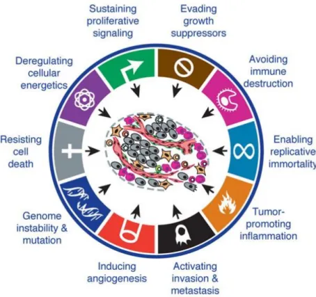
The hallmarks of cancer are the different biological capabilities that cancer cells acquire during tumour development and progression (Figure 8) (170). Genomic instability is the most important driver of the cancer cells alterations, which affect several cellular process, such as proliferation, senescence, survival signalling and apoptosis (170, 171). Metabolic deregulation is an established hallmark of cancer. To support the enhanced proliferation and uncontrolled cell division, most cancer cells exhibit metabolic adaptations that promote their survival and progression under non physiological conditions. Therefore, although cellular transformation occurring in different cell type arises from many different pathways, the metabolic reprogramming of cancer cells is similar (172). The requirements
Dr. Marina Bacci
Metabolic reprogramming of oestrogen receptor positive breast cancer in endocrine therapy resistance. PhD Course in Life Sciences and Biotechnologies XXIX cycle - Università degli Studi di Sassari.
33
of proliferative cancer cells are essentially to generate energy, in the form of adenosine 5-triphosphate (ATP), and to sustain macromolecules biosynthesis, while managing the high oxidative stress levels that accompany a rapid cell growth. Cancer cells are surrounded by different cell components of the tumour microenvironment that contribute to the acquisition of hallmarks traits. The tumour microenvironment influences tumour metabolism by exerting additional selective pressure on the cancer cells to adapt to harsh conditions, such as hypoxia, acidity and/or nutrient starvation.
Figure 8. Hallmarks of cancer: the next generation. There are now 10
established hallmarks of cancer, including inflammation, metabolism and genomic instability (taken from 170).
5.1 Glucose metabolism
Under aerobic conditions, differentiated cells metabolise glucose to pyruvate via glycolysis in the cytosol and then the pyruvate enters the mitochondrial tricarboxylic acid (TCA) cycle for its complete oxidation. This reaction produces NADH (nicotinamide adenine dinclueotide NAD+,
Dr. Marina Bacci
Metabolic reprogramming of oestrogen receptor positive breast cancer in endocrine therapy resistance. PhD Course in Life Sciences and Biotechnologies XXIX cycle - Università degli Studi di Sassari.
34
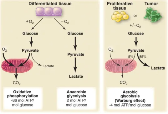
maximize the production of energy form ATP molecules. In the absence of oxygen, glycolysis is favoured and a small amount of pyruvate undergoes OXPHOS, whereas a high pyruvate quantity is converted in lactate in a process called fermentation. Otto Warburg first reported that in the presence of oxygen, proliferating cancer cells could reprogram their glucose metabolism, and thus their energy production, consuming glucose at a surprisingly high rate compare to normal cells by an increase of glycolysis and subsequent lactate release, in a state that has been termed “aerobic glycolysis”. This phenomenon is also known as the “Warburg effect” (figure 9) (173).
Figure 9. Comparison of glycolysis between a normal tissue and tumour/ proliferated tissue ( taken from 174).
Warburg originally hypothesized that cancer cells developed a defect in mitochondria that led to an impairment in aerobic respiration and a subsequent reliance on glycolytic metabolism (173). However, subsequent works showed that mitochondrial function was not impaired in most cancer cells (175-177). The highly glycolytic rate provides several advantages for proliferating cells. First, an increase of glycolysis allows cells to use the most abundant extracellular nutrient, glucose, to produce ATP. Although the quantity of ATP produced by glycolysis is lower compared to that
Dr. Marina Bacci
Metabolic reprogramming of oestrogen receptor positive breast cancer in endocrine therapy resistance. PhD Course in Life Sciences and Biotechnologies XXIX cycle - Università degli Studi di Sassari.
35
obtained via OXPHOS, if the glycolytic flux is high enough, the percentage of cellular ATP produced by glycolysis can exceed that produced from aerobic respiration (173, 178). To allow an increase in glucose uptake, many cancer cells upregulate the glucose transporters, (GLUTs), which can contribute to a substantial increase in glucose import into the cytoplasm (179-181). Indeed, markedly increased uptake and utilisation of glucose have been documented in many human tumours using positron emission tomography (PET) with radiolabeled analogue of glucose (18
F-fluorodeoxyglucose, FDG). Furthermore, during cell growth and proliferation, cells need a large quantity of nucleotides, amino acids and lipids to create biomass. Glucose could be used to generate biomass as well as ATP. Degradation of this metabolite provides cells with intermediates needed for biosynthetic pathways, such as glycerol and citrate for lipids, non-essential amino acids for protein synthesis and through the oxidative pentose phosphate pathway, ribose sugars for nucleotides and NADPH (182). The switch from OXPHOS to glycolysis, with its concomitant accumulation of lactate produced and released in tumour microenvironment results in an increased acidity in the tumour microenvironment, which promotes tumour cells adaptation and contributes to the evolution of the tumour niche (182, 183). The availability of biosynthetic precursors is enhanced by regulation of the last rate-limiting step of glycolytic pathway, which is catalysed in normal cells by pyruvate kinase M1 (PKM1). PK catalyses the conversion of phosphoenolpyruvate (PEP) to pyruvate, with concomitant phosphorylation of ADP to ATP. It also exists an alternative splice form of PK, PKM2, which has reduced catalytic activity. This isoform is predominant in proliferating and cancer cells and can be allosterically activated by fructose-1-6-biphosphate (FBP) (184, 185). This activation can be countered by either phosphotyrosine binding to PKM2 or by phosphorylation of a specific tyrosine residue (Y105) of PKM2 by the activation of signalling pathways downstream of receptor tyrosine kinases. PKM2 can exist as both dimer and tetramer form (186, 187). The PKM2 dimer is less active compare to its tetrameric form in converting PEP to ATP and pyruvate (187, 188). While tetrameric PKM2 favours ATP production through TCA cycle, dimeric PKM2 plays a critical role in aerobic glycolysis (187). This reduced catalytic activity allows the reduction of the