phd_unisi_076750_1.pdf
73
FIRMATO DIGITALMENTE DA: Nome: NICLA
Cognome: LORITO Data: 18 febbraio 2021
Università degli Studi di Siena
Department of Biotechnology, Chemistry and Pharmacy
Research Doctorate in Biochemistry and Molecular Biology
BIBIM2.0
Cycle XXXIII°
Coordinator: Prof. Lorenza Trabalzini
Metabolic determinants of therapy resistance in
estrogen receptor positive breast cancer
Scientific-disciplinary sector: BIO/10
PhD student: Nicla Lorito
Co-tutor: Dr. Andrea Morandi
Supervisor: Prof. Elisa Giannoni
INDEX
AbbreviationsAbstract 1
Chapter I. Introduction 2
1. Breast cancer 2
1.1 Molecular subtypes of breast cancer 3
1.2 Estrogen receptor positive breast cancer 5
1.2.1 Estrogen and breast cancer risk 6
1.2.2 Estrogen receptor and mechanism of action 7
2. Cell cycle 10
2.1 Control of the cell cycle: phases and checkpoints 10
2.2 Role of the cyclin D1/cyclin-dependent kinases 4/6 complex in the cell cycle 12
3. Breast cancer therapy 14
3.1 Endocrine therapy 14
3.1.1 Tamoxifen 15
3.1.2 Fulvestrant 16
3.1.3 Aromatase inhibitors 18
3.2 Cyclin-dependent kinases 4/6 inhibitors 19
3.2.1 Palbociclib 21
4. Therapy resistance 23
4.1 Endocrine therapy resistance 23
4.1.1 Crosstalk between estrogen receptor and growth factor receptors 24
4.1.2 Cell cycle checkpoint alterations 25
4.1.3 Autophagy 26
4.2 Cyclin-dependent kinases 4/6 inhibitor resistance 26
5. Tumor metabolism 31
5.1 Glucose metabolism 32
5.2 Amino acid metabolism 36
6. Tumor metabolic reprogramming 39
6.1 Deregulated metabolism and therapy resistance in breast cancer 42
6.2 Metabolic targeting in breast cancer 45
Chapter II. Materials and Methods 49
1. Materials 49
1.1 Cell lines 49
1.2 Mouse models and care 50
1.4 Drugs, compounds, and reagents 51
1.5 Antibodies 51
2. Methods 52
2.1 General culture conditions 52
2.1.1 Long-term cell frozen storage 52
2.2 Immunofluorescence 52
2.3 Protein manipulation 53
2.3.1 Immunoblotting 54
2.3.2 Immunoprecipitation 55
2.4 Protein de novo synthesis assay 55
2.5 RNA manipulation 55
2.6 Quantitative Real-Time Polymerase Chain Reaction 56
2.7 Cell viability and survival assays 56
2.7.1 Crystal violet assay 56
2.7.2 MTT assay 57
2.8 Colony formation assay 57
2.9 Transwell motility and invasion assay 57
2.10 RNAi transfection 58
2.11 Radioactive assays 58
2.11.1 Radiolabeled glucose and amino acid uptake 58
2.11.2 Incorporation of radiolabeled amino acids into proteins, lipids, and DNA 58
2.12 High-Performance Liquid Chromatography 59
2.13 Gas Chromatography-Mass Spectrometry 59
2.14 13C-tracing experiments using Liquid Chromatography-Mass Spectrometry 60
2.15 Metabolomics data analysis 60
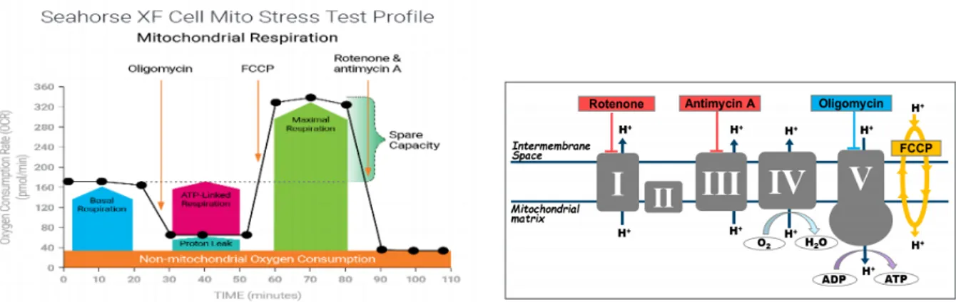
2.16 Seahorse XFe96 metabolic assays 61
2.16.1 Cell Mito Stress Test 61
2.16.2 Glycolytic Rate Assay 62
2.17 Lung retention assay 63
2.18 Gene and microRNA expression analysis 63
2.19 Bioinformatic analysis 63
2.20 Analysis of human datasets 64
2.21 Statistical analysis 64
Chapter III. Reprogramming of amino acid transporters to support aspartate and
glutamate dependency sustains endocrine resistance in breast cancer 66
1. Introduction 66
2.1 Global genome analysis reveals a deregulated metabolic miR-23b-3p/SLC6A14 node with prognostic value in endocrine therapy resistant ER+ breast cancer 67 2.2 Expression levels of miR-23b-3p and SLC6A14 are deregulated in endocrine
therapy resistant cells 69
2.3 Endocrine therapy resistant cells display enhanced autophagic flux essential
for their survival 71
2.4 Aspartate and glutamate intracellular levels correlate with the aggressive
features of endocrine therapy resistant cells 74
2.5 Aspartate and glutamate confer metabolic plasticity to endocrine therapy
resistant cells 76
2.6 Impairing the transport of aspartate and glutamate affects the metastatic
potential of endocrine therapy resistant cells in vivo 79
3. Discussion 82
Chapter IV. Glucose metabolic reprogramming of ER+ breast cancer in acquired
resistance to the CDK4/6 inhibitor palbociclib 85
1. Introduction 85
2. Results 85
2.1 Palbociclib affects the expression of key metabolic players implicated in
glucose catabolism 85
2.2 Palbociclib resistant cells show growth rates similar to the sensitive
counterpart while enhancing glucose uptake 87
2.3 HER2 status impacts on glucose catabolism and defines distinct glucose dependencies during acute and chronic drug administration 89 2.4 Targeting glucose catabolism re-sensitizes ER+/HER2+ palbociclib resistant
cells to the drug 93
2.5 Targeting glycolysis increases the effect of palbociclib on ER+/HER2-parental cells 94
2.6 Metabolomic analysis shows a different intracellular metabolite profile
between HER2- and HER2+ palbociclib resistant cells 97
2.7 High expression levels of HK2 identify a subset of patients with poor
prognosis 99
3. Discussion 100
References 103
Appendix 118
Bacci, M.; Lorito, N.; Ippolito, L.; et al. Reprogramming of Amino Acid Transporters to Support Aspartate and Glutamate Dependency Sustains Endocrine Resistance in Breast Cancer. Cell Rep. 2019
Lorito, N.; Bacci, M.; Smiriglia, A.; et al. Glucose Metabolic Reprogramming of ER+ Breast Cancer in Acquired Resistance to the CDK4/6 Inhibitor Palbociclib. Cells. 2020
Bacci, M; Lorito, N.; et al. Fat and Furious: Lipid Metabolism in Anti-Tumoral Therapy Response and Resistance.
Abbreviations
2-DG, 2-deoxyglucose α-KG, alpha-ketoglutarate acetyl-CoA, acetyl-coenzyme A ACLY, ATP-citrate lyase ADP, adenosine diphosphate AI, aromatase inhibitors ANOVA, analysis of variance AP1, activator protein one
APC/C, anaphase promoting complex/cyclosome ASL, argininosuccinate lyase
ASS, argininosuccinate synthetase ATCC, American type culture collection ATP, adenosine 5’-triphosphate AURKA, aurora kinase A BCA, bicinchoninic acid BRCA, breast cancer gene BSA, bovine serum albumin CA IX, carbonic anhydrase nine CAF, cancer associated fibroblasts CBP, CREB binding protein CCCA, complete cell cycle arrest CDH1, cadherin one
CDK, cyclin-dependent kinases CKI, CDK inhibitors
CQ, chloroquine CSC, cancer stem cells
DABS, 4-N,N-dimethylaminoazobenzene-4’-sulfonyl chloride DBD, DNA binding domain
DCA, dichloroacetate
DCC, dextran charcoal stripped FBS DFS, disease free survival
DMEM, dulbecco’s modified eagle medium DMSO, dimethyl sulfoxide
E1, estrone E2, 17-β-estradiol E3, estriol
ECAR, extracellular acidification rate ECM, extracellular matrix
EGFR, epidermal growth factor receptor EMA, European Medicines Agency EMT, epithelial-mesenchymal transition ER, estrogen receptor
ERE, estrogen response elements ET, endocrine therapy
ETC, electron transport chain F26BP, fructose-2,6-bisphosphate FASN, fatty acid synthase FBP, fructose-1,6-bisphosphate FBS, fetal bovine serum FBXO4, F-box protein four
FCCP, carbonyl cyanide-4 (trifluoromethoxy) phenylhydrazone FDA, Food and Drug Administration
FDG, 18F-fluorodeoxyglucose
FDG-PET, fluorodeoxyglucose positron emission tomography FGFR1, fibroblast growth factor receptor one
FH, fumarate hydratase FOXM1, forkhead box M one
FSH, follicle stimulating hormone FULVR, fulvestrant resistance
FZR1, fizzy-related protein homolog one G6P, glucose-6-phosphate
GAPDH, glyceraldehyde-3-phosphate dehydrogenase GC-MS, gas chromatography-mass spectrometry GDH, glutamate dehydrogenase
GF, growth factor GLS, glutaminase
GLUT, glucose transporters
glycoPER, glycolytic proton efflux rate GnRH, gonadotropin-releasing hormone GPx1, glutathione peroxidase one GSA, glutamic-γ-semialdehyde GSEA, gene set enrichment analysis GSH, glutathione
HC, hydroxycholesterol HCQ, hydroxychloroquine HDAC, histone deacetylases
HER2, epidermal growth factor receptor two HIF1-α, hypoxia-inducible factor one alpha HK, hexokinase
HPLC, high-performance liquid chromatography HRP, horseradish peroxidase
HRT, hormone replacement therapy HSP, heat shock proteins
IC50, half maximal inhibitory concentration IFN, interferon
IGF-1, insulin-like growth factor one LBD, ligand binding domain
LC3, microtubule-associated protein 1A-1B-light chain three LC-MS, liquid chromatography-mass spectrometry
LD, lipid droplets
LDH, lactate dehydrogenase LH, luteinizing hormone
LTED, long-term estrogen deprived MAPK, mitogen-activated protein kinase MCT, mono-carboxylate transporters MDM2, mouse double minute two homolog
MSTFA, N-trimethylsilyl-N-methyl trifluoroacetamide mTOR, mammalian target of rapamycin
NADH, nicotinamide adenine dinucleotide
NADPH, nicotinamide adenine dinucleotide phosphate NCOR, nuclear receptor corepressor
NF-κB, nuclear factor kappa-light-chain-enhancer of activated B cells NO, nitric oxide
NR, nuclear receptor
Nrf2, nuclear factor erythroid related factor two OCR, oxygen consumption rate
OS, overall survival
OXPHOS, oxidative phosphorylation P5C, 1-pyrroline-5-carboxylate PBS, phosphate buffered saline PCA, principal component analysis PD, palbociclib
PDH, pyruvate dehydrogenase
PDK1, 3-phosphoinositide-dependent protein kinase one PDK1, pyruvate dehydrogenase kinase one
PDR, palbociclib resistance PDX, patient-derived xenografts
PEP, phosphoenolpyruvate PFK1, phosphofructokinase one
PFKFB3, 6-phosphofructo-2-kinase/fructose-2,6-biphosphatase three PFS, progression free survival
PHGDH, 3-phosphoglycerate dehydrogenase
PIK3CA, phosphatidylinositol-4,5-bisphosphate 3-kinase alpha PKB, protein kinase B
PKM2, pyruvate kinase M2
PPARγ, peroxisome proliferator-activated receptor gamma PPP, pentose phosphate pathway
PR, progesterone receptor PRODH, proline dehydrogenase PTEN, phosphatase and tensin homolog PYCR, P5C reductase
qRT-PCR, quantitative real-time polymerase chain reaction Rb, retinoblastoma
RET, rearranged during transfection RFS, relapse free survival
ROS, reactive oxygen species RTK, tyrosine kinase receptor SB, sample buffer
SDS-PAGE, sodium dodecyl sulphate-polyacrylamide gel electrophoresis SERD, selective ER downregulators
SERM, selective ER modulators
SHMT, serine hydroxy-methyltransferase SLC1A2, solute carrier family 1 member 2 SLC6A14, solute carrier family 6 member 14
SMAD3, mothers against decapentaplegic homolog three SP1, specific protein one
SRC, steroid receptor coactivator
SREBP, sterol regulatory element-binding transcription factor STAT3, signal transducer and activator of transcription three TAMR, tamoxifen resistance
TCA, tricarboxylic acid TCA, trichloroacetic acid
TGF-β, transforming growth factor beta TNBC, triple negative breast cancer
TNFAIP3, tumor necrosis factor alpha-induced protein three TNFR, tumor necrosis factor receptor
TRAF6, TNFR-associated factor six UTP, uridine-5’-triphosphate
1
Abstract
The majority of breast cancers are estrogen receptor positive (ER+) and epidermal growth factor receptor two negative (HER2-) and are dependent on estrogens for their growth and survival. Endocrine therapy (ET), which acts by targeting the ER signaling pathway, is the standard of care for these tumors. Unfortunately, ~40% of women relapse with ET resistant disease and understanding the metabolic reprogramming underlying such resistance is an important need. In the first part of this thesis, we performed a global gene expression analysis in ET resistant compared to parental cells revealing a downregulation of the neutral and basic amino acid transporter SLC6A14 governed by enhanced miR-23b-3p expression, resulting in impaired amino acid uptake. Biochemical and biological assays showed that this deregulation of the amino acid metabolism is supported by autophagy activation and increased import of acidic amino acids (i.e., aspartate and glutamate) mediated by the cognate SLC1A2 transporter in ET resistant cells. We then analyzed aspartate and glutamate destiny by radioactive tracing assay and LC-MS, and we observed that both amino acids (i) fuel lipid, protein, and nucleotide biosynthesis and (ii) enhance 13C-labelled TCA cycle
intermediates (e.g., citrate, α-ketoglutarate, succinate, fumarate, and malate) together with uridine-5'-triphosphate (UTP, DNA synthesis) and glutamine (protein synthesis) levels, indicating that both glutamate and aspartate boost TCA and interrelated anaplerotic pathways in ET resistant cells compared to the parental counterpart. Interestingly, Seahorse analysis showed that the concomitant deprivation of aspartate and glutamate in ET resistant cells significantly impaired oxygen consumption rate and subsequent oxidative potential, whereas the withdrawal of each single amino acid has no effect, suggesting that the mitochondrial-dependent catabolism is sustained by either one or the other amino acid. The clinical relevance of these findings is validated by multiple orthogonal approaches in large cohorts of ET treated patients and in patient-derived xenografts (PDX). Targeting amino acid metabolic reprogramming re-sensitizes ET resistant cells to the therapy and impairs their aggressive features (e.g., proliferation, invasion, clonogenicity/stemness), including their metastatic ability in in vivo experiments.
In the second part of the thesis, we decided to broaden the investigation of therapy resistance in ER+ breast cancer, based on the notion that, recent clinical trial have shown that a superior clinical outcome is achieved in a subset of ER+/HER2- metastatic breast cancer patients receiving a combination of a cyclin-dependent kinases 4 and 6 (CDK4/6) inhibitor (e.g., palbociclib, PD) together with the standard ET. Moreover, CDK4/6 inhibitors have also been tested in ER+/HER2+ preclinical breast cancer models and reported encouraging results. Despite the clinical advances of a combinatorial therapy using ET plus CDK4/6 inhibitors, potential limitations (i.e., PD resistance) could emerge and investigating the metabolic adaptations underlying such resistance warrants further elucidations. Thus, we subjected a panel of ER+ breast cancer cells sensitive to PD (PDS) and their resistant derivatives (PDR) to a metabolic profiling using an array of complementary high-end techniques including 14C-radioactive glucose tracing, western blotting, and qRT-PCR analysis of
key metabolic enzymes, together with Seahorse analysis coupled to gas chromatography-mass spectrometry (GC-MS). This approach revealed a differential metabolic behavior of PDR cells when compared to PDS, independently of their proliferative status. Moreover, the metabolic phenotype of the PDR cells showed significant differences between cells that are HER2+ and HER2-. Specifically, ER+/HER2+ PDR cells are characterized by enhanced glucose dependency in both basal and under metabolic stress conditions compared to PDS cells. Conversely, ER+/HER2- PDR cells exhibit a decreased glycolytic phenotype compared to their parental counterpart. We have therefore targeted these glucose dependencies using 2-deoxyglucose, glucose deprivation, galactose-containing medium, and HK2 (hexokinase two) silencing. Crucially, glycolysis inhibition re-sensitizes ER+/HER2+ PDR cells to PD as well as potentiates the response of ER+/HER2- PDS cells to the therapy. Finally, HK2 higher-expressing ER+/HER2+ breast cancers show a worse prognosis when compared to the lowering-expressing patients, even in multivariate analysis, suggesting that HK2 may characterize a subset of tumors more susceptible to therapy resistance and subsequent relapse.
In conclusion, our results suggest that the deregulated tumor metabolism could represent a strategic mechanism that sustains therapy resistance and offer a series of predictive biomarkers and potential targetable pathways to be exploited to combat or delay ET resistance in ER+ breast cancer.
2
Chapter I. Introduction
1. Breast cancer
Breast cancer is the second most frequently diagnosed cancer in the world and the leading cause of cancer-related death in women with almost 2 million cases and 600,000 deaths in 2018. Breast cancer incidence varies widely, ranging from less favorable trends in Northern America, Western Europe, and Australia (e.g., 92 per 100,000 in Northern America) to lowest rates in Africa, Southern Asia, and Central America (e.g., 27 per 100,000 in Middle Africa). In Italy, nearly 60,000 new cases have been diagnosed in 2019. In terms of mortality, the trend is opposite with lowest rates in high- and middle- income countries. This dichotomy is largely influenced by life expectancy, breast cancer screening campaigns, and better diagnostic technologies together with an array of therapeutic opportunities allowing early diagnosis and tailored treatment, thus resulting in improved prognosis of breast cancer patients [1, 2]. Breast cancer predisposition is influenced by reproductive-related factors such as advanced maternal age for the first pregnancy, early menarche, and late-onset menopause, together with modifiable risk factors including obesity, physical inactivity, and alcohol abuse, and also involves familiarity and genetic predisposition. About 10% of breast cancers are inherited and associated with a family history. Mutations in two high-penetrance tumor suppressor genes,
BRCA1 and BRCA2 (breast cancer gene one and two), are associated with a 70% probability to
develop breast cancer by the age of 80 years [3].
Adenocarcinoma is the most common type of breast cancer representing more than 95% of mammary tumors and arises from the glandular tissue. Depending on the site of origin, these cancers are classified as ductal and lobular carcinomas that originate from either the breast ducts or lobules, respectively. Both ductal and lobular carcinomas are defined in situ when confined within the primary site, and invasive when metastasize to adjacent and/or distal tissues. The majority of breast cancers are ductal carcinomas with only a 10% of invasive lobular carcinoma, that is the most frequent in women between 45 and 55 years old [4]. Figure
3 Figure 1. Histological subtypes of breast cancer (adapted from E. Wong (2012) Breast cancer pathogenesis and
histologic vs. molecular subtypes. Pathophysiology Review).
From a clinical point of view, the current management of breast cancer comprises a selection of treatment modalities including surgery, chemotherapy (e.g., anthracyclines, taxanes), radiotherapy, hormonal therapy (endocrine therapy, ET), and targeted treatments (e.g., the antibody against the epidermal growth factor receptor two, HER2, trastuzumab). Most breast cancers are removed surgically by either local excision or mastectomy (i.e., complete removal of the breast). Patients can be treated with neo-adjuvant hormonal therapy or chemotherapy with the aim of reducing the tumor size prior to surgery. Following clinical excision, breast cancers are assessed for their size, grade, and the presence of local lymph vascular invasion together with the genetic and molecular profile, all information needed for cancer staging and subsequent therapeutic intervention.
1.1
Molecular subtypes of breast cancer
Breast cancer is a highly heterogeneous disorder with an extremely variable biological and clinical behavior, and understanding such histological and molecular vulnerability is crucial to determine the appropriate therapy. Over the last decades, genomic, transcriptomic, and proteomic analyses have been applied to identify innovative molecular markers with prognostic and/or predictive value. Initial studies of gene expression profile identified four major groups of breast cancer associated to specific phenotypic characters and clinical outcomes, according to the expression of a series of genes that could be reconducted to the presence of estrogen receptor (ER) and/or progesterone receptor (PR), and the overexpression of HER2. The main
4 molecular subsets classified are luminal-like, HER2+, basal-like, and normal-like. Additionally, the luminal subtype can be further categorized into three different subgroups, according to their distinct expression profiles: luminal A, that are mostly ER+, HER2-, and/or PR+, characterized by high expression of ER-related genes and low levels of the proliferation marker Ki-67; luminal B that are ER+ and/or PR+, can express HER2 and HER2-related genes and, in addition, express a consistent set of genes related to proliferation and cell cycle; and the new heterogeneous subtype luminal C showing a more aggressive progression than luminal A or B
[5]. The biological behavior of these cancers is correlated with their gene expression profile: luminal A tumors are the most commonly diagnosed, typically low grade, and usually responsive to ET with a positive prognosis; in contrast, luminal B have a worse prognosis with reduced overall survival (OS) [5]. The HER2+ subset is characterized by the overexpression and/or amplification of HER2, that is a member of the EGFR (epidermal growth factor receptor) family with tyrosine kinase activity, and is usually ER- and PR-. The amplification or overexpression of HER2 approximately occurs in 15–30% of breast cancers. This kind of tumors have a significantly higher therapeutic response compared to HER2- cancers thanks to the introduction of the HER2-targeted therapy [6], while showing poorer prognosis than luminal subtypes. Basal-like cancers are characterized by the absence of ER, PR, and HER2 and the upregulation of genes expressed by basal/myoepithelial cells, and are more common in women with BRCA1 mutations. These tumors are typically high grade and show poor response, thereby resulting in an early metastatic disease [7]. Finally, the normal-like subtype is ER+, PR+, and HER2-, has low levels of the protein Ki-67 and a slightly worse prognosis than luminal A tumors [5].
In summary, the classification into ER+, HER2+, and triple-negative (ER, PR and HER2-) breast cancer (TNBC) is clinically relevant, since the current management is centered on anti-estrogen treatment, HER2 targeted agents, and chemotherapy. These molecular subtypes have different prognostic index and clinical outcome, thus requiring distinct therapeutic regimes [8], described in Figure 2.
5 Figure 2. Molecular subtypes of breast cancer (adapted from E. Wong (2012) Breast cancer pathogenesis and
histologic vs. molecular subtypes. Pathophysiology Review).
More recently, further studies of gene expression profiling identified additional and rare subtypes of breast cancer including the “claudin-low”. This subtype is characterized by the absence of hormonal receptors and HER2, low levels of proteins involved in the cell-cell adhesion known as claudins, and high expression of stem cell features, immune-related proteins (e.g., CD79b and CD14), and markers of migration and angiogenesis (e.g., integrin α5 and vascular endothelial growth factor A, VEGFA, respectively) [9].
1.2
Estrogen receptor positive breast cancer
Approximately 80% of breast cancers are positive for ER and/or PR and negative for HER2 [10]. In the normal human breast, 10-15% of mammary luminal epithelial cells express ER at detectable levels but do not proliferate, although they are in close proximity to proliferating cells. Interestingly, estrogen stimulation of ER+ breast cells induces the release of paracrine factors which in turn promote the proliferation of the surrounding ER- epithelial cells [11]. In contrast, ER+ human breast cancer cells promptly proliferate and, since they are dependent on estrogens for their growth and survival [10], inhibiting this dependency with the ET that targets ER pathway is the standard of care for this subset of tumors. ER+ breast cancers are well differentiated, less aggressive, and associated with better outcome after surgery when compared with ER- tumors [12]. Indeed, about two-third of ER+ breast cancers regress after estrogen deprivation mediated by hormonal therapy [13].
6
1.2.1 Estrogen and breast cancer risk
Estrogens belong to the steroid hormone class synthesized from cholesterol and mainly secreted by ovaries. The three main forms of estrogens are: estrone (E1), estradiol (E2, or 17β-estradiol), and estriol (E3). E2 is the major product derived from the biosynthetic process and the most potent estrogen in premenopausal women. Such female sexual hormones play a key role in the development and maintenance of the reproductive function and regulate physiological processes in the cardiovascular, skeletal, immune, and central nervous system
[14]. Furthermore, estrogens are also involved in the development and progression of breast cancer. Estrogen physiological and pathological effects are generally mediated by ER, which acts as a transcription factor regulating the expression of ER-dependent genes [15].
Ovary and adipose tissue represent the principal source of estrogens, although the site of production differs between pre and postmenopausal women. In premenopause, estrogen synthesis predominantly occurs in the ovary under the control of the hypothalamic-pituitary-ovarian axis. Indeed, the hypothalamic-pituitary-ovarian production is stimulated by the hypothalamic gonadotropin-releasing hormone (GnRH) which, in turn, induces the pituitary release of luteinizing hormone (LH) and follicle stimulating hormone (FSH). LH stimulates androgen production by theca cells while FSH upregulates the aromatase enzyme which converts androgen into estrogen in the granulosa cells. Estrogens are then released into blood circulation and reach distal estrogen responsive tissues. Ovarian synthesis of estrogens stops at menopause when distal organs including adipose tissue, bone, vascular endothelium, aortic smooth muscle, and brain become the main source of production [16]. This localized and peripheral production plays an important role in tumor progression in postmenopausal women, in which the intratumoral concentration of E2 is more than 20-fold higher of that present in the plasma due to the conversion of androgens into estrogens that occurs in breast tumor and surrounding tissue [17].
Estrogens play a key role in the etiology of breast cancer thanks to their proliferative effect and the exposure to estrogens is associated with an increased risk of breast cancer [18]. The molecular mechanisms underlying this increased risk related to estrogen exposure are not fully elucidated. Factors associated with a higher incidence of breast cancer include early menarche, late menopause, late first full-term pregnancy, and the use of the hormone replacement therapy (HRT). Such strong association between estrogen and breast cancer risk may be explained by two hypotheses: (i) estrogen binding to ER promotes the transcription of genes involved in cell proliferation enhancing errors and mutations generated during DNA replication thus resulting in an altered cellular division which sustains the neoplastic transformation [19]; (ii) estrogens can be transformed into genotoxic compounds (e.g., quinone derivatives), which directly bind and damage DNA by acting as free radicals [20]. In addition, the prolonged exposure to other hormones involved in estrogen signaling, such as prolactin [21], progesterone [18], and testosterone [22], may also exert a role in increasing breast cancer risk.
7
1.2.2 Estrogen receptor and mechanism of action
Estrogen effects are mediated by two ERs, estrogen receptor α (ERα) and estrogen receptor β (ERβ), that belong to the nuclear receptor (NR) superfamily and act as transcription factors [23]. ERα is mainly expressed by gonadal organs and at low levels in bone, liver, kidney, and adipose tissue; in contrast, non-gonadal tissues such as colon, lung, and brain express ERβ. Co-expression of both receptors occurs in mammary glands, thyroid, epididymis, bone, and regions of the brain. ERα and ERβ are two distinct proteins of 595 and 530 amino acids respectively, encoded by distinct genes located on different chromosomes: ESR1 on chromosome 6 and ESR2 on chromosome 14, respectively [24].
Mechanistically, estrogens bind to ER and trigger a series of conformational changes allowing its transactivation consisting of receptor dimerization, as either homodimer (ERα/ERα or ERβ/ERβ) or heterodimer (ERα/ERβ), translocation to the nucleus, recruitment of and interaction with coactivators and other transcription factors. The dimeric receptor binds to small palindromic sequences known as estrogen response elements (ERE), located within the promoter of the target genes. This interaction and the subsequent recruitment of coactivators induce the transcription of ER-dependent genes. In particular, the p300/CREB binding protein (CBP) coactivator synergistically cooperates with ER to increase the efficacy of the ligand-dependent transcriptional activation [25, 26].
ERs are structurally related and evolutionarily conserved and contain several structural domains, defined by the putative functions enclosed in each region (Figure 3). The highly conserved DNA binding domain (DBD, region C) contains two zinc finger motifs and is involved in DNA binding and receptor dimerization, whereas the ligand binding domain (LBD, region E) mediates the estrogen binding and the subsequent receptor dimerization, nuclear translocation, and interaction with transcriptional coactivators and corepressors. Region F contains a ligand activable transcriptional domain AF-2, whereas the N-terminal A/B region contains a ligand-independent domain AF-1 that regulates the transcription in response to phosphorylation events orchestrated by growth factor (GF) signaling, including MAPK (mitogen-activated protein kinase) and AKT or PKB (protein kinase B) pathways. Finally, the hinge domain (D-H) includes the nuclear localization signal of the receptor and provides the flexibility of the two moieties containing DNA and ligand binding sites [27-29].
ERα and ERβ show a sequence homology of >95% in DBD, 60% in LBD, and <25% in the N-terminal domain (Figure 3), thereby explaining the different response of the two receptors [30, 31]. They have similar affinity for estrogen and bind to the same DNA response elements. However, the biological role of ERβ is still controversial and knockout experiments in mice revealed a new unique role for ERβ. Indeed, while ERα induces the transcription of pro-proliferative and anti-apoptotic genes thus promoting cell proliferation and leading to carcinogenesis and tumor progression, ERβ stimulates the transcription of anti-proliferative and
8 pro-apoptotic genes, potentially playing a protective role against breast cancer [32, 33]. Here I will limit my discussion to ERα, hereafter called ER.
Figure 3. Schematic representation of the structural and functional domains of human ERα and ERβ (adapted
from [34]).
The ligand-dependent ER mechanism of action can be classified into genomic and non-genomic signaling (Figure 4). The genomic pathway is the classical mechanism in which estrogens bind to the cytoplasmic ER and induce a conformational modification of the receptor causing its dissociation from the HSP (heat shock proteins), which typically maintain ER in the inactive form, and the consequent receptor dimerization and activation. The estrogen-ER complex translocates into the nucleus where binds to ERE thus promoting the transcription of ER-dependent genes. The indirect genomic signaling (known as tethered signaling) also exists and provides the interaction between estrogen activated ER and transcription factors, thus affecting gene transcription in the absence of a direct binding to the DNA. Indeed, several genes do not contain ERE in the promoter region but can be regulated by estrogen thanks to the interaction between ER and transcription factors such as AP1 (activator protein 1), SP1 (specific protein 1), and NF-κB (nuclear factor kappa-light-chain-enhancer of activated B cells). Conversely, the non-genomic pathway consists in the estrogen binding to a plasma membrane form of ER which activates a signaling cascade via second messengers also leading to a physiological response [28, 35]. Finally, as mentioned above, ER can be activated by ligand-independent mechanisms mediated by GF receptors. Indeed, the GF binding to the cognate receptor can activate downstream signaling events promoting the phosphorylation of the AF-1 domain hence activating ER in absence of the ligand and promoting the transcription of ER-dependent genes through the recruitment of different transcriptional coregulators [28].
9 Figure 4. Schematic illustration of the key ER signaling pathways.
Among these, SRC (steroid receptor coactivator) proteins interact with the AF-2 domain and exert acetyltransferase activity mediating the transcriptional activation by the direct histone acetylation (i.e., major transcription activity). The p300/CBP complex further modifies chromatin through histone acetylation and/or methylation to enhance the ER-mediated transcription [15]. ER action is also regulated by several factors that act as corepressors. The nuclear receptor corepressor one (NCOR1) and two (NCOR2) interact with ER in absence of the ligand and induce the recruitment of histone deacetylases (HDAC), thus suppressing the transcription [36]. Many of the ER-dependent genes are involved in cell proliferation, apoptosis, angiogenesis, invasion, and metastasis formation. For example, the nuclear transcription factor c-Myc promotes the cell cycle progression and exerts a central role also in protein biogenesis, cell adhesion, metabolism, and signal transduction thus representing a crucial oncogenic driver and an attractive therapeutic target. Indeed, c-Myc is often deregulated in cancer and it has been found overexpressed in 20-30% of breast cancers. Inhibition of c-Myc prevents cellular proliferation induced by estrogens [37]. CCND1 is a further important ER target gene which encodes for the cyclin D1 protein. Cyclin D1 binds to and activates cyclin-dependent kinases (CDK) 4 and 6 promoting cell cycle progression through the phosphorylation (i.e., inactivation) of cell cycle inhibitory substrates, such as retinoblastoma (Rb) protein [38]. Numerous efforts have been made to impair cyclin D1 activity and one the most effective consists in targeting the cyclin D1-associated kinases [38, 39], as I will describe later. Another approach to treat cyclin D1-dependent cancers is based on the notion that CCND1 expression is also mTOR (mammalian target of rapamycin)-dependent, suggesting that mTOR inhibitors might reduce cyclin D1 abundance and block cell cycle progression, more effectively in a combinatorial treatment with CDK4/6 inhibitors [40].
10
2. Cell cycle
Cell cycle is a complex series of events in which cellular components are synthesized, DNA is duplicated, and the replicated chromosomes are accurately separated into two daughter cells before cell division. The duration of the cell cycle changes among different human cells. A typical highly proliferating cell divides approximately every 24 hours. Other types, such as embryonic cells, can replicate more rapidly; in contrast, some adult cells may stop or divide occasionally just to replace cells lost due to damage or death. The correct progression of the cell cycle is regulated in a both spatial and temporal manner by a conserved regulatory system. This system consists of phases and checkpoints which are essential for pausing a cell cycle phase if the conditions are not suitable for the cell division (e.g., genetic damage or abnormal cell size) but also serves to translate in mitogenic signals the extracellular inputs that control cell proliferation [41].
2.1
Control of the cell cycle: phases and checkpoints
Cell cycle consists of two consecutive processes divided into four interrelated phases: interphase which includes G1 (gap 1), S (synthesis), and G2 (gap 2) phases, and M (mitosis) phase (Figure 5). The two gaps separate and coordinate phases S and M. Importantly, these phases are not periods of cell inactivity but serve to produce the biomass required for cell division. Specifically, G1 is the interval between a previous mitosis and the following DNA replication, in which the cell is metabolically working and constantly growing. In this phase, the cell prepares DNA synthesis, tightly controlled by the first checkpoint, and starts the transcription of cell cycle-related genes. DNA replication occurs in S phase and is followed by the G2 phase during which cell growth continues and a second check on size and DNA duplication errors occurs. During M phase consisting of prophase, metaphase, anaphase, and telophase, chromatin is condensed and chromosomes are segregated into two diploid cells. Alterations in M phase can have severe outcomes, leading to cell death or deregulated cell proliferation [42]. Cytokinesis is the last step of cell division and ends when the cytoplasm of a single cell is separated into two daughter cells that are genetically identical to the parental cell. After completing one cycle of division, the cell may either resume with G1 phase or stay quiescent into G0 phase. In the G0 phase, cells are metabolic active but do not grow unless appropriate extracellular GF stimuli appear [43].
11 Figure 5. Schematic representation of cell cycle phases and checkpoints (taken from [44]).
Three main checkpoints closely regulate the different stages of the cell cycle and ensure that the complete genome is transmitted to daughter cells. Indeed, it is essential to prevent the cell entry into the next step of the cell cycle until the conclusion of the previous phase. Alternatively, daughter cells would fail to inherit the complete genetic material and undergo the so-called catastrophic cell division. The coordination of the various cell cycle phases is dependent on CDK and the timed-expression of their activating proteins known as cyclins [45] (Figure 5). CDK are a family of serine/threonine kinases that are activated at specific points of the cell cycle and modulated by the interaction with cyclins and CDK inhibitors (CKI). Currently, more than 20 CDK have been identified [46] and five of them play a major role in the cell cycle progression (i.e., CDK4, CDK6, and CDK2 in phase G1, CDK2 during S phase, and CDK1 in mitosis), a process that could be altered in cancer [47, 48].
CDK expression remains stable during the cell cycle and their activation depends on the protein levels of their regulatory cyclins, that are synthesized and degraded by ubiquitin-mediated proteolysis in response to growth stimuli and, in this manner, they regularly activate CDK. Distinct cyclins are required according to the different phases of the cell cycle and act by controlling kinase activity and substrate specificity. Particularly, in response to mitogenic signals, three cyclin D (cyclin D1, D2, D3) types bind to and activate CDK4 and CDK6. The cyclin D-CDK4/6 complex is essential for the entry in G1 [49] and catalyzes the phosphorylation and subsequent inactivation of Rb, inducing the release of the transcription factor E2F and resulting in the induction of E2F responsive genes required for cell cycle progression [50]. Among these, CCNE encodes cyclin E which activates CDK2 by binding and completes the Rb phosphorylation further promoting the transcription of genes required for the entrance into S phase and the passage through the restriction checkpoint G1/S. CDK2 also plays an important
12 role in S phase progression by binding cyclin A. This complex phosphorylates proteins involved in DNA replication [51]. During G2/M checkpoint and mitosis, CDK1 binds first to cyclin A and then to cyclin B to regulate the transition from late G2 until the exit from mitosis [52]. Finally, an additional checkpoint controls the M phase and is the spindle checkpoint that prevents the transition to anaphase when the spindle fibers are not correctly connected to the chromosome kinetochores. In summary, each cyclin-CDK complex harbors unique functions limited to a specific phase of the cell cycle. Additional regulatory CDK cooperate for the correct cell cycle progression such as CDK7 which acts in combination with cyclin H as a CDK activating kinase [53].
Activity and function of the cyclin-CDK complexes are regulated by two families of CKI: the INK4 family (e.g., p16INK4a, p15INK4b) that specifically inactivates CDK4 and CDK6, and the Cip/Kip family (e.g., p21Cip1, p27Kip1) that inhibits cyclin E-, cyclin A-CDK2, as well as cyclin A-, cyclin B-CDK1 [54].
2.2
Role of the cyclin D1/cyclin-dependent kinases 4/6 complex in the cell cycle
CDK4/6 associate with and are activated by D-type cyclins to drive the transition from G1 to S phase of the cell cycle. The most important targets of this complex are the tumor suppressor Rb and the Rb-related proteins p107 and p130. In absence of GF stimuli, Rb couples with and inhibits the transcription factor E2F. CDK4/6-mediated Rb phosphorylation gradually induces the release of E2F, allowing the transcription of E2F target genes, and is a crucial step in driving the G1/S transition and ultimately promoting cell cycle progression [55] (Figure 6).Unlike other cyclins that are regularly produced and degraded during the cell cycle, cyclin D is controlled by extracellular mitogenic signaling. In particular, it has been reported that ER can induce G1 phase progression and sustain cell proliferation by enhancing the expression of cyclin D1 encoded by CCND1 that is one of the most important ER target genes [56] (Figure 6).
13 Figure 6. Mechanism of action of the cyclin D1-CDK4/6 complex.
Alterations such as cyclin D overexpression, CDK4/6 amplification, and loss of negative regulators potentiate the activity of the cyclin D-CDK4/6 complex which hyperphosphorylates Rb leading to uncontrolled cell proliferation. Thus, CDK4/6 targeting has emerged as a promising anti-cancer therapy [39].
Interestingly, cyclin D-CDK4/6 may drive proliferation and survival also through Rb-independent mechanisms. Indeed, CDK4/6 can phosphorylate other substrates such as the transcription factor FOXM1 (forkhead box M one) [57], the APC/C (anaphase promoting complex/cyclosome) activator CDH1 (cadherin one) [58], and a mediator of the TGF-β (transforming growth factor beta) anti-proliferative signaling, SMAD3 (mothers against decapentaplegic homolog three) [59], to drive cell cycle progression. In addition, cyclin D-CDK4/6 complex acts by removing the cell cycle inhibitors p21 and p27 from the complexes containing CDK2 [60].
CDK4/6 are more than mere regulators of the cell cycle. A growing body of literature shows that they are multifunctional proteins involved in cell cycle-independent processes. For example, the cyclin D3-CDK6 complex can phosphorylate and inactivate key glycolytic enzymes such as phosphofructokinase one (PFK1) and pyruvate kinase M2 (PKM2) fueling NADPH (nicotinamide adenine dinucleotide phosphate) and glutathione (GSH) production to reduce ROS (reactive
14
3. Breast cancer therapy
Clinically, breast cancer is classified into three main classes categorized according to the expression of ER and PR and the overexpression/amplification of HER2. The three subtypes have different risk profiles and care strategies that include surgery, radiotherapy, chemotherapy, ET, and targeted therapy.
Eradicating tumor with surgery is the principal approach for treating early-stage and localized breast cancer and could be preceded by neo-adjuvant (pre-operative) therapy to reduce tumor mass. Moreover, surgery is usually followed by adjuvant (post-operative) therapy to guarantee the recovery and prevent or minimize the risk of metastatic recurrence. Specifically, cancer cells that survive surgical resection can be removed directly with high levels of radiation or chemotherapy. The choice of the systemic adjuvant therapy for not metastatic breast cancer should be determined by the intrinsic molecular phenotype: it is recommended ET for endocrine responsive tumor and HER2 targeted therapy for HER2+ tumor with some patients requiring also chemotherapy, while patients with TNBC receive chemotherapy. Otherwise, the therapeutic goal for women with inoperable metastatic breast cancer is to prolong life and symptom palliation using the same set of therapies [62, 63].
3.1
Endocrine therapy
ER+ breast cancer is the most prevalent subtype of mammary tumors and is dependent on estrogen for growth and survival. ET, that acts by interfering with ER signaling thus resulting in tumor growth inhibition, is the standard of care for this type of tumors. Currently, ET is the most effective therapy for ER+ breast cancer and exerts its inhibitory effect either by blocking estrogen action through a competitive antagonist of ER or by depriving tumor of estrogens [64]. Originally, endocrine ablation by ovariectomy in premenopausal women was used to prevent tumorigenesis and contrast tumor recurrence [65] and has now been replaced by three different classes of anti-hormonal agents, selective ER modulators (SERM), selective ER downregulators (SERD), and aromatase inhibitors (AI), together with drugs used to suppress estrogen production (e.g., goserelin that is a GnRH agonist).
SERM have been intensively studied over the past and have shown efficacy in treating different conditions related to postmenopause such as osteoporosis and ER+ breast cancer [66, 67]. They act by binding intracellular ER as a result of the structural similarity with estrogen and the mechanism of action depends on their tissue-selective ER agonist or antagonist activity [68]. The first developed SERM was MER25, a nonsteroidal anti-estrogen agent able to block estrogen action [69]. However, MER25 failed due to toxicity issues (e.g., hallucinations) and the first successful estrogen antagonist to enter the clinic was tamoxifen (ICI46,474) [70]. Although
15 SERM offer several therapeutic benefits, they also have some potentially serious adverse effects, such as thromboembolic disorders and uterine cancer.
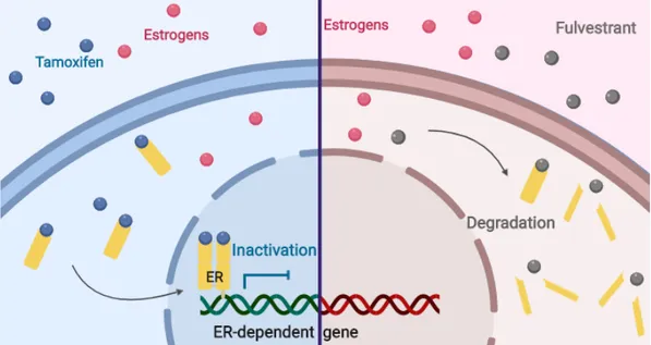
SERD are a class of ER antagonists that induce ER downregulation by selectively degrading the receptor. Fulvestrant (Faslodex or ICI182,780) is a high affinity competitive antagonist of ER promoting its proteasome-dependent degradation [71] and is now considered to be a first-in-class SERD for patients with metastatic ER+ breast cancer [72]. Unfortunately, fulvestrant has substantial pharmaceutical limits (e.g., requiring intramuscular injection) which negatively impact on its extensive usage [73]. More recently, the development of orally bioavailable SERD, some of which have been clinically evaluated in clinical trials in breast cancer patients who have progressed on standard ET, may provide the possibility of blocking ER signaling in advanced metastatic breast cancer [74].
Finally, AI reduce estrogen levels by preventing their peripheral synthesis through the inhibition of the aromatase enzyme, which catalyzes the conversion of androgens into estrogens. Three generations of AI have been developed with the third generation showing a more favorable tolerability and selectivity profile compared to first- and second-generation agents. Currently, AI represent the gold standard for the treatment of early and advanced ER+ breast cancer in postmenopausal women [75, 76].
3.1.1 Tamoxifen
Tamoxifen belongs to SERM and is a pioneering drug for the treatment of ER+ breast cancer. In particular, it is the most commonly used endocrine agent as first-line adjuvant therapy in premenopausal women with ER+ tumors [70]. Structurally, tamoxifen is a nonsteroidal triphenylethylene derivative anti-estrogen that acts by competing with estrogen and binding ER, thus blocking the downstream molecular signaling and reducing breast cancer cell proliferation [77] (Figure 7).
The anti-cancer activity of tamoxifen occurs via its active metabolites, 4-hydroxy-tamoxifen and 4-hydroxy-N-desmethyltamoxifen. The affinity of 4-hydroxy-tamoxifen for ER is comparable to that of E2 [78]. More specifically, the anti-tumoral effect of tamoxifen is mediated not only by the competitive inhibition of ER but also by inducing an ER conformational change that prevents the binding of coactivators and induces the recruitment of transcriptional corepressors [79]. Consequently, the transcription of estrogen-dependent genes is blocked resulting in the cell cycle arrest in G1 phase. Furthermore, it has been shown that tamoxifen may also directly induce apoptosis through the production of ROS, changes in cell membrane fluidity, and induction of mitochondrial permeability [80]. The active metabolite 4-hydroxy-tamoxifen has been reported to induce the accumulation of autophagic vacuoles leading to apoptosis, suggesting a possible role for autophagy in the regulation of 4-hydroxy-tamoxifen-induced cell
16 death [81]. Additionally, tamoxifen has been described to have inhibitory effect on the mitochondrial respiratory capacity [82, 83].
Depending on the tissue, tamoxifen can act both as estrogen agonist and antagonist. In particular, it acts as an ER antagonist in breast, while it exerts agonistic effect in other sites such as vascular system and endometrium [84], where it has been reported that tamoxifen promotes the expression of ER target genes that do not contain a classical ERE, such as c-Myc and IGF1 (Insulin-like Growth Factor 1), both implicated in the regulation of cell proliferation, survival, and malignant transformation [85]. This agonistic activity is related to serious side effects such as thromboembolic events and uterine cancer [86, 87]. Whether tamoxifen acts as antagonist or agonist of the ER signaling depends on the cellular context. As discussed in paragraph 1.2.2, ER contains two domains that regulate the transcriptional activation, AF-1 and AF-2 (Figure 3). Since ER activity in breast is mainly driven by the ligand-dependent AF-2 domain, tamoxifen acts largely as an antagonist. Conversely, in other organs such as uterus, ER activity is also controlled by the ligand-independent AF-1 domain, resulting in greater agonistic tamoxifen activity [88].
Compelling data have demonstrated a significant OS benefit in ER+ breast cancer patients, with the tamoxifen treatment resulting in a 51% reduction in tumor recurrence and a 28% reduction in death as well as in improved life quality for patients with metastatic disease [89]. Tamoxifen has also been shown to be effective in reducing the incidence of breast cancer in patients at risk for developing the disease [90] and in women with ductal carcinoma in situ [91].
Despite these recognized benefits, not all patients with ER expressing tumors respond to this endocrine agent (de novo resistance) and a substantial group of responsive patients experience disease progression or recurrence (acquired resistance). Most patients with metastatic disease and 40% of early-stage breast cancer patients treated with adjuvant tamoxifen relapse with a tamoxifen resistant disease [92]. The biological mechanisms underlying resistance remain still unclear, but several hypotheses have been formulated, including the loss of ER expression and function, altered expression patterns of coregulatory proteins, the crosstalk between ER and GF receptors, and cell cycle checkpoint alterations [93].
3.1.2 Fulvestrant
Although adjuvant ET is an effective treatment for ER+ breast cancer, the majority of patients with an advanced disease will most likely exhibit resistance to the individual therapy [94]. Moreover, an initial response to the first endocrine treatment is generally indicative of a positive response to further alternative endocrine agents [95]. Consequently, the therapeutic options for ER+ breast cancer have been extended beyond SERM.
Fulvestrant represents a possible addition to the variety of endocrine treatments for postmenopausal women with advanced ER+ breast cancer and is the most commonly used
17 second-line agent after prior therapy with tamoxifen or AI [96, 97]. Fulvestrant belongs to the class of SERD and is a steroidal anti-estrogen compound that exerts its anti-tumoral role by preventing the estrogen-ER interaction, thus abrogating the estrogen-regulated transcriptional activity. Specifically, fulvestrant impairs the receptor dimerization and the energy-dependent nucleus-cytoplasm shuttling, thereby blocking the nuclear translocation of ER. Moreover, the ER-fulvestrant complex is instable and susceptible to a rapid proteasome-dependent degradation [98] (Figure 7).
In contrast to tamoxifen, which exclusively blocks the AF-2 domain and also exhibits partial agonistic activity, fulvestrant is considered a “pure” anti-estrogen that displays a higher ER affinity [99, 100] and induces a conformational change of ER resulting in the impairment of both AF-2 and AF-1 related transcriptional activities. Therefore, fulvestrant exhibits full ER antagonism and no agonistic effects [101].
Fulvestrant shows similar efficacy to tamoxifen as first-line therapy in patients with advanced ER+ breast cancer and to the AI anastrozole as second-line therapy in patients whose disease has progressed on prior ET [102]. Moreover, as monotherapy, fulvestrant may have superior efficacy than AI in patients who have not received adjuvant ET and in patients with inoperable locally or advanced breast cancer. However, despite clinical benefits, resistance to fulvestrant also frequently occurs [103]. Recent studies have shown numerous molecular mechanisms involved in fulvestrant resistance such as PIK3CA (phosphatidylinositol-4,5-bisphosphate 3-kinase α) and ESR1 mutations [104]. The main side effects related to fulvestrant are nausea, asthenia, pain, vasodilatation, and headache [96].
18
3.1.3 Aromatase inhibitors
AI administration is restricted to women without functional ovaries. Indeed, in premenopausal patients, AI ineffectively repress estrogen production [105] and should not be used alone. Conversely, in postmenopausal women, AI significantly decrease estrogen levels by blocking their peripheral synthesis. This is due to the fact that in premenopause the major estrogen source is the ovarian production, whereas after menopause estrogens derive exclusively from non-glandular tissues, in particular from the subcutaneous fat.
Aromatase is a member of the cytochrome P450 superfamily, encoded by CYP19A1 gene located on the chromosome 15, and catalyzes the rate-limiting final step of the estrogen synthesis that is the conversion of androgenic precursors (i.e., androstenedione and testosterone) to estrogens (Figure 8). Aromatase expression is elevated in ovarian granulosa cells and depends on the cyclical gonadotropin stimulation. It is also present in several extra-gonadal tissues, including subcutaneous fat, liver, muscle, brain, bones, vascular endothelium, and normal breast adipose tissue [106]. Furthermore, it has been demonstrated that aromatase expression and activity are significantly increased in malignant compared to normal breast tissue, resulting in a higher intratumoral estrogen concentration [107, 108].
Blocking the conversion of androgen into estrogen, AI deprive tumor of estrogen thus reducing the ER signaling and causing a significant reduction of the estrogen-dependent proliferation in cancer cells [109] (Figure 8).
AI are classified into three generations according to their time of development and entry into the clinical setting, efficacy and mechanisms of action. The first- (e.g., aminoglutethimide) and second-generation AI (e.g., fadrozole and vorozole) were less selective and they also decreased aldosterone and cortisol production. The utility of these compounds was limited due to poor tolerability and adverse effects resulting in inadequate clinical efficacy [110]. Third generation AI are highly selective for the aromatase enzyme and well tolerated from patients showing only minor side effects. Two classes of third generation AI have been described: the nonsteroidal inhibitors (e.g., anastrozole and letrozole) that reversibly bind aromatase and the steroidal AI (e.g., exemestane) that irreversibly inhibit the enzyme [111].
Third generation orally available AI have now surpassed tamoxifen as first-line adjuvant therapy for postmenopausal women with metastatic ER+ breast cancer [112], showing significantly greater disease free survival (DFS), lower metastatic recurrence, and reduced incidence of contralateral breast cancer. Indeed, AI have no (partial) agonist activity thus reducing adverse effects as thromboembolic events, vaginal bleeding, and endometrial cancer [113]. However, despite clinical benefits, resistance emerges in approximately 50% of adjuvant treated patients and is inevitable in metastatic breast cancer [114].
19 Figure 8. Aromatase inhibitor mechanism of action.
3.2
Cyclin-dependent kinases 4/6 inhibitors
Despite considerable advances in treating ER+ breast cancer patients with ET, resistance still remains the major cause of morbidity and mortality ultimately raising the need for new therapeutic approaches. The uncontrolled cell proliferation is an established hallmark of cancer [115] and the detailed understanding of cell cycle regulation and imbalance has contributed to the development of innovative and promising anti-cancer therapies. In particular, the dysregulation of the cyclin D1-CDK4/6-Rb axis signaling drives the phosphorylation of tumor suppressors and transcription factors ultimately contributing to a cell cycle unchecked progression [116]. In consideration of this pivotal role, extensive studies have been conducted to evaluate CDK4/6 as effective targets in cancer, especially in breast cancer.
The opening results with the first generation CDK inhibitors (e.g., flavopiridol, R-roscovitine, and UCN-01) were mostly disappointing due to their broad specificity in blocking a consistent number of CDK (pan-CDK inhibitors) and inadequate clinical activity observed in vivo resulting in poor efficacy, significant toxicity, and insuperable adverse effects [117]. Furthermore, the lack of applicable patient selection and the absence of predictive biomarkers may also have impacted on the initial failure. Flavopiridol is the most extensively investigated first generation CDK inhibitor that is not specific for a single CDK while acts as a pan-CDK inhibitor showing repressive effect on CDK1, CDK2, CDK4, CDK6, CDK7, and CDK9 [118, 119]. This compound not only exerts a cytostatic effect mediated by the cell cycle inhibition but also causes apoptosis, autophagy, transcriptional repression, and endoplasmic reticulum stress [120-122]. Phase I and II studies enrolling flavopiridol have shown low efficacy and off-target effects in healthy tissues providing severe toxicities typical of cytotoxic agents such as neutropenia, hyperglycemia, gastrointestinal, cardiac, and pulmonary disfunction [123, 124].
20 The second generation class showed superior specificity for preferential CDK with little or no suppression of other kinases and randomized clinical trials have validated that inhibitors highly selective for CDK4/6 are effective for the treatment of ER+/HER2- locally advanced or metastatic breast cancer in combination with ET [125]. The use of the CDK4/6 inhibitors has become common for this subset of patients and will certainly increase in the future.
Three CDK4/6 inhibitors, palbociclib (PD0332991, Ibrance, Pfizer), ribociclib (LEE011, Kisqali, Novartis), abemaciclib (LY2835219, Verzenio, Lilly), have received the approval by Food and Drug Administration (FDA) and European Medicines Agency (EMA) and are currently used in the clinical management of ER+/HER2- breast cancer patients. More specifically, palbociclib and ribociclib have a similar prescription. They are used as first-line therapy in postmenopausal patients with ER+/HER2- locally advanced or metastatic breast cancer in combination with an AI or in women previously treated with ET in combination with fulvestrant [FDA. Ibrance
Prescribing Information. Available from:
https://www.accessdata.fda.gov/drugsatfda_docs/label/2017/207103s004lbl.pdf; FDA. Kisqali
Prescribing Information. Available from:
https://www.accessdata.fda.gov/drugsatfda_docs/label/2017/209092s000lbl.pdf]. Abemaciclib is the only CDK4/6 inhibitor that can be administered as monotherapy in patients with ER+/HER2- metastatic breast cancer previously treated with ET and chemotherapy as well as in combination with fulvestrant as second-line therapy in women with ER+/HER2- advanced or metastatic breast cancer [FDA. Verzenio Prescribing Information. Available from: https://www.accessdata.fda.gov/drugsatfda_docs/label/2017/208716s000lbl.pdf]. All are orally administered and act as selective ATP competitive inhibitors of CDK4 and CDK6, exhibiting little or no effect on other CDK.
Structurally, palbociclib and ribociclib are analogous [126] and show high specificity for CDK4 with an IC50 (half maximal inhibitory concentration) of 11 nM and for CDK6 with an IC50 of 16 nM [127, 128]. Abemaciclib has a different structure and a higher CDK4/6 binding activity than the other two drugs, with an IC50 of 2 and 10 nM for CDK4 and CDK6, respectively [129]. Moreover, palbociclib should be taken concomitantly with food because its effectiveness may be reduced with an empty stomach [130]; conversely, the exposure of ribociclib and abemaciclib is not influenced by food intake. More consideration will be reserved in the next section to palbociclib that is the CDK4/6 inhibitor used in the thesis project.
Several randomized clinical trials have proved efficacy and safety of these CDK4/6 inhibitors and favored their approval. Ribociclib is the second CDK4/6 inhibitor that was approved based on the results of the phase III studies MONALEESA-2 and MONALEESA-3 comparing the combination of ribociclib plus letrozole and ribociclib plus fulvestrant versus placebo plus letrozole or fulvestrant alone, respectively [131, 132], and showing a significantly high progression free survival (PFS) in the combination groups. More recently, the phase III clinical trial MONALEESA-7 revealed a considerable prolongation in both PFS and OS in
21 premenopausal patients with ER+/HER2- breast cancer receiving a combination of ribociclib, ET, and goserelin [133]. Ribociclib is recommended at a dose of 600 mg/day on a 3/1 schedule (3 weeks on/1 week off) and dose reduction is allowed if resistance does not occur [131]. Abemaciclib, the third CDK4/6 inhibitor approved, has been designed as monotherapy based on the outcome from the MONARCH-1 study showing a clinical benefit of the single agent in patients with pretreated refractory metastatic ER+ breast cancer [134]. In addition, worldwide phase III studies MONARCH-3 and MONARCH-2 evaluated the combination of abemaciclib with letrozole and fulvestrant in patients with ER+/HER2- metastatic breast cancer as first- and second-line therapy respectively, demonstrating a significantly improved PFS [135, 136]. Abemaciclib is administrated continuously twice daily at 150 mg if combined and at 200 mg as monotherapy [137].
Despite efficacy and tolerability shown by these drugs, they are not immune to side effects such as neutropenia that is the most dangerous [138-141]; therefore, two next-generation CDK4/6 inhibitors (i.e., G1T28 and G1T38) associated with lesser myelosuppression have been developed and are currently tested in clinical trials [142, 143].
3.2.1 Palbociclib
Palbociclib is the first highly selective ATP competitive CDK4/6 inhibitor synthesized and approved by FDA and EMA for the treatment of ER+/HER2- locally advanced or metastatic breast cancer in combination with AI in postmenopausal women as first-line therapy and in combination with fulvestrant in patients that were previously treated with ET [FDA. Ibrance
Prescribing Information. Available from:
https://www.accessdata.fda.gov/drugsatfda_docs/label/2017/207103s004lbl.pdf]. Palbociclib has been also approved by FDA for the treatment of ER+/HER2- metastatic male breast cancer [144]. The recommended dosing regimen consists of 125 mg/day of orally administrated drug on a 3/1 schedule (3 weeks on/1 week off) in combination with letrozole (2.5 mg/day) or fulvestrant (500 mg/month intramuscularly injected) and, if resistance does not occur, doses can be reduced to 100 or up to 75 mg/day [FDA. Ibrance Prescribing Information. Available from: https://www.accessdata.fda.gov/drugsatfda_docs/label/2017/207103s004lbl.pdf].
ER+ breast cancers are characterized by high expression of the ER-dependent gene cyclin D1 (CCND1) that mediates cell cycle entry through CDK4/6 by the phosphorylation and subsequent inactivation of Rb that uncouples from E2F. The release of this transcriptional factor promotes the transcription of genes involved in the G1/S checkpoint. Therefore, palbociclib inhibition of CDK4/6 decreases the E2F-dependent gene expression that regulates mitotic entry, thus suppressing DNA replication and causing a cell cycle arrest in the G1 phase [145, 146] (Figure 9). Consequently, the biological function and anti-tumoral effect of this drug are limited by the presence of an active Rb protein [127].
22 Palbociclib does not exert any effects on apoptosis [147] according to its cytostatic action. Additionally, palbociclib inactivates the transcription factor FOXM1 which is involved in the induction of cell division [57] and, besides the well characterized anti-proliferative effect, it has been shown to promote epithelial-mesenchymal transition (EMT) and tumor invasion [148].
Figure 9. Mechanism of action of CDK4/6 inhibitors (taken from [149]).
Palbociclib anti-tumoral effect has been validated in several human cancers such as hepatocellular carcinoma [146], neuroblastoma [150], pancreatic ductal adenocarcinoma [148], melanoma [151], and especially breast cancer [152]. Three pivotal randomized clinical trials have allowed palbociclib entry into the clinic. PALOMA-1 and PALOMA-2 are phase II and phase III trials that proved efficacy and safety of palbociclib in combination with letrozole versus letrozole alone as firs-line therapy in the postmenopausal metastatic setting, showing a significant prolongation in the PFS [138, 153]. PALOMA-3 is a phase III study that investigated the combination of palbociclib plus fulvestrant versus fulvestrant alone in pre and postmenopausal women with disease progression during or after prior hormonal therapy and exhibited improved efficacy over ET alone [154].
The clinical efficacy of palbociclib has been explored also in the neo-adjuvant setting and in HER2+ breast cancer. NeoPalAna is a single-arm phase II study testing the combination of anastrozole plus palbociclib in stage II/III ER+ breast cancer. The trial showed a significantly higher complete cell cycle arrest (CCCA) than monotherapy [155]. The phase II trial NA-PHER2 investigated the combination of palbociclib, fulvestrant, and trastuzumab in the neo-adjuvant setting in women with ER+/HER2+ breast cancer and showed a complete pathological response in the combination arm [156]. The combination of palbociclib plus trastuzumab has already demonstrated a synergistic effect in human breast cancer cell lines [157] and both CDK4 and cyclin D1 are required for murine breast cancer growth [158-160]. Therefore, further

![Figure 9. Mechanism of action of CDK4/6 inhibitors (taken from [149]).](https://thumb-eu.123doks.com/thumbv2/123dokorg/4631154.41010/30.892.217.714.234.550/figure-mechanism-action-cdk-inhibitors-taken.webp)
![Figure 10. Schematic representation of the principal molecular mechanisms underlying endocrine resistance in ER+ breast cancer (taken from [165])](https://thumb-eu.123doks.com/thumbv2/123dokorg/4631154.41010/32.892.261.657.480.865/schematic-representation-principal-molecular-mechanisms-underlying-endocrine-resistance.webp)
![Figure 12. Cell cycle-related mechanisms of resistance to CDK4/6 inhibitors (taken from [212])](https://thumb-eu.123doks.com/thumbv2/123dokorg/4631154.41010/36.892.108.811.656.1103/figure-cell-cycle-related-mechanisms-resistance-inhibitors-taken.webp)
![Figure 14. The hallmarks of cancer: the next generation (adapted from [229]).](https://thumb-eu.123doks.com/thumbv2/123dokorg/4631154.41010/39.892.239.682.748.1112/figure-hallmarks-cancer-generation-adapted.webp)
![Figure 11. Metabolic alterations in hormone-dependent and -independent breast cancer (taken from [332])](https://thumb-eu.123doks.com/thumbv2/123dokorg/4631154.41010/48.892.115.854.230.577/figure-metabolic-alterations-hormone-dependent-independent-breast-cancer.webp)
![Figure 13. Metabolic plasticity in ER+ breast cancer resistant to ET (taken from [353])](https://thumb-eu.123doks.com/thumbv2/123dokorg/4631154.41010/51.892.225.690.91.555/figure-metabolic-plasticity-er-breast-cancer-resistant-taken.webp)