research article
The introduction of alternative implant surfaces to the well known and experimented turned surface,
improperly called “smooth” has been motivated by better biological responses which the “rough” sur-faces seemed to produce, especially in a bone of poor quality and/or associated to regenerative ther-apies (Hansson and Norton, 1999; Cooper, 2000; Hansson, 2000; Van Stenberghe et al., 2000; Rocci
C
OMPARING THE
T
I
O
BLAST AND
O
SSEOSPEED SURFACES
.
H
ISTOMORPHOMETRIC AND
HISTOLOGICAL ANALYSIS IN HUMANS
M. ROCCI*, A. ROCCI*, M. MARTIGNONI**, T. ALBREKTSSON***,
A. BARLATTANI****, M. GARGARI****
* Private practice in Chieti, ** Private practice in Rome *** MD, PhD, Odhc
**** Tor Vergata University, Rome
SUMMARY
Comparing the TiOblast and Osseospeed surfaces. Histomorphometric and histological analysis in hu-mans
The aim of the present study was to compare two implant surfaces, the TiOblast (Astra Tech) surface, manufactured by blasting the surface and already present in literature and the Osseospeed (Astra Tech) surface, manufactured by blasting and treating the surface with fluoride ions and re-cently launched onto the market with the modified surfaces of the latest generation. This study is part of a more exten-sive research project whose protocol required the insertion of 10 couples of implants; thus in the present discussion partial data are being taken into consideration, with an eye at collecting more data in the future, regarding both mi-croscopy and histomorphometric histological analysis on 5 couples of implants. The purpose of the study is to investi-gate how the modified surfaces of the latest generation can guarantee a greater osseointegration both from a qualitative and quantitative level compared to the surfaces presently used and that they may represent the first example of “bioactivity”, that is, an active interaction with the processes of new bone formation and tissue healing.
Key words: sandblasted surface, fluoride, histology, histo-morphometry, microthreads, macrothreads.
RIASSUNTO
Comparazione tra la superficie TiOblast e la superficie Osseospeed. Analisi istologica ed istomorfometrica nell’umano
Il presente studio è finalizzato alla comparazione tra due superfici implantari, la superficie TiOblast (Astra Tech) ot-tenuta per sabbiatura e già presente in letteratura e la su-perficie Osseospeed(Astra Tech), ottenuta per sabbiatura ed implementazione con fluoro in forma ionica, di recente introduzione sul mercato nell’ambito delle superfici modifi-cate di ultima generazione. Questo studio è parte di un progetto di ricerca più ampio, il cui protocollo ha previsto l’inserimento di 10 coppie di impianti; pertanto nella pre-sente discussione vengono presi in considerazione dati parziali, che ci riserviamo di ampliare, riguardanti l’analisi istologica al microscopio ottico ed istomorfometrica su 5 coppie di impianti. L’obiettivo della ricerca è quello di veri-ficare che le superfici modificate di nuova generazione possano garantire un’osteointegrazione qualitativamente e quantitativamente maggiore rispetto alle superfici attual-mente in uso e che dunque possano rappresentare un pri-mo esempio di “bioattività”, cioè di interazione attiva con i processi di neoformazione ossea e guarigione tissutale.
Parole chiave: superficie sabbiata, fluoro, istologia, isto-morfometria, microspire, macrospire.
Background and literature’s
review
©
CIC
EDIZIONI
research article
et al., 2005). Even though histological tests on hu-mans are not numerous in literature, they have con-firmed that there is a bigger integration which is ex-pressed in bone-implant contact (BIC) percentage values greater than those obtainable with the turned surfaces (Albrektsson et al., 1993; Ivanoff et al., 2001; Rocci et al., 2002: Ivanoff et al., 2003; Rocci et al., 2003; Schüpbach et al., 2005). This is the rea-son why different surface typologies have come to light with a modified microtopography as an evolu-tion of the Brånemark turned surface, achieved by using different manufacturer’s methodologies. Grouping these surfaces according to the methods used for manufacturing them in order to have those “blasted” characteristics, it can all be narrowed down to two main groups: surfaces roughend either by subtraction or by addiction. The subtraction of a minimum amount of titanium from the surface of an implant can be made by chemical means (acid etch-ing), physical means (blasting or shot peening) or by a combination of the two. The adding techniques see the addition of materials of various nature to the titanium surface, such as hydroxylapatite, titanium dioxide in a plasma-spray form and the titanium dioxide obtained with an anodic oxidation.
Another possible classification is that which takes into consideration the microtopography as a dis-criminatory parameter, that is the surface geometry of the implant on a micrometric level; following this concept, both Albrektsson and Wennerberg classify the implants as follows: smooth, minimally rough, moderately rough and rough based on the surface area or Sa value. Smooth implants are the ones with an Sa value inferior to 0.5 µm: surfaces having these characteristics are those of the healing abutments, with values ranging between 0.1 and 0.3 µm. Implants minimally rough show an Savalue within 0.5 and 1.0 µm and are represented by the Bråne-mark and Astra Tech turned fixtures and those 3i acid-etched. Implants moderately rough all have Sa values ranging between 1.0 and 2.0 µm and practi-cally include all modern implants, such as Astra Tech TiOblast™ and Osseospeed™, Nobel Biocare TiUnite™, Straumann SLA and Dentsply Cellplus. Rough implants are the ones with Savalues superi-or then 2.0 µm, represented by surfaces treated with plasma-spray, and among the modern implants
Dentsply Frialit-2 (Albrektsson and Wennerberg, 2004a, 2004b).
The present study has focused its investigation on the implants already equipped with two of the sur-faces above mentioned: TiOblast™ and Os-seospeed™, manufactured by Astra Tech Dental AB, Mölndal, Sweden.
The Tioblast surface is manufactured with a physi-cal subtractive procedure, or rather by sandblasting with spheric particles (shot peening) of titanium dioxide the surface, under controlled conditions and with no possibility of contamination. The surface structure is characterized by a well defined topogra-phy with a high density of pit of optimized dimen-sion which shows an Savalue of 1.1 µm. A further development of the TiOblast surface is represented by the Osseospeed surface, which is obtained as well as with the sandblasting procedure also with a procedure of chemical type: fluoridation. The titani-um surface once sandblasted, is treated with fluoride ions. In vivo tests have demonstrated that the pres-ence of dioxide titanium with a negative charge, favours the deposition of calcium ions onto the im-plant, which in turn show a great affinity with the phosphate groups contained in many organical mol-ecules (proteins, glycans, etc.). the presence of fluo-ride ions on the implant surface facilitate and strengthen such biological mechanisms; fluoride as a matter of fact (being highly electronegative), in-creases the speed of sedimentation of the calcium ions and causes an increase in the density of the bone trabecular structure, by stimulating the activity of the osteoprogenitor cells and the alkaline phop-shatase, too. It is demonstrated, in vitro, the presence of weak secondary bonds between calcium ions and groups of phosphate on a TiOblast surface; whilst such bonds become of a strong covalent type if the surface itself is coated with fluoride ions which are released in the surrounding space following the es-tablishment of such bond (Ellingsen et al., 2000).
Objectives
The objective of the present study is to analyse with a light microscope the potential differences
©
CIC
EDIZIONI
of the bone response, in humans, comparing a sandblasted implant surface (TiOblast) and a sand-blasted surface implemented with fluoride ions (Osseospeed).
Materials and methods
It has been used in the present study implants com-mercially available on the market but of a smaller dimension (3.5 × 8.0 mm) with a cylindrical profile, screw like morphology and self-tapping, with a de-veloped macrothread on the top up to about 3 mm from the coronal end: this portion has a microthread part, instead. Half of the implants had a micro-rough surface obtained by sandblasting the surface with titanium dioxide particle (TiOblast), the other half had the same surface but enriched with fluoride ions (Osseospeed). The sample of the candidates taking part in the study was made up by 7 male pa-tients with an age ranging between 40 and 68 years old (mean 60.4), who did not suffer of any patholo-gies controindicating surgery, nonsmokers and parafunctional free.
The study protocol planned the insertion of one or two couples of implants one next to the other, one for each surface typology, in the inferior maxillary areas edentulous for at least 3 months, within the insertion procedure of placing the implants for the intended rehabilitation. After eight weeks of heal-ing, during the second stage surgery, the biopsies of the fixtures for the study were retrieved togeth-er with the ptogeth-erimplant bone tissue by coring with a trephine. The implants, retrieved with the sur-rounding tissues were immediately placed in a 4% formaldehyde solution and sent to the Department of Biomaterials of the University of Gothenburg, Sweden on the same day. All samples have been dealt with according to the directions of the De-partment for the histologic procedure of the unde-calcified specimens. For this purpose was used the Donath’s tecnique of cutting and abrasion (1993) using the Exact system (Exact Apparatebau Co, Nordstedt, Germany). The sections thus obtained were further ground to a 10-15 µm thickness in different phases, and dyed in toluidine blue O and pyronine G. Histologic analysis were done with a
light microscope (Eclipse 600 Nikon, Japan) and the hystomorphometric evaluations processed with an image analysis software. Evaluations for each section were performed according to the per-centage of bone-implant contact (BIC), in a “blind” manner, with the analyzer not knowing the type of surface being analyzed.
Results
The histologic and hystomorphometric analysis was carried out on five couples of implants which were just a part of the couples considered in the original study protocol. The observation in a light microscope at a low magnification, showed the close contact between the implants’ surfaces and the bone tissue (Figs. 1 and 2).
research article
Figure 2
Hystologic section of an Osseospeed implant.
Figure 1
Hystologic section of a TiOblast implant.
©
CIC
EDIZIONI
research article
By increasing the magnifications, it was obvious to note the integration process that took place due to both the growth of the osteotomy walls (dis-tant osteogenesis) and to the direct apposition on the implant surfaces (contact osteogenesis) (Figs. 3 and 4). Distant osteogenesis was characterised by bone apposition on the wound margins in the pre-existing bone, contact osteogenesis often ap-peared as thin linear zones which followed the threads profile.
Cell migration signs from the marrow tissue to-wards the mineralization front could be identified with larger magnifications, thus indicating recruit-ment and differentiation of the immature cells
from pre-osteoblasts to osteoblasts (Figs. 5 and 6). Haversian systems were identifiable, too.
The bone-implant contact percentage analysis was carried out separately for the two different implant macrogeomtries: the coronal portion (microthread) and the apical one (macrothread). As far as the bone response pertains the zone with the micro-thread, results are shown in tables 1 and 2 with BIC mean values equal to 24.6% for the TiOblast and 34.6% for the Osseospeed (Fig. 7); in three cases the Osseospeed surface has shown a response numerically strong and in two cases the TiOblast surface proved superior (Fig. 8).
Figure 4
Hystologic section of a TiOblast implant portion.
Figure 3
Hystologic section of a microthreaded Osseospeed implant portion.
Figure 6
Hystologic section of a microthreaded Osseospeed implant portion.
Figure 5
Hystologic section of a TiOblast implant portion.
©
CIC
EDIZIONI
research article
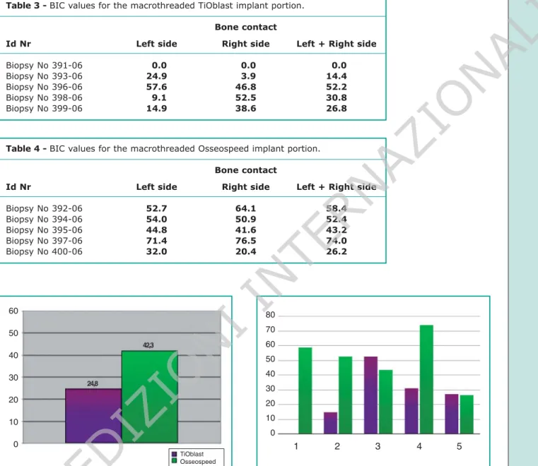
Hystomorphometric data concerning the apical parts of implants are shown in Tables 3 and 4. The TiOblast surface has shown a BIC median of 24.8% as the Osseospeed surface a BIC median of 48.3% (Fig. 9). In three cases the Osseospeed sur-face has shown a response decisevely higher, in two cases the TiOblast surface has proved slightly superior (Fig. 10).
Discussion
Within the limits of this study it is possible to con-firm what is already in literature about the good performance of modified microtopography sur-faces, in terms of bone tissue integration, as previ-ously mentioned. The two surfaces in question, Table 1 - Values for the microthreaded TiOblast implant portion.
Bone contact
Id Nr Left side Right side Left + Right side
Biopsy No 391-06 13.1 10.0 16.6
Biopsy No 393-06 13.5 14.1 13.8
Biopsy No 396-06 14.4 43.4 28.9
Biopsy No 398-06 67.0 57.4 62.2
Biopsy No 399-06 43.1 10.0 21.6
Table 2 - BIC values for the nine microthreaded Osseospeed implant portions. Bone contact
Id Nr Left side Right side Left + Right side
Biopsy No 392-06 12.2 14.8 13.5 Biopsy No 394-06 22.8 50.8 36.8 Biopsy No 395-06 54.8 23.8 39.3 Biopsy No 397-06 36.8 46.7 41.8 Biopsy No 400-06 43.3 60.0 51.7 Figure 7
BIC values for the microthreaded portion of each couple of implants.
Figure 8
Mean BIC values for the portion of microthreaded implants. 60 50 40 30 20 10 0 80 70 60 50 40 30 20 10 0 TiOblast Osseospeed 1 2 3 4 5
©
CIC
EDIZIONI
INTERNAZIONALI
research article
have expressed to a microscopic level, distant and contact new bone apposition (osteogenesis). Ob-servation with the light microscope would confirm that the Osseospeed implants show a bigger bone deposition, quantitatively-wise, in contact with the surface. Despite the fact that the study has been carried out on a small number of samples, the analysis performed on the mean BIC values for the
coronal portion of the implants (microthread), a difference numerically inferior has been noted be-tween the two surfaces compared to that achieved between the mean BIC values for the macrothread portion. According to this it can be assumed that – nonetheless the small number of samples – there are biological mechanisms which could explain the homogeneity of the results at the interface. The Table 3 - BIC values for the macrothreaded TiOblast implant portion.
Bone contact
Id Nr Left side Right side Left + Right side
Biopsy No 391-06 10.0 10.0 10.0
Biopsy No 393-06 24.9 13.9 14.4
Biopsy No 396-06 57.6 46.8 52.2
Biopsy No 398-06 19.1 52.5 30.8
Biopsy No 399-06 14.9 38.6 26.8
Table 4 - BIC values for the macrothreaded Osseospeed implant portion. Bone contact
Id Nr Left side Right side Left + Right side
Biopsy No 392-06 52.7 64.1 58.4 Biopsy No 394-06 54.0 50.9 52.4 Biopsy No 395-06 44.8 41.6 43.2 Biopsy No 397-06 71.4 76.5 74.0 Biopsy No 400-06 32.0 20.4 26.2 Figure 9
BIC values for the macrothreaded portion of each couple of implants.
Figure 10
Mean BIC values for the macrothreaded portion of implants. 60 50 40 30 20 10 0 80 70 60 50 40 30 20 10 0 TiOblast Osseospeed 1 2 3 4 5
©
CIC
EDIZIONI
INTERNAZIONALI
coronal portion of the implant is in contact with bone of a cortical kind, which both for the histo-logical characteristics and for the position in the surgical site, shows healing mechanisms of its own different from the ones which affect the medullar compartment. Cortical bone is charac-terised by a dense cancellous structure and makes the tissue particularly hard and little elastic; to these characteristics it can be added a scarce cellu-larity and very little vascularization all elements which favor slower healing processes. This phe-nomenon is enhanced by the mechanical stress which the cortical bone undergoes both during site preparation for surgery and while inserting the fix-ture; in biological terms the postinsertion bone re-modelling will be greater at the interface. Further-more, both a scarce vascularization and a scarce cellularity, delay the start of the early healing phases even because osteoblasts are cells inca-pable to migrate and replicate autonomously. Os-teoblast cells activate themselves as a response to the presence of precursor cells deputed to osteoge-nesis (DOPC, Determined Osteogenic Precursor Cells) (Friedestein, 1973) and are commonly lo-cated in proximity of the blood vessels and close to the bone surface. Bearing this picture in mind, it is necessary to remember that the role fluoride plays is fundamental during the early phase of the healing process as being highly electronegative and enhances the initial Calcium ion deposition on the implant’s surface more rapidly and thus guar-anteeing the formation of a strong covalent bond between the titanium and calcium dioxide. By forming this bond fluoride enters in solution and its action is exhausted. Due to the precociousness of this mechanism and considering the longer times in which the peri-implant cortical rstorative processes take place, it can be assumed that fluo-ride does not play a role such as to greatly affect the different times of osteointegration in the coro-nal portion of the implant. What appears interest-ing is the analysis performed on the BIC values for the apical portion (macrothread). The difference between the mean values was decisively higher (the mean BIC values for the test surface have val-ues almost the double compared to the valval-ues of the control surfaces), therefore it can be assumed
that the role of fluoride is probably more relevant in this area. The cancellous bone tissue is highly cellular and vascular compared to the compact one, and is characterized by a bigger trabecular meshwork structure that gives the tissue a higher elasticity.
Considering the fact that the mechanical stress in the apical portion of the bone is definitely smaller both during the preparation of the implant site and the insertion of the fixture, and the histological characteristics confer a greater capacity to endure stress, it can be easily understood how the neo bone apposition processes can start faster and therefore the fluoride presence might positevely influence the healing times and thus those of the osteointegration.
Conclusions
Considering all the limits this study has, and in a short while it will be completed by analysing five more pairs of Tioblast and Osseospeed implants, preliminary results achieved allow us to make hy-pothesis and reasonings susceptible of future con-firmation.
The most interesting hypothesis could be that by implementing the implant surfaces with fluoride, represents a first significant step towards making bioactive prosthesis devices and not just biocom-patible or “biotolerated”.
It seems evident, should the hypothesis drawn from the data produced in this study be confirmed in the future, that there will most probably be im-portant clinical implications concerning the reduc-tion in time’s procedures for the funcreduc-tionalizareduc-tion and immediate rehabilitation of those situations of poor bone quality (D3 and D4) and of biologic challenge such as bone grafting procedures.
References
11. Adell R, Lekholm U, Rockler B, Brånemark PI. A 15-year study of osseointegrated implants in the treat-ment of the edentulous jaw. Int J Oral Surg 1981; 10: 387-416.
research article
©
CIC
EDIZIONI
research article
12. Adell R, Eriksson B, Lekholm U, et al. A long-term follow-up study of osseointegrated implants in the treatment of totally edentulous jaws. Int J Oral Maxil-lofac Implants 1990; 5:347-359.
13. Albrektsson T, Eriksson AR, Friberg B, Lekholm U, Lindahl L, Nevins M, Oikarinen V, Roos J, Sennerby L, Astrand P. Histologic investigations on 33 retrieved Nobelpharma implants. Clin Mater 1993; 12(1):1-9.
14. Albrektsson T e Wennerberg A. Oral implant surfaces: Part 1-review focusing on topografic and chemical properties of different surfaces and in vivo responses to them. Int J Prosthodont 2004a; 17(5):536-543.
15. Albrektsson T e Wennerberg A. Oral implant surfaces: Part 2-review focusing on clinical knowledge of dif-ferent surfaces. Int J Prosthodont 2004b; 17(5): 544-564.
16. Brånemark PI, Breine U, Adell R, Hansson BO, Ohls-son A. Intraosseous anchorage of dental prostheses. Part I: experimental studies. Scand J Plast Reconstr Surg Hand Surg 1969; 3:81-100.
17. Brånemark PI, Hansson BO, Adell R et al. Osseointe-grated implants in the treatment of the edentulous jaw. Experience from a 10-year study period. Scand J Plast Reconstru Surg 1977; 16:1-132.
18. Carlsson LV, Albrektsson T, Jacobsson CM, MacDo-nald W. Osseointegration of a surface engineered or-thopaedic implant. Applied Osseointegration Re-search 2006; 5:45-49.
19. Cooper L, Masuda T, Whitson SW, Yliheikkilä P, Fel-ton D. Formation of mineralizing osteoblast cultures on machined, Titanium oxide grit-blasted and plasma-sprayed Titanium surfaces. Int J Oral Maxillofac Im-plants 1999; 14: 37-47.
10. Cooper L. A role for surface topography in creating and maintaining bone at titanium endosseous im-plants. J Prosthet Dent 2000 Nov; 84(5):522-34. 11. Davies JED. Mechanisms of endosseous integration.
Int J Prosthodont 1998; 11:391-401.
12. Donath K. Preparation of histologic sections by cut-ting-grinding technique for hard tissue and other ma-terials not suitable to be sectioned by routine me-thods. Morderstedt: Exakt-Kulzer publications 1993; 1-16.
13. Ellingsen JE. Pre-treatment of Titanium implants with Fluoride improves their retention in bone. J Mat Sci-ence Mat Medicine 1995; 6:749-753.
14. Ellingsen JE. On the properties of surface-modified Titanium. In: Davies JE (ed). Bone engineering; em Squared Inc, Toronto, Canada. 2000; 183-189. 15. Ellingsen JE, Johansson CB, Wennerberg A, Holmén
A. Improved retention and bone-to-implant contact with Fluoride-modified Titanium implants. Int J Oral & Maxillofac Implants 2004; 19:659-666.
16. Ellingsen JE. The development of a bone regenerati-on promoting implant surface. Applied Osseointegra-tion Research 2006; 5:18-23.
17. Esposito M, Hirsch JM, Lekholm U, et al. Biological factors contributing to failures of osseointegrated im-plants. (I). Success criteria and epidemiology. Eur J Oral Sci 1998; 106:527-551.
18. Esposito M, Hirsch JM, Lekholm U, et al. Biological factors contributing to failures of osseointegrated im-plants. (II). Ethiopatogenesis. Eur J Oral Sci 1998; 106:721-764.
19. Esposito M, Worthington HV, Coulthard P, Thomsen P. Maintaining and re-establishing health around os-seointegrated oral implants: a Cochrane systematic review comparing the efficacy of various treatments. Periodontal 2003; 33: 204-12
20. Farley SM, Libanati CR, Mariano-Menez MR, Tud-tud-Hans LA, Schulz EE, Baylink DJ. Fluoride thera-py for osteoporosis promotes a progressive increase in spinal bone density. J Bone Miner Res 1990 Mar; 5 Suppl 1:S37-42.
21. Friedenstein AJ. Determined and inducible osteogenic precursor cells. In: Hand Tissue Growth Repair and Remineralisation. Aba Foundation Symposium 1973; 11: 169-181.
22. Gotfredsen K, Nimb L, Hjörting-Hansen E et al. Hi-stomorphometric and removal torque analysis for TiO2-blasted Titanium implants. An experimental study on dogs. Clin Oral Impl Res 1992; 3:77-84. 23. Hansson S, Norton M. The relation between surface
roughness and interfacial shear strength for bone-an-chored implants. A mathematical model. J Biomecha-nics 1999; 32:829-836.
24. Hansson S. Surface roughness parameters as predic-tors of anchorage strength in bone: a critical analysis. J Biomechanics 2000; 33:1297-1303.
25. Ivanoff C-J, Hallgren C, Widmark G et al. Histologic evaluation of bone integration of TiO2-blasted and turned titanium microimplants in humans. Clin Oral Impl Res 2001; 12:128-134.
26. Ivanoff C-J, Widmark G, Johansson c, Wennerberg A. Histologic evaluation of bone response to oxidi-zed and turned titanium micro-implants in human jawbone. Int J Oral Maxillofac Implants 2003; 18:341-348.
27. Lazzara RJ, Testori T, Trisi P, Porter S, Weinstein RL. A human histologic analysis of Osseotite and machi-ned surfaces using implants with two opposing surfa-ces. Int J Periodontics Restorative Dent 1999; 19:117-129.
28. Piattelli A, Scarano A, Piattelli M. Histologic obser-vations on 230 retrieved dental implants: 8 years’ ex-perience (1989-1996). J Periodontol 1998; 69(2): 178-184.
29. Rasmusson L, Kahnberg K-E, Tan A. Effects of plant design and surface on bone regeneration and im-plant stability: an experimental study in the dog man-dible. Clin Impl Den Rel Res 2001; 1:2-8.
30. Rasmusson L, Roos J, Bystedt H. A 10-year follow-up
©
CIC
EDIZIONI
research article
study of Titanium dioxide-blasted implants. Clin Im-plant Dent Rel Res 2005; 7:36-42.
31. Resch H, Libanati C, Farley S, Bettica P, Schulz E, Baylink DJ. Evidence that fluoride therapy increases trabecular bone density in a peripheral skeletal site. J Clin Endocrinol Metab 1993 Jun; 76(6):1622-4. 32. Rocci A, Martignoni M, Sennerby L, Gottlow J.
Im-mediate loading of a Branemark system implant with the TiUnite surface: histological evaluation after 9 months. Applied Osteointegration Research 2002; 3 (1):25-28.
33. Rocci A, Martignoni M, Burgos PM, Gottlow J, Sen-nerby L. Histology of retreived immediately and ear-ly loaded oxidized implants: light microscopic obser-vations after 5 to 9 months of loading in the posterior mandible. Clin Implant Dent Relat Res 2003; 5 (Suppl 1):88-98.
34. Rocci A, Martignoni M, Gottlow J. Immediate loa-ding of branemark system TiUnite and machined-sur-face implants in the posterior mandible: a randomized open-ended clinical trial. Clin Implant Dent Rel Res 2003; 5 (Suppl 1):57-63.
35. Schüpbach P, Glauser R, Rocci A, Martignoni M, Sennerby L, Lundgren A-K, Gottlow J. The hman bone-oxidized Titanium implant interface: a light microscopic, scanning electron microscopic, back-scatter electron microscopic, and energy-dispersive
x-ray study of clinically retrieved dental implants. Clin Implant Dent Rel Res 2005; 7 (Suppl 1):536-543.
36. Testori T, Szmukler-Moncler S, Francetti L, Del Fabbro M, Scarano A, Piattelli A, Weinstein RL. Im-mediate loading of Osseotite implants: a case report and histologic analysis after 4 months of occlusal loading. Int J Periodontics Restorative Dent 2001; 21(5):451-9.
37. Testori T, Szmukler-Moncler S, Francetti L, Del Fab-bro M, Trisi P, Weinstein RL. Healing of Osseotite implants under submerged and immediate loading conditions in a single patient: a case report and inter-face analysis after 2 months. Int J Periodontics Resto-rative Dent 2002; 22(4):345-53.
38. Trisi P, Lazzara R, Rebaudi A, Rao W, Testori T, Por-ter SS. Bone-implant contact on machined and dual acid-etched surfaces after 2 months of healing in the human maxilla. J Periodontol 2003; 74(7):945-56. 39. van Steeberghe D, De Mars Greet., Quirynen M et al.
A prospective split-mouth comparative study of two screw-shaped self-tapping pure Titanium implant sy-stems. Clin Oral Impl Res 2000; 11:202-209. 40. Wei SHY, Wefel JS. Fluoride agents solutions, gels
and coatings. pagg 1-14; In: Smith D.C. & Williams D.F. (eds.). Biocompatibility of dental materials, vol. II. CRC.
Correspondence:
Dott. Antonio Rocci Cunidental
V.le Benedetto Croce, 158 - 66100 Chieti Tel. 0871 560574 - Fax 0871 560904 E-mail: [email protected]