ABSTRACT
Myelofibrosis (MF) is a clonal disorders of hematopoietic stem cells. Mutations in 3 genes (JAK2, CALR, MPL) and chronic inflammation are the hallmark of MF. Triple negative patients (TN) are negative to all three mutations. In addition to molecular aberrations, MF is characterized by specific abnormalities in the development of megakaryocytes and platelet activation and immune dysfunction. In this scenario, infectious complications are the leading cause of morbidity and mortality. Therapy with Ruxolitinib (RUX), a JAK1/2 inhibitor, suppresses both clonal myeloproliferation and release of proinflammatory cytokines, reducing splenomegaly and constitutional symptoms in around 50% of patients (pts). RUX exerts also immunosuppressive activity, resulting in increased infectious risk in RUX-treated MF pts.
Inflammation plays a role in cancer and MF. However, the crosstalk between normal hemopoietic stem/progenitor cells HSPC) and their inflammatory microenvironment is largely elusive. Circulating microvesicles (MVs; 0.1-1 µm), which are part of the inflammatory network, are small vesicles deriving from the cell plasma membrane with a role in intercellular communication. They are increased in inflammation and cancer, including MF. Most of circulating MVs are of platelet (PLT) and megakaryocyte (MK) origin. However, their pathogenetic role in the inflammatory microenvironment of MF is still elusive.
Furthermore, even though previous studies described immune dysfunction in pts with MF, it is still unknown whether the atypical infectious events are caused by specific deficit in the innate or adaptive immune response and whether RUX therapy may impact the monocyte (MO) Based on this background, the main aim of my PhD project was the functional characterization of the immune/inflammatory microenvironment of MF. Specific aims were: 1) to analyze the effects of inflammation on the functional behavior of normal HSPC; 2) to characterize the bio-molecular profile of circulating MVs in MF; 3) to functionally characterize the circulating immune microenvironment of MF and 4) to investigate the role of circulating MO in the inflammatory microenvironment of JAK2V617F mutated MF pts and evaluate whether and to what extent RUX may
influence their in vitro/in vivo behaviour.
Focusing on the functional effects of the inflammatory microenvironment on the HSPC compartment, we show that various combinations of inflammatory cytokines promote the in vitro survival of CD34+ cells from umbilical cord and increase proliferation/clonogenicity and in vitro migration of CD34+ cells from G-CSF-mobilized peripheral blood. We demonstrated that normal CD34+ cells from two different sources show distinctive response to inflammatory factors and that the balance between pro/anti-inflammatory signals play a very important role in the functional behaviour of normal CD34+ cells.
Focusing on the functional role of circulating MVs in MF, the results show that 1) the circulating MK/PLT-MVs profile is altered; 2) according to IPSS score, Intermediate 2/high risk pts show respectively reduced/increased MK/PLT-MVs proportion as compared with the intermediate1/low risk pts; 3) at baseline spleen-responders (SR) pts show a significant increased MK-MVs proportion as compared with the non-responder (NR) counterparts; importantly, a cut-off value below 19.95% of MK-MVs predicted the NR pts. Interestingly, RUX therapy restores the normal MK/PLT-MVs profile in SR pts only. On this basis, the circulating MK/PLT-MVs could have a diagnostic and prognostic role in MF.
Finally, focusing on the immune microenvironment of MF, the results of this thesis demonstrate that 1) there are phenotypic/functional alterations in key immune cell subsets such as reduced ability of monocyte to differentiate into dendritic cells, reduced plasticity of Th17 lymphocytes and reduced functional capacity of Innate Lymphoid Cells. Furthermore, selected immune defects were mainly associated with the presence of the JAK2V617F or CALR mutation; 2)
produce/secrete inflammatory cytokines in response to an infectious stimulus. Importantly, at variance with previous studies on T cells, RUX improves intracellular pro-inflammatory cytokines production of MF-MO and promotes the release of inflammatory cytokines associated with MO-derived-MVs in response to an infectious stimulus. Overall, these immune system abnormalities could contribute to the development of an immunodeficiency state with the potential to promote immune evasion, cancer progression and increased susceptibility to infection. These findings further contribute to better understand immune biology in the setting of the MF and refines the biological effects of RUX, suggesting that RUX activity is cell type-dependent.
Alma Mater Studiorum Università di Bologna
DOTTORATO DI RICERCA IN
SCIENZE BIOMEDICHE e NEUROMOTORIE
Ciclo XXXII
Settore Concorsuale di afferenza: 06/D3
Settore Scientifico disciplinare: MED/15
THE INFLAMMATORY/IMMUNE SIDE OF
MYELOFIBROSIS:
A BIOLOGICAL UPDATE
Presentata da:
Martina Barone
Coordinatore Dottorato
Supervisore
Prof. Pietro Cortelli
Dott.ssa Lucia Catani
Co-Supervisore
Prof.ssa Matilde Yung Follo
CONTENTS
Introduction...4
1. Myelofibrosis...5 1.1. Clinical features...5 1.2 Molecular pathogenesis...10 2. Inflammatory pathogenesis ...16 2.1 Extracellular Microvesicles ... 20 3. Immune dysregulation in MF... 22 3.1 Monocyte ... 25 3.1.1 Monocytes physiology... 25 3.1.2 Monocytes and MF... 4. Ruxolitinib ...30Aims of the thesis...34
Results I ... 37
ABSTRACT ...39 INTRODUCTION ... 40 RESULTS ...42 DISCUSSION ...55 CONCLUSIONS MATERIALS AND METHODS ... ..59Results IIa ...62
ABSTRACT 4 INTRODUCTION ... ....65
RESULTS ...66
CONCLUSIONS ...72
MATERIALS AND METHODS ...73
SUPPLEMENTARY MATERIALS...75
Results IIb...79
INTRODUCTION ...81
RESULTS ...81
CONCLUSIONS 8
MATERIALS AND METHODS ... 89
Results III ...90
ABSTRACT 2 BACKGROUND ...93 RESULTS ...94 DISCUSSION ... 107 CONCLUSIONS 09 MATERIAL AND METHODS ...110Results IV...114
ABSTRACT 6 INTRODUCTION ...117
RESULTS ...119
DISCUSSION ...137
MATERIAL AND METHODS ...140
Conclusion ...145
1.Myelofibrosis
1.1 Clinical features
The Myeloproliferative Neoplasms (MPN), including Essential Thrombocythemia (ET), Polycythemia Vera (PV) and Myelofibrosis (MF), are clonal disorders of the hematopoietic stem cells characterized by a myeloid proliferation driven by at least one somatically acquired driver mutation in JAK2, MPL, and CALR genes. Regardless of driver mutations, the JAK-STAT signalling pathway is hyperactivated in all MPNs (1). The World Health Organization (WHO) classification system for hematopoietic tumors was recently revised and the 2016 document recognizes several major categories of myeloid malignancies including acute myeloid leukemia (AML) and related neoplasms, myelodysplastic syndromes (MDS), MPN, MDS/MPN overlap, d neoplasms with recurrent mutations involving PDGFRA, PDGFRB, FGFR1, and , and myeloid neoplasms with germline predisposition. Within the WHO MPN category, PV, ET and primary (PMF) are grouped together .
. Somatic mutations in MPN, including s; the former include mutations in JAK2, CALR and MPL genes and the latter mutations in genes mainly regulating methylation and splicing. It is generally believed that driver mutations are essential for the MPN phenotype whereas the
t contribute to disease progression and leukemic transformation (2).
MF is a blood cancer with an incidence of about 0.58 new cases per 100.000 person-years, but with a much higher prevalence because of a chronic and disabling course leading always to death due to progression, disease-related or treatment-related complications. MF patients suffer from debilitating systemic symptoms, progressive splenomegaly and transfusion-dependent cytopenias. They also experience increased risk of thrombosis, second neoplasia, and evolution to acute leukemia. It is mainly characterized by a clonal myeloproliferation and medullary fibrosis, with consequent insufficiency and delocalization of medullary hemopoiesis at the spleen and liver level (splenomegaly and hepatomegaly). Bone marrow fibrosis is mainly caused by the expansion of monocytes, which secrete pro-angiogenetic factors (3) and megakaryocyte contribution ( ).. Inefficient hematopoiesis leads to a lower production of red blood cells (anemia), an impaired
megakaryocytopoiesis (thrombocytopenia or thrombocytosis), platelet activation, an increase of immature granulocytes and the appearance of myeloid precursors in the peripheral blood.
Constitutional symptoms such as fatigue, dyspnoea, night sweats and fever are also observed (4). The diagnosis of MF is based on the observation of bone marrow morphology and on the search for the three "driver mutations" (JAK2, MPL, CALR). Based on histological analysis of the bone marrow, a pre-fibrotic state and a fibrotic state are also distinguished (5).
Fig 1: Examples of bone marrow sections of patients with MF (6).
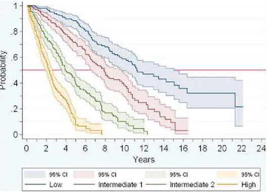
The International Prognostic Scoring System (IPSS) risk scorewas designed in 2009 for use in the initial diagnosis and risk stratification of PMF patients according to the following prognostic factors: 1- age (above 65 years); 2- presence of constitutional symptoms; 3-hemoglobin levels (lower than 10 g/dL); 4- leukocyte count (higher than 25×109/L); 5- percentage of circulating blasts
Based on these factors, MF patients can be subdivided into 4 categories:
(0 adverse factors)
(1 adverse factors)
(2 adverse factors)
( )
and the corresponding median survivals were reported at 11.3, 7.9, 4 and 2.3 years (7).
Fig 2: Stratification of patients according to the IPSS risk score (7).
The IWG-MRT subsequently developed a dynamic prognostic model (DIPSS) that utilizes the same prognostic variables used in IPSS but can be applied at any time during the disease course and used to predict the survival of the patients during the follow-up (8). The DIPSS prognostic score was then further extended to the DIPSS plus model, which also considers the patient's karyotype, platelet count and transfusion status (9).
Fig 3: Prognostic models in MF (10).
In recent years two new prognostic scores have been developed: GIPSS (genetically-inspired prognostic scoring system) and MIPSS70 + version 2.0 (mutation- and karyotype-enhanced international prognostic scoring system) (5).
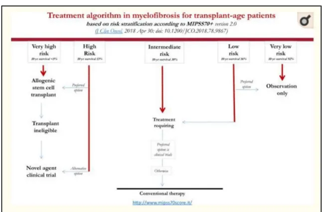
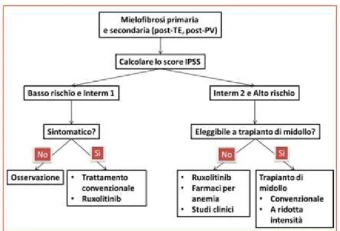
The GIPSS prognostic score estimates the risk based on the mutational status. The cytogenetic aspects, the presence of the three driver mutations (JAK2, MPL, CALR) and other additional mutations (ASXL1, SRSF2, U2AF1Q157) are also taken into consideration (11). Finally, the MIPSS 70+ prognostic score, used for patients with age eligible for hematopoietic stem cell transplant, includes the presence of various mutations (such as ASXL1, SRFR2, EZH2, IDH1, IDH2, CALR) and, at the same time, six clinical parameters (hemoglobin level, lymphocyte count, percentage circulating blasts, degree of medullary fibrosis, constitutional symptoms) (12). Based on the risk estimation, a different therapeutic approach is applied. A therapeutic option is the allogeneic stem cell transplant, which is recommended for high risk subjects. Patients with intermediate and low risk are treated with conventional therapy, including thalidomide, danazol, hydroxyurea and inhibitors of JAK1/2 (5).
Fig 4: The therapeutic approach to MF based on the risk assessment using the MIPSS70 + method (13).
1.2 Molecular pathogenesis
The molecular pathogenesis of MF relies on 3 driver mutations in JAK2, MPL and CALR genes. The first identified mutation (in 2005) was the JAK2V617F, a gain-of-function mutation, caused by the
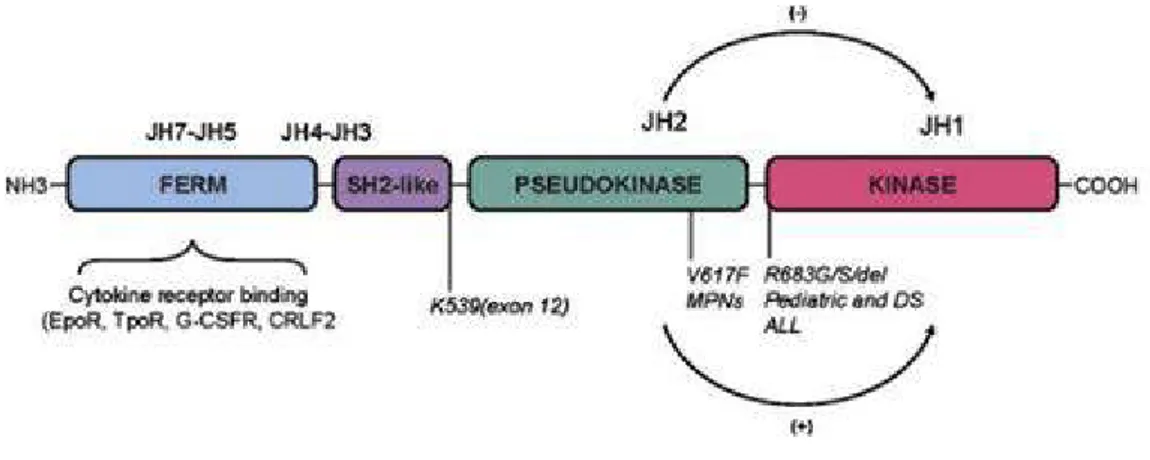
replacement of a valine with a phenylalanine in position 617 (V617F) (14). JAK2 is a tyrosine kinase belonging to a large family of similar proteins that, in mammals, includes JAK1, JAK2, JAK3, JAK4 (15). The gene coding for JAK2 is placed in position 9p24.1 26 and has 26 exons (16). Physiologically, JAK plays a fundamental role in signal transduction that occurs when a cytokine or a growth factor interacts with its receptor. When the JAK kinase is activated, it phosphorylates the STAT substrate by starting a fundamental signal pathway (JAK/STAT) involved into proliferation, survival and inhibition of apoptosis. JAK2 can also activate other downstream pathways, such as Ras and the PI3K/Akt pathways, which regulate these important cellular mechanisms (17).
To perform its crucial task, the kinase JAK2 is formed by two protein domains: a kinase domain (JH1 domain), which phosphorylates the downstream substrates allowing signal transduction and a pseudokinase domain (JH2 domain) with an inhibitory role against the kinase domain.According to some recent studies, the pseudokinase domain is able to phosphorylate two residues of the JAK2 protein (Ser523 and Tyr570), generating a conformational change that makes the kinase inactive. So, the JAK2 protein has an important feedbeck-negative regulation system (18).
Fig 7: JAK2 protein domains (19)
The JAK2V617F mutation falls at the level of this regulatory domain (specifically in exon 14), thus
causing a constitutive hyperactivation of the kinase. Therefore, the cell carrying this type of mutation is essentially independent of the stimulation of cytokines and growth factors, with consequent clonal proliferation (20). Other mutations have also been reported in other positions of the JAK2 gene, such as a mutation in exon 12 (21). At the cellular level, the effect of these mutations is substantially the same as the JAK2V617F mutation.
The second driver mutation identified in MPN was detected in the MPL gene. This gene, placed in 1p34.2 position of the human genome, encodes the Thrombopoietin receptor, the most important regulatory factor of megakaryocytopoiesis and platelet formation. Once the ligand binds its receptor, there is an MPL receptor dimerization and the activation of the JAK/STAT pathway (22).
Fig 8: The TPO/MPL pathway (23).
The MPL mutation consists in the substitution of a tryptophan in 515 position with another aminoacid
cases, there is an abnormal constitutive activation of the MPL receptor (in the absence of the ligand) with consequent activation of the JAK/STAT pathway (24).
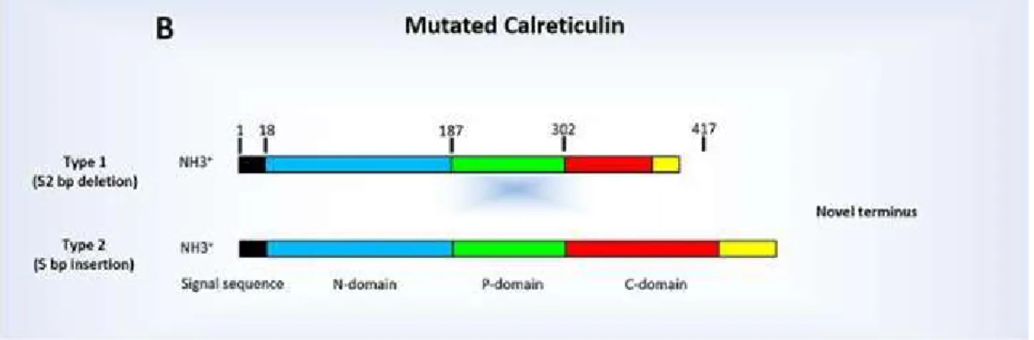
Finally, another mutation in the Calreticulin gene (CALR) was recently discovered. This gene, which is on chromosome 19p13.13, encodes a protein implicated in intracellular calcium homeostasis (transport of calcium from the endoplasmic reticulum) and in the correct "folding" of proteins; however, it can also play a role as a transcription factor (25). The Calreticulin protein is present at the intracellular, extracellular or transmembrane level. It is a very versatile protein, with roles in the regulation of proliferation, apoptosis and in the control of the immune response (26).
Fig 9: The structure of the Calreticulin protein (27)
Mutations in the CALR gene are generally deletions or insertions that usually fall into exon 9. There are two types of mutations of the CALR gene:
- Type 1: deletion of 52 base pairs - Type 2: insertion of 5 base pairs
Both mutations affect the C-terminal domain of the protein (28).
Fig 10: The mutated Calreticulin: type 1 and 2 (29)
According to Araki M. et al. and Elf S et al., the mutated Calreticulin undergoes a conformational change that allows it to interact with the MPL receptor, inducing a signal transduction via JAK/STAT in the absence of the ligand (Thrombopoietin). In particular, the mutated Calreticulin
would bind the extracellular part of the MPL receptor, determining its constitutive activation (30, 31).
Therefore, constitutive activation of the JAK/STAT signalling pathway is key to the development of the MF phenotype in all mutant backgrounds. JAK2V617F mutations can drive MF through activation
of erythropoietin receptor (EPOR), thrombopoietin receptor (MPL) and granulocyte-colony stimulating factor receptor (G-CSFR) receptors present on different stages of a maturing myeloid cell. Clonal dominance of homozygosity or heterozygosity of JAK2V617F, the presence and order of
acquisition of co-operating mutations and additional factors such as iron deficiency and gender can impact on the resulting phenotype. CALR and MPL mutations result in a PMF phenotype through activation of the MPL receptor. All drivers mutations appear to be largely mutually exclusive although bi-clonal disease can occur. JAK2 V617F and CALR mutations are detectable in the long
term haematopoietic stem cell (LT-HSC) population and in all maturing stages of the haematopoietic hierarchy. Yet, these JAK2 V617F LT-HSC population appear to exhibit reduced self-renewal and are skewed towards expansion of the progenitor pool instead (32).
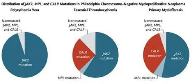
In addition to the 3 driver mutations (JAK2, MPL and CALR), a range of genes are repeatedly found to be mutated in MF. These co-operating oncogenic mutations found alongside the driver mutations include genes involved in cell signalling pathways (LNK, CBL, NRAS and NF1), epigenetic regulation (ASXL1, EZH2, TET2, DNMT3A, IDH1 and IDH2), transcriptional regulation (TP53, RUNX1) and mRNA processing (SF3B1, SRSF2, U2AF1, ZRSR2) (32). The most mutated genes are TET2, ASXL1, DNMT3A, CBL, LNK, IDH1/2, IKF1, EZH2, TP53, SRSF2 (28). This creates a general genetic instability that characterizes and complicates the MF framework. As shown in Fig. 10, around 60% of MF patients carry the JAK2V617F mutation, 30% are CALR mutated and 8% are MPL mutated (33). However, there is a small group of patients (around 10%) in which none of the driver mutations has been observed; these patients are called triple negative .
Fig 11: The distribution of the driver mutations in MPN (34).
As demonstrated by Tefferi et al., the mutational spectrum of MF patients is correlated with survival. The triple-negative patients have the lowest survival (with an average of 2.3 years), while the CALR mutated patients have the highest (with an average of 15.9 years). Mutations in JAK2 and MPL genes respectively confer an average life expectancy of 5.9 years and 9.9 years (35).
Fig 12: Survival of patients with MF according to mutational status (35).
In addition, the allelic burden of the JAK2 mutation (more than 56.7%) is associated with increased disease severity and increased risk of thrombosis in patients with MF (3, 36).
2. Inflammatory pathogenesis in MF
Chronic inflammation is the hallmark of MF. We know that inflammation has a protective role; however, in some cases, it can become harmful. Some authors describe "oncoinflammation", referring to the relationship between tumour cells and inflammatory microenvironment (37).
Cytokines are soluble proteins, commonly known for their immunomodulatory functions, that orchestrate both innate immunity and adaptive immunity. In addition to classically defined cytokines, such as interleukins and interferons, a variety of other soluble factors, including a range of growth factors, have been often classified as cytokines. Generally, cytokines are produced in response to cellular stresses including pathogen infections, inflammation, or injury. Their release exerts effects on different types of somatic cells by modulating different types of response. In case of infections or inflammations, monocytes, macrophages and neutrophils infiltrate and secrete numerous cytokines locally, including a variety of angiogenic factors, growth factors, and proteases. This results in a variety of cellular responses including increased angiogenesis, cell proliferation, migration of cells, and haematopoiesis (38) The immune response is mediated by the early reactions of innate immunity and other later ones of adaptive immunity. The innate immunity consists of cellular and biochemical defence mechanisms pre-existing to infection and ready to react quickly. In the context of innate immunity, the cytokines guarantee a rapid response from the leucocytes but also from the parenchymal cells, which are able to identify a pathogen by toll like receptor (TLR) expression. The main cytokines in this category are Interleukins (IL)-1, 6, 12, 18 (CXCL8), Tumor necrosis factor (TNF)- , Granulocyte/Granulocyte-macrophage colony stimulating factor (G-CSF, GM-CSF). They activate a series of fundamental cells for innate immunity (monocytes, dendritic cells, T cells, NK etc.) as well as the processes aimed at the elimination of the pathogen (chemokine release, increase of adhesion molecules expression at the endothelial level, increased fluidity of blood etc.) (39).This category of cytokines is also called inflammatory cytokines because they induce an inflammatory state. Regarding adaptive immunity, one of the fundamental roles of cytokines is to guide the T-cell response, for example IL-12 induce a TH1-type response, resulting in the production of effector cytokines (interferon (IFN)-
-induced by IL-4 production with consequent activation of the humoral response and IgE production (40).
Some cytokines have a negative regulatory role of immunity. They can be defined as anti-inflammatory cytokines. The suppression of the immune response is mainly driven by IL-10 and Transforming Growth Factor (TGF)-
They are produced by many cell type (monocytes, dendritic cells, T cells, regulatory T-cell (Treg) NK, etc) (41).
The released cytokines, both pro-inflammatory and anti-inflammatory, act to control cellular stress and minimize tissue damage. In general, after the resolution of the lesion or the inflammatory state, the cytokines return to the homeostatic levels. However, it is increasingly clear that chronic inflammation, resulting in abnormal production and dysregulation of cytokine levels, contributes to the pathogenesis of various diseases including cancer (42).
MF is typically characterized by a high level of proinflammatory cytokines both in the bone marrow and in the system. The constitutive activation of the JAK/STAT pathway leads to the excessive production of pro/anti-inflammatory cytokines. Therefore, a complex inflammatory microenvironment, supported by the activation of the JAK/STAT pathway, is created (43). These pro-inflammatory cytokines result from both mutant haematopoietic MPN clones and non mutant haematopoietic cells as a direct result of JAK/STAT signalling (44).
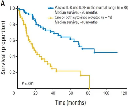
Recent studies have examined the circulating levels of pro/ anti-inflammatory cytokines in patients with MF and have studied their prognostic significance. Various pro-inflammatory cytokines such as TNF-alpha, IL-6, IL-8, IL- are elevated in the circulation. It has also been shown that IL-8 and IL-2 receptor (IL-2R) are prognostic indicators of reduced survival and leukemic transformation (45). In particular, IL-8 can contribute to the development of the tumour microenvironment through its important role in angiogenesis and in the proliferation of endothelial cells (46).
Fig 13: Increased survival of patients with normal levels of IL-8 and IL-2R (blue) as compared to patients with one / both cytokines increased in plasma (yellow)(45).
Skov et al., based on the study of the gene expression profile of whole blood in patients with MF, has shown upregulation of genes involved in inflammation and immunity, including Vascular Endothelial Growth Factor (VEGF), Hepatocyte Growth Factor (HGF), G-CSF, monokine induced by IFN- MIG). In particular, high levels of growth factor such as Platelet Derived Growth Fcator (PDGF), Fibroblast Growth Factor (FGFB) and VEGF have been implicated in fibrosis and angiogenesis. Furthermore, the expression of bone morphogenetic proteins (BMP), such as BMP1, BMP6, and BMP7, were higher in patients with advanced stages of MF (47). Increased BMP6 expression was also observed in the pre-fibrotic phases of MF. It has been suggested that progressive medullary fibrosis may be promoted by synergism between fibronectin and proinflammatory cytokines such as TGF- 1 and IL-1. Hoermann et al. has shown that the level of oncostatin M, a pleiotropic cytokine involved in a variety of physiological contexts including hematopoiesis, is elevated in JAK2V617F-positive patients and this is mutation-linked. The authors
concluded that overexpression of oncostatin M plays a role in bone marrow fibrosis and neo-angiogenesis of the bone marrow microenvironment and furthermore, amplifies cytokine production contributing to the cytokine storm observed in patients with MF (48).
Splenomegaly in MF is associated with the migration of CD34+ stem/progenitor cells from the bone marrow to the spleen and it is supposed to be the result of the clonal expansion of neoplastic stem cells, associated with high levels of cytokines. Consistently, high number of CD34+ cells is observed in the peripheral blood of MF patients. The presence of the JAK2V617F mutation in both the
bone marrow cells and the spleen confirms clonality. In addition, TNF-alpha, a cytokine known to be associated with clonal evolution and the selection of preleukemic stem cells in Fanconi anemia, is also associated with clonal expansion in MF and therefore splenomegaly. Many other cytokines, including HGF, MIG and IL-1RA have been associated with marked splenomegaly (49).
Frequent symptoms reported by patients with MF, such as night sweats and itching, are caused by high levels of cytokines, and more particularly, high circulating IL-8 levels have been associated with severe constitutional symptoms.
Based on in vitro studies and animal models, it has been hypothesized that chronic inflammation plays an important role in the initiation and progression of MF. It has been hypothesized that chronic inflammation can promote genetic instability and mutations (50) and may contribute to select the malignant clone and promote disease progression to leukemia (51).
More recently, it has been observed that, in addition to TNF- the Tissue Inhibitor of Metalloproteases (TIMP1-inhibitor of metalloproteases) are present at high levels in the blood of patients with MF (both JAK2 and CALR mutated). When combinations of these three factors are
added in culture in vitro, a significant increase in survival, migration and clonogenic capacity of circulating CD34+ cells of patients with MF is observed (52).
Interestingly, pre-existing inflammatory diseases, such as Crohn's disease or autoimmune diseases, can significantly increase the risk of developing MF. In addition, patients with MF have a high risk of developing secondary tumours, both haematological and non-haematological, and this risk is probably related to the JAK2V617F mutation (50).
2.1. Extracellular microvesicles
Among the possible mechanisms of inflammation development/propagation, it has been described that the contribution of extracellular microvesicles (EVs) is crucial. EVs, which are composed of microvesicles (MVs, 150-1000 nm) and exosomes (30-150 nm), are released by a wide variety of cells during homeostasis and cellular activation with pleiotropic effects on signalling between cells (53). EVs express antigens and contain constituents from the source cell including microRNAs. This mechanism supports cellular communication because, proteins, lipids and nucleic acids (DNA, miRNA, etc.) can be found within EVs with the potential to affect the short and long distance microenvironment. Therefore, they have the unique ability to transport membrane and cargo molecules between cells and quickly spread cellular information without the need for cell migration (53, 54).
The biogenesis of MVs occurs by extroflection of the cell membrane, which in turn, is made possible by the action of proteins and lipids that are able to modify the rigidity of the membrane itself. Subsequently, the detachment of the nascent vesicle from the plasma membrane takes place thanks to the actin-myosin contractile system and with ATP consumption. Their development is regulated by small GTPases from the ARF, Rab and Rho families. Contrary to MVs, the exosomes are released by the multivascular bodies of the cells (55).
Peripheral blood contains MVs resulting from platelets/megakaryocytes, red blood cells, leukocytes and endothelial cells; however, platelet/megakaryocytes-derived MVs are the most abundant (56). Specifically, Flaumenhaft et al identified the circulating MVs of megakaryocyte and platelet origin. According to this study, the megakaryocyte MVs are CD41+/CD62P- and express phosphatidylserine on the surface. Furthermore, these MVs are characterized by the presence of Filamin A. The platelet MVs are instead CD41, CD62P and LAMP-1 positive (57).
Numerous studies indicate that EVs, due to their cargo in lipids, inflammatory cytokines/proteins and nucleic acid, have pivotal role in the initiation, propagation and regulation of inflammatory diseases and might be used as biomarkers. They likely play a role in modulating inflammatory and autoimmune diseases, such as arthritis, diabetes and lupus. MVs enhance inflammation through secretion or surface expression of pro-inflammatory cytokines that promote an inflammatory microenvironment and drive immunomodulatory/immunosuppressive activities (58).
Fitzgerald et al. have systematically analyzed the association between 33 cytokines and EVs in eight in vitro, ex vivo and in vivo biological systems (cultured T cells, cultured monocytes, explants of tonsillar, cervical, placental villous, and amnion tissues, amniotic fluid, and blood plasma of healthy volunteers).
They found that a cytokine could be released predominantly either in soluble or in EV-associated form depending on the biological system. These two systems are not strictly separated, as many cytokines in vitro, ex vivo, and in vivo are released in EV-encapsulated forms and can elicit biological effects upon contact with sensitive cells. Moreover, upon stimulation, the pattern of encapsulation changes depending on the stimulus. This suggests that the encapsulation of cytokines in EVs is not simply the property of a particular cytokine, but rather a tight biological process that can be changed upon system activation. Such a targeting would require that EV-associated cytokines are biologically active, and they provided evidence of such activity. Their experiments demonstrated that the biological activity of the EV-encapsulated cytokines was the same whether they released the cytokines or provided them in EVs.
Multiple biologic meanings have been suggested to loading EVs with cytokines: (1) could be a mechanism to dispose of products when they are over-produced and simultaneously protecting the releasing cell from an autocrine effect; (2) EVs protect cytokines from environmental degradation. Indeed, EV-entrapped cytokines are protected from trypsin digestion; (3) may be a mechanism whereby the cytokine expressing cell could expand its sphere of influence to concentrate cytokines at the surface of other cells that might not otherwise be targeted by cytokines in solution (59). These studies show that deciphering the regulatory mechanisms of EV encapsulation could lead to a better understanding of cell-cell communications in health and disease.
Based on the bidirectional transfer of molecules between tumour cells and the microenvironment, EVs are emerging players. Recent evidence suggests that EVs have crucial roles in cancer development, including pre-metastatic niche formation and metastasis, angiogenesis and suppression of the immune system. Cancer cells are now recognized to secrete more EVs than their non-malignant counterparts and EVs have strong potential as blood-based biomarkers for the diagnosis, prognostication and surveillance of cancer. Thus, EVs play a key role in the regulation of immunity/inflammation and cancer, which in turn can contribute to the further release of EV (60, 61).
Few studies have shown elevated circulating EVs levels in patients with MPN (62, 63). Trappenburg et al have shown that patients with ET have higher number of circulating microparticles with platelet and endothelial markers, suggesting an ongoing platelet and endothelial activation and a role of microparticles in thrombosis of ET (64). Furthermore, Timari et al found that EVs released by MSCs from patients with MPN were found to be selectively enriched in miR155, and they induced an increase in colony forming unit (CFU) ability of neoplastic CD34+ cells (65). However, the role of EVs in MPN, including MF, has yet to be addressed. A deepening
of their role in MPNs would be useful to better understand the mechanisms underlying the disease and to identify new therapeutic strategies.
3. Immune dysregulation in MF
The survey of US Surveillance, Epidemiology, and End Results, (SEER)-Medicare database (1,017 MPNs cases) has documented that autoimmune conditions, overall, are associated with an increased risk of MPN with respect to healthy controls. Autoimmune conditions can cause an immune-related and inflammation-driven tumorigenesis that may result in MPN. Furthermore, therapies given to patients with autoimmune disease (anti-inflammatory and immunosuppressive agents) can play a role in the risk of developing MPN. Moreover, there might be a shared common genetic and/or environmental susceptibility in autoimmune diseases and MPN (37).
Several studies provided evidence that T, B, and NK cell lineages could be involved by the MPN mutations, suggesting that the target cell in MPN is a myelo-lymphoid progenitor. Anyway, the consequences of the involvement of B, T, and NK cells by the somatic mutation that drives the clonal proliferation are still unclear, but they could underline some of the immunologic abnormalities of MPN patients.
Few studies analyzed lymphocyte subsets and their possible correlations with the immune/inflammatory features of MPNs. First of all, Cervantes et al have documented a reduced absolute circulating lymphocyte count in MF patients. Despite this, there was an increase of cytotoxic T cells (CD3+/ CD56+) in most of PMF patients and 10% of them showed an increased CD19+/CD5+ B cell subpopulation (66).
In another study, the presence of the MPLW515K gene mutation in CD4+ lymphocytes was observed, suggesting that the lymphoid compartment may also be affected by the mutation (67). Regulatory T lymphocytes (Treg), which are a subpopulation of T helper lymphocytes with the role of maintaining immune tolerance, show altered number. Zhao et al investigated the frequency/function of Tregs (CD4+CD25+ FOXP3+) in PV patients and healthy donors. Tregs were significantly increased in patients and the expression of FOXP3, the master regulator of the immunesuppressive activity of Tregs, was increased (37,68). Conversely, Keohane et al reported low levels of Tregs in 50 MPN patients as compared to healthy donors (69). It has also been observed that the mutational status can influence the proportion between the regulatory T lymphocyte subpopulations. If we consider the three regulatory T-subpopulations identified by Miyara et al, the "triple negative" MF patients show an increase in the circulating population I
(CD3+CCD4+ CCD45RA+ CD25+ CD127low); conversely, patients with the JAK2V617F mutation show an increase in circulating population III (CD3+CD4+CD45RA-CD25 + CD127-) (70).
Regarding other subset of immune cells, dysregulations have also been observed in the monocyte/macrophage population. Thiele et al. have revealed an expansion of the monocyte/macrophage cell population in the bone marrow of PMF patients, with a significant increase in the number of mature CD68+ macrophages. These macrophages are also morphologically altered (71). Anyway, monocytosis in MPNs is a rare event, restricted to PMF, and is associated with rapid disease progression. Furthermore, it has been described that the monocytes of patients with PMF are hyperactivated, because there is an increased production of proinflammatory cytokines and transforming growth factor-beta
(TGF-mechanism (72).
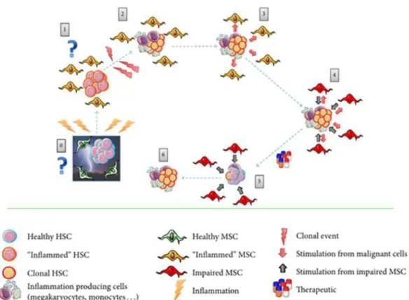
Myeloid-derived suppressor cells (MDSCs) is a subpopulation of immune cells that plays an important role in the immune system and tumorigenesis. This population is part of a larger and more complex group of cells that are fundamental for the mechanisms of immunological tolerance towards the tumor: they guarantee an immunosuppression that allows the neoplastic clone to progress undisturbed (37). MDSCs are part of a generalized circuit of immunosuppression in which other tolerogenic cells are active participants. It is not surprising, therefore, that in MPN the MDSCs (CD11b+CD14-CD33+) are significantly increased, as demonstrated by Kundra et al. (73). The network regulating the relationship between hematopoietic stem/progenitor cells and immunoregulatory cells has not been completely elucidated in MPN. There are many outstanding questions on the role of immunomodulation in the generation and progression of MPN that deserve to be addressed. For example, mesenchymal stem cells (MSCs) are key cells for immunoregulation and inflammation. MSCs are a key component of the hematopoietic niche, where they support the proliferation and differentiation of HSC. Furthermore, MSCs have a fundamental role in immune regulation, suppressing the proliferation of T cells and favoring the immunosuppressive function of regulatory T lymphocytes (Treg) (74). The bone marrow of MF patients has both cellular and extracellular changes due to inflammation/fibrosis that modifies the hematopoietic niche; for example, alterations of fibroblasts, osteoblasts, endothelial cells and even MSCs are found (75). The MF inflammatory microenvironment shows a high production of FGF and VEGF, which
stimulate neo-angiogenesis and marrow fibrosis, and of PDGF and TGF- ce an
increase in matrix proteins such as proteoglycans, fibronectin and collagen (76). Chronic inflammation therefore creates a microenvironment that facilitates the release of many molecules from the cells of the microenvironment and from the hemopoietic clone, that leads to an
"inflammatory storm" in the bone marrow (75). Due to the overproduction of these factors, the MSCs are stimulated to proliferate and to explicate their immunoregulatory and immunosuppressive role.
Fig 14: The vicious circle that is created between cells and inflammatory microenvironment in the bone marrow of patients with MF. (75)
The concept of onco-inflammation" and immunoregulation in MPN offered further suggestions for therapeutic strategies. Approaches with anti-inflammatory/immunomodulatory drugs have been designed as promising drug therapies in MPN, including JAK1/2 inhibitors (such as Ruxolitinib), IFN, statins or specific anti-TGF beta agents or IL-8 antagonists (77).
3.1. Monocytes
3.1.1 Monocytes physiology
Monocytes are immune cells that are part of the reticuloendothelial system, which is a system playing a role of sentinel of the organism for protection from what is foreign. Monocytes are circulating cells that can be recruited from tissues and mature on macrophages. Alternatively, they can mature to dendritic cells (Monocyte derived DCs) (78). They derive from a common myeloid precursor and develop in the primary lymphoid organs (bone marrow in adults, liver in fetal life). Starting from this precursor, a factor called M-CSF (macrophage-colony stimulating factor) is essential for their terminal maturation. In the blood, mature monocytes account for about 5-10% of the circulating cells (79, 80).
Although monocytes are generally referred to as circulating precursors of macrophages, today their role in the immune system has been expanded. Thanks to the characterization of surface antigens, in fact, it is possible to identify the various phases of their development and to distinguish the different subsets that are present in humans.
In the murine model, circulating monocytes are LY6C + and lose this protein once activated and migrated into lymph nodes or tissues. In this second phase, there is also an increase in the transcriptional levels of
IL-LY6C monocytes control the tissue environment and are able to present a possible antigen to the T lymphocytes, even without differentiating to macrophages (78).
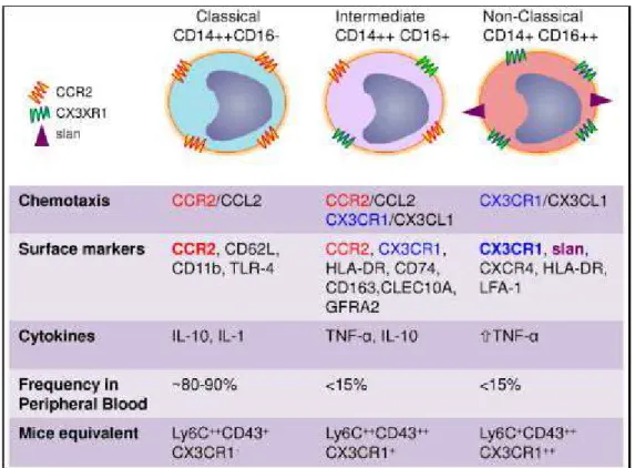
In humans, LY6C + monocytes are defined as "classic" because they are the most represented population in the peripheral blood (80-90%). From the point of view of antigenic expression, classical monocytes are CD14 ++/CD16-. LY6C- monocytes, on the other hand, are defined as "non-classical" and are CD14+/CD16 ++. They can also be referred to as "patrolling subset" because their role is to patrol the vessels, checking the integrity of the endothelium (82). More recently, a third class of monocytes has been identified: these are the "intermediate" monocytes, with characteristic antigenic CD14 ++/CD16+ expression. Although numerically smaller than the other subset, they are interesting because they are able to respond to the lipopolysaccharide (LPS)
stimulus by TNF- is the LPS
receptor 83).
In addition to the expression of CD14 and CD16, the three subsets can also be distinguished by the different secretory capacities. Following stimulation with LPS, it appears that:
- Classic monocytes produce high levels and a wide variety of cytokines (G-CSF, 10, CCL2,
IL-6 TNF- -6)
- Intermediate monocytes produce high levels of pro-inflammatory cytokines such as TNF- , and IL-6
- Non-classical monocytes produce the same cytokines as the other subset, but generally they mainly produce anti-inflammatory cytokines (such as IL10).
The same study also highlighted other differences regarding the phenotype of the three sub-sets: - CCR2, CXCR1, CXCR2 and CD62L are highly expressed by classical monocytes.
- CD64, CCR1, CCR2, CX3CR1, CD11b, CD33 and CD115 are expressed at intermediate levels in intermediate monocytes; CD40, CD54 and HLA-DR are expressed instead in this subset at high levels.
Fig 16: Phenotypic and biological characteristics of the three subsets of monocytes (85)
Based on this information, each of the three sub-sets can be assigned a specific role. Classic monocytes are the first to be recruited and come out from the bone marrow to go into the bloodstream. They have high phagocytic capacity, thanks to the production of peroxidase and
produce high levels of Reactive oxygen species (ROS), IL10, IL1-
-stimulated by LPS and they differentiate into intermediate and non-classical monocytes. They are involved in angiogenesis and coagulation.
Intermediate monocytes, on the other hand, are typically inflammatory and have a reduced phagocytic capacity and peroxidase activity but produce higher levels of TNF- - -6 under inflammatory stimulus. From the bloodstream they reach the tissues and sites of inflammation by CX3CR1 and CCR5 receptors that mediate the accumulation of monocytes in inflammatory sites and that bind CCL3, expressed by macrophages in inflammatory sites, and CCL5 (RANTES), expressed and secreted by T cells to recruit leukocytes to the inflammatory sites. They are increased in diseases associated to chronic inflammation. They also express CD40 for the activation of T lymphocytes.
Non-classical monocytes patrol the vessels and from the bloodstream invade the tissues damaged by inflammation through the expression of CX3CR1, which is the receptor for chemokine CX3CL, and
contribute to angiogenesis and fibrosis, favouring the production of collagen (IL10 and TGF -relate). They are in fact defined as "patrolling". They therefore have an anti-inflammatory function.
They can produce IL1- - se to nucleic acids (86).
3.1.2 Monocytes and MF
Previous studies have partially characterized the monocyte population in the course of MF. It has been shown that monocytes of patients with MPN are more functionally active and therefore produce large amounts of cytokines (TGF- -1 and substance P) (72);
In 2016, a possible role of monocytes was shown in the development of one of the major clinical features of MF: marrow fibrosis. In fact, high percentages of a particular monocyte-derived cell type, namely fibrocyte, has been identified in patients with MF at the fibrotic stage (87). The monocyte-fibrocyte transition is mediated by the Pentraxin-3 (PTX3), released by macrophages and
88).
To carry out their task in the context of the immune system, monocytes are recalled at the site of inflammation thanks to some chemokines, including the most important Monocyte chemoattractant protein 1 (MCP-1). It has been recently described that a given polymorphism of MCP-1-2518A/G, may predispose to the development of MF. This polymorphism had already been associated with other pathological conditions (autoimmune disorders, atherosclerosis, chronic infections), which places monocytes at the centre of the etiopathogenesis of various immune defects (89).
To investigate their role in MPN, monocyte lines with stable JAK2V617F mutation were developed. They were then used as a study model and the levels of pro and anti-inflammatory factors were evaluated. The results show that the mutated cells produce a greater quantity of metalloproteases (and their inhibitors), growth factors and other crucial substances (such as PTX3) compared to "wild type" cells (90).
In addition to being predictive of disease development and as study model for the underlying etiopathogenetic mechanisms, it was tested whether monocytes could be a prognostic index for MF. In a recent study, patients were stratified based on absolute monocyte counts and it was shown that monocytosis is associated with a poorer prognosis (91).
It has also been shown that monocytes are able to express the receptor for angiopoietin 2, namely Tie-2, and therefore they can promote angiogenesis in an autocrine manner (92). In fact, patients with MF have an increased concentration of Tie-2+ monocytes in the peripheral blood. In this case we are dealing with monocytes of the "intermediate" subset (CD14 ++ CD16 +) (93).
However, monocytes expressing the same receptor for angiopoietin 2 have also been identified in -classical monocytes (CD14 + CD16 ++) (94). This last evidence is very relevant, as an increased
neo-demonstrated (95). Monocytes therefore seem to be responsible for this phenomenon.
It has also been shown that the expression of IL- could be associated with an increased risk of thrombosis in patients with MF who have the JAK2V617F mutation (96).
Based on these observations, monocytes may have a leading role in the development and maintenance of MF. Nevertheless, additional studies are needed to understand the mechanisms underlying their action within the inflammatory microenvironment of MF.
4.Ruxolitinib
MF is still a treatment-orphan disease that may be cured only by allogeneic stem cell transplant in younger selected patients. However, as above described, regardless of the type of mutation, patients with MF have hyperactivation of the JAK/STAT pathway. An effective therapeutic approach, therefore, would be to inhibit the action of the JAK kinase. The first drug developed and approved for the treatment of patients with MF was Ruxolitinib (or INCB018424): a JAK1 / 2 inhibitor (97).This drug was approved by the EMA in 2012 and is marketed under the trade name of JAKAVI (98). The approval of the drug came thanks to two main clinical studies:
- COMFORT I: in which Ruxolitinib was compared with placebo (99)
- COMFORT II: in which Ruxolitinib was compared with the best available therapy (100)
Thanks to these two trials, Ruxolitinib has been approved for patients with intermediate (1-2) or high-risk MF who are not eligible for hematopoietic stem cell transplantation.
Ruxolitinib reduces inflammatory cytokine production (JAK1-driven) and exhibits myelosuppression (JAK2-driven).
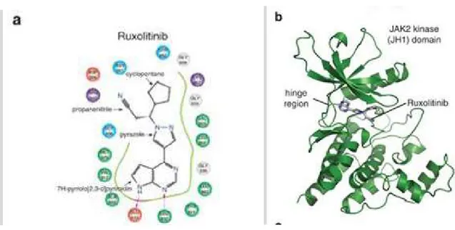
Regarding the mechanism of action, Ruxolitinib is able to inhibit the kinase activity of JAK1/2 by binding to the ATP "binding domain" of the protein (the site where ATP normally resides). In particular, the Ruxolitinib molecule has a double ring system, through which it forms two hydrogen bonds with JAK, at the level of its kinase domain (102).
Fig 18: Molecular interaction between the JAK2 kinase and its inhibitor Ruxolitinib. (102) The binding to the ATP "binding domain" turns out to be effective in inhibiting the activity of JAK1/2, since the ATP is not only used by the kinase to obtain phosphate groups, but it also seems to have a role in stabilizing the pseudokinase domain in the absence of stimulus. Ruxolitinib, therefore, would act as an ATP mimetic, stabilizing the inhibited form of JAK1/2 (103).
In these randomised controlled trials it has demonstrated efficacy in spleen volume reduction and symptom burden reduction with a moderate improvement in overall survival of PMF patients (104). Despite these benefits, there is limited impact to induce complete haematological remission with normalisation of blood counts, reduce the mutant allele burden or reverse bone marrow fibrosis. Clonal evolution has been observed on ruxolitinib therapy and transformation to acute leukaemia can still occur.
Spleen size reduction occurs in more than 50% of patients with MF and a significant reduction in constitutional symptoms is also observed. This can be related to the reduction of pro-inflammatory cytokines. Furthermore, this finding demonstrates how the inhibition of the JAK/STAT pathway has important anti-inflammatory implications (97).
Although important therapeutic effects have been demonstrated following treatment with Ruxolitinib, numerous side effects have however been reported. These are due to the fact that the drug is not selective for the mutated kinase, and therefore also acts on the wild type form. The clinical study COMFORT I showed that neutropenia, urinary tract infections and herpes zoster were observed in patients treated with Ruxolitinib (99). These evidences were confirmed following a 5-year follow-up in the context of the clinical study COMFORT II, with an increased risk of developing pneumonia, sepsis and tuberculosis (100). Interestingly, new drugs are being studied with increased inhibitory capacity against JAK and with greater selectivity towards the mutated protein (105).
Fig 19: Adverse effects observed in patients with MF treated with Ruxolitinib or with the best available therapy (100)
The risk of infections is a serious problem for individuals with MF, as it represents about 10% of the causes of death for these patients. This is due to the profound deregulation of the immune system in these subjects, which affects both the cellular component and mediators such as cytokines (106). The question then arose of verifying whether treatment with Ruxolitinib could exacerbate this situation of immunodepression in treated patients, since the JAK/STAT pathway plays a key role in many immune-related processes.
According to studies conducted both in vivo and in vitro on dendritic cells, Ruxolitinib is able to inhibit their differentiation capacity, the ability to produce IL-12, the migration and expression of activation markers (107). Dendritic cells are fundamental for the antigen presentation process to T cells and, moreover, are able to produce various cytokines (such as IL12, IL23), which in turn drive the Th1 and Th17 response. For this reason, reduced functionality of dendritic cells results in dysfunction of the immune system (108).
The effects of Ruxolitinib on natural killer cells were also studied. Natural killer cells are effector cells with a critical role in defence against viral infections and cancer cells. They are able to produce IFN- - 108). In a recent study, an important reduction in the frequency of these cells were demonstrated in patients with MF treated with Ruxolitinib. This may probably be due to a defective maturation process. In vitro, a reduced capacity to produce cytokines and lytic activity typical of this cell type was then highlighted (109).
Tregs are reduced in MF. In a recent study, it was observed that this decrease is even more pronounced in patients treated with Ruxolitinib. These authors have in fact demonstrated a reduction of CD4+CD127low CD25high FOXP3 + cells in the peripheral blood of patients. They also demonstrated, in vivo and in vitro, a functional block of these cells (110).
Fig 20: Effects of Ruxolitinib on crucial cells of the immune system. (108).
Of note, although monocytes are key players of the inflammatory microenvironment, in MF their pathogenetic role, both at baseline and following treatment with Ruxolitinib, is far from being defined.
This thesis is based on four projects aiming to address the
pathogenetic
role
of
the
immune/inflammatory
microenvironment in MF.
Specific aims were:1. To analyze the role of inflammation on the functional behaviour of normal hemopoietic stem/progenitor CD34+ cells. It has been hypothesized that the sustained inflammatory microenvironment of MF can alter crucial biological processes, leading to genomic instability and cancer progression. To mirror the in vivo inflammatory microenvironment, here we investigated the in vitro functional effects and role of combined crucial proinflammatory cytokines (IL- - -6, and tissue inhibitor of metalloproteases (TIMP-1)) on the functional behaviour of normal CD34+ cells from neonatal umbilical cord blood (CB) and adult normal G-CSF-mobilized peripheral blood (mPB) in the presence or absence of bone marrow MSCs. Specifically, we analysed the effects of these selected inflammatory mediators on the viability, proliferative activity, clonogenic potential and migration capability of CD34+ cells. Results of these project have been published on Mediators Inflammation 2018 Jul 4;2018:5974613. doi: 10.1155/2018/5974613. eCollection 2018 .
2. To characterize the bio-molecular profile of circulating MVs in MPN and particularly MF. Circulating MVs, as biomarkers of disease/malignancy and as contributors of the inflammatory network in MPN, are an open question. Here we investigated: 1) the profile of MVs in MF and ET; 2) whether MVs proportions could be related to severity of disease; 3) the role of inflammation on MVs frequency in MF; 4) the effects of Ruxolitinib on MVs proportion in MF; 4) the microRNA (miR) cargo of circulating MVs from MF patients. Results of these projects have been published on British Journal of Haematology , 2019 Jun;185(5):987-991. doi: 10.1111/bjh.15682. Epub 2018 Nov 18 and presented to the following Congresses: American Society of Hematology (ASH) 2017: Blood 2017 130:4220; European Hematology Association (EHA) 2017: abstract n. E1309; XV Congress of the Italian Society of Experimental Hematology (SIES) Rimini, Italy, 18-20 October 2018, Haematologica Abstract n° PO047; American Society of Hematology (ASH) 2018: Blood 2018 132:4334; doi:
3. To characterize the circulating immune microenvironment of MF. Infectious complications are the leading cause of morbidity and mortality constituting more than 10% of all patient deaths. In order to understand whether the infectious events are caused by deficits in the innate or adaptive immune response, a comprehensive analysis of key immune cells is required. Based on this background and considering the essential role of the JAK/STAT pathways in shaping the immune response, we enumerated and functionally characterized key immune-cell subsets including (dendritic cells (DCs), T-helper (Th) 17 cells , regulatory T cells (Tregs) and innate lymphoid cells (ILC)) with the aim to investigate their putative role in immunosurveillance in MF. Results of these project have been published on Oncoimmunology 2017 Jul;6(10):e1345402. doi: 10.1080/2162402X.2017.1345402. eCollection 2017 .
4. To investigate the role of circulating monocytes in the inflammatory microenvironment of MF and to evaluate whether and to what extent Ruxolitinib may influence their in vitro / in vivo behavior. Monocytes play a key role in the inflammatory microenvironment of MPN. Ruxolitinib improve the therapeutic scenario of MF by reducing splenomegaly and systemic symptoms in a significant fraction of patients. Nonetheless, Ruxolitinib is burdened by hematological and extra-hematological toxicity (i.e.: infections). The in vitro and ex vivo inhibitory effects of Ruxolitinib on number/function of dendritic cells and T-cells (including Tregs) have been previously described. However, to date, in MF the effects of Ruxolitinib on monocytes biology have never been investigated. Based on this evidence and considering the essential role of the JAK/STAT pathways in shaping the immune response, the main purpose was: 1) to phenotypically and functionally characterize circulating monocytes in MF before and after 6 months of in vivo treatment with Ruxolitinib; 2) to address monocyte interaction with the inflammatory microenvironment and 3) to investigate whether and at what extent Ruxolitinib affects the in vitro/in vivo behaviour of circulating monocytes from MF patients. The driving hypothesis of the present proposal is that the analysis of the in vitro/in vivo biological effects of Ruxolitinib on the monocytes compartment will contribute to clarify the role of JAK1/2 inhibition in the modulation of the immune landscape of MF. Results of these project have been submitted for publication Frontiers in Immunology" and presented to the following Congress European Hematology Association (EHA) Congress 2018: abstract n. PS1345.
Published on
HindawiMediators of Inflammation
Volume 2018, Article ID 5974613, 14 pages
https://doi.org/10.1155/2018/5974613
Mobilized Peripheral Blood versus Cord Blood: Insight into
the Distinct Role of Proinflammatory Cytokines on Survival,
Clonogenic Ability, and Migration of CD34+ Cells
Dorian Forte, 1,2 Daria Sollazzo,1 Martina Barone,1 Marisole Allegri,1 Angela di Martella Orsi,1
Marco Romano,3 Barbara Sinigaglia,1 Giuseppe Auteri,1 Nicola Vianelli,1 Michele Cavo,1
Francesca Palandri,1 and Lucia Catani 1
1Department of Experimental, Diagnostic and Specialty Medicine, University of Bologna, Bologna,
Italy;
2Wellcome Trust-Medical Research Council Cambridge Stem Cell Institute and Department of
Haematology, University of Cambridge and National Health Service Blood and Transplant, Cambridge Biomedical Campus, CB2 0PT Cambridge, UK;
3
London, UK Correspondence should be addressed to Lucia Catani; [email protected]
Received 21 February 2018; Revised 24 May 2018; Accepted 31 May 2018; Published 4 July 2018 Academic Editor: Elena Dozio
Copyright © 2018 Dorian Forte et al. This is an open access article distributed under the Creative Commons Attribution License,which permits unrestricted use, distribution, and reproduction in any medium, provided the original work is properly cited.
ABSTRACT
Inflammation may play a role in cancer. However, the contribution of cytokine-mediated crosstalk between normal hemopoietic stem/progenitor cells (HSPCs) and their (inflammatory) microenvironment is largely elusive. Here we compared survival, phenotype, and function of neonatal (umbilical cord blood (CB)) and adult (normal G-CSF-mobilized peripheral blood (mPB)) CD34+ cells after in vitro exposure to combined crucial inflammatory factors such as interleukin-(IL-) 1 , IL-6, tumor necrosis factor- (TNF-) , or tissue inhibitor of metalloproteinases-1 (TIMP-1). To mimic bone marrow (BM) niche, coculture experiments with normal BM stromal cells (BMSCs) were also performed. We found that combined inflammatory cytokines increased only the in vitro survival of CB-derived CD34+ cells by reducing apoptosis. Conversely, selected combinations of inflammatory cytokines (IL-1 + TNF- , IL-6 + TNF- , and IL-1 + TNF- + TIMP-1) mainly enhanced the in vitro CXCR4- driven migration of mPB-derived CD34+ cells. TNF- , alone or in combination, upregulated CD44 and CD13 expression in both sources. Finally, BMSCs alone increased survival/migration of CB- and mPB-derived CD34+ cells at the same extent of the combined inflammatory cytokines; importantly, their copresence did not show additive/synergistic effect. Taken together, these data indicate that combined proinflammatory stimuli promote distinct in vitro functional activation of neonatal or adult normal HSPCs.
INTRODUCTION
Hemopoietic stem/progenitor cell (HSPC) activation and retention are modulated by the bone marrow (BM) niche where they are located. In response to inflammation and/or BM injury, long-term quiescent hemopoietic stem cells (HSCs) are efficiently recruited into the cell cycle progression returning back to quiescence after reestablishment of homeostasis [1, 2]. Inflammation is a fundamental response that protects tissues from damage and preserves internal homeostasis. However, chronic inflammation may hinder functionality of different tissues and has been suggested to cover a key role in cancer [3].
Proinflammatory cytokines are emerging as key regulators of steady-state and infection-driven hemopoiesis. Recent findings contributed to highlight how HSPC fate could be dictated by inflammatory factors in the BM microenvironment as HSPCs may actively respond to danger signals and proinflammatory cytokines [4, 5]. However, excessive chronic signalling can have negative effects on HSPC regulation and function [6]. Moreover, abnormalities in the inflammatory signalling pathways have been discovered in both preleukemic and leukemic diseases [7]. BM mesenchymal stromal cells (BMSCs) are one of the most important components of the BM microenvironment. They respond to various microenvironment stimuli by changing their secretory capacity and displaying immune-suppressive activity through direct or indirect production of prostaglandin E-2, indoleamine 2,3-dioxygenase, interleukin- (IL-) 10 [8 10], and soluble receptors for IL-1 and tumor necrosis factor-
-stromal cells may also create a proinflammatory environment that promotes malignant transformation and disease progression [12]. In such process, several factors and pathways have been implicated but it is not clear how inflammation could affect or transform HSPCs. Understanding the direct cellular target(s) of proinflammatory cytokines is a critical step to better clarifying how HSCs/HSPCs are regulated in the BM niche.
Granulocyte colony-stimulating factor- (G-CSF-) mobilized peripheral blood (mPB) and umbilical cord blood (CB) are two of the current sources of HSPCs for transplantation in hematological malignancies [13]; however, insights into the effects mediated by inflammation on neonatal and adult HSPCs are still elusive. In the last years, several phenotypic and functional differences between CB and mPB-derived HSPCs have been described [14 19]. However, so far, studies analysing the adaptations of HSPCs from these two sources to inflammatory cytokines were focused on a limited number of cytokines which were individually tested [20 24].
To mirror the in vivo inflammatory microenvironment, here we investigated the role of combined crucial proinflammatory cytokines (IL- - -6, and tissue inhibitor of
metalloproteases (TIMP-1)) on the in vitro functional behavior of CB- or mPB-derived CD34+ cells in the presence or absence of BMSCs.
RESULTS
1. Selected Combinations of Proinflammatory Cytokines Promote the In Vitro Survival of CB-Derived CD34+ Cells.
To test the role of proinflammatory factors on HSPCs, we firstly evaluated the in vitro survival of CB- and mPB derived CD34+ cells in the presence of IL-6, IL- - -1, at concentrations previously shown by us to be effective in dose-response experiments [20].
Spontaneous survival rate of CB-derived CD34+ cells was higher as compared to mPB .05; Figures 1(a) and 1(b)).
As shown in Figure S1, CB-derived CD34+ cell survival was further enhanced by TIMP-1,
IL- - Compared to
untreated cells (control), TIMP-1, IL- -6 alone poorly promoted the survival of mPB- and
CB derived CD34+ cells with the notable exception of TNF- .05)
increased mPB-derived CD34+ cell survival.Therefore, based on these results and on data reported in literature [20, 25, 27], we hypothesized that combinations of cytokines can make CB- or mPB-derived CD34+ cells more responsive to inflammatory stimuli. As shown in Figure 1(a), when cytokines were two-by-two combined, we found that IL- - .01), 6 +
IL-0.05), or TNF- .01) or TIMP- .01) significantly increased the percentage of viable CB-derived CD34+ cells as compared with the untreated counterparts. In contrast, only
IL-- - .001).
Testing multiple cytokine combinations (Figure 1(b)), the survival of CB-derived and mPB-derived CD34+ cells was significantly increased in the presence of IL- - -0. .001, resp.) as compared with untreated cells. When we compared CB and mPB (Figures 1(a) and 1(b)), the survival rate of CB-derived CD34+ cells was promoted in the presence
of IL- -1 and IL-6 + TNF- .05).
These data suggest that CD34+ cells from CB are more actively responsive to inflammatory cues than their mPB counterparts; however, multiple combinations are required to promote their survival.
Subsequently, we examined whether a combination of proinflammatory factors would trigger CD34+ progenitor cell differentiation. In selected experiments, freshly isolated CD34+ cells were cultured in RPMI medium supplemented with or without additional proinflammatory factors for 24 hours. The expression of selected myeloid-specific markers (CD11c, CD13, CD14, and CD45) along with HSPC markers (CD38, CD133) or specific marker for cell adhesion/proliferation (CD44) was analyzed by flow cytometry. The expression of CD11c, CD14, CD38, CD45, and
CD133 was not significantly affected by inflammatory factor treatment (data not shown). By contrast, after treatment with combined inflammatory cytokines, mPB- and CB-derived CD34+ cells upregulated the expression of CD13 and CD44 (Figures 1(c) and 1(d) and Supplementary Table 2). After treatment with IL-6 + IL- - -1, CB-derived CD34+ cells showed a 5-fold increase in geometric mean fluorescence intensity (gMFI) of CD13 as compared to untreated .001). Accordingly, a statistically significant difference was also found in the
presence of IL- - or IL-6 + TNF- - - and 4.64- .001, resp.).
A similar pattern was also found when mPB CD34+ cells were tested (Figure 1(c)).
Consistent with CD13 expression, the combination of IL- - .01) and IL-6 +
IL- - .05) ± TIMP- .01) induced a significantly higher CD44 expression in
CB-derived CD34+ cells (>2-fold increase, respectively; Figure 1(d)). When we evaluated the mPB-derived CD34+ cells, CD44 expression markedly increased in the presence of IL-
-.001), IL-6 + TNF- .01), and the combination of cytokines .001). Importantly,
TNF-.001).
Taken together, these results demonstrate that selected combinations of inflammatory cytokines, along with the promotion of the survival of CB-derived CD34+ cells, stimulate the expression of CD13, which is an early and late myeloid marker.
Figure 1: Survival and phenotype of CD34+ cells from CB or mPB in the presence of combined proinflammatory cytokines. (a) Percentage of live CD34+ cells from CB (indicated as negative for Annexin V and PI (black columns, n=9) or mPB (grey columns, n=8) in vitro treated for 24 hours with a two-by-two-factor combination and assessed using Annexin V/PI staining, as described in Methods. (b) Percentage of live CD34+ cells in the presence of multiple combinations of proinflammatory cytokines. (c d) Box-plot graphs with fold change of gMFI for CD13 and CD44 expression in CD34+ cells after treatment with different combinations of inflammatory cytokines. Dot lines were used to mark control samples without any treatment. All data are presented as mean ± SEM of n (as above described) experiments performed in
duplicate ( .01, and . .05 CB versus