A
A
l
l
m
m
a
a
M
M
a
a
t
t
e
e
r
r
S
S
t
t
u
u
d
d
i
i
o
o
r
r
u
u
m
m
-
-
U
U
n
n
i
i
v
v
e
e
r
r
s
s
i
i
t
t
à
à
d
d
i
i
B
B
o
o
l
l
o
o
g
g
n
n
a
a
DOTTORATO DI RICERCA IN
Oncologia e Patologia Sperimentale
Ciclo XXVIII
Settore Concorsuale di afferenza: 06/A2
Settore Scientifico-Disciplinare di appartenenza: MED/04
AIR POLLUTION AND HUMAN HEALTH RISK:
EVALUATION OF CARCINOGENIC POTENTIAL
OF URBAN AIRBORNE PARTICULATE MATTER
Tesi di dottorato di ricerca presentata da:
Dott.ssa Stefania Serra
Coordinatore Dottorato Relatore
Chiar.mo Prof. Pier-Luigi Lollini Chiar.mo Prof. Sandro Grilli
Contents
Introduction and aim of the study
1Chapter 1: Alternative tests in vitro
51.1 Introduction 5
1.2 Cell transformation assay test 7
1.2.1 BALB/c 3T3 A31-1-1 Cell Transformation
Assay 10
1.2.2 BHAS 42 Cell Transformation Assay 10
1.3 Application of the CTAs in various industry sectors 13
1.3.1 Pharmaceutical industry 13
1.3.2 Chemical industry 13
1.3.3 Cosmetic industry 14
1.3.4 Food industry 14
Chapter 2: Risk Assessment
152.1 Risk assessment 15
Chapter 3: Particulate Matter
273.1 General characteristics of Particulate Matter 27
3.3 Effects of Particulate Matter 35 3.3.1 PM exposure and health effects 37 3.4 Polycyclic aromatic hydrocarbons (PAHs) 41 3.4.1 Carcinogenic effects of PAHs 43
Chapter 4: Materials and Methods
464.1 Experimental design 46
4.2 Air samples collection 49
4.3 Preparation of treatment solutions 50
4.4 BALB/c A31-1-1 Cells 51
4.5 BALB/c A31-1-1 Cell Transformation Assay 52
4.5.1 Cytotoxicity test 52
4.5.2 Morphological Cell Transformation Assay 53
4.5.3 Evaluation Criteria for Morphological Cell
Transformation 54
4.5.4 Classification criteria 55
4.5.5 Statistical analysis
56
4.6 Bhas 42 Cells 57
4.7 Bhas 42 Cell Transformation Assay 58
4.7.2 Cell transformation test 59
4.7.3 Counting of transformed foci and statistical
analysis 61
4.8 Risk assessment 63
Chapter 5: Results
675.1 Cell Transformation Assay in BALB/c 3T3
A31-1-1 cells 67
5.2 Evaluation of toxicity in BALB/c 3T3 A31-1-1 71
5.3 Evaluation of the transforming potential in BALB/c 3T3 A31-1-1 cells.
81
5.4 Bhas 42 Cell Transformation Assay 84
5.4.1 Initiation assay: results from the autumn
campaign 2012 85
5.4.2 Promotion assay: results from the autumn
campaign 2012 87
5.4.3 Initiation assay: results from the summer campaign 2013
89
5.4.4 Promotion assay: results from the summer
campaign 2013 91
5.5 Risk Assessment 92
5.5.1 Chemical characterization 92 5.5.2 Cancer risk: PAHs and NPAHs 99
Chapter 6: Discussion and Conclusions
1036.1 Cell Transformation Assays 103
References
1111
Introduction and aim of the study
Air pollution constitutes a major public health concern because of its ubiquity and of its potential short-term and long-term health impact. Polluted air contains a complex mixture of particles and gas phase pollutants so that individuals are exposed to many air pollutants at once. Most pollutants are highly correlated to each other, so that an additive or synergic effect cannot be excluded. Fine particulate matter (PM2.5, PM1), which is generated by
combustion, has been suggested to induce mutagenic and toxic effects related to particle size and PM concentration and composition. The effects of inhaled PM on human health have been widely studied in humans as well as in animal models and include asthma, cardiovascular issues and premature death due to respiratory diseases and probably lung cancer (Pope and Dockery, 2006; WHO 2000; Rueckerl et al., 2011; Teixeira et al., 2012) Recently, the International Agency for Research on Cancer (IARC) classified outdoor air pollution as “carcinogenic to humans” (Group 1) (Loomis et al., 2013; Hamra et al., 2014). The IARC classification was based on the evidence of lung tumours as a consequence of lifetime exposure to 10-30 µg/m3 PM2.5.
It has been also suggested that PM short and long term effects are related to particles concentration, chemistry and size (Valavanidis
et al., 2008). The field of mixture toxicology and the experimental
analysis of chemical mixtures have undergone a significant expansion, which is driven by the need to clarify the effects of exposure on human health. Historically, this approach is being superseded in favour of investigating the effects of complex
2
mixtures and understanding of the interactions between different chemicals.
Predicting the toxicological risk associated with the exposure to environmental samples, such PM extracts, still shows some critical issues. The environmental samples are characterized by the simultaneous presence of a large number of pollutants, showing different mechanisms of action and toxicity profiles.
The current EU regulation establishes the list of chemicals that should be identified and characterized in the airborne PM and set the acceptable concentration levels for reference compounds, whose toxicological profile has been evaluated in standard tests (EU, 2008). The acceptable concentration levels are derived from experimental studies. Below these levels, the exposure is considered as safe. However, this approach may be inadequate to estimate the real risk from several environmental carcinogens co-present in a complex mixture at low doses.
Among all of the strategies seeking for the relationships between exposure to chemicals and the effects on human health, the predictive toxicology approach has the potential to better identify biological effects from exposure to environmental mixtures and predict the final health outcome, by using in vitro methods supported by high throughput approaches and linking them to known key steps in disease progression.
Alternative methods to animal testing are considered as suitable tools to support hazard identification and are also of growing interest for predicting the toxicological risks. The simultaneous presence of a huge number of different chemicals at low concentrations could often result in misleading characterization of
3
the hazard associated with complex mixtures, leading to underestimation of the risk, since the possible additive or more than additive interactions among chemicals could not be properly identified (Jarvis, et al., 2014).
Airborne particulate matter (PM) could be regarded as the prototypical example, as it is nearly impossible to identify and measure all components in the PM extracts. Moreover, individual pollutants in airborne PM samples are often under the acceptable concentration level established by the current legislations and near or even under the method detection limits. In this context, the establishment of in vitro methods able to characterize the toxic effects and the carcinogenic potential of mixtures could be relevant for hazard and risk assessment.
Cancer may arise from the exposures to these environmental mixtures as the consequence of the interaction among single chemicals, each one affecting one (or more) cancer hallmarks. The adverse outcome may be reached at doses much lower than those at which the effect has been observed in traditional toxicological studies. Also, not all components in a complex mixture may be equally identified and characterized (Vaccari et al., 2015).
In recent years, a shift from in vivo costly and time consuming animal studies to short term in vitro assays has been proposed to assess the hazard of single chemicals or complex mixtures (Collins
et al., 2008; Kohonen et al., 2014). The UE current regulations on
the Registration, Evaluation, Authorization and Registration of Chemicals (REACH) are prompting the use of alternative test methods including in vitro methodologies (EU, 2003, 2006).
4
Among in vitro tests reproducing several stages of the multistep process of carcinogenesis, the cell transformation assays (CTAs) appear to be the most suitable tools to predict the carcinogenic properties of chemicals (Lilienblum et al., 2008; Vasseur and Lasne, 2012) and to evaluate the carcinogenic risk associated with environmental samples (Colacci et al., 2007; Mascolo et al., 2010; Colacci et al., 2014)
The aim of this thesis is to highlight the toxic and carcinogenic potential of airborne particulate matter from different seasons at a site that is located in the northern area of the city of Bologna by using alternative in vitro tests, such as the cell transformation assay with BALB/c 3T3 -clone A31-1-1- and Bhas 42 cells. The purpose is also to evaluate the lifetime cancer risks associated with air inhalation in different sites, (rural and urban) by using the relative potency of compounds belonging to the same chemical class (PAHs and nitro-PAHs) and the specific unit of carcinogenic risk.
5
CHAPTER 1
Alternative tests in vitro
1.1 Introduction
Carcinogenesis is a multistage process that can take many years before clinical symptoms are manifested. The prediction and the assessment of the carcinogenic potential of a new compound is thus an essential component of toxicity testing. Historically, the evaluation of cancer hazard and potency is assessed using the chronic carcinogenicity bioassay in rodents (OECD, 2009) and based upon the expectation that the potential to induce tumours in rodent can be extrapolated to humans. However, rodent carcinogenicity assays are costly, time consuming and use a high number of animals and the extrapolation of the results to man is a challenging and often imprecise exercise (Mascolo et al., 2010). For the last 20 years the scientific community has paid great attention to alternative strategies in compliance with common moral and ethical values. The European Union began a policy of development and use of alternative methods, defined as “systems that can be used to replace, reduce or refine the use of animal testing in biomedical research, testing and education”.
6
This definition goes back to a text of 1959 (Russell and Burch, 1959), and is commonly known as the definition of the “3 R:
replace, reduce, refine”. An alternative to animal testing is defined
as any technique that:
- replace animals with non-sentient alternatives (replacement);
- reduce to a minimum the number of animals used (reduction);
- refine experiments which used animals so that they caused the minimum pain and distress (refinement).
Recently, the new European chemical regulation, aiming at the Registration, Evaluation and Authorization of Chemicals (REACH), which strongly supports the development and use of alternative tests to reduce and eventually replace animal bioassays, gave added momentum (Reg.EC 1907/2006) and recommends that the registration of chemicals is achieved through the least possible use of animal testing, to be considered as a last “resort” for obtaining information (Lilienblum et al., 2008).
In vitro studies have considerable advantages for the study of
carcinogens, such as speed, cost-effectiveness and reproducibility, in addition to the possibility of evaluating the dose-response relationship.
7
1.2 Cell transformation assay test
Among in vitro testing methods, cell transformation assay (CTA) appears to be one of the most appropriate approaches to predict the carcinogenic properties of single chemicals, complex mixtures and environmental pollutants (Lilienblum et al., 2008; Mascolo et al, 2010; Corvi et al, 2012; Vanparys et al, 2012; Vasseur and Lasne, 2012).
They are proposed as a second-level screening for carcinogens and as a screening test of choice for non-genotoxic carcinogens, which are not detected in mutagenicity assays.
In vitro Cell Transformation Assays (CTAs) have been shown to
involve a multistage process that closely models some stages of the
in vivo carcinogenesis. The cellular and molecular changes that are
involved in the in vitro process of cell transformation are similar to those of in vivo carcinogenesis and arise from cellular responses to direct and indirect damage to DNA, genes and cellular systems (Vanparys et al., 2012; Vasseur and Lasne, 2012).
Several authors have defined the in vitro cell transformation assay as “unique system that offers potential benefit in such screening” (Kakanuga, 1985; Montesano et al., 1986; Sakai, 2007).
These assays measure the induction of malignant features in mammalian cells after the treatment with the tested chemicals and entail morphological, biochemical and molecular changes in behaviour and growth control of cultured cells, such as alteration of cell morphology, disorganized pattern of colony growth, and
8
acquisition of anchorage-independent proliferation (Combes et al., 1999).
Later on, transformed cells become able to grow in semi-solid agar (anchorage-independent growth), produce autocrine growth factors and can cause tumours when injected in susceptible animals (Combes et al, 1999; Sakai, 2007).
The transformed cells acquire the ability to divide indefinitely (immortalization) which is associated with other alterations like aneuploid karyotype and altered genetic stability.
Chemical carcinogens can be classified into two categories according to their ability to interact directly or indirectly with DNA:
genotoxic carcinogens (or their metabolites) are able to initiate
cells to carcinogenesis via direct interaction with DNA. These interactions result in DNA damages and/or structural/numerical chromosomal aberrations which can be detected by genotoxicity tests. Generally, an evaluation of genotoxic potential focuses on the assessment of gene mutations and structural/numerical chromosomal aberrations;
non-genotoxic carcinogens are carcinogenic agents that are, at least
initially, devoid of direct interaction with DNA. The indirect modifications to DNA structures, amount or function may induce altered gene expression and/or signal transduction.
The evaluation of the genotoxic potential of a compound can be assessed by a variety of tests that focus on gene mutations and chromosomal damage (structural and numerical aberrations). These genotoxicity tests are not suitable for the detection of the carcinogenic potential of non-genotoxic carcinogens that exhibit
9
indirect modifications to DNA structures/functions and alterations in signal transduction pathways and cellular communication. It has been shown that the established in vitro cell transformation assays are responsive to chemicals acting via genotoxic or non-genotoxic mechanisms (Sakai, 2007).
Various types of cell transformation assays have been developed for the detection of the carcinogenic potential of chemicals. The
Syrian hamster embryo cells (SHE) CTA is a primary cell system,
while C3H 10T1/2, BALB/c 3T3 and Bhas 42, derived by BALB A31-1-1, are established cell lines.
Syrian hamster embryo cells are “normal cells” since they are
diploid, genetically stable, non tumorigenic cells and are theorized to measure the initial stages of transformation, whereas the BALB/c 3T3 and C3H 10T1/2 are aneuploid, immortalized cells and are hypothesized to measure later stages in the carcinogenic process. Bhas 42 cells are supposed to be initiated cells.
International validation studies of SHE and BALB/c 3T3 CTAs were performed by the European Union Reference Laboratory for alternatives to animal testing (EURL-ECVAM). Afterwards an international validation study of Bhas 42 CTA was performed by the Japanese Centre for the Validation of Alternative Methods (JaCVAM) in conjunction with the New Energy and Industrial Technology Development Organization (NEDO). This validation study ensured the use of standardized Bhas 42 CTA protocol, confirmed its transferability between and within laboratories, and established its intra and inter laboratory reproducibility.
10
1.2.1 BALB/c 3T3 A31-1-1 Cell Transformation Assay
BALB/c 3T3 Cell Transformation Assay is one of the three available models to investigate the cell transformation in vitro as a consequence of the exposure to possible carcinogens. This model has been reported to show good predictability of mammalian carcinogenicity (IARC/NCI/EPA Working Group 1985, Mascolo et
al., 2010, Creton et al., 2012). Moreover, a modified protocol of
the CTA on BALB/c 3T3 has been validated in the EURL-ECVAM (Sasaki et al., 2012 a,b).
BALB/c 3T3 cells are immortalized embryonic mouse fibroblast. These aneuploid, contact-inhibited cells are able to grow as a monolayer culture until confluent. The chemical transformation of 3T3 cells results in the induction of morphologically aberrant foci, shaped with cells that do not stop proliferating at confluence but grow over contact-inhibited normal cells. Only foci that show basophilic dense multilayering of cells, random orientation at the focus edge, invasion into the surrounding contact-inhibited monolayer and domination of spindle shaped cells are recognized as positive transformed foci (Kakanuga, 1985; Sakai, 2007)
1.2.2 Bhas 42 Cell Transformation Assay
The Bhas 42 cell line was established by the transfection with a plasmid pBR322 containing vHa-ras oncogene into the BALB/c 3T3 A31-1-1 cell line (Sasaki, et al., 1988; Sasaki et al, 2010). Similar to the parental BALB/c 3T3 cell line, untransformed Bhas 42 cells grow to confluence forming a contact-inhibited monolayer and lack tumorigenicity upon transplantation in vivo. After exposure to carcinogenic stimuli, Bhas 42 cells can become
11
morphologically altered and form independent aberrant colonies, referred to as transformed foci, capable of invading the surrounding non-transformed contact-inhibited monolayer.
The Bhas 42 cell transformation assay has recently been evaluated by comparing it with the BALB/c 3T3 transformation assay (Muramatsu, et al., 2009), by analyzing the performance of detection of chemicals carcinogenicity (Ohmori, et al., 2004; Sakai,
et al.,2010) in interlaboratory collaborative studies (Ohmori, 2005;
Tanaka, et al., 2009), and in an international validation study (Sakai, et al., 2011).
These studies show that the Bhas 42 cell transformation assay is reproducible and reliable and has the following advantages, when compared to the BALB/c 3T3 cell transformation assay:
it is a simple assay: treatment with a tumour initiator can be omitted to detect tumour promotors
it is a short – term assay: the culture period is shortened from 4 to 6 weeks to 2.5 to 3 weeks;
it is a sensitive assay: specificity is high
it is economical assay: six wells of 6-wells plates are required for each dose, instead of 8 to 12 60-mm dishes (Ohmori, et al., 2009).
Several comprehensive studies were performed to assess the relevance and predictive reliability of the Bhas 42 CTA. These included:
extensive analysis of 98 chemicals (Sakai et al., 2010), a multi-laboratory collaborative study (Ohmori et al., 2005), a prevalidation study (Tanaka et al., 2009),
12
two international validation studies (Sakai, et al., 2011) The results of all of these studies confirmed the applicability, transferability, reproducibility and reliability of the Bhas 42 CTA protocol and the assay was found to be sufficiently sensitive to predict both initiating activity and promoting activity of carcinogens.
13
1.3 Application of the CTAs in various industry
sectors
1.3.1 Pharmaceutical industry
For carcinogenicity testing, the rodent bioassay is still a required method within the pharmaceutical field. Long-term rodent carcinogenicity studies are associated with high cost and time making them unsuitable for carcinogenic screening (Vanparys et
al., 2012). For these reasons, the cell transformation assays (CTAs)
have been used by some in pharmaceutical industry. The CTAs are mentioned in the FDA guidance as a recommended approached for the integration of genetic toxicology study results (FDA, 2006; Vanparys et al., 2012). Cell transformation assays could be considered in the pharmaceutical industry as another tool for screening for potential carcinogens during early phases of drug development. They can also be used to show similarities or differences through compounds of the same family.
1.3.2 Chemical industry
The development of the Registration, Evaluation, Authorization and Restriction of Chemicals (REACH) policy requires the registration of thousands of individual substances (EU, 2006). CTAs are used in the chemical industry to investigate the carcinogenic potential of genotoxic compounds or to determine whether a non genotoxic chemical is a potential non genotoxic carcinogen (Vanparys et al., 2012).
14
1.3.3 Cosmetic industry
Due to the 7th Amendment to the EU Cosmetics Directive 76/768/EEC testing ban that was promulgated March 2009 (EU, 2003) it is not possible to perform acute in vivo genotoxicity testing for cosmetic ingredients. However, until 2013, in vivo genotoxicity testing may be still conducted if integrated in repeated dose toxicity studies (Pfuhler et al., 2009). In the absence of the possibility of performing in vivo testing after 2013, CTAs provide a useful approach to obtain additional hazard information with the data obtained in the conventional in vitro genotoxicity test batteries (Adler et al., 2011).
1.3.4 Food industry
The European Food Safety Authority (EFSA) published in 2011 a scientific opinion on genotoxicity testing strategies applicable to food and feed safety assessment (EFSA, 2011). The EFSA opinion also suggests that CTAs can be useful for investigation of substances with structural alerts for carcinogenicity and to demonstrate similarities or differences across chemical categories of food ingredients (Vanparys et al., 2012).
15
CHAPTER 2
2.1 Risk assessment
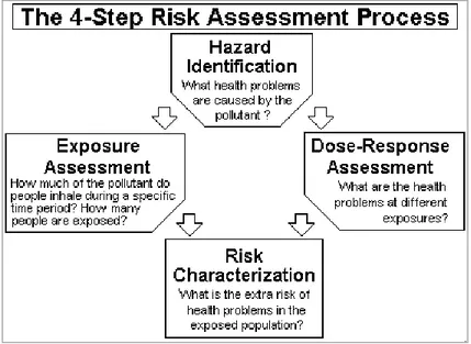
Risk assessment is the process that scientists and government officials use to estimate the increased risk of health problems in people who are exposed to different amounts of toxic substances. Risk assessment is a four-step process, including hazard
identification, dose-response modelling, exposure assessment and risk characterization. (Figure 2.1)
Figure 2.1: The four-step process in the risk assessment source http://www3.epa.gov/airtoxics/3_90_024.html
The dose-response modelling and the exposure assessment are combined to quantify the risk associated with current and anticipated exposures. The risk characterization step presents both
16
the qualitative likelihood that the hazard will occur and the quantitative estimates of risk.
The hazard identification is the description of the toxic potential of the analyzed agent. It is based on the evaluation of all available data (e.g., epidemiology, animal bioassay studies, and in vivo and
in vitro studies) to characterize the strength of evidence indicating
potential health effects that might occur in exposed human populations.
The first step in cancer risk assessment is to determine the carcinogenicity to humans of a specific exposure. The degree of uncertainty in the classification process depends on the availability of adequate and acceptable data. The best type of evidence comes from human studies.
The international institutions which are mainly involved in identifying and classifying the risk of carcinogenicity of agents and exposures are:
- the International Agency for Research on Cancer (IARC) which publishes the Monographs on the Evaluation of Carcinogenic Risks
to Humans,
- the US National Toxicology Program (NTP) which publishes every two months, the Report on Carcinogens,
- the U.S. Environmental Protection Agency (U.S. EPA) that develops assessment of carcinogenic risk.
17
The IARC categorizes agents, mixtures and exposures into five categories (IARC, 2010b):
Group 1: “the agent (mixture) is carcinogenic to humans. The
exposure circumstance entails exposures that are carcinogenic to humans”. This category is used when there is sufficient evidence of
carcinogenicity in humans. Exceptionally, an agent (mixture) may be placed in this category when evidence of carcinogenicity in humans is less than sufficient but there is sufficient evidence of carcinogenicity in experimental animals and strong evidence in exposed humans that the agent (mixture) acts through a relevant mechanism of carcinogenicity.
Group 2 A “the agent (mixture) is probably carcinogenic to
humans. The exposure circumstance entails exposures that are probably carcinogenic to humans”. This category is used when
there is limited evidence of carcinogenicity in humans and sufficient evidence of carcinogenicity in experimental animals. In some cases, an agent (mixture) may be classified in this category when there is inadequate evidence of carcinogenicity in humans and sufficient evidence of carcinogenicity in experimental animals and strong evidence that the carcinogenesis is mediated by a mechanism that also operates in humans.
Group 2 B: “the agent is possible carcinogenic to humans”. This
category is used for agents, mixtures and exposure circumstances for which there is limited evidence of carcinogenicity in humans and less than sufficient evidence of carcinogenicity in experimental animals. It may also be used when there is inadequate evidence of
18
carcinogenicity in humans but there is sufficient evidence of carcinogenicity in experimental animals.
Group 3: “the agent (mixture) is unclassifiable as to
carcinogenicity in humans”. This category is used most commonly
for agents, mixtures and exposure circumstances for which the evidence of carcinogenicity is inadequate in humans and inadequate or limited in experimental animals. Exceptionally, agents (mixtures) for which the evidence of carcinogenicity is inadequate in humans but sufficient in experimental animals may be placed in this category when there is strong evidence that the mechanism of carcinogenicity in experimental animals does not operate in humans.
Group 4: the agent (mixture) is probably not carcinogenic to
humans. This category is used for agents or mixtures for which there is evidence suggesting lack of carcinogenicity in humans and in experimental animals.
The Italian CCTN classification (CCTN, 1991) was similar to that from IARC and EPA. The CCTN classification distinguished, until recently, 5 groups:
Group A: carcinogenic to humans
Group B: probably carcinogenic to humans (B1 limited evidence of carcinogenicity in epidemiological studies; B2 sufficient evidence of carcinogenicity in experimental animals)
Group C: possible carcinogenic to humans
Group D: unclassifiable as to carcinogenicity in humans.
Group E: probably not carcinogenic to humans, based on animals studies
19
The general categories recognized by the 1986 EPA guidelines were (U.S. EPA, 1986):
Group A - Carcinogenic to Humans:
Group B - Probably Carcinogenic to Humans: Agents with sufficient evidence (i.e., indicative of a causal relationship) from animal bioassay data, but either limited human evidence (i.e., indicative of a possible causal relationship, but not exclusive of alternative explanations; Group B1), or with little or no human data (Group B2).
Group C - Possibly Carcinogenic to Humans: Agents with limited animal evidence and little or no human data.
Group D - Not Classifiable as to Human Carcinogenicity: Agents without adequate data either to support or refute human carcinogenicity.
Group E - Evidence of Non-carcinogenicity for Humans: Agents that show no evidence for carcinogenicity in at least two adequate animal tests in different species or in both adequate epidemiologic and animal studies.
The 2005 EPA Guidelines recommend expressing Weight of Evidence (WOE) by narrative statements rather than only hierarchical categories, and expressing them separately for the oral and inhalation routes. The statements are (U.S. EPA, 2005):
Carcinogenic to Humans
Likely to be Carcinogenic to Humans
Suggestive Evidence of Carcinogenic Potential
Inadequate Information to Assess Carcinogenic Potential Not Likely to be Carcinogenic to Humans
20
The evaluation of the dose-response analysis, through the calculation of the carcinogenic potential, is required to look at the quantitative aspect of the cancer risk. Dose-response analysis determines the relationship between the dose and the type of adverse response and/or the probability or the incidence of the effect (dose-response assessment). The complexity of this step in many contexts derives mainly from the need to extrapolate results from experimental animals (mouse, rat) to humans, and/or from high to lower doses, including the extrapolation from high acute occupational levels to low chronic environmental levels.
In the absence of human data to describe low-dose effects, two different approaches are most frequently used for dose-response analysis:
- approach for “threshold” endpoint; - approach for “non threshold” endpoint.
For non-cancer effects, it has been assumed that at low doses the body's natural protective mechanisms repair any damage caused by the pollutant, so a dose may exist below the minimum health effect level for which no adverse effects occur. The first dose of an agent which induces a response above zero (or above the control response) is usually referred to as a “threshold-dose”. The safe doses of chemicals associated with human diseases that are thought to act by a “threshold dose mechanism” are most frequently defined by applying safety factors to the no-observed-effect levels (NOEL) in animal bioassay studies. These so-called safe exposure levels are reported as acceptable daily intakes (ADIs) or reference doses (RfD).
21
For non-threshold effects (cancer), meaning that there may be some risks associated with any exposure, dose-response relationships based on observed incidence in humans and animals exposed at high doses are established and then extrapolation models are used to estimate the cancer risk associated with low-dose exposure. Essentially all chemicals can cause non-cancer adverse health effects if given at high enough doses. However, when the dose is sufficiently low, typically no adverse effect is observed. Thus, in characterizing the non-cancer effects of a chemical, the key parameter is the threshold dose at which an adverse effect first becomes evident. Doses below the threshold are considered to be safe, while doses above the threshold are likely to cause an effect (Grilli, 1992).
The threshold dose is typically estimated from toxicological data (derived from studies of humans and/or animals) by determining the highest dose that does not produce an observable adverse affect and the lowest dose which produces an effect. These are referred to as the “no-observed-adverse-effect-level” (NOAEL) and the “lowest-observed-adverse-effect-level” (LOAEL), respectively. However, in order to be conservative (protective), non cancer risk evaluations are not based directly on the threshold exposure level, but on a value referred to as the Reference Dose (RfD). The RfD is an estimate of a daily exposure to the human population that is likely to be without an appreciable risk of deleterious effects during a lifetime.
22
The RfD is derived from the NOAEL, (or LOAEL if a reliable NOAEL is not available) by dividing the NOAEL by an “uncertainty factor”. If the data were from studies in humans and the observations were considered to be very reliable, the uncertainty factor may be as small as 1.0. However, the uncertainty factor is normally at least 10 and can be much higher if data are limited. The purpose of dividing the NOAEL or the LOAEL by an uncertainty factor is to ensure that the RfD is not higher than the true threshold level for adverse effects. Thus, there is always a “margin of safety” built into an RfD, and doses equal to or less than the RfD are nearly certain to be without any risk of adverse effect. Doses higher than the RfD may carry some risk, but because of the margin of safety, a dose above the RfD does not mean that an effect will necessarily occur.
For cancer effects, the toxicity assessment process has two components. The first one is a qualitative evaluation of the weight of evidence that the chemical does or does not cause cancer in humans. For chemicals that are believed to be capable of causing cancer in humans, the second part of the toxicity assessment is to describe the carcinogenic potency of the chemicals. This is done by quantifying how the number of tumours observed in exposed animals or humans increases as the dose increases.
Usually, it is assumed that the dose-response curve for cancer has no threshold (there is no dose other than zero that does not increase the risk of cancer), arising from the origin and increasing linearly until high doses are reached. Thus, the most convenient descriptor of cancer potency is the slope of the dose-response curve at low dose (where the slope is still linear). Estimating the cancer slope
23
factor is often complicated by the fact that observable increases in cancer incidence usually occur only at relatively high doses, frequently in the part of the dose-response curve that is no longer linear. It is necessary to use mathematical models to extrapolate from the observed high dose data to the desired slope at low doses. For carcinogen pollutants, such as dioxins (PCDDs/PCDFs), polycyclic aromatic hydrocarbons (PAHs), polychlorinated biphenyls (PCBs) and some heavy metals (HMs), the EPA model (2005) assumes there is no safe threshold dose below which there is no health risk. The health risk, measured as the probability of developing cancer during the entire lifetime (assumed to be 70 years), is estimated by multiplying the exposure dose by the Cancer Slope Factor (CSF), which represents the estimate of the carcinogenic potency of the chemical.
Three approaches have been recommended by WHO (WHO/IPCS, 1998) for the quantitative assessment of cancer risk by inhaled PAHs:
1. Benzo(a)pyrene (B(a)P) Toxicity Equivalence Factors (TEFs) approach, based on individual potencies relative to B(a)P in order to obtain a benzo(a)pyrene equivalent ,
2. Benzo(a)pyrene surrogate epidemiological approach, which assumes that B(a)P is an indicator of all PAHs in the mixture, 3. comparative Potency Approach, which does not identify or
quantify the individual compounds.
The current study was based on the first approach. To evaluate the risk for human health of mixtures of PAHs, international and national Regulatory Agencies suggest to use equivalence factors (Potency Equivalence Factors, PEFs) (Table 2.1), which are
24
derived from carcinogenicity studies in small rodents and, where available, correlate the carcinogenic potential of each PAH or Nitro-PAH (NPAH) to that of B(a)P. The carcinogenic potential of B(a)P is assumed as 1 (WHO/IPCS, 1998).
B(a)P was chosen as the reference compound as it is considered the most appropriate indicator for the carcinogenic fraction of the large number of PAHs in air (WHO, 2000).
The potency of many individual PAHs and NPAHs relative to that of B(a)P was estimated in order to obtain B(a)P equivalents (B(a)Peq) (WHO/IPCS, 1998). The B(a)Peqs represent the relative carcinogenic potential of the corresponding mixture of PAHs and NPAHs to B(a)P.
The B(a)P equivalents are calculated multiplying the concentration of each component of the mixture by its PEF. Then the carcinogenic potency of the complete mixture of PAHs and NPAHs is estimated as the sum of each individual B(a)Peq with the following equation:
B(a)Peq = ∑ (PAHi x PEFi) + ∑ (NPAHi x PEFi)
In Table 2.1 the list of PEFs, which are currently available, is reported:
25 PAH PEF Benzo(a)pyrene 1 Anthracene 0.28* Benz(a)anthracene 0.1 Benzo(c)Phenanthrene 0.023* Benzo(b)fluoranthene 0.1 Benzo(j)fluoranthene 0.1 Benzo(k)fluoranthene 0.1 Benzo[rst]pentaphene 1.1* Benzo(g.h.i)perilene 0.01# Ciclopenta(c.d)pyrene 0.012* Chrysene 0.01 Dibenzo(a.h)acridine 0.1 Dibenzo(a.j)acridine 0.1 Dibenzo(a.h)anthracene 1# 7H-Dibenzo(c.g)carbazole 1 Dibenzo(a.e)pyrene 1 Dibenzo(a.h)pyrene 10 Dibenzo(a.i)pyrene 10 Dibenzo(a.l)pyrene 10 Phenanthrene 0.00064* Indeno[1.2.3-c.d]pyrene 0.1 5-Methylcrhysene 1 NPAH PEF Benzo(a)pyrene 1 1,6-Dinitropyrene 10 1,8-Dinitropyrene 1 6-Nitrochrysene 10 2-Nitrofluorene 0.01 1-Nitropyrene 0.1 4-Nitropyrene 0.1
Table 2.1: Potency Equivalency Factors (PEFs) for PAHs and NPAH in B(a)P
equivalent. Data from California EPA 1999 except (*) from MOE (Ministry of the Environment- Toronto-Canada) 1997 and (#) from CCME (Canadian Council of Ministers of the Environment) 2008.
26
The Cancer risk was assessed for each carcinogenic compound detected in the samples, whose Unit Risk (UR) value has been established by International Agencies (WHO, 2000 and CA-EPA, 2005).
The lifetime cancer risk (LCR) attributable to inhalation exposure was calculated by multiplying each UR estimated value by the average daily concentration of each compound:
LCRi = Ci x URi
The UR of each chemical corresponds to the excess lifetime cancer risk calculated as a result of the continuous exposure to B(a)Peq at a concentration of 1 ng/m3 in air over a lifetime of 70 years (U.S. EPA, 2011).
The UR used for calculations are reported in Table 2.2.
B(a)P Cancer UR 1,1 x 10⁻⁶/ng/m3 (CA EPA, 2005) B(a)P equivalents Cancer UR 8,7 x 10⁻⁵/ng/m3 [rounded to 1 x 10⁻⁴/ng/m3] (WHO, 2000) 1 x 10⁻⁴/ng/m3 (CCTN, 1991)
27
CHAPTER 3
Particulate Matter
3.1 General characteristics of Particulate Matter
Air pollution is already known to increase risks for a wide range of diseases, such as respiratory and heart diseases (Pope C.A., et al., 2009). Studies indicate that in recent years exposure levels have increased significantly in some part of the world, particularly in rapidly industrializing countries with large populations. The most recent data indicate that in 2010, 223.000 deaths from lung cancer worldwide resulted from air pollution (Straif, K., et al. 2013). In terms of the global burden of all diseases attributable to air pollution, lung cancers account for less than 7% of the 3.22 million estimated deaths (Hamra, G.B., et al, 2014).
Airborne air pollution is made up of particulate matter (PM) and hundreds of chemicals from natural sources and human activities (Brauer, M., et al, 2012; Fajerztajn et al., 2013)
In October 2013, the International Agency for Research on Cancer (IARC) classified outdoor air pollution and particulate matter from outdoor air pollution as carcinogenic to humans (Group 1) (Loomis, D., et al, 2013) based on sufficient evidence of carcinogenicity in humans and experimental animals and strong mechanistic evidence.
Particle pollution (also called particulate matter or PM) is the term for a mixture of solid particles and liquid droplets found in the air. PM is an air-suspended mixture of solid and liquid particles that
28
vary in time and space, in dependence on the source of emission, by the chemistry of the atmosphere and weather conditions (Pope and Dockery, 2006; Schauer et al., 1996). Among these particles, there are different components, such as sand, ash, dust, soot, acidic substances of various nature, plant substances, metal compounds, natural and artificial textile fibres, salts and elements such carbon or lead.
Among the chemical-physical characteristics of particles, the size is the one used for classification due to its intrinsic importance. In fact the atmospheric deposition rates of particles, their residence times in the atmosphere and deposition patterns within the lung are strongly influenced by particles size.
The behaviour of particles in the atmosphere and within the human respiratory system is determined largely by the physical properties, which have a strong dependence on size, varying from a few nanometres to tens of micrometres.
The aerodynamic properties of particles determine how they are transported in air and how they can be removed from it. These properties also govern how far they get into the air passages of the respiratory system. Additionally, they provide information on the chemical composition and the source of particles.
Airborne particles have irregular shapes and their aerodynamic behaviour is expressed in terms of the diameter of an idealised spherical particle known as aerodynamic diameter. The sampling and description of particles is based on this aerodynamic diameter, which is usually simply referred to as “particle size”. The aerodynamic diameter of a particle is the diameter of a spherical
29
particle having unit density and the same settling velocity from an airstream as the particle under study (Schlesinger 1985).
Based on size, particulate matter is often divided into two main groups:
coarse fraction contains the larger particles with a size ranging from 2.5 to 10 µm (PM10 – PM2.5).
fine fraction contains the smaller ones with a size up to 2.5 µm (PM2.5).
The particles in the fine fraction which are included in the 1 µm – 0.1 µm range are called ultrafine particles (UFP or UP).
One of the most important distinctions of particulate pollution is based on how the particles are introduced into the atmosphere: - primary particles, that are released directly from their source into the atmosphere. The main sources of primary PM are the land, the sea through soils carried by the wind and the generation of marine aerosol particles by the bursting of air bubbles in breaking waves. Road transport and industrial process are also major sources of primary particles. Primary PM and the precursor gases can have both man-made (anthropogenic) and natural
(non-anthropogenic) sources. Anthropogenic sources include combustion engines (both diesel and petrol), solid-fuel (coal, lignite, heavy oil and biomass) combustion for energy production in households and industry, other industrial activities (building, mining, manufacture of cement, ceramic and bricks, and smelting), (Schwarze et al., 2006) and erosion of the pavement by road traffic and abrasion of brakes and tyres. Agriculture is the main source of
30
ammonium. Natural sources include sea salt, naturally suspended dust, pollen and volcanic ash (EEA, 2012).
- secondary particles are subsequently formed within the atmosphere as a result of chemical reactions, producing substances of low volatility, which consequently condense into solid or liquid phase, becoming PM. Examples include sulphates and nitrates, formed from the oxidation of sulphur dioxide (SO2) and nitrogen
dioxide (NO2) in the atmosphere to acid, which are then neutralised
by atmospheric ammonia derived from agricultural sources. Carbonaceous particulates also contain a secondary fraction formed from the oxidation of volatile organic compounds (VOCs). Compared to primary particles, the chemicals processes involved in the formation of secondary ones are relatively slow and their persistence in the atmosphere is prolonged. The secondary particles are composed almost exclusively of particles of diameter less than 2.5 µm (Kelly et al., 2012).
The process by which secondary particles are formed is termed nucleation (Hamed et al., 2007);this term refers to the processes by which molecules of low volatility condense to form solid or liquid matter. There are two distinct types of nucleation process (Hinds, 1999). The first is heterogeneous nucleation. Most secondary particle formation in the atmosphere occurs by this process, newly formed substances condense onto existing particles causing the growth of those particles. The condensation processes are most effective on small and medium sized particles, causing these to grow larger. The second process is called homogeneous nucleation. The best known process of homogeneous nucleation occurs when sulphuric acid (H2SO4) is formed from the atmospheric oxidation
31
particles is the coagulation, which interact with those produced by nucleation. Coagulation can also affect products from homogeneous reactions occurring in the atmosphere.
Figure 3.1 shows, in schematic form, the typical size distribution
of airborne particles. Sizes range over several orders of magnitude. The smallest, freshly nucleated, particles are only 1–2 nm in diameter and contain only tens of molecules. At the other extreme, particles may be up to ~100 µm in diameter, which is comparable to a human hair. Particles as large as this rapidly settle out of the air and are of minor health significance because, although they can be inhaled, they do not generally penetrate beyond the nose and mouth. Consequently, air pollution research generally focuses on particles ≤10 µm in diameter, although it is important to recognise that larger particles are also present and contribute mass to ‘total suspended’ particles (TSP).
Figure 3.1 Schematic diagram of the size distribution of airborne particles
Adapted from: United Kingdom Department of Environment, Food, and Rural Affairs, Expert Panel on Air Quality Standards, 2004
32
3.2 Composition of Particulate Matter
Airborne particles have a very diverse chemical composition that varies both in time and space. Air samples of particulate matter from urban areas from around the world typically show the same major components, although in considerably different proportions according to the sampling location (Harrison and Yin 2000). Airborne particles contain both major and minor components. The major components, normally, include the following:
Sulphate – arises mainly as a secondary component from
atmospheric oxidation of SO2, although there may be a small
primary component that arises from sea salt or mineral matter such as gypsum;
Nitrate – normally present as NH4NO3, which results from
the neutralisation of HNO3 vapour by NH3, or as sodium nitrate
(NaNO3), due to displacement of hydrogen chloride from NaCl by
HNO3 vapour;
Ammonium – generally present in the form of ammonium
sulphate ((NH4)2SO4) or NH4NO3;
Sodium and chloride – in sea salt;
Elemental carbon – black, graphitic carbon formed during
the high temperature combustion of fossil and contemporary biomass fuels;
33
Organic carbon – carbon in the form of organic
compounds, either primary, resulting from automotive or industrial sources, or secondary, resulting from the oxidation of volatile organic compounds (VOCs);
Mineral components – crustal materials (rock and soil) are
rich in elements such as aluminium, silicon, iron and calcium. These are generally present in coarse dusts that arise from, for example, wind-driven entrainment processes, quarrying, construction and demolition processes;
Water
Biological materials - bacteria, spores, pollens, debris and
plant fragments; generally coarse in size, considered as part of the organic
Carbon component in most studies rather than as a separate
biological component.
Minor components include the following:
Trace metals – many metals such as lead, cadmium,
mercury, nickel, chromium, zinc and manganese are used in metallurgical processes. Some occur as impurities or additives in fuels and others are used in industrial products. These and other uses cause emissions to the atmosphere, but concentrations are generally very small;
Trace organic compounds – although the total mass of
organic compounds can comprise a significant part of the overall mass of particles, it is made up of a very large number of individual organic compounds, each of which is present at a very low concentration. Such organic compounds vary greatly in
34
composition and include aliphatic and aromatic hydrocarbons, heterocyclics and oxygenates such as aldehydes, ketones and carboxylic acids. Generally speaking, organic compounds that arise directly from fuel combustion processes have relatively high hydrogen to carbon ratio, whereas secondary organic compounds are more oxidised and polyfunctional carbonyl and carboxylic acids species are often present (Figure 3.2).
Figure 3.2: Composition (%) of particulate typical of urban source (a), natural (b) and rural (c). (source Ministry of the Environment, Land and Sea)
35
3.3 Effects of Particulate Matter
The respiratory system is the primary target of airborne particles which are inhaled and tend to accumulate in the airways. The comprehension of PM dosimetry is important to understand the toxic potential of PM. The respiratory tract can be divided into three main regions: the extrathoracic region, the tracheobronchial
region and the alveolar or pulmonary region, where gas exchange
occurs (U.S. EPA, 2004; Figure 3.3). The most important PM property driving deposition rate in the airways is particles size: in fact inhaled particles penetrate the respiratory system depending on mechanisms, such as inertial impact, sedimentation, diffusion, electrostatic attraction, which are related to particles size (U.S. EPA, 2004). Each region of the respiratory system presents typical clearance mechanisms that protect the organism from inhaled external agents. Filtration of the nasal passages is the main protective mechanism in the head region, while mucus secretion and ciliated cells act to remove particles and other external bodies in the tracheobronchial region. At the alveolar surface the clearance machinery is constituted by macrophages and type II pneumocytes that perform particles removal through their phagocytic activity. Particular attention should be finally paid to ultrafine particles which, thanks to their tiniest dimensions, may be able to escape macrophages removal and penetrate the alveolar-capillary barrier (Elder et al, 2006).
The size of the particle is a main determinant of where in the respiratory tract the particle will come to rest when inhaled. Simple nomenclature is used to indicate the different degrees of relative penetration of a PM particles into the cardiovascular system
36
(Figure 3.3). Larger particles are generally filtered in the nose and throat via cilia and mucus (inhalable fraction). The particulate matter smaller than about 10 micrometers, referred to as PM10, can
settle in the bronchi and lungs (thoracic fraction). Particles smaller than 2.5 micrometers, PM2.5, tend to penetrate into the gas
exchange regions of the lung (alveolus) and very small particles (<100 nanometers) may pass through the lungs to affect other organs (respirable fraction)(Nemmar et al, 2002).
37
3.3.1 PM exposure and health effects
Numerous epidemiological studies have established an association between short or long-term exposure to various airborne particulates and human mortality and morbidity (Chen and Lippmann, 2009; Rueckerl et al, 2011; Schwarze et al, 2006). Studies have found a significant correlation between excess mortality and short-term exposure to high concentration of ambient particulate matters (Bell and Davis, 2001; Schwartz and Markus 1990). A population-based study on data collected in 6 US cities suggested an association between long-term exposure and human mortality (Dockery et al. 1993; Laden et al., 2000). Studies conducted in European and United States cities reported short-term exposure to PM10 to increase mortality of a value ranging from
0.46 to 0.62 % (mean 0.5 %) for each 10 μg/m3 increment in the
daily concentration (Cohen et al., 2004; Katsouyanni et al., 2001). Pope and colleagues (Pope et al., 2009) reported that PM2.5
concentrations fell by a third from the early 1980s to 1990s across major US metropolitan areas, with each 10 µg/m3 reduction associated with an increase in life expectancy of 0.61 years (Pope et al., 2009).
38
A report from the prospective Cancer Prevention II study of the American Cancer Society, including about 500.000 participants, showed that each 10 µg/m3 increase in fine particulate air pollution was associated with a 6% increase in all-cause mortality, and with a 9% increase in risk of cardiopulmonary mortality and 14% increase in risk of lung cancer (Pope et al., 2002).
Recent long-term studies show associations between PM and mortality at level well below the current annual WHO air quality guideline level for PM2.5 (10 µg/m3). Scientific evidence does not
suggest a threshold of exposure to PM below which no adverse health effects would be anticipated (WHO, 2006a, 2006b, 2013). Another important epidemiological project, ESCAPE (European Study of Cohorts for Air Pollution Effects), was recently published. In the first study the association between long-term exposure and lung cancer was confirmed unequivocally (Raaschou-Nielsen et al., 2013) whereas the second study was focused on the relationship between long-term mortality and air pollution (Beelen et al., 2013). In the first study the authors did not explore the causes of death. Recently it was reported an 18% increase in lung cancer incidence for each 5 µg/m3 increase in PM2.5 concentration in this cohort
(Raaschou-Nielsen et al., 2013). The second study presents an analysis of pooled data from 22 longitudinal cohort studies across Europe, including more than 360.000 people followed up for an average of 13.9 years (Beelen et al., 2013). The authors show a 7% increase in natural cause mortality with each 5 µg/m3 increase in PM2.5 concentration (Beelen et al, 2013). These results are
39
diverse European populations exposed to PM2.5 concentration
below the limits recommended in existing guidelines.
Toxicological studies widely described the genotoxic effect of PM in humans (Møller et al., 2008; Billet et al., 2008; Oh et al., 2011). These results, together with the epidemiological evidences of lung cancer mortality for long-term exposure to PM2.5, suggest a role for
DNA damage in PM related health effects (U.S. EPA, 2009). In agreement with epidemiology, fine PMs usually have a higher genotoxic potential compared to coarse fractions, in in vitro and in
vivo models (Chackra et al., 2007; de Kok et al., 2005; Park et al.,
2005); moreover, PMs from industrialized, traffic and wood/biomass combustion areas have been found to exert a stronger effect on DNA (Shi et al., 2006; U.S. EPA, 2004).
The high genotoxicity of these PMs is due to the particles composition and source, since particles derived from combustion processes have been found to be rich in metals and organic compounds, such as polycyclic aromatic hydrocarbons (PAHs), which are notorious genotoxic elements (Mehta et al., 2008). Actually, different mechanisms have been reported for PM-induced DNA damage, which include oxidative stress, organics metabolites formation and direct interaction of particles organic compounds with the DNA (Møller et al., 2008). Generation of ROS and consequent oxidative stress is widely recognized as an important toxicological mechanism in PM induced DNA double strand breaks/oxidation and possibly lung cancer (Shi et al., 2006; Møller
40
High level of ROS production could lead to cell apoptosis and tissue damage, which may contribute to lung injury as seen in human or animal models following acute exposure to PM. ROS are able to initiate a series of redox signalling cascades to induce inflammatory responses and cytokine production found in both acute and chronic PM exposures (Akthar et al., 2010; Diabate et
al., 2011). It has been demonstrated that transition metals (Cr, Cd,
Ni and arsenic) and aldehydes present in PM can inhibit DNA damage repair mechanisms and this effect may enhance the genotoxic effect of PM and contribute to lung carcinogenesis (Mehta et al., 2008).
41
3.4 Polycyclic aromatic hydrocarbons (PAHs)
A major group of chemicals that are found in complex mixtures are the polycyclic aromatic hydrocarbons (PAHs), a family of more than 1500 compounds (NTP, 2012) comprised of two or more fused aromatic rings, found both in their native and substituted form. PAH molecules are made up of three or more benzene rings. PAHs are ubiquitous environmental contaminants that are formed as a result of incomplete pyrolytic processes and to which humans are exposed through inhalation, ingestion and dermal absorption. In nature, PAHs are found in coal tar, crude oil, being also produced during combustion processes, such as forest fires and volcano eruptions (Rajput and Lakhani, 2009). In urban atmospheres, where the occurrence of PAHs in ambient air is of a particular concern, the majority of PAHs come from vehicular traffic emissions (Castellano et al, 2003; Gaga et al, 2012; Omar et
al, 2002); however, PAHs are also emitted by trains and aircrafts.
Stationary sources such as domestic heating, various industrial processes, waste incineration and energetic production systems are other significant contributors of PAHs to outdoor atmosphere (WHO, 1998). Due to their persistence in environmental matrices, as well as their adverse effects on human health, PAHs have been classified as priority pollutants by the US EPA – United States Environmental Protection Agency (U.S. EPA, 2004). PAHs are generally insoluble in water but can be solubilised in organic solvents or organic acids. This means that in aqueous environments PAH are generally found adsorbed on particulates and on humic matter; PAHs have an affinity for environmental matrices, such as
42
sediments and soils due to their greater hydrophobicity and through bioaccumulation and thus magnified in the food chain (WHO – IARC, 1998; Senthilkumar et al., 2008).
Once released into the atmosphere, PAHs are partitioned between the PM and gas phases depending on their molecular weight (Chen
et al., 2005; Furuuchi et al., 2007). As a result, larger molecular
weight PAHs (< 4 rings) are almost exclusively bound to particulate matter, while low molecular weight PAHs (3 rings) can also be found dissolved in water (Wang et al., 2007; Khaiwal et al., 2006).
Links between human exposure to complex PAH mixtures and development of diseases including cancer, and respiratory and cardiovascular diseases have been described previously (IARC, 2010; Pope et al, 2002). Despite their structural similarities, PAHs vary greatly in their carcinogenic potency, with both individual and complex mixtures of PAHs classified as possible or probable human carcinogens by the International Agency for Research on Cancer (IARC) (IARC, 2010).
Subsequent to exposure, PAHs cross through cell membranes and, being lipophilic, undergo intracellular accumulation by concentrating in liquid droplets (Murphy et al., 2008). Studies from Ramesh et al have shown that PAHs can initiate and accelerate atherosclerosis and aneurysm in experimental animals through a cascade of biochemical events that involve oxidative stress and inflammation (Ramesh et al, 2015; Prins et al., 2012; Wang et al., 2009).
43
3.4.1 Carcinogenic effects of PAHs
The binding of PAHs to DNA, and the associated effects that occur as a result, is considered the major mechanism of PAH-induced mutagenesis and carcinogenesis. Like chemical carcinogens, PAHs require activation through a series of enzymatically-catalyzed reactions to form their active metabolites (Conney et al., 1982; Huberman et al.,1976; Sims et al., 1974). The cytochrome P-450 family of enzymes, in particular CYP1A1, CYP1A2 and CYP1B1, are primarily involved in bioactivation of PAHs and formation of their reactive intermediates (Pelkonen and Nebert, 1982; Shimada and Fujii-Kuriyama, 2004). Many PAHs are ligands for the aryl hydrocarbons receptor (AhR), which has different roles in metabolism including regulation of the different bioactivating enzymes (Baird et al., 2005; Pàlkovà et al, 2015). The aryl hydrocarbon receptor (AhR) is a basic helix-loop-helix/Per-Arnt-Sim (bHLH/PAS) transcription factor essential for adaptive responses to xenobiotics (Hao et al., 2013). Polycyclic aromatic hydrocarbons and halogenated aromatic hydrocarbons (HaHs) are by far the most common classes of AhR ligands (Denison et al., 2003). For example, B(a)P is the most widely studied PAH. It is usually chosen as a marker for evaluating the toxicity of PAH mixtures, since the available toxicological data provide a sufficient basis for the conventional risk assessment, in which it can be assumed that the toxicity of all PAHs is equivalent to that of B(a)P. As a result of recent toxicological research, B(a)P has become classified as human carcinogen, group 1 (IARC 2012).
44
B(a)P is activated through a three step enzymatic mechanism involving (Jarvis et al., 2014) initial metabolism by CYP enzymes to B(a)P-7,8-epoxides, followed by conversion to B(a)P-7,8-diols by epoxide hydrolase, and final transformation to the ultimate reactive B(a)P-7,8-diol-9,10-epoxide metabolites, again by CYP enzymes (Conney, 1982) (Figure 3.4). This mechanism of activation has also been shown for other PAHs.
45
The tumorigenic and mutagenic activities of many PAHs have been linked to the ability of their diol epoxide metabolites to bind covalently to exocyclic amino groups on purine bases to form either stable bulky adducts or depurinating adducts which are released from DNA, leaving abasic sites (Rogan et al., 1993; Szeliga and Dipple, 1998). Following bioactivation and reaction with DNA, the PAH metabolites can induce mutation that either activate oncogenes or inactivate tumour suppressor gene as part of their carcinogenic mechanism (Jarvis et al., 2014).
46
CHAPTER 4
Materials and Methods
4.1 Experimental design
Six different monitoring campaigns were performed by Arpa ER in the period 2011-2013 (Table 4.1).
For each campaign, the toxicological characterization in BALB/c 3T3 cells and the cancer risk assessment were performed. During my training period at Harlan Cytotest Research, (Rossdorf, Germany), I have had the opportunity to perform the Bhas 42 CTA. Two different campaigns (Winter 2012 and Summer 2013) were evaluated with both cell transformation assays in order to compare the results obtained by the two testing methods.
47
The experimental design for the toxicological characterization is reported in Figure 4.1.
Figure 4.1 Study design for the toxicological characterization. Green lines: activity performed by Arpa ER
48
In Figure 4.2, the experimental workflow for cancer risk assessment is reported.
Figure 4.2 Experimental workflow for cancer risk assessment. Green lines: activity performed by Arpa ER.