a v a i l a b l e a t w w w . s c i e n c e d i r e c t . c o m
j o u r n a l h o m e p a g e : w w w . i n t l . e l s e v i e r h e a l t h . c o m / j o u r n a l s / d e m a
Electric device improves bonds of simplified
etch-and-rinse adhesives
Guido Pasquantonio
a, Franklin R. Tay
b, Annalisa Mazzoni
c, Pietro Suppa
c,
Alessandra Ruggeri Jr.
c, Mirella Falconi
c, Roberto Di Lenarda
d, Lorenzo Breschi
d,∗aDepartment of Dental Sciences, University of Tor Vergata, Rome, Italy bDepartment of SAU & FAL, University of Bologna, Bologna, Italy
cDepartment of Oral Biology & Maxillofacial Pathology, School of Dentistry, Medical College of Georgia, Augusta GA, USA dDepartment of Biomedicine, School of Dentistry, University of Trieste, Trieste, Italy
a r t i c l e
i n f o
Article history:
Received 19 April 2005 Received in revised form 23 February 2006 Accepted 8 March 2006
Keywords:
Dentin bonding systems Electric field
FEISEM Microtensile Dentin
a b s t r a c t
Objectives. This study investigated the effects of an electric field produced by a new device
for the application of etch-and-rinse adhesives on demineralized dentin surfaces.
Methods. Three simplified etch-and-rinse adhesives (Single Bond, Prime&Bond NT and
One-Step) were applied with the electric device and compared with controls prepared with disposable sponges. Specimens were processed for microtensile bond strength test and nanoleakage investigation using high resolution SEM.
Results. Microtensile testing revealed higher bond strengths (p < 0.05) for all adhesives tested
when electricity was used. Adhesive interfaces prepared with electric impulses exhibited very homogenous hybrid layers with minimal nanoleakage compared with the controls.
Significance. The use of electricity produced by a new electronic device during the application
of dentin adhesives may increase adhesive adaptation to the dentin substrate and improve dentin hybridization due to the substrate modifications induced by an electric field on the demineralized dentin organic matrix.
© 2006 Academy of Dental Materials. Published by Elsevier Ltd. All rights reserved.
1.
Introduction
Despite the recent developments in adhesive dentistry to reduce the number of working steps and to simplify the clin-ical procedure, the new simplified adhesives do not produce better results in in vitro tests[1]or improve clinical efficacy[2]. Ironically, the most user-friendly simplified adhesives, the so-called self-etching one-step adhesives, exhibited the lowest bond strengths and the least predictable clinical performances over time when compared with the multi-step etch-and-rinse and self-etch systems[2,3].
Previous reports have shown that one of the major disad-vantages of the etch-and-rinse systems is incomplete
infiltra-∗Corresponding author at: UCO of Dental Sciences, University of Trieste, Via Stuparich, 1, I-34129 Trieste, Italy. Tel.: +39 040 3992192; fax: +39 040 912579.
E-mail address:[email protected](L. Breschi).
tion of the exposed dentin matrix due to the collapse of the collagen fibrils after removal of the mineral phase[4–6]. A layer of disrupted collagen fibrils may interfere with adhesive pen-etration and the formation of the hybrid layer. Incompletely infiltrated voids within these hybrid layers may be revealed with a tracer such as silver nitrate using transmission (TEM) or scanning electron microscopy (SEM). Tracer infiltration in the absence of a physical interfacial gap has been referred to as nanoleakage[4], which may occur in interfaces bonded with either etch-and-rinse or self-etch adhesives. Field emission-SEM (FE-emission-SEM) and TEM studies showed that entrapment of water within adhesive interfaces may create additional voids or tracks that could be revealed by these tracers[7–9].
0109-5641/$ – see front matter © 2006 Academy of Dental Materials. Published by Elsevier Ltd. All rights reserved. doi:10.1016/j.dental.2006.03.009
There is a general consensus that the quality of dentin hybridization is more important than the actual thickness of the hybrid layers in establishing the long-term seal of bonded restorations[1]. The existence of incompletely infiltrated col-lagen fibrils within the hybrid layers and additional entrapped water within the polymerized adhesives may expedite the degradation of resin–dentin bonds, resulting in clinical and visibly detectable microleakage[10–12].
All dentin adhesives are currently applied mechanically to tooth structures using either disposable sponges or brushes. Recently, a technique has been introduced that utilizes an electric field to enhance resin infiltration into the deminer-alized collagen matrices of acid-etched dentin[13]. This elec-trical device (ElectroBond, Seti, Rome, Italy) is incorporated in a handpiece to which a small application sponge is attached
[14]. It creates an electric potential difference between the etched tooth substrate and the adhesive-filled sponge that produces a constant electric flow during adhesives applica-tion. The purpose of this in vitro study was to examine the quality of dentin hybridization achieved with the use of this electrical application technique for the bonding of three etch-and-rinse adhesives to crown dentin. The null hypothesis tested was that there is no difference in the quality of dentin hybridization achieved between a conventional mechanical adhesive application technique and the use of an electric impulse assisted adhesive application technique.
2.
Materials and methods
Thirty non-carious human third molars were used in this study after informed consent had been obtained for their use for research, under a protocol approved by the Human Assur-ance Committee of the University of Bologna, Italy. The teeth were stored in a 0.5% chloramine T solution at 4◦C and used within 1 month after extraction. Occlusal enamel and root dentin were removed perpendicular to the long axis of each tooth by a low speed diamond saw under water irrigation (Micromet, Remet, Bologna, Italy). A standardized smear layer was created on the exposed coronal dentin with 180-grit wet silicon carbide paper. Each tooth was longitudinally sectioned into two halves (experimental and control halves) to create two similar bonding substrates. The specimens were etched for 15 s with 35% phosphoric acid gel (3M EPSE, St. Paul, MN, USA) and rinsed for 15 s. Excess water was removed using lint-free tissues, in accordance with the wet bonding technique.
The specimens were randomly assigned (N = 10) to three simplified etch-and-rinse adhesives: Single Bond (3M ESPE), Prime&Bond NT (Dentsply DeTrey, Konstanz, Germany) and One-Step (Bisco Inc., Schaumburg, IL, USA). For experimental halves of each tooth, the adhesive was applied using Elec-troBond (Seti, Rome, Italy) that delivered a direct current (dc) between the acid-etched dentin (working as cathode) and the adhesive-filled sponge (working as anode)[14]. As specimens showed different electrical resistances to dc, the tested Elec-troBond automatically induced an electric flow over 20A throughout the adhesive interface during the application pro-cedure. To permit electricity conduction under in vitro bond-ing conditions, the tooth to be bonded was fitted into a copper ring that was wired to the electric application device and
bond-ing was performed by holdbond-ing the copper rbond-ing with ungloved fingers. For the control halves of each tooth the selected adhe-sive was applied in the same manner, but with the electric current switched off. A single blind study design was used, in which the operator performing the bonding procedure was not aware of the operating state of the electrical device (i.e. switched-on mode or switch-off mode). The electric applica-tor was used with a brushing motion, continuously supply-ing energy (experimental halves). To ensure consistency in all the experimental groups, two adhesive coats were applied for 10 s each, irrespective of the adhesive selected, and air-dried for 5 s to evaporate the respective solvent. Each adhesive was cured for 20 s with a quartz–tungsten–halogen light-curing unit (Curing Light 2500, 3M ESPE). A 2-mm thick layer of microhybrid resin composite (Z250, 3M ESPE) was subse-quently placed over the bonded dentin surface and polymer-ized for 20 s.
2.1. Microtensile bond strength evaluation
The pulp chamber of each tooth half was bonded with Single Bond in accordance with the manufacturer’s instruction and filled with resin composite (Z250). Sticks with surface areas of approximately 0.9 mm2were created from each specimen using a low speed saw under water irrigation. The dimen-sion of each stick was individually measured with a digital caliper to the nearest 0.01 mm and the calculated area was recorded for subsequent bond strength calculation. The spec-imens were observed using a stereomicroscope to avoid the inclusion of sticks containing residual enamel during bond testing. The sticks were stored in deionized water for 24 h, attached to a modified jig for microtensile testing and stressed under tension until failure with a universal testing machine at a crosshead speed of 1 mm/min. The failure modes were evaluated at 50× (Stemi 2000-C, Carl Zeiss Jena GmbH, Ger-many) and classified as cohesive (C), adhesive (A), or mixed (M) failures. The number of prematurely debonded sticks per group during specimen preparation was also recorded. As val-ues were not normally distributed (Kolmogorov–Smirnof test), a Mann–Whitney test was used to compare the data with sta-tistical significance set at˛ = 0.05.
2.2. FE-SEM nanoleakage evaluation
Bonded sticks from the center of each bonded tooth half were employed for nanoleakage evaluation. The specimens were covered with nail varnish, leaving 1 mm free at the interface. They were immersed immediately in a 50 wt.% ammoniacal AgNO3solution that was prepared according to the method described by Tay et al.[7]. After immersion in the tracer solu-tion for 24 h, the specimens were photodeveloped to reduce the diamine silver ions ([Ag(NH3)2]+) into metallic silver grains. The silver-impregnated sticks were polished with 1200-grit silicon carbide papers to remove the surface silver deposits and expose the bonded interfaces. They were dehydrated and dried in accordance with the technique reported by Suppa et al.[9]. Dehydrated, uncoated specimens were examined using the in-lens mode of a FE-SEM (JSM 890, JEOL, Tokyo, Japan) at 7 kV and 1× 10⁻¹²A. Images were obtained with both sec-ondary electron (SE) and back-scattered electron (BS) signals.
Fig. 1 – Percentages of the failure modes after microtensile test analyzed using stereomicroscope. Fracture were classified as: A, adhesive; CC, cohesive in composite; CD, cohesive in dentin; M, mixed. N = numbers of specimens.
Additional silver-impregnated sticks were fixed and dehy-drated as described previously and embedded in epoxy resin (Epon 812, Fluka, Switzerland). Undemineralized sections con-taining the resin–dentin interfaces were obtained with an ultramicrotome (Ultra-Cut S, Leica, Austria) and examined with the FE-SEM for comparison with the previously exam-ined, unembedded bulk specimens.
3.
Results
Significant increases in microtensile bond strength were observed for the three adhesives when they were bonded to dentin using the electric impulse assisted application tech-nique (Table 1; p < 0.05). Premature failures due to prepara-tion procedures were not included in the statistical analy-sis (Table 1). No differences were observed in failure mode between the control and the electric impulse assisted appli-cation groups (Fig. 1).
FE-SEM images of the bonded specimens revealed the pres-ence of electron-dense silver grains that had been confirmed by energy-dispersive X-ray analysis [9]. Unembedded bulk specimens and resin-embedded specimen sections exhib-ited similar leakage patterns using FE-SEM. Low magnifica-tion images revealed significant reducmagnifica-tions in the number
and dimensions of silver clusters using the electric impulse assisted application technique when compared to the con-trols (Figs. 2 and 3). In particular, nanoleakage from the basal part of the hybrid layers created under an electric impulse was reduced even if silver deposits in adhesive and the superficial part of the hybrid layers still remained (Figs. 2a and c and 3a and c). Clusters of silver deposits could be also identified in proximity to the dentinal tubule orifices (Figs. 2c and 3d), which were probably created by the entrap-ment of water derived from outward fluid flux.
4.
Discussion
Dentin consists of mineralized type I collagen fibrils embed-ded in a highly hydrophilic matrix of proteoglycans, other non-collagenous proteins and water[15]. Ideally, after acid-etching, the adhesive should completely infiltrate the delicate fibrillar network. Optimal resin infiltration is crucial to maximize bond strength[5,6]and bond durability[1], as unprotected collagen fibrils may be hydrolyzed over time[11,16,17].
The results of the present study showed that ElectroBond
[14]was able to improve bonding efficacy, as shown by the increased microtensile bond strength when compared with the control application technique. The bond strength data were further supplemented by FE-SEM findings that revealed reduced nanoleakage in bonded interfaces that were created by adhesive application under an assisted electrical impulse.
Interpretations of the infiltration phenomenon that accom-panied the use of an assisted electric impulse may be achieved with different hypotheses: the difference in electric potential between the etched dentin and adhesive could have either enhanced the penetration of these adhesive monomers, or could have altered the wetting characteristics of the etched dentin surface, thereby improving the spreading of the adhe-sives. Low-frequency electric currents cause dielectric disper-sion in tissues, which is associated with enhanced ionic diffu-sion and interfacial polarization, although the extent of such improvements is dependent upon the complexity of the sub-strate[18].
The use of electrical currents to facilitate the rate of per-meation of ionized substances (such as different drugs, anes-thetics, etc.) through dentin has been previously reported
[19–21]. For example, delivery of drugs through intact and caries-affected dentin is enhanced with the use of iontophore-sis[20]. Likewise, resin monomer infiltration through a par-tially demineralized collagen network may be accelerated via the use of electric currents. This should not be related
Table 1 – Microtensile bond strength data obtained applying the adhesives on parts A (experimental) and B (control) of the same teeth with the experimental electric device or in accordance with the manufacturers’ instructions (control)
Adhesive Electric impulse assisted
adhesive applicationa (MPa)
Control, conventional sponge applicationa(MPa)
Prime&Bond NT 38.2 (4.0) [68] A 26.6 (5.2) [48] B
One-Step 38.8 (4.1) [51] A 28.7 (5.1) [57] B
Single Bond 44.2 (7.1) [63] A 29.4 (8.6) [52] B
Fig. 2 – FE-SEM micrographs of bonded interfaces in Single Bond that were created using the electric impulse assisted application technique (experimental) or conventional sponge application technique (control). (a) An epoxy resin-embedded experimental specimen section that was cut with diamond knife revealing dentin (D), hybrid layer (H) adhesive (A) and composite (C). Small clusters of silver grains with a diameter of 3–6m were seen within the hybrid layer, created using the application (pointers). (b) An epoxy resin-embedded control specimen section revealing extensive nanoleakage within the hybrid layer (H); dentin (D), adhesive (A), composite (C). (c) A high magnification view of the silver grains that were found within the adhesive layer (pointers) in an unembedded experimental section. (d) An unembedded control specimen revealing extensive silver deposits (asterisk) within the entire thickness of the hybrid layer. The insert depicts a high magnification of these silver clusters. Similar regions with extensive nanoleakage were scattered along the interface and separated by regions with less nanoleakage.
to an increase in the intrinsic dentin permeability but to a faster rate of impregnation as ionic monomers are mov-ing across the dentin with increasmov-ing ion mobilities that are caused by the imposed electrical gradient[21]. Adhesive infil-tration may be positively influenced by the application of electric currents, since polar resinous components such as polyelectrolyte (polyalkenoic acid copolymer), HEMA, PENTA and BPDM are present and may interact with the electric field generated by the device. Single Bond contains polyalkenoic acid polymer that is charged. One-Step contains biphenyl dimethacrylates (BPDM) that contains two ionisable carboxylic acid groups. Prime&Bond NT contains dipentaerythritol-pentaacrylate phosphate ester (PENTA), which is a phosphate-substituted methacrylate. Thus, iontophoresis may accelerate the movement of ions across dentin[19]. Iontophoresis may also remove excess water from the hybrid layer. Since it is likely that “water-saturated” demineralized dentin also con-tains salt ions from collagen and dentinal fluid, iontophoretic electric fields may induce electro-osmotic fluid movement. That is, the attraction of ions to the applied electrode osmot-ically obligates fluid to follow the ions. This may occur at the same time when the oppositely charged monomers are being driven into the hybrid layer. The use of electric currents may also influence the dentin substrate by increasing its wettabil-ity and rate of water substitution by the adhesive monomers, as the affinity between these molecules and water dipoles is modified.
We speculate that apart from the polarization effect con-curred upon the polar functional groups of the adhesive monomers, these molecules may also be physically attracted by the electrical field, thus increasing the flow throughout the intricate layer of demineralized dentin.
Application of electricity may also transiently alter the bio-chemical characteristics of the dentin organic matrix, since collagen fibrils are polar in nature by virtue of the 3.7 Debye dipole moment present in the peptide unit of the triple helix
[22]. Moreover, the dipoles of water molecules contribute to the polar nature of collagen[23], as they exist in a hydrated state during the process of wet bonding. Molecular model-ing of collagen with adjacent charged residues predicted that side-chains participate in the formation of intra-chain and inter-helix ion pairs[24,25]. Moreover, proteoglycans associ-ated with dentin collagen fibrils are characterized by lateral glycosaminoglycans chains with negatively charged residues that are responsible for the high water binding and tis-sue swelling behavior of this collagen–proteoglycan complex
[26,27]. Indeed, the latter represents a polyelectrolyte hydro-gel with a diffusion coefficient of ions that approximates to the diffusion coefficient of free, unbound water[28].
Since both collagen and proteoglycans are polar, the authors speculate that an electric impulse assisted adhesive application technique influences the three-dimensional arrangements of the demineralized dentin network, which in turn, may favorably influence adhesive infiltration. By
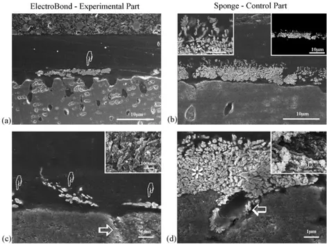
Fig. 3 – FE-SEM micrographs obtained from epoxy-embedded specimens that were sectioned with a diamond blade. (a) Bonded interface obtained after the application of One-Step with the electrical device, revealing silver clusters within the hybrid layer (pointer). (b) Bonded interface obtained when One-Step was bonded to acid-etched dentin without the use of an electrical impulse (control). Extensive silver deposits were visible along the entire thickness of the hybrid layer, with an increasing gradient of silver deposits along the base of the hybrid layer. Left insert reveals high magnification features of the silver infiltration. Right insert shows a back-scattered image of the same area. (c) Bonded interface created with Prime&Bond NT that was applied using the electrical device, revealing minimal nanoleakage (pointers) within the hybrid layer. Insert illustrates high magnification features taken from an unembedded specimen, with silver grains identified along the exposed collagen fibrils. (d) Bonded interface created when Prime&Bond NT was applied to acid-etched dentin without the use of an electrical impulse (control). Extensive silver deposits (asterisk) could be seen within the base of the hybrid layer and around the dentinal tubules (open arrow). Insert reveals uninfiltrated collagen fibrils that were covered with silver grains.
suspending this highly polar organic matrix in a water envi-ronment, such as with the use of a wet bonding technique, not only are the dipoles of individual molecules modified, but the quaternary protein structure may also be altered due to the existence of intra- and interfibrillar hydrogen bonds among the collagen fibrils [29] and the partially retained proteoglycans [27]. Moreover, collagen fibrils have also been demonstrated to possess piezoelectric capability that enable then to alter their three-dimensional arrangement
[19,30,31]in relation to the amount of adsorbed water[32]. For these reasons, the application of an electric current to a demineralized, wet dentin surface may alter the dentin surface characteristics, inducing subtle orientation changes within the organic fibrillar network that favors adhesive infiltration.
In conclusion, this study represents a preliminary attempt to identify the improvements in microtensile bond strengths and reduction in nanoleakage that are associated with the use of an electric impulse assisted application technique for the bonding of simplified etch-and-rinse adhesives to wet, acid-etched dentin. The null hypothesis tested has to be rejected since the use of an electric impulse assisted adhe-sive application technique improved the quality of dentin hybridization and bond strength values when compared with a conventional mechanical adhesive application technique. Further studies are required to quantify the effect of electric
currents on adhesive infiltration, for instance, by correlat-ing the voltage with the solubility parameter for hydrogen bonding of adhesive monomers with the use of a model-ing approach in which all available adhesive constituents are known. When this additional information is available, this electric impulse assisted application technique may be considered ready for in vivo testing.
r e f e r e n c e s
[1] De Munck J, Van Landuyt K, Peumans M, Poitevin A, Lambrechts P, Braem M, et al. A critical review of the durability of adhesion to tooth tissue: methods and results. J Dent Res 2005;84:118–32.
[2] Tay FR, Pashley DH. Have dentin adhesives become too hydrophilic? J Can Dent Assoc 2003;69:724–31.
[3] Van Meerbeek B, De Munck J, Yoshida Y, Inoue S, Vargas M, Vijay P, et al. Buonocore memorial lecture. Adhesion to enamel and dentin: current status and future challenges. Oper Dent 2003;28:215–35.
[4] Sano H, Shono T, Takatsu T, Hosada H. Microporous dentin zone beneath resin-impregnated layer. Oper Dent 1994;19:59–64.
[5] Spencer P, Swafford JR. Unprotected protein at the dentin adhesive interface. Quintessence Int 1999;30:501–7. [6] Yoshida Y, Van Meerbeek B, Snauwaert J, Hellemans L,
characterization of adhesive tooth-biomaterials interfaces. J Biomed Mater Res 1999;47:85–90.
[7] Tay FR, Pashley DH, Yoshiyama M. Two modes of
nanoleakage expression in single step adhesives. J Dent Res 2002;81:472–6.
[8] Li H, Burrow MF, Tyas MJ. The effects of thermocycling regimens on the nanoleakage of dentin bonding systems. Dent Mater 2002;18:189–96.
[9] Suppa P, Breschi L, Ruggeri A, Mazzotti G, Prati C, Chersoni S, et al. Nanoleakage within the hybrid layer: a correlative FEISEM/TEM investigation. J Biomed Mater Res B: Appl Biomater 2005;73B:7–14.
[10] Yoshida Y, Nagakane K, Fukuda R, Nakayama Y, Okazaki M, Shintani H, et al. Comparative study on adhesive
performance of functional monomers. J Dent Res 2004;83:454–8.
[11] Hashimoto M, Tay FR, Ohno H, Sano H, Kaga M, Yiu C, et al. SEM and TEM analysis on water degradation of human dentinal collagen. J Biomed Mater Res 2003;66:287–98. [12] Breschi L, Prati C, Gobbi P, Pashley DH, Mazzotti G, Teti G, et
al. Immunohistochemical analysis of collagen fibrils within the hybrid layer: a FEISEM study. Oper Dent 2004;29:538–46. [13] Pasquantonio G, Suppa P, Tay FR, Biasotto M, Contardo L,
Breschi L. Bond strength of adhesives applied with an electric device. J Dent Res 2005;84(Spec Iss A):Abs 152, www.dentalresearch.org.
[14] Pasquantonio G, Breschi L, Petrone A. Method and device for preparing the hard structures of teeth for the application of dental restorative materials. US Patent 6,641,396; November 4, 2003.
[15] Marshall GW, Marshall SJ, Kinney JH, Balooch M. The dentine substrate: structure and properties related to bonding. J Dent 1997;25:441–58.
[16] Burrow MF, Satoh M, Tagami J. Dentin bond durability after three years using a dentin bonding agent with and without priming. Dent Mater 1996;12:302–7.
[17] Hashimoto M, Ohno H, Kaga M, Endo K, Sano H, Oguchi H. In vivo degradation of resin-bonds in humans over 1 to 3 years. J Dent Res 2000;79:1385–91.
[18] Jastrzebska M, Kocot A. Ionic diffusion and space charge polarization in structural characterization of biological tissues. Eur Phys J 2004;14:137–42.
[19] Pashley DH, Livingston MJ, Outhwaite WC. Dentin
permeability: changes produced by iontophoresis. J Dent Res 1978;57:77–82.
[20] Puapichartdumrong P, Ikeda H, Suda H. Facilitation of iontophoretic drug delivery through intact and caries-affected dentine. Int Endod J 2003;36:674–81. [21] Padula C, Colombo G, Nicoli S, Catellani PL, Massimo G,
Santi P. Bioadhesive film for the transdermal delivery of lidocaine: in vitro and in vivo behavior. J Control Release 2003;88:277–85.
[22] Pethig R. Dielectric properties of body tissues. Clin Phys Physiol Meas 1987;8(Suppl A):5–12.
[23] Jayasuriya AC, Scheinbeim JI, Lubkin V, Bennett G, Kramer P. Piezoelectric and mechanical properties in bovine cornea. J Biomed Mater Res 2003;66A:260–5.
[24] Kaltz EP, David CW. Energetics of intrachain salt linkage formation in collagen. Biopolymers 1990;29:791–8. [25] Kaltz EP, David CW. Unique side-chain conformation
encoding for chirality and Azimuthal orientation in the molecular packing of skin collagen. J Mol Biol 1992;228: 963–9.
[26] Bourdon MA, Oldberg A, Perschbacher M, Ruoslahti E. Molecular cloning and sequence analysis of a chondroitin sulfate proteoglycan cDNA. Proc Natl Acad Sci
1985;82:1321–5.
[27] Breschi L, Gobbi P, Lopez M, Mazzotti G, Falconi M, Teti G. Immunohistochemical analysis of dentin: a double labeling technique. J Biomed Mater Res 2003;67A:11–7.
[28] Comper WD, Laurent TC. Physiological function of connective tissue polysaccharides. Physiol Rev 1978;58:255–315.
[29] Ramachandran GN, Chandrasekharan R. Interchain hydrogen bonds via bound water molecules in the collagen triple helix. Biopolymers 1968;6:1649–58.
[30] Ghosh S, Mei BZ, Lubkin V, Scheinbeim JI, Kramer P, Bennett G, et al. Piezoelectric response of scleral collagen. J Biomed Mater Res 1998;39:453–7.
[31] Marino AA, Gross BD. Piezoelectricity in cementum, dentine and bone. Arch Oral Biol 1989;34:507–9.
[32] Ueda H, Fukada E. Piezoelectric relaxations in collagen gelatin system. Polym J 1979;11:103–11.