Alma Mater Studiorum
Alma Mater Studiorum –
– Università di Bologna
Università di Bologna
DOTTORATO DI RICERCA IN
ONCOLOGIA E PATOLOGIA SPERIMENTALE
Ciclo XXIV Settore Concorsuale di afferenza:
SCIENZE BIOLOGICHE, BIOMEDICHE E BIOTECNOLOGICHE
TITOLO TESI
RECONSTRUCTIVE MICROSURGERY AND TISSUE
ENGINEERING IN MUSCULO-SKELETAL ONCOLOGY –
INNOVATIVE TECHNIQUES
Presentata da: Dr.ssa CLAUDIA DI BELLA
Coordinatore Dottorato Relatore
Prof SANDRO GRILLI Prof PIETRO RUGGIERI
INDEX 1. INTRODUCTION
1.1. Surgery in musculo-‐skeletal oncology 1.1.1. Bone
1.1.2. Soft tissues
1.2. Surgery reconstruction after bone sarcoma resection 1.2.1. Prosthetic bone reconstructions
1.2.2. Biologic bone reconstructions
1.3. Plastic surgery and reconstructive microsurgery in musculo-‐skeletal oncology
1.3.1. Soft tissue reconstructions 1.3.2. Free flaps in sarcoma surgery
1.3.3. Vascularized flaps and the role of innervated reconstructions
1.4. Tissue Engineering in orthopaedics
1.4.1. Tissue Engineering for bone reconstruction after tumor resection
2. GOAL OF THIS PROJECTS AND THESIS
2.1. Bone regenerative and reconstructive techniques 2.2. Soft tissue microsurgical reconstructive strategies
after tumor resections
3. BONE REGENERATION PROJECT
3.1. Background 3.2. Previous studies 3.3. Experimental design 3.4. Materials and methods
3.4.1. Surgical procedure 3.4.2. Analysis of results 3.5. Results
3.5.1. Animal model
3.5.2. Clinical complications
3.5.3. Transplanted vessel evaluation 3.5.4. Imaging
3.5.5. Histology 3.6. Discussion
4. SOFT TISSUE REGENERATION PROJECT
4.1. Background
4.2. Patients and Methods 4.3. Results 4.4. Discussion 5. CONCLUSIONS 6. BIBLIOGRAPHY
1. INTRODUCTION
1.1 SURGERY IN MUSCULO-‐SKELETAL ONCOLOGY
Thanks to the recent and constant improvements in imaging techniques, chemotherapy protocols, radiotherapy and surgery, the possibility to perform wide tumor resection and subsequent functional reconstruction has become more and more achievable, and now it represents the starting point for the great part of treatment approach in muscolo-‐skeletal tumor surgery. Compared to 30-‐40 years ago, ablative surgery (amputation) is reserved to very rare cases, in which the size of the lesion or the neuro-‐vascular structures involvement influence the treatment choice leading to an absolute impossibility to achieve reconstructive options (1-‐5). For the majority of the cases, instead, the surgical gold standard is a wide resection followed by reconstruction. Reconstruction techniques are evolving
from purely anatomical to functional for both bone and soft tissues, in some cases with the inclusion of vessels and nerves.
In sarcoma surgery the concept of “margin” is of paramount importance for an adequate treatment. In general, the base principle in the resection of malignant tumors of bone and soft tissues is to obtain “wide” margins, which means excision of the tumor surrounded by a reasonable amount (usually at least 2 cm all around) of healthy tissue with no evidence of tumor (6-‐9). In fact, any other type of resection, such as “marginal” or “intra-‐lesional”, is considerate inadequate for sarcoma surgery, as the most likely outcome would be a recurrence of the lesion and possible metastatic spread (10-‐13). The gold standard, therefore, is the wide excision. This type of resection was considered inadequate in the early ages of sarcoma surgery definitions, while the “radical” excision was considered the best possible treatment. The radical margin, or compartimental, is a resection of the lesion together with the entire compartment in which the lesion is located. It is clear therefore, that in this case the aggressiveness of the surgery is massive and that many times this surgery was synonymous of amputation (Fig.1).
It has been demonstrated over the years that, in terms of overall survival and disease free survival, the outcome of a wide excision are comparable with the outcome of a radical excision, therefore sarcoma surgery has moved towards the concept of “limb sparing surgery” with a decrease of morbidity at the level of the resection (14). Despite
bone or the soft tissue is still quite extensive, and generally there is the necessity for reconstruction in order to guarantee the best possible outcome to the patient. It is therefore extremely important having reconstruction techniques which allow not only a good anatomic and aesthetic coverage of the defect, but even an acceptable functional recovery, especially now that the long term survivorship is definitely longer than in the past.
-‐ Bone sarcomas
Many type of bone sarcomas are known, and the majority involves young adults. Osteosarcomas (OS) and Ewing sarcomas (ES), in fact, are, among the others, the most common primary bone sarcomas, and the majority of the patients affected are in their 2nd
decade of life. On the other hand, Chondrosarcomas (CS), are more commonly diagnosed in adult patients, in general older than 40 years of age. In terms of treatment strategies, while OS and ES usually respond relatively well to chemotherapy (and radiotherapy in the case of ES), CS are not responsive to adjuvant therapies and surgery constitutes the main treatment option. In any case, regardless of the help of chemotherapy or radiotherapy, surgery remains one of the corner stones of treatment, which implies removal of the diseased bone and, therefore, subsequent necessity for reconstruction.
When a sarcoma involves the bone, the main choices for the surgeon are: 1) mega-‐prosthesis, which can replace the lost bone with adjacent joint, and 2) bone or osteoarticular allograft, which are used when a biologic reconstruction is preferred to a prosthetic one. The advantages of the biologic reconstruction compared to a prosthetic one are to be seen in the view of an improved and longer outcome, especially in the young patient, together with the possibility to re-‐ attach structures such as tendons and ligaments which otherwise would be lost. Therefore some authors believe that this technique, if
superior quality of the reconstruction compared to the prosthetic technique (15, 16).
It is well known, however, that the risks associated with a biologic reconstruction are generally superior compared to a prosthetic one, and in fact many complications can occur and are understandably discouraging, especially because they increase with the increase of the follow-‐up (16-‐18). It is therefore absolutely necessary an improvement of this technique, especially in the possible strategies that allow a re-‐vitalization of the allograft. In this optic, tissue engineering could represent the ideal solution, and researchers around the world are working to define the best possible strategy to obtain the perfect and most durable biologic reconstruction.
-‐ Soft tissues sarcomas
Soft tissue sarcomas (STS) are malignant lesions arising in the muscle or subcutaneous tissue of the body, or more rarely in a nerve or in the joint space. As it happens for bone sarcomas, there is a wide variety of different pathologic types of sarcomas, which are defined on the base of the type of cells that they express the most from the histologic point of view; the most common STS are liposarcoma, Malignant Fibrous Histocytoma (MFH), Leyomiosarcoma, Pleyomorphic sarcoma, Schwannoma. All these lesions are more commonly diagnosed in adult patients, in general after the 4th decade
of life, and the vast majority of them responds relatively well to adjuvant radiotherapy. From the surgical point of view, depending on where the lesion is, what is its size and which compartment of the body is involved, the excision could be quite extensive and could involve large part of the skin and/or the subcutaneous tissue. In the majority of the cases the surgery is “conservative” enough to guarantee a good primary closure of the wound. In some cases, however, there is the necessity of cutaneous or musculo-‐cutaneous flap in order to be able to close the wound, and this happens when the lesion oblige the surgeon to excise big part of the superficial tissue with the impossibility of a primary wound closure.
Thanks to plastic surgery techniques, it is possible now to excise extremely big soft tissue lesions without the necessity to amputate the limb, as long as the main neuro-‐vascular bundle is not involved and can be safely dissected.
The use of plastic flaps, however, is able to guarantee only a coverage of the defect left after the tumor excision, without the possibility of regain the function of the lost muscle(s). The use and advantages of innovative plastic reconstruction techniques is the goal of the second part of this study.
1.2 SURGICAL RECONSTRUCTION AFTER BONE SARCOMA RESECTION
After bone loss for sarcoma resection, there is the necessity to replace the defect. To do this, the surgeon can choose between a prosthetic reconstruction and a biologic reconstruction.
-‐ PROSTHETIC BONE RECONSTRUCTIONS
After massive bone resections in limb sparing surgery, there is clearly the necessity for anatomic and functional reconstruction. When the lesion, and therefore the resection, involves an area of the bone particularly close to, or at the level of the joint, the gold standard reconstruction technique is the use of a mega-‐prosthesis (19-‐23). These type of special prosthesis are particularly useful in tumor surgery, because they not only replace the joint, as a normal orthopaedic prosthesis does in case of, for example, osteoarthritis, but they allow the replacement of the resected bone as well (21, 24, 25). Sometimes these prosthesis can reconstruct the entire length of the bone, as in the case of a total femur replacement, in which both the knee and the hip joint are replaced together with the entire femur (22, 26-‐28). Mega-‐prosthesis are the best possible strategy when there is the necessity to sacrifice a joint, because they allow a hypothetical
return to function when the muscles that govern the replaced joint are still in place and functional (29-‐33).
The main quality of mega-‐prosthetis, together with their ability to replace the missing bone and joint, is that they guarantee a relatively long functional life and the possibility for the patient to start mobilizing in the immediate days after the surgery (29, 31, 32).
Despite the numerous advantages in the use of mega-‐prosthesis,
Fig. 2 Example of bone sarcoma located to the proximal femur, treated with proximal femur resection and reconstruction with proximal femur mega-‐prosthesis
limit their success: 1) The risk of infection is very high, and in particular is much higher compared to joint replacement prosthesis for arthritis (34). This is because the bone-‐cartilage resection is much bigger, the length of the surgery is longer with longer exposure of the wound, usually patients are immune-‐compromised due to neo-‐ adjuvant chemotherapy or radiotherapy and, finally, the soft tissue coverage of the prosthesis might be insufficient. In a recent study, infection has been proven to be the most common mode of failure for this type of prosthesis (35); 2) The achievement of complete function is generally somehow limited due to the loss of muscle and the difficulty and sometimes impossibility to re-‐attach musculo-‐tendinous insertions into the prosthesis (25, 36); 3) In the young patient, the longevity of the prosthesis is usually insufficient, with the need of multiple revisions and loss of bone stock, which increases the morbidity of this treatment together with the risk of subsequent infection; 4) Aseptic loosening has been considered one of the most common cause of failure in both cemented and non cemented implants, and is associated with reduction of the longevity of the reconstruction; at the same time, this complication limits the possible surgical options for revision because of the decrease of available bone stock (35).
Taking into account all these aspects, it is clear how an improvement in the current techniques appears necessary, because there is the absolute need to improve the clinical and functional
outcome of our reconstructions and, at the same time, the quality of life of these already proven patients.
-‐ BIOLOGIC BONE RECONSTRUCTIONS
When the bone segment involved by the tumor is relatively far from the articular joint, a biologic reconstruction can be taken in consideration. The reason for the relative exclusion criteria in case the joint is involved refers to the early degeneration of the cartilage, which leads to early osteoarthritis and consequent necessity for relatively early new joint replacement intervention (18, 37-‐40). In some occasion, however, especially when the patient is very young (usually up to the adolescence), a biologic reconstruction can be attempted even to replace a joint, with the hope that re-‐ vascularization and synovial fluid re-‐constitution would be able to give enough nutrients to the replaced cartilage to survive (41, 42).
The most common material used for biologic reconstruction is allogenic bone (or bone allograft). This type of tissue is provided by the so-‐called “bone banks”, which are institutes that store bones harvested from organs donors. One of the most important qualities of bone and cartilage allografts compared to other tissues or organs is that there is no need for immune-‐suppressive therapy for the recipient, because this tissue does not have enough surface antigens to
The main advantages in the use of bone allografts are to be seen in the possibility to replace significant bone and sometimes osteo-‐ chondral defects maintaining all the main bone-‐tendon and bone-‐ ligament attachments, therefore allowing a better and more anatomical function compared to a mega-‐prosthesis (44, 45). If satisfactory results are obtained, the reconstruction can last for many years with a great functional advance for the patient. (Fig.3). Potentially the implication of a biologic reconstruction, therefore, is extremely attractive, however there are still many reasons why the use of allograft is limited to selected cases and maintain the use of prosthesis as a gold standard in the majority of the cases.
The main disadvantages in the use of a biologic reconstruction are: 1. Risk of non-‐union or pseudo-‐arthrosis (46). The integration
between the host bone and the allograft is one of the weak points of this technique. The allograft, in fact, is a structure that is constituted by dead bone/cartilage, therefore the integration process occurs only in one direction, from the host to the graft, and depends only on the quality of the host bone. The process ideally should, with time, replace the entire allograft with host bone, but in reality the integration between the 2 structures involves only few millimiters of the implant, while the rest of the allograft remains constituted by non-‐vital bone (47-‐49). As a consequence of this limited and slow integration process, the rest from any activity has to be prolonged for at least 3 months, and sometimes
patients require up to 12 months before they can start weight bear on the operated limb (50, 51).
2. Risk of infection (52). The causes are the same present for prosthesis, such as the long length of the surgery and the fact
Fig. 3 Case of a 6-‐year-‐old boy with a diagnosis of osteosarcoma in whom a transepiphyseal resection and reconstruction with an intercalary allograft was performed. (A) The anteroposterior radiograph of the knee after neoadjuvant chemotherapy shows the osteosarcoma compromises the medial cortex with a varus deformity. (B) A coronal T1-‐weighted MRI image shows the metaphyseal and diaphyseal extension. (C) A schematic drawing shows the preoperative planning of the reconstruction. (D) The anteroposterior radiograph shows the intercalary allograft after 2 years of followup. (REF: Muscolo DL, Ayerza MA, Aponte-‐Tinao L, et al. Allograft reconstruction after sarcoma resection in children younger than 10 years old. Clin Orthop Relat Res. 2008;466:1856-‐1862, with kind permission from Springer Science+Business Media.)
aspect of the allograft biology needs to be added. The allograft, in fact, being for the main part non-‐vascularized and non-‐vital, can represent an ideal environment for micro-‐ organisms.
3. Risk of fracture (53). This risk is again due to the avascularity and non vitality of the bone, which cannot undergo to the usual mechanism of micro-‐cracks and remodelling of normal bone. In this situation, in fact, the micro-‐cracks will occur without repair and with time this will lead to a proper fracture.
4. Risk of early osteoarthritis. In the case of osteo-‐articular allografts, cartilage remains constituted by entrapped and not-‐vital chondrocytes, which cannot receive any nutrient, nor from the synovial fluid nor from the subchondral bone. This will inevitably lead to degenerative changes and, in the long term, early onset of osteo-‐arthritis (54, 55).
When all these possible risks of failure are analysed together the fear on the use of massive allografts is understandable. However, with a closer analysis, it is clear how every single element of risk is strictly correlated to the vitality of the graft: an allograft that cannot be incorporated and, therefore, remains constituted by dead bone, will have all of these risks which, moreover, will increase overtime. On the other hand, if a strategy to guarantee early re-‐vascularization and re-‐ vitalization of the graft is found, all these risks will decrease and potentially disappear, and this will greatly improve the treatment
options that can be offered to patients with bone tumors. It is evident, therefore, how a technique to allow bone allograft re-‐vitalization could potentially represent an ideal solution for these patients and a valid option to prosthetic replacements.
1.3 PLASTIC SURGERY AND RECONSTRUCTIVE MICROSURGERY IN MUSCULO-‐SKELETAL ONCOLOGY
When the tumor involves the soft tissues (muscles, ligaments and tendons, adipose tissue, skin), there are mainly two strategies of wound closure after the resection: 1. Primary closure. 2. Plastic reconstruction, from simple skin grafts to more complex techniques such as free or rotational flaps. The indications depend on the amount of tissue that needs to be replaced, the available skin coverage and the necessity or not to cover prosthesis or allografts.
-‐ SOFT TISSUE RECONSTRUCTIONS
In many cases, when the tumor involves muscles or subcutaneous tissue, there is the chance for the orthopaedic surgeon to suture the wound without the need of the plastic surgeon. This happens only when the resection, being wide, is still limited and allows a
general rule, the main goal of the wound closure, is to re-‐suture muscular groups with the same function or, in general, without an opposition function. In many cases, a substantial portion of the muscle where the lesion was has been resected, leaving the surgeon with a small fragment of tissue that helps only as “gap filler”. Usually, the function of the limb is severely impaired, because there is no reconstruction of the functional units lost with the resection, and other muscles, with time, tend to hypertrophy to partially replace the lost function. In addition to this, aesthetic is also compromised because of asymmetry with the contra-‐lateral side and “depression” at the level of the resection. In many cases this is only a minimal discomfort for the patient, but sometimes, in areas such as the adductor compartment of the thigh, following the resection an abundant seromatous/haematic collection follows (56).
Plastic reconstructive surgery is the solution when the defect is too big to be covered with primary closure; plastic techniques allow a more accurate anatomic reconstruction, usually with a better accepted aesthetic results (57-‐59). Plastic reconstructive techniques are numerous and depend on the size of the defect, the area that needs to be replaced, the amount of muscle that has to be replaced, and the quality of the skin. In the case of big sarcoma resections, typically a free or rotational muscolo-‐cutaneous flap is the surgeon’s choice because it provides adequate coverage despite the bulkiness that sometimes remains at the level of the flap (60).
-‐ FREE FLAPS IN SARCOMA SURGERY
The use of free or rotational flaps is an extremely valuable resource in soft tissue reconstruction; in the most recent years, the use of this type of flaps is not anymore restricted to the cases in which there is the necessity to cover the prosthetic or biologic bone reconstruction or in cases in which the primary closure cannot be achieved. In fact, the use of flaps has became more and more common in sarcoma reconstruction, with particular indications in which, despite the possibility of a primary closure, a flap is preferred because of the decrease of post-‐operative complications. This is the case, for example, of the lesions in the adductor compartment of the thigh. In this area, in fact, a wide excision of the lesion often ends with the neurovascular bundle isolated and dissected free of the lesion and of the muscles; even when the primary closure is achievable, the large empty space and the loss of the lymphatic drainage together with a big number of the venous branches, brings to the formaton of a large haematoma/seroma which cannot be promptly re-‐absorbed by the body with a severe increased rick of post-‐operative infection (56). Furthermore, the radiation therapy given preoperatively will modify the quality of the surrounding tissues, with a high prevalence of fibrosis and a further decrease of the ability to re-‐absorb the collection. In a second time, this collection eventually tends to
recurrence results difficult, with an understandable stress for the patient and the clinician. Being able to avoid this post-‐operative trend is of paramount importance, therefore very often now a free or transposed flap is the reconstruction of choice when the lesion is localized in that area or in areas with similar outcomes. Musculo-‐ cutaneous flaps, in fact, not only have the ability to fill the space with autogenous tissue, but, being vascularized, can guarantee the survival of the tissue and the decrease of the morbidities (Fig. 4)
Various techniques are available to the plastic surgeon when there is the need to fill the empty space following tumor resection. In general, a muscolo-‐cutaneous flap is the most common method used in this type of reconstruction (61). The flap can be rotated from an adjacent area (as, for example, in the gastrocnemius rotation to cover the proximal tibia mega-‐prosthesis), or can be removed from a different part of the body and positioned where necessary (free flap). This last scenario is one of the most common. The most used free flaps are: parascapular, gracilis and TRAM (transverse rectus abdominis muscle) (62).
To survive, the free flaps need to be secured to an artero-‐vascular pedicle with micro-‐anastomosis at the level of the vascular bundle in the recipient area, through termino-‐terminal anastomosis, or, when a major vessel is involved, through a termino-‐lateral anastomosis. The ability of the micro-‐surgeon to perform these sutures is extremely important for the success of the procedure, because if these fail the flap will not survive and a new procedure will be necessary.
Fig. 4 Example of post radiotherapy soft tissue sarcoma in a 42 yo man located in
the antero-‐lateral compartment of the lower leg. The wide resection lead to important muscle sacrifice, with extensive soft tissue loss and bone exposure. Note in quadrant 3 the inner margin of the resection, showing the healthy muscle covering the lesion.
-‐ VASCULARIZED FLAPS AND THE ROLE OF INNERVATED RECONSTRUCTIONS
Musculo-‐cutaneous flaps are therefore a very important resource for the reconstruction, however the implanted muscle is not functional. When a large soft tissue lesion is excised, together with the tumor a large amount of muscle is lost because has been replaced by the sarcoma or because its resection is required to obtain wide margins. This functional unit cannot be reconstructed with a traditional flap, because the muscle that is re-‐implanted will be alive but not functional. In many cases this represents a minor problem, because other fellow muscles can partially replace the lost function, but when the major component of a joint mechanism is lost, such as, for example, the entire extensor mechanism of the knee, then the possibility of having a functional reconstruction becomes very appealing In the case of the anterior compartment of the thigh, in fact, a wide excision usually involves the complete loss of the knee extensors (quadriceps) which act as knee stabilizers as well; in this situation the patient is not able to put weight on the affected limb without an appropriate support which stabilizes the knee in extension.
It is clear, therefore, how important is to find new advanced techniques for soft tissue reconstructions which allow not only the coverage of the defect with viable tissue, but even the ability to re-‐gain
at least partially the lost function in order to achieve the best possible outcome.
Recently, advances in plastic reconstruction techniques lead to the development of a new procedure that will permit not only the anatomical filling of the defect with vital tissue, but the function of this tissue in synergy with the other muscles around. The innervated free flap, in fact, is a free flap that is harvested not only with its vascular pedicle, but even with its nerve pedicle for motor function. Therefore, once positioned, these flaps can be connected to the nerve that has been resected during the tumor procedure and, hence, thanks to the reinsertion into the remaining muscle or directly into the bone/prosthesis, these flaps recover their contractile mechanism and can improve the patient functional outcome.
Innervated flaps have been attempted for the first time in animal studies by Tamai in 1970 (63), where the feasibility of free neurovascular muscle transplantation has been showed. These studies began the era of microvascular flap innovation, and since then few other reports described this technique in clinical situations. In orthopaedic surgery, innervated flaps has been used for sensate reconstruction of the heel and the ankle, in which the medial plantar flap has been used (64), and has been shown to be a versatile flap capable of providing immediate sensate coverage for heel and ankle defects. Rigorous neurologic testing has shown that when carefully dissected with preservation of its sensory branch, normal sensation
innervated flaps has been used for foot and hand reconstructions with different results (65, 66), and it has been demonstrated that while reinnervating a muscle flap with a sensory nerve will permit reinnervation of the muscle and the overlying skin, it is still unclear whether this provides a superior result in durability and, in the case of the foot, gait (67).
On the other hand, there is not a wide literature with regards to motor innervated flaps for the restoration of a muscle function rather than sensation. In sarcoma surgery, this would be the main goal after the resection of the mass, especially when it involves muscular groups important for ambulation, such as the extensor mechanism of the knee or the hip, or when it involves the upper limb and the daily living activities. The first case reports have been presented started form the mid 90s, describing the use of free muscle transplantation for the reconstruction of the anterior compartment of the thigh (68), and more recently the surgical technique has been throughoutly described (69). Starting from the reconstruction of the quadriceps, more muscular functions have been proposed to be restored with this technique, and in 1999 Ihara described the use of innervated flaps for reconstruction after resection of soft tissue sarcomas achieving very good aesthetic and functional results (70, 71). This procedure, although complex and time expensive, is very promising in the view of improving as much as possible the outcome in limb salvage procedures, and is the focus of the second part of this thesis.
1.4 TISSUE ENGINEERING IN ORTHOPAEDICS
Tissue Engineering is a branch of Regenerative Medicine that involves the creation of an organ or a tissue in laboratory, and its subsequent transplant in vivo.
Musculoskeletal tissues, such as bone and cartilage, are since many years the focus of a very wide number of studies in tissue engineering, because they represent a relatively “easy” tissue to reproduce in laboratory and with a very large clinical demand.
Fig. 5 Representation of Stem Cells and their possibilities
The cells derived from the bone marrow with proliferative capacity toward the mesenchymal lineage are called Mesenchymal Stem Cells (MSC) and are considered multipotent cells. Multipotency represents the characteristic of these cells to reproduce themselves and, at the same time, to differentiate toward a selective lineage for different tissue and organs (Fig. 5).
Fig 6. Differentiation abilities of MSC
By using special culture media with selected growth factors, these cells have the ability to grow and to become more and more differentiated toward the chosen lineage (Fig. 6).
When osteogenic growth factors are chosen, for example, these cells after few passages in culture start to loose their proliferative capacity and gain the ability to differentiate themselves, and therefore become progressively osteoblasts and finally osteocytes with the capacity to produce bone matrix and mineralize. With this progression, the more the cells are moving towards the end of the differentiating line, the more they lose the ability to replicate themselves. On the other hand, when the cells are in a very undifferentiated status, they theoretically can replicate with no end. Growth factors used in laboratory to promote differentiation are chemical constituent that act by miming the function of autologous growth factors; it is however possible to use directly the autologous growth factors when they are collected from the cells that biologically constitutes their origin and their reservoir.
With respect to osteogeneic tissue, the most used growth factors are the ones harvested from platelets, such as Platelet Derived Growth Factor (PDGF) and Transforming Growth Factor β (TGF-‐β). While platelets are obtained from the peripheral blood and subsequent centrifugation and exclusion of the red component, to obtain Mesenchymal Stem Cells (MSC) a bone marrow aspiration is necessary because they are one of the component of the red marrow.
It is well demonstrated now that MSC are found not only in the bone marrow, but potentially in every tissue, because it is postulated that in each tissue there is a reservoir of progenitor cells (72-‐74). This has been specifically demonstrated in adipose tissue, which currently represents a widely used source of MSC (called Adipose Derived Stem Cells, ADSC) especially because of the simplicity of the harvest and the consistency of the results (75-‐77).
Stem cells, as specified before, regardless of their origin, when cultured in vitro with osteogenic media, can differentiate and transform themselves in cells forming bone (78, 79). To make this process, growth factors and cells are not enough; in fact, there is the necessity of a third element that allows cells to growth in a 3D construct, which represents the in vivo structure that they will need to reproduce (80). The third element is a scaffold, which act as a tridimensional support for the proliferation and, at the same time, direct the differentiation toward the correct line. Many materials can be used as scaffolds, going from simple organic fluid such as collagen, to more complex 3D structures constructed with highly advanced technology, such as nano-‐materials. With respect to bone, materials that can possibly be used as scaffolds need to have specific characteristics such as: a strength modulus similar to bone, adequate porosity, biocompatibility and the ability to function as osteoconductive and osteoinductive material. Some of the most used scaffolds used in laboratory are Hydroxyapatyte, Ceramic, Poly-‐α-‐ hydroesters and other natural polymers as collagen and Chitosan.
Overall, however, the best possible scaffold for bone reconstruction is bone itself, which has already the required 3D structure and can be demineralized to further stimulate the osteogenesis.
Stem Cells, Growth Factors and Scaffold are the three fundamental elements in every tissue-‐engineering project. Stem cells are the primary elements from which the new tissue will form, Growth Factors are the “fuel” that stimulate these cells into the desired direction and differentiation, and the Scaffold is the 3D structure in which the cells can growth (Fig 7 and 8).
Fig. 7 Scaffold, Cells and Growth Factors are the 3 fundamental element in every tissue engineering process. With an adequate substrate and environment the final outcome is the achievement of the two most important processes in bone regeneration: osteo-‐induction and osteo-‐conduction
Fig. 8 The three elements together (left of the picture), when in adequate culture, can create a tissue that is suitable for bone reconstruction and, ideally, after implantation, can reproduce healthy bone.
1.4.1 Tissue Engineering for bone reconstruction after tumor resection
Massive bone allografts are widely used in orthopedic reconstructive surgery to replace bone defects due to trauma or oncologic resections. The effectiveness of the clinical outcome depends on bone healing time and type of graft–host integration: the larger the amount of bone to be replaced, the more difficult the integration process becomes (81). This process may involve only 20% of the graft in 5 years, as shown by studies on retrieved allografts (82,
83). In these studies it was shown that revascularization plays a key role to achieve good allograft repair: a delay or absence in revascularization may impair new bone formation and influence the implant outcome. Therefore, accelerating and increasing this process may be critical to allow bone healing and integration.
Numerous techniques have been proposed in order to increase the revascularization, and therefore the regeneration, of a bone allograft. These include the use of stem cells, the use of growth factors (such as BMPs), or the combination of the two.
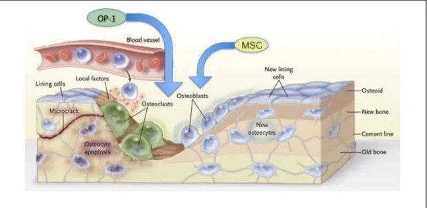
Bone morphogenetic proteins (BMPs) are a family of pleiotrophic factors, members of the Transforming Growth Factor β super family (TGF-‐β), that can influence the migration, proliferation, and differentiation of bone progenitors cells (84). It was postulated that the addition of BMPs to an allograft could increase its integration with the host bone by increasing the vascularization. Several studies demonstrated new bone formation when OP-‐1 alone or in combination with bone marrow is added to the allograft (85-‐88). BMP-‐2, BMP-‐3, and BMP-‐7 have osteoinductive capacity in vitro and in vivo. In particular, BMP-‐7, also known as OP-‐1, has been shown to be able to accelerate bone remodeling, thanks to its osteoinductive and osteoconductive characteristics (89, 90). However, some authors reported that BMPs are able to up-‐ regulate osteoclast-‐like activity in vitro, thus leading to a higher allograft porosity and resorption when BMPs are added to a massive bone allograft in vivo (55, 85, 91, 92).
Bone marrow contains limited amount of subpopulation of plastic-‐adherent, fibroblast-‐like osteoblast progenitor cells, commonly referred as mesenchymal stem cells (MSCs) (93-‐95). MSCs are used in regenerative medicine to repair bone, due to their ability, demonstrated both in vitro and in vivo, to differentiate into active osteoblasts (96, 97). MSCs isolated from the mononuclear cell fraction after separation by density gradient centrifugation can be expanded ex vivo to reach clinically relevant numbers. It was demonstrated by pre-‐ clinical and clinical studies that MSCs can induce bone repair when associated with natural and synthetic biomaterials (98-‐104).
1.4.2. Tissue Engineering for soft tissue reconstruction
Plastic surgical techniques continue to evolve to deal with problematic wounds following soft tissue sarcoma resection. Important advances in how tissue is transferred have allowed most wounds to be closed following extirpation; the emphasis is now placed on refining these transfers while minimizing donor site injury. Reconstructive microsurgery has emerged as a frequently preferred way to resurface wounds after sarcoma resection, particularly in patients who have received radiotherapy or previous surgery. Free flaps provide well-‐vascularized tissue to fill dead space, cover exposed vital structures, and provide structural support and contour. These procedures demonstrate a high success rate of over 90% and often
can ensure a healed wound in a single-‐stage operation (59, 62, 105). Creative use of the versatile rectus abdominis or latissimus dorsi myocutaneous flaps can reconstruct the majority of extremity, and head and neck soft tissue defects. Endoscopic harvest of muscle flaps has minimized donor morbidity and scarring. The use of "fillet flaps" is an important concept that spares a patient donor site (60, 106, 107). The future holds great promise for sarcoma reconstruction because tissue engineering is rapidly closing in on techniques that can duplicate tissues in the laboratory for ultimate use in reconstruction, thus sparing the donor site from disease (59). In 2007 a group from Singapore has proposed to combine tissue-‐engineering techniques together with flap prefabrication techniques to generate a prefabricated vascularized soft tissue flap in the nude mice. They have demonstrated that PLGA-‐c scaffolds, enveloped by a cell sheet composed of fibroblasts, can serve as a suitable scaffold for generation of a soft tissue flap. A ligated arteriovenous pedicle can serve as a vascular carrier for the generation of a tissue engineered vascularized flap (108). In more recent experimental studies, promising new method of generating significant amounts of mature, vascularized, stable, and transferable adipose tissue for permanent autologous soft-‐ tissue replacement have been shown, although this still represents a future prospective (109).
While tissue engineering techniques for soft tissue reconstruction are under study, there is the need for more advanced
2. GOAL OF THIS PROJECTS AND THESIS
The goal of this thesis is to improve the outcome of the current reconstructive therapies for bone and soft tissue pathologies applying tissue engineering strategies. This thesis, therefore, combines two projects that cover:
1. Bone regenerative and reconstructive techniques 2. Soft tissue microsurgical reconstructive strategies
2.1 Bone regenerative and reconstructive techniques
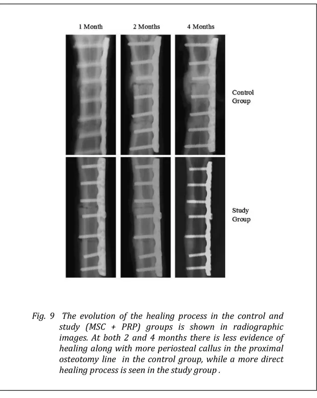
The goal of this project is to improve the outcome of massive bone allograft reconstruction after diaphyseal tumor resection. After the encouraging results of our previous study in the large animal with the use of non-‐vascularized allograft added with MSC and OP-‐1, the main goal of this experiment is to obtain a re-‐vascularization of the
allograft. In our previous study (110) we have demonstrated the superiority of ADSC and OP-‐1 added to the allograft compared to the allograft alone in the graft regeneration and integration. However, the amount of new bone formation in the allograft remained un-‐ satisfactory. The addition of an artero-‐venous bundle inside the canal would permit the early re-‐vascularization of the allograft, allowing the survival of the implanted stem cells and the allograft itself. This, in theory, would end in a graft that can be revitalized and eventually completely replaced by host bone, which will substantially decrease the amount of possible allograft complications. Therefore, the main goal of this project is to accelerate and improve the regeneration of a massive bone allograft through the insertion of a vascular bundle which should guarantee the early and direct re-‐vascularization of the implant.
2.2 Soft tissue microsurgical reconstructive strategies after tumor resections
The quality of microsurgical plastic techniques for soft tissue reconstruction has improved dramatically over the past years. The goal of this clinical part of thesis is to obtain satisfactory functional,
muscle/tendon for tumor. This will be achieved using motor re-‐ innervated free flaps that should allow the flap to sustain the function of the resected muscle(s) and/or tendon(s).
To our knowledge this is the first study that evaluates the functional outcome of innervated muscle transfer in the setting of irradiated limbs for soft tissue sarcomas.
3. BONE REGENERATION PROJECT
3.1 Background
Bone allografts are widely used to replace bone defects caused by traumas or by surgical operations for congenital malformations, tumor, infections or prosthetic failures (51, 55, 81, 111-‐115). The incorporation of the allograft requires a cooperative interaction between the allograft itself and the host bone (116, 117). The most important role in the incorporation process is done by the vascular bed which provides not only the cells responsible for the regeneration of this bone (MSCs), but even the fundamental factors for the maturation of these cells and their differentiation into osteoblast precursors (48, 85, 86, 101, 118, 119). The allograft incorporation can therefore be seen as a process that involves both re-‐vascularization and re-‐vitalization at the same time, because these two actions are strictly connected one to each other. A slow or insufficient re-‐ vascularization can impair the implant incorporation or, in some