Alma Mater Studiorum
Alma Mater Studiorum –
– Università di Bologna
Università di Bologna
DOTTORATO DI RICERCA IN
Oncologia e Patologia Sperimentale
Ciclo
XXII°
TITOLO TESI
MECHANISMS OF p53-MEDIATED APOPTOSIS
IN NEUROBLASTOMA
Presentata da: Eveline Barbieri
Coordinatore Dottorato
Relatore
Prof. Sandro Grilli
Prof. Andrea Pession
1
To Carlos, Pietro, and my family
To my mentors: Prof. Andrea Pession, Prof David Poplack, and Prof Enzo Piccinini
2
MECHANISMS OF p53-MEDIATED APOPTOSIS IN
NEUROBLASTOMA
3
INDEX
1. INTRODUCTION 4
2. MATERIALS AND METHODS 16
3. RESULTS 20
4. DISCUSSION AND CONCLUSIONS 29
5. FIGURES AND TABLES 35
4
1. INTRODUCTION
Neuroblastoma
Neuroblastoma accounts for more than 7% of malignancies in patients younger than 15 years and around 15% of all pediatric oncology deaths. It is the most common extra cranial solid tumor in childhood and the most frequently diagnosed neoplasm during infancy. This disease is remarkable for its broad spectrum of clinical behavior. Although substantial improvement in outcome of certain well-defined subsets of patients has been observed during the past few decades, the outcome for children with a high-risk clinical phenotype has improved only modestly, with long-term survival still less than 40% (1).
The genetic aberration most consistently associated with poor outcome in neuroblastoma is genomic amplification of MYCN (2, 3, and 4). MYCN amplification occurs in roughly 20% of primary tumors and is strongly correlated with advanced stage disease and treatment failure. Its association with poor outcome in patients with otherwise favorable disease patterns such as localized tumors or INSS stage 4S disease underscores its biological importance.
Although initially responsive to chemotherapy, neuroblastoma often becomes drug resistant, leading to relapse, that is the most common cause of death. Little progress has been made with regards to survival rates over the past 5 to10 years, despite intensive chemotherapy and radiation intervention at maximum levels. Improving metastatic neuroblastoma's dismal survival rates remains a major challenge in pediatric oncology and will only result from specifically targeted molecular therapies founded on a better understanding of the molecular determinants of neuroblastoma.
Amplification of MYCN occurs in a substantial subset of neuroblastomas with high mortality, although the basis for this effect is enigmatic. Compelling data demonstrate the ability of deregulated MYCN to sensitize cells to apoptosis following loss of mitogenic signals or genotoxic damage. Therefore, it is speculated that evasion of MYCN primed apoptosis is mandatory for neoplastic progression in neuroblasts with MYCN amplification. This evasion is likely to result from early cooperating mutations that activate survival pathways or inhibit apoptosis pathways. These signaling pathways have not been fully characterized with deregulated MYC, and are even less well understood for MYCN. Understanding this process, and how it is evaded in neuroblasts with MYCN
5
amplification, may have far ranging implications since traditional anti-neoplastics engage cellular apoptosis machinery to exert their cytotoxic effects. Defects in the regulation or engagement of apoptosis, as selected for in neoplastic cells with deregulated MYCN, can render these cells intrinsically resistant to therapy. Novel biological therapeutics targeting these pathways may restore sensitivity to both the inherent death-priming function of deregulated MYCN and to traditional anti-neoplastic agents.
Neuroblastoma detection in early life or in utero suggests that early disruption of normal developmental processes plays a part in tumour initiation. Insight into the molecular regulation of noradrenergic development has shed light on many facets of neuroblast biology and might lead to the identification of novel targets.
Similarly, an emerging appreciation for the role of cancer stem cells in propagating malignant disease could prove especially helpful in understanding and treating embryonal tumours such as neuroblastoma. Evidence suggests that malignancies can arise or be maintained in a stem cell compartment with the attributes of limitless replication and self-renewal. The Notch, Sonic hedgehog, and Wnt/β-catenin developmental programmes play a crucial part in stem cell determination and renewal in diverse tissues and misappropriation of these pathways seems to be a recurring theme in embryonal tumourigenesis (5, 6). β-catenin signaling is involved in the maintenance and expansion of neural crest stem cells and neural progenitors. An engineered gain-of-function β-catenin allele targeted to neural tissues causes marked neural progenitor expansion by promoting cell cycle entry at the expense of differentiation (7). This pathway might contribute to maintenance of neuroblastoma stem cells as well, raising the question of whether emerging inhibitors of this pathway might have therapeutic usefulness.
Overall, improved understanding of normal neurodevelopment of the sympathicoadrenal system will help us identify the key mutational events that initiate neuroblastoma tumorigenesis. Defining these events, as well as those that reliably predict for the acquisition of a high-risk phenotype, might ultimately direct us to the key pathways that can be exploited therapeutically.
6 P53 and Neuroblastoma
The p53 tumor surveillance network constitutes the core defense mechanism of the cell against loss of genomic integrity and malignant transformation. Evasion of p53 activity is, therefore, a prerequisite for tumor cells to survive and thrive, and this is attainable either through mutation of the TP53 gene or through defects in the molecular components that govern or execute the various aspects of the p53 response. Elucidation of the mechanisms by which tumor cells override the p53-orchestrated failsafe program is not only important to gain insight into the ontogenesis of a tumor, but may also point to preferable modes of therapeutic intervention.
Approximately half of human cancers have inactivating mutations of p53 (known as TP53 in human), and most of the remaining malignancies deactivate the p53 pathway by increasing its inhibitors, reducing its activators or inactivating its downstream targets. p53 is best characterized as a transcription factor that binds to specific DNA sequences and transactivates a number of genes with a variety of functions including cell cycle arrest, apoptosis, causing changes in metabolism and others.
In addition to this nuclear activity, p53 also possesses biological activities that are cytosolic and transcription-independent. Several years ago it was noted that overexpression of a mutant p53, lacking most of the DNA-binding domain (DBD) and completely deficient in transactivation function, could nonetheless trigger apoptosis. Indeed, overexpression of a variety of transactivation-incompetent p53 mutants can efficiently induce apoptosis in human cells (8).
A striking feature of the childhood cancer neuroblastoma is the low frequency (<2%) of TP53 mutations at diagnosis (9). There is considerable evidence that TP53 mutations may be acquired during chemotherapy and malignant progression of neuroblastoma.
Accordingly, an increased frequency of TP53 mutations is observed in multidrug-resistant neuroblastoma cell lines and in neuroblastoma cell lines established at relapse, but even in this context, the majority of cell lines remain characterized by a wild-type TP53 gene. Furthermore, many studies indicate that the p53 signal transduction pathway is intrinsically intact in neuroblastoma (10, 11, and 12), suggesting that circumvention of the p53 barrier in this tumor entity relies primarily on an inappropriately
7
increased activity of inhibitors of p53 signaling or, alternatively, on a loss of positive regulators of p53 activity.
Aberrant cytoplasmic localization of wild-type p53 has been proposed as one of the mechanisms for p53 inactivation in neuroblastoma cells. Although controversy exists on the frequency and functional relevance of this phenomenon, it has been extensively documented that cytoplasmic p53 sequestration does occur in at least some cases of neuroblastoma. Interestingly, as will be discussed below, cumulating evidence indicates that an increased activity of MDM2 or a dysfunction of its functional counterpart HAUSP, a principal p53-deubiquitinating enzyme, lies at the molecular basis of cytoplasmic p53 retention in neuroblastoma, further underscoring the importance of MDM2 deregulation as a means to escape from p53 control.
An initial study found cytoplasmic p53 sequestration in 96% of undifferentiated neuroblastoma tumors; whereas this phenotype was absent in differentiated neuroblastoma tumors (13). However, other studies have reported a predominant nuclear localization of p53 in undifferentiated neuroblastoma tumors, and both cytoplasmic and nuclear p53 in differentiating neuroblastoma (14, 15, and 16).
Conflicting results also exist for neuroblastoma cell lines, as the subcellular localization of p53 has been reported to be exclusively cytoplasmic (e.g. in IMR-32 and SK-N-SH cells), primarily cytoplasmic and weakly nuclear (e.g. in IMR-32 and SK-N-SH cells), equally cytoplasmic and nuclear (e.g. in SK-N-SH cells), predominantly nuclear (e.g. in IMR-32 cells), and completely nuclear (e.g. in IMR-32 and SK-N-SH cells). Some of the discrepancies may be explained by cross-reactivity of the antibodies used to detect p53 and by different methods of tissue fixation and cell preparation. Nonetheless, it is generally accepted that some cytoplasmic p53 does exist in neuroblastoma, although the prevalence and importance of cytoplasmic p53 sequestration remain a subject of debate. It has been reported that abnormal cytoplasmic p53 localization may attenuate the DNA damage–induced G1 checkpoint function and the apoptotic activity (17, 18) of wild type p53 in some neuroblastoma cells. On the contrary, many studies have shown that the DNA-binding and transactivation capacity of p53 and the p53 signal transduction pathway are intact in neuroblastoma cells with wild-type p53 (9, 10, and 11), indicating that cytoplasmic retention of wild-type p53 is either an infrequent anomaly or a relative block on p53 that can be overcome by appropriate p53-inducing stimuli. Proposed mechanisms for abnormal p53 accumulation in the cytoplasm of neuroblastoma cells include hyperactive nuclear export of p53, cytoplasmic tethering of
8
p53, resistance of p53 to proteasomal degradation, and possibly impaired nuclear re-import of p53. A unifying theme common to these diverse mechanisms may be the involvement of a disrupted MDM2/HAUSP regulation of p53. It has been firmly established that cytoplasmic p53 sequestration in neuroblastoma cells is at least in part caused by enhanced nuclear export and that MDM2 plays an important role in this nuclear exclusion of p53. Once transported to the cytoplasm, p53 may be held in this compartment by a cytoplasmic anchor protein, such as Parc (19). A comparable cytoplasmic anchoring function may be exerted by the large T antigen from human polyomavirus BK (20), by the glucocorticoid receptor (21), and by the MDM2-related protein MDM4 (also known as MDMX) (22). It has also been shown that p53 in neuroblastoma cells is aberrantly ubiquitinated because of an impaired interaction between p53 and the deubiquitinating enzyme HAUSP, and that this hyperubiquitination contributes to cytoplasmic p53 sequestration (23). In keeping with the deregulation of MDM2/HAUSP, interference with p53 hyperubiquitination by targeted inhibition of the p53–MDM2 interaction in neuroblastoma cells has been shown to relocate p53 from the cytoplasm to the nucleus and to restore the transcriptional and apoptotic activities of p53 (23).
The rarity of TP53 mutations in neuroblastoma has been a puzzling issue to many investigators given the potent antitumor capacity of wild-type p53 protein. A substantial number of alternative p53-inactivating lesions have been identified in neuroblastoma during the past few years, many of which interfere with proper functioning of the p14ARF-MDM2- p53 axis. Full characterization of the nature and relative importance of the different blocks on the p53 pathway in neuroblastoma cells awaits genome-wide experimental approaches in well-controlled model systems.
The p14ARF-MDM2-p53 Axis and Lesions at the MDM2 and CDKN2A (p16INK4a/p14ARF) Loci in Neuroblastoma
The MDM2 oncoprotein, a human homolog of the ‘mouse double minute 2’ gene product that was originally identified in a spontaneously transformed mouse cell line with double minute chromosomes (24), is a critical negative regulator of p53 stability and activity. It has been well established that p53 and MDM2 mutually control their cellular levels and form a tight auto regulatory feedback loop (Figure 1a). Under normal
9
physiological conditions, p53 protein levels are very low because of MDM2-dependent proteasomal degradation. Exposure of cells to harmful stimuli, such as DNA damage, hypoxia, telomere erosion, ribonucleotide depletion, or oncogene activation, results in a number of modifications on the p53 protein (e.g. phosphorylation and acetylation), which suppress the binding of p53 to MDM2 and which lead to accumulation and increased transcriptional activity of p53 (25). In addition to inducing expression of target genes involved in cell-cycle arrest, DNA damage repair, senescence, and apoptosis, p53 also transactivates the MDM2 gene (Figure 1b). The resulting increase in MDM2 expression limits the duration and intensity of a non-lethal stress response. There are several mechanisms by which MDM2 is capable of counteracting p53 activity and stability (Figure 1c). First, MDM2 binds to the transactivation domain of p53 and, therefore, directly interferes with recruitment of the basal transcriptional machinery and transcriptional coactivators (26, 27, and 28). Second, MDM2 acts as an E3 ubiquitin ligase for p53 in a dosage-dependent manner. Low levels of MDM2 promote p53 monoubiquitination, which may both stimulate nucleocytoplasmic shuttling of p53 because of unmasking of a nuclear export signal and decrease p53 transactivation capacity owing to unavailability of the ubiquitinated lysine residues for acetylation. At higher levels, the activity of MDM2 results in polyubiquitination and subsequent proteasomal degradation of p53 (29, 30). Third, MDM2 also induces monoubiquitination of histone proteins in the vicinity of p53-responsive promoters, resulting in transcriptional repression (31). Fourth, MDM2 has been reported to inhibit p53 transcriptional activity by promoting conjugation of the ubiquitin-like protein NEDD8 to p53 (32). Fifth, MDM2 may also contribute to p53 inactivation by recruiting several corepressor proteins, such as HDAC1, CTBP2, YY1 and KAP1 (33-36).
A central negative regulator of MDM2 is the tumor suppressor protein p14ARF, which is an alternate reading frame product of the CDKN2A locus on chromosome 9p21. This locus encodes two structurally unrelated growth-inhibitory proteins, p16INK4a and p14ARF, that govern the activities of the pRb and p53 tumor suppressor pathways, respectively. The p14ARF protein serves as a key sensor of hyperproliferative signals generated by activated oncogenes and engages both dependent and p53-independent pathways to protect cells from malignant transformation (37, 38).
The importance of p14ARF-mediated signaling of oncogene activity in the p53 tumor surveillance network is underscored by observations in mice models that the cancer-protective activity of p53 is abolished in the absence of the murine homolog
10
p19ARF (39, 40). The physical interaction between p14ARF and MDM2 is in large part responsible for the ability of p14ARF to stabilize and activate p53. P14ARF prevents MDM2 from targeting p53 for degradation by inhibiting the E3 ubiquitin ligase activity of MDM2 (41) and by blocking nuclear export of MDM2 and p53 (42, 43). It has also been established that p14ARF, which is predominantly a nucleolar protein, is capable of mobilizing MDM2 into the nucleolus, and it has, therefore, been proposed that p14ARF releases nucleoplasmic p53 from the inhibitory grip of MDM2 by inducing nucleolar sequestration of MDM2 (44).
Not surprisingly, many forms of cancer develop defects in MDM2 or p14ARF to escape from p53 control. Genetic aberrations of the MDM2 locus as well as genetic or epigenetic disruption of the CDKN2A (p16INK4a/p14ARF) locus may account for inactivation of the p53 pathway in a subset of neuroblastoma tumors, mainly at relapse. Amplification of chromosome 12q–derived sequences encompassing the MDM2 gene has been described almost exclusively in neuroblastoma tumors and cell lines that simultaneously have amplification of the MYCN oncogene on chromosome 2p24, and is associated with attenuated p53 transcriptional function and multidrug resistance (45, 46).
The CDKN2A (p16INK4a/ p14ARF) locus at 9p21 is the most frequent target of homozygous deletion in both neuroblastoma cell lines and primary tumors, and has been found to be silenced by methylation in several neuroblastoma cell lines (47,48, and 49). It has been estimated that approximately half of all neuroblastoma cell lines established at relapse are subject to a genetic or epigenetic lesion of the MDM2 or CDKN2A (p16INK4a/p14ARF) locus, but these findings await confirmation in a study that takes also neuroblastoma tumor samples into account (50). A recent line of evidence supporting a role for MDM2 activity in the development and malignant behavior of neuroblastoma stems from epidemiological studies of a T4G single nucleotide polymorphism in the MDM2 promoter (SNP309; rs2279744). The presence of this polymorphism increases the affinity of the MDM2 promoter for a transcriptional activator, Sp1. This results in enhanced transcription of MDM2, overexpression of the MDM2 protein, attenuation of the p53 pathway, and may eventually lead to accelerated tumor formation. Both individuals homozygous for SNP309 (G/G) and subjects heterozygous for SNP309 (T/G) have an increased risk for the development of neuroblastoma, and neuroblastoma patients carrying the SNP309 variant (G/G or T/G) present with a more advanced clinical stage and have a shorter 5-year overall survival than patients homozygous for the wild-type allele (T/T) (51, 52).
11
These findings suggest that an increased activity of MDM2 due to the presence of SNP309 has an adverse effect on the development, aggressiveness, and outcome of neuroblastoma, and provide a direct incentive for the development of novel therapeutic strategies aimed at MDM2 inhibition.
Transactivation of MDM2 Expression by MYCN
Amplification of the MYCN oncogene plays a central role in the pathophysiology and clinical behavior of high-risk neuroblastoma. This genetic aberration is found in approximately 22% of all neuroblastoma tumors and is highly correlated with advanced stages of disease, rapid progression, treatment failure, and fatal outcome (53, 54). MYCN amplification results in overexpression of the MYCN protein, which is a bHLH transcription factor that operates in a heterodimeric complex with Max family proteins to promote cell growth and proliferation (55). The oncogenic effects of MYCN overexpression have been established in a variety of model systems. Enhanced expression of MYCN elicits neoplastic transformation of mammalian cells, induces autocrine growth factor activity and increases proliferative potential, accelerates cell-cycle progression, enhances tumor cell motility and invasiveness, evokes genomic instability through disruption of the regulation of centrosome replication, diminishes expression of angiogenesis inhibitors, and promotes immune escape in neuroblastoma by inhibiting the chemoattraction of natural killer T cells (56, 65).
Direct evidence for a causative role of MYCN amplification in the pathogenesis of neuroblastoma is derived from the observation that transgenic mice with targeted expression of MYCN in normal neuroblasts develop tumors with a phenotype very similar to human neuroblastoma (66). Importantly, the mouse model of neuroblastoma faithfully recapitulates the p53 wild-type status, chemosensitivity, and p53-dependent apoptotic responses of human neuroblastoma.
However, aberrant MYCN expression also potently sensitizes neuroblastoma cells to drug- and stress-induced apoptosis, and, therefore, needs to be accompanied by a collateral impairment of the cell death program to provide a selective advantage for the tumor. This counterbalance to the intrinsic apoptosis-sensitizing effect of MYCN may be delivered by an increased activity of MDM2. The study conducted in our laboratory with a ChIP cloning approach combined with oligonucleotide pull-down and luciferase reporter assays has successfully identified MDM2 as a direct transcriptional target of
12
MYCN in neuroblastoma cells (67). In this study, endogenous MDM2 mRNA and MDM2 protein levels were rapidly upregulated on induction of MYCN in MYCN-conditional neuroblastoma cell lines, whereas targeted inhibition of MYCN in MYCN-amplified neuroblastoma cells resulted in reduced MDM2 levels with stabilization of p53 and induction of apoptosis.
These data suggest that MYCN driven expression of MDM2 may constitutively debilitate the p53 pathway in MYCN-amplified neuroblastoma cells, providing both a possible mechanism for evasion of MYCN-primed apoptosis and an explanation for the low frequency of TP53 mutations in these cells. This view is further strengthened by evidence that the closely related MYC (c-MYC) oncoprotein also relies on MDM2 to restrain p53-mediated apoptosis, as Myc-induced lymphomagenesis in mice is profoundly suppressed by haploinsufficiency of Mdm2 because of a drastic increase in p53-dependent apoptosis (68).
Based on the central role of MYC oncogenes in neuroblastoma tumorigenesis and the necessity of inhibiting wild-type p53 activity to prevent apoptosis, our laboratory hypothesized that MDM2-mediated suppression of p53 is a vital component of MYCN-driven tumor initiation and progression.
To test this hypothesis, we crossed the well-characterized pTHMYCN transgenic model of neuroblastoma and the Mdm2 haploinsufficient mouse model (69) and compared tumor latency and incidence in the resulting animals. We found that Mdm2+/−MYCN+/+ transgenics had markedly delayed tumor development and had a lower overall incidence of tumors, strongly implicating Mdm2-mediated blockade of p53 as an essential step in the pathogenesis of neuroblastoma. Analysis of the resulting tumors demonstrated high levels of p53 and dramatically decreased levels of Arf. We further confirmed the impact of MDM2 levels on neuroblastoma growth in xenograft models using conditional short hairpin RNA (shRNA)–mediated knockdown of MDM2. Based on our data, we propose that both MYCN-mediated activation of MDM2 and suppression of ARF contribute directly to the pathogenesis of neuroblastoma. Further work designing molecular targeted therapies that modulate these pathways should provide clinically effective novel therapeutic approaches for neuroblastoma.
13 MDM2 Inhibition in Neuroblastoma
The search for specific small molecule inhibitors of MDM2 is an active area of research. Recently, Lyubomir T. Vassilev and his group at Hoffman-La Roche characterized a group of cis-imadazoline analogues (termed Nutlins) that displace p53 from its binding pocket in the N-terminus of MDM2. These compounds have active (a) and inactive (b), enantiomer forms. Nutlin-3a binds with 150-fold higher affinity than does Nutlin-3b.
The outcome of p53 activation in cells ranges from a reversible cell-cycle arrest and induction of DNA repair to more drastic responses, such as cell death by apoptosis or senescence. p53 transactivates genes that induce apoptosis, such as FAS, BBC3 (PUMA), PMAIP1 (Noxa), and BAX, as well as genes that induce cell-cycle arrest, such as CDKN1A (p21) and GADD45. The choice of biological response to p53—death or survival— is strongly influenced by the presence of death or survival signals, and such signals act both upstream and downstream of p53. However, the exact molecular mechanism by which the switch between arrest and apoptosis is implemented remains largely unknown.
In a recent study, Nutlin-3a was shown to have potent in vitro and in vivo effects against tumor cells with wild-type p53 (70). As neuroblastoma tumors are universally p53 wild type at diagnosis, we sought to characterize the effects of Nutlin-3a on neuroblastoma cells. To test our hypothesis that altered p53 stabilization contributes to the robust response of neuroblastoma to MDM2 inhibition, we monitored the kinetics of apoptosis in response to Nutlin 3a and evaluated the levels of p53 at early time points after treatment. We found that p53 levels responded to Nutlin in neuroblastoma with a time course quite distinct from the pattern of comparable solid tumor cell lines and previously published studies.
We demonstrate here a very rapid stabilization of p53 in response to MDM2 inhibition in neuroblastoma cells associated with rapid apoptotic response. Additionally, MDM2 inhibition combined with etoposide induce a robust apoptotic response in neuroblastoma cell lines and primary tumor cultures. As expected these results are specific for the active –3a enantiomer and confirmed that the effects seen on proliferation and apoptosis were due to p53 dependent effects of MDM2 inhibition. We believe that MDM2 inhibition dramatically enhances the activity of genotoxic drugs in neuroblastoma and should be considered as an adjuvant to chemotherapy for this aggressive pediatric cancer and for possibly other p53 wild-type solid tumors.
14
P53 and Metabolism- Efficient combination of MDM2 and mTOR inhibition
It has almost become a truism to say that cancer cells have acquired distinctive characteristics that distinguish them from their normal counterparts, but it is important remembering that among the very first of these differences to be recognized were the changes in tumour cell. The importance of metabolism in cancer has revived enthusiasm for the study of how these pathways are controlled, revealing some interesting contributions from well-known oncoproteins and tumour suppressor proteins. Among these is p53, which is now also emerging as an important player in the response to and the regulation of metabolic stress.
Compared with our understanding of the functions of p53 in controlling cell cycle progression and apoptosis, this is a new and burgeoning area of influence for p53, with some confusing and apparently contradictory results highlighting gaps in our understanding. However, these activities of p53 are indisputably interesting and probably extremely important. Furthermore, it is clear that the role of p53 in responding to and effecting alterations in metabolism will have consequences beyond cancer, influencing various other aspects of disease and normal life (Table 1).
Metabolic pathways in normal cells are tightly regulated to allow cell growth or survival, depending on the conditions. Nutrient availability supports the synthesis of proteins, lipids and nucleic acids for cell growth and proliferation, whereas starvation triggers a series of responses to restrict cell proliferation, maximize energy production (by switching to the breakdown rather than the synthesis of macromolecules) and help cell survival.
One important node in these responses is mTOR, which promotes protein synthesis and suppresses the induction of autophagy (a mechanism that can mobilize alternative energy sources and is discussed in more detail below). Key regulators of mTOR are AKT, which is stimulated by growth factors to activate mTOR, and AMP-activated protein kinase (AMPK), which responds to an increased AMP/ATP ratio under conditions of low energy to repress mTOR. These growth regulatory cascades intersect with the metabolic pathways that control energy production and biosynthesis, in which AKT can promote the anabolic, energy-consuming pathways (such as fatty acid synthesis) that are necessary for cell growth and AMPK drives the catabolic, energy-producing responses that are needed under conditions of metabolic stress. Layered over
15
this complexity are the pathways that regulate cell division and survival; virtually all parts of this intricate network can be profoundly perturbed in cancer cells.
The mammalian TOR protein (mTOR) forms two distinct signaling complexes, called mTORC1 and mTORC2. The mTORC1 complex, which, in addition to mTOR, consists of raptor, PRAS40, and mLST8, is responsible for control of cell growth and protein synthesis (71).
Genotoxic stress was suggested to inhibit mTORC1 activity through p53-dependent upregulation of negative regulators such as PTEN, TSC2, and AMPKb1 (72). p53 also increases the phosphorylation of the AMPKa subunit, leading to AMPK activation. However, the precise physiological mechanism by which p53 activates AMPK and inhibits mTOR was not established.
In our attempt to identify additional pathways to potentiate the p53 response, we use oligonucleotide microarray analysis to explore the molecular mechanism of the Nutlin-induced p53 biological response. Two p53 target genes genes, Sestrin1 (Sesn1)/PA26 and Sestrin2 (Sesn2)/Hi95, were significantly affected by Nultin treatment. Previous studies have shown that Sesn1 and Sesn2, whose expression is induced upon DNA damage and oxidative stress, may have a cytoprotective function based on regeneration of overoxidized peroxiredoxins (73). Recently has been also demonstrated that Sesn1 and Sesn2 are negative regulators of mTOR signaling and that they execute this redox-independent function through activation of AMPK and TSC2 phosphorylation. The important study of Budanov et al (74), provide in vivo evidence for the functional importance of Sesn2 in inhibition of mTOR signaling in mice treated with a p53-activating alkylating agent. Hence, Sesn1 and Sesn2 link genotoxic stress, p53, and mTOR signaling.
Mammalian target of rapamycin (mTOR) signaling pathway is active in neuroblastoma and mTOR inhibition has demonstrated antiproliferative effects in vitro and in vivo in aggressive MYCN amplified neuroblastoma. In addition, signaling through mTOR is elevated in neuroblastoma tumor-initiating cells (TICs) and mTOR can increase hypoxia-inducible factor (HIF) translation.
For this reason we investigated the role of Sestrin1 and Sestrin2 in p53-mediated apoptosis in neuroblastoma and the interaction of mTOR and p53 pathways after their simultaneous blockade using the mTOR inhibitor, Temsirolimus and the MDM2 inhibitor, Nutlin 3a.
16
2.MATERIALS AND METHODS
Mice Breeding
Mice were handled in accordance with an approved protocol from the Animal Research Committee of Baylor College of Medicine. Mdm2+/− mice (C57b strain; Dr. Lozano, MD Anderson Cancer Center) were first backcrossed for seven generations with wild-type 129/Svj mice to gainMDM2 heterozygousmice with 129/Svj strain (Mdm2+/−MYC−/−).
The neuroblastoma model is highly strain-dependent, and to ensure equivalent comparisons, Mdm2+/− and Mdm2+/+ mice were all derived from the same founding mice and littermates used for all analyses. For genotyping PCR, tail DNA was prepared using DNeasy kit and genotyping for MDM2 performed as previously published (69). MYCNheterozygosity or homozygosity was confirmed by quantitative PCR (qPCR) for the human pTH-MYCN transgene using the following primers.
Implantation of Neuroblastoma Xenograft
Five million ZC21 or SJ9 cells were resuspended together with 100 µl of Matrigel and injected subcutaneously in 4-week-old severe combined immunodeficient (SCID) mice with mixed gender. One week after injection, mice were separated into two groups that were fed with drinking water either containing 2 mg/ml of doxycycline or not. Fresh water with or without doxycycline was served to the mice once every 2 days until the experiments were complete. The sizes of tumors in each mouse were measured daily starting from 10 days after cell injection. The data were plotted using Kaplan-Meier method to analyze the tumor growth.
In vivo xenografts and administration of mTOR inhibitor
One million SY5Y were orthotopically injected in 4-week-old nude mice. Tumors were measured daily with a digital caliper and tumor volume calculated as length x width2 x0.44. At a tumor volume of > 0.2 ml the animals were randomized to receive
17
either Temsirolimus, CCI-779, (20 mgkg) intraperitoneally daily for 12 days or no treatment (controls). Tumor materials were fixed in 4% paraformaldehyde and frozen in liquid nitrogen and stored at 4 or -80 C respectively.
Cell Lines and Cell Culture
The ZC21 and SJ9 conditional subclones were generated from human neuroblastoma cells IMR32 (American Type Culture Collection, Manassas, VA) or SJ3-12 (a gift from Dr. Dirk Geerts, University of Amsterdam, the Netherlands) by transfection with pSuperior plasmids containing 64-bp shRNA hairpin loop constructs cloned into its unique Bgl II and HindIII sites. The MDM2 messenger RNA (mRNA) sequence targeted with small interfering RNA (siRNA) was gccattgcttttgaagtta. The expression of shRNA is driven by an inducible (Tet-ON) H1 promoter, which is activated by the presence of doxycycline. After transfection, cells were grown under puromycin selection for 2 weeks. Single clones were isolated, expanded, and characterized for their cellular MDM2 levels 72 hours after doxycycline treatment. Cell lines ZC21 and SJ9, showing remarkably decreased level of MDM2 as determined by Western blot, were used for the xenograft study.
The JF, IMR32, HCT 116, MCF7 lines were maintained in RPMI 1640, MEMa, McCoy’s 5A, DMEM plus 1% insulin respectively, supplemented with 10% fetal bovine serum and 1% penicillin and streptomycin. The SJSA-1 cells were grown in RPMI 1640 medium supplemented with 10mM HEPES, 1 mM sodium pyruvate, 4.5 g/L glucose, 1.5 g/L sodium bicarbonate, 10% fetal bovine serum and 1% penicillin and streptomycin. All three primary neuroblastoma lines (p202, p218, pH) were grown in IMDM, supplemented with 20% fetal bovine serum, 0.1% ITS (bovine insulin, human transferrin and sodium selenite) and 1% penicillin and streptomycin. The p53-mutant cell line SJ3-12 (21 amminoacids deletion in the DNA binding domain) was provided by Ham Geertz and used as control. Nutlin 3a was provided by Dr. Vassilev (Roche, Nutley, NJ).
Western Blot
Antibodies used are as follows: a-p53, a-MDM2, p21, a-actin monoclonal antibodies and horseradish peroxidase –conjugated goat a-mouse antibody were used for Western blot analysis as described below. Cells were directly lysed with H-1 lyses
18
buffer (50 mM HEPES, 250 mM NaCl, 0.5% NP-40) for 20 minutes on ice and proteins were quantified by Bradford assay before electrophoresis. Actin was used for loading control in all experiments. Cycloheximide (CHX) (25 ug/ml) (Sigma) was used as protein synthesis inhibitor and added to the cells for the duration of Nutlin A treatment.
Terminal Deoxynucleotidyl Transferase Apoptosis Assay
IMR32, JF, p218, p202, pH, HCT 116 +/+ and +/-, MCF7, SJSA cells were plated in 10-cm dishes at 2 million cells per dish. The cells were then exposed to Nutlin A for 16, 24, and 48 hours. Briefly, cells were harvested in PBS and incubated in 3 mL of 1% formaldehyde-PBS for 15 minutes on ice. The cells were then fixed in 70% ethanol-PBS overnight at – 20 C. 50uL of the terminal deoxynucleotidyl transferase (TdT) reaction mix were added to each sample consisting of 10uL of 5 X reaction buffer, 1.5uL TdT, 5uL CoCl2, 0.5uL biotin-16-dUTP and 33uL dH2O. The samples were incubated for 1 hour at 37C and then washed with PBS. For fluorescence-activated cell sorting analysis, 100uL avidin FITC buffer was then added to each sample. The samples were incubated in the dark at room temperature for 30 minutes and washed in 1X PBS + 0.1% Triton X-100. Fluorescence-activated cell sorting analysis was done using CellQuestPro and standard software. All the experiments were done in triplicate.
3-(4,5-Dimethylthiazol-2-yl)-2,5-Diphenyltetrazolium Bromide (MTT) Assay
Briefly, 0.5 x 10⁵ cells/mL were plated into 96-well microtiter plates, incubated at 37 C, and cultured overnight. Stock solutions of Nutlin 3a or 3b in DMSO were diluted in PBS and added to the appropriate concentration by serial dilutions. PBS alone, medium alone, and medium plus DMSO were control wells for each dilution. Plates were incubated for 72 hours and then centrifuged for 5 minutes at 1,000 rpm. Half the medium (135 uL) was removed and replaced with fresh medium. Then, 15 uL MTT were added to each well. Plates were shaken in the dark for 5 minutes, incubated for an additional 4 hours, and read at 550 nm in the plate reader.
19 Real-time quantitative PCR
SY5Y and IMR32 cells were treated with 5 or 10 uM of Nutlin-3a for up to 8 hours. RNA was prepared from cells using a RNeasy Mini Kit (Qiagen, Valencia, CA, USA), and first-strand cDNA was generated using random hexamers (SuperScript III First-Strand Synthesis SuperMix) from 1 mg total RNA. The mRNA expression levels of p53 and 18S were quantified using TaqMan gene expression assays.
Quantitation of intracellular proteins by flow cytometry
For intracellular phospho S6 detection, cells were fixed with 2% paraformaldehyde, permeabilized with 100% ice-cold methanol and incubated for 1 h at 4 C with antibody against phosphoS6 Alexa Fluor 647 or its isotypic control, according to the manufacturer’s instructions.
Oligonucleotide Microarray Data Analysis
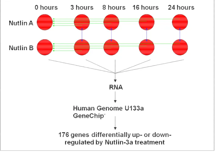
P202 cells were exposed to the treatment with Nutlin 3a or the inactive enantiomer Nutlin 3b for 8, 16 and 24 hours. Biological duplicates of the experiment were performed. We hybridized cDNA prepared from p202 cells untreated or treated with 10 uM of Nutlin 3a at different time points to Affymetrix hgu133a chips containing approximately 22,000 probe sets mapping to 15,000 known human genes.
Raw data from the hgu133a2 chips were normalized using the robust multiarray average with GC correction (GCRMA) procedure, implemented in the R package gcrma (quantile-based normalization with background correction based on gene chip content of probes). Per gene hypothesis testing was performed using the significance analysis of microarrays (SAM) procedure, implemented in the R package siggenes, using linear models for microarray data. Pathway analysis was performed using a modified version of the procedure in Tian et al. (75).
20
3. RESULTS
Mdm2 Deficiency Extends the Tumor Latency and Decreases Incidence in MYCN Transgenic Mice
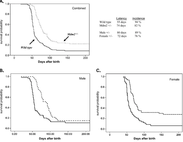
To investigate the role of Mdm2 in neuroblastoma pathogenesis in vivo, we generated MYCN+/+ transgenic mice (carrying two copies of the targeted transgene) with either wild-type Mdm2+/+ or a heterozygous knockout Mdm2+/−. The strategy for animal breeding is shown in Figure 2a. In pTH-MYCN transgenics, the human MYCN gene is driven by the tyrosine hydroxylase promoter, which is active in migrating cells of neural crest during early development and in the resulting peripheral sympathetic nervous tissue from which neuroblastoma arises (76). pTH-MYCN transgenic mouse (MYCN+/+,129/Svj strain) spontaneously develops neuroblastoma in a MYCN dose-dependent manner (77, 78). Both cohorts contain two copies of human MYCN gene, express elevated levels of MYCN in neural crest precursors and developing sympathetic ganglia, and are expected to develop tumors efficiently, permitting us to study the role of Mdm2 on MYCN-driven tumors by comparing the two cohorts. Consistent with previous studies (69, 78), we found no difference in animal weights, activity, oral intake, or fertility between cohorts. As expected, both Mdm2+/+ and Mdm2+/− mice developed large abdominal neuroblastomas. Tumors from both groups had an aggressive undifferentiated phenotype with indistinguishable histologic diagnosis, morphology, and fine structure containing neurosecretory granules (Figure 2 b and c).
Overall, 110 Mdm2+/− and 117 Mdm2+/+ mice were observed for up to 1 year. Because no mice died of tumors after 110 days, we present Kaplan-Meier survival analysis taken out to 200 days (Figure 3). Mice were monitored at least twice a week for tumor formation and changes in health status. Mice were killed within 10 days of the detection of tumors to be examined in accordance with our institutional review board– approved animal protocol. Figure 3a illustrates the statistically significant differences in cumulative survival probabilities between Mdm2+/− and Mdm2+/+ mice (as analyzed by Mantel-Cox and Wilcoxon methods). We found a dramatic difference in the median survival time (defined as time of sacrifice), which is closely correlated with tumor latency because these tumors, once fully established, are rapidly fatal. For Mdm2+/+ mice, median latency was 55 days (95% confidence intervals [CI], 52-57 days), and for Mdm2+/− mice, tumor latency was 74 days (95% CI, 72-81 days; P < .001). Thus, the
21
mean time to tumor onset was delayed approximately 20 days (38%) in the Mdm2 haploinsufficient mice, supporting our contention that Mdm2-mediated suppression of p53 is a rate-limiting step in MYCN-driven neuroblastoma tumorigenesis.
Overall, the MYCN transgenic mice wild type for Mdm2 had a very high incidence of tumors with 94% (110/117) of mice developing tumors, in line with previously published studies of the pTHMYCN tumor model in the Svj-129 background (79). However, the Mdm2 haploinsufficient mice had a significantly lower incidence of 80% overall (88/110 total; P < .001). This difference was sex dependent (Figure 3b and 3c). Seventy-six percent of female Mdm2+/− mice (47/62 animals) developed tumors compared with 93% (55/ 58 animals) of female Mdm2 wild-type mice (P < .001). For male mice, Mdm2 haploinsufficiency had an insignificant influence on overall tumor incidence, with 89% (43/48 animals) of the Mdm2+/− mice compared with 95% (55/59 animals) of the Mdm2 wild-type mice developing tumors (P value not significant). Further analysis of tumor latency according to sex revealed a trend toward increased latency in the male Mdm2+/− mice compared with the female Mdm2+/−, but this was not significant by Cox regression analysis. No sex-dependent difference in latency or incidence was seen in the Mdm2 wild-type pTH-MYCN mice.
The significant decrease in tumorigenesis in the female Mdm2-deficient mice may be secondary to interaction between MDM2 and the estrogen-dependent signaling in these mice.
Because a major role of MDM2 is inhibition of p53 activity, it is possible that the delay to tumor formation and the reduced incidence reflect the need for additional genetic events to overcome increased p53-mediated tumor suppression. Since decreased Mdm2 and increased baseline p53 levels could lead to selective pressure to mutate p53 during tumorigenesis, we analyzed the sequences of p53 complementary DNA in a representative sample of tumors. Sequence data from 15 Mdm2+/− and 8 Mdm2+/+ tumors revealed only wild-type p53, consistent with the almost complete absence of p53 mutations found in primary human neuroblastomas.
Mdm2 Deficiency Alters p53 Regulation and Activity in Neuroblastoma
The data shown in the previous section suggest that Mdm2 is critical for the development of MYCN-driven tumors and that a 50% decrease in Mdm2 significantly alters tumorigenesis. In cultured neuroblastoma cells, siRNA-mediated knockdown of
22
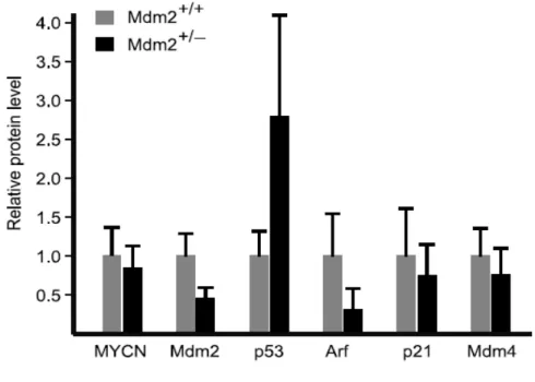
MDM2 leads to a significant increase of p53 and sensitization to apoptotic stress. To better understand the regulation of p53 and related proteins in Mdm2+/− and Mdm2+/+ tumors, we performed semiquantitative Western blot and qRT-PCR analysis of Mdm2, p53, p21, Mdm4, p19Arf, and MYCN in 12 tumors from each cohort, as shown in Figure 4 . As expected, Mdm2 mRNA and protein levels are reduced to approximately 50% of wild type in the Mdm2 haploinsufficient mice. Western blots demonstrate that p53 protein levels are remarkably higher in Mdm2+/− tumors, whereas the mRNA levels do not differ. This is consistent with decreased Mdm2-mediated ubiquitination of p53 and relative stabilization of the protein compared with Mdm2+/+ tumors.
A primary transcriptional target of p53 is p21CIP1/WAF1, which encodes the cyclin-dependent kinase inhibitor, p21, a central cell cycle regulator. Consistent with observations of increased p53 protein and presumably its transcriptional activities, the average p21 mRNA in Mdm2+/− tumors was approximately 80% higher than that in Mdm2+/+. However, a trend toward decreased p21 protein levels is observed in Mdm2+/− tumors, suggesting that p21 protein is destabilized in these tumors. Interestingly, it has been shown that Mdm2 can inhibit p21 by promoting its ubiquitin-dependent and –inubiquitin-dependent proteosomal degradation (80, 81). However, decreased p21 protein levels would not be expected to correlate with decreased Mdm2 levels in Mdm2+/− mice, and an alternative mechanism for p21 protein reduction is likely invoked in this cohort.
We also analyzed levels of Mdm4 (MdmX), a homolog of Mdm2 with overlapping and distinct regulatory effects on p53 (82). Mdm4 has potent cell cycle effects independent of Mdm2 (83) and he binding of Mdm4 to p53 at its N-terminal domain suppresses its transactivation activity. Although a trend toward decreased levels of Mdm4 can be seen, no significant change in Mdm4 protein or mRNA levels were detected. This suggests that Mdm4 levels do not compensate for low Mdm2 levels in Mdm2+/− tumors.
In the data presented in this study, we provide the first in vivo evidence that MDM2 contributes to MYCN-driven tumorigenesis in neural crest–derived neuroblastoma with both Mdm2 haploinsufficient transgenic mice and human neuroblastoma xenografts in SCID mice using cell lines with shRNA-mediated conditional knockdown of MDM2. Both tumor latency and tumor incidence are significantly altered by the reduction in Mdm2 levels in the transgenic model.
23
We also demonstrate a strong influence of sex on tumor incidence in the Mdm2 haploinsufficient mice with overall tumor rate, decreasing more than 18% in the female Mdm2+/− mice (P < .001). The estrogen receptor α (ERα) interacts with Mdm2 and p53 and can inhibit Mdm2-mediated ubiquitination of p53. Subsequent increased p53 activity may be sufficient to further suppress tumorigenesis in female Mdm2 haploinsufficient mice. However, ERα may also act to increase Mdm2 levels indirectly and alter glucocorticoid receptor signaling, and Mdm2 can enhance ERα activity in breast cancer cells (84). It is also possible that p53-independent functions of Mdm2 or mouse strain– specific effects account for the observed inhibition of tumorigenesis in the female mice. Neuroblastoma is almost exclusively found in premenarchal children, and clinical and mechanistic implications of these findings will require additional investigation.
Both MYCN and MYCC activate apoptosis and stimulate proliferation simultaneously in culture cells. How the neural crest–derived precursors of neuroblastoma respond to aberrant MYC activation clearly depends on direct MYC transcriptional targets and on indirect and parallel oncogenic stimuli. Recent studies comparing the sympathetic ganglia of pTH-MYCN and wild-type mice have shown that MYCN expression leads to the persistence of proliferative rests of neuroblasts and that this correlates with tumorigenesis in a MYCN gene dose-responsive manner. The aberrant expression of MYCN likely disrupts apoptosis and differentiation favoring the proliferation of these “neuroblast” cells. We demonstrate that haploinsufficiency of Mdm2 clearly prolongs tumor latency and decreases the incidence of tumor development in this same pTH-MYCN model. Lower levels of Mdm2 may weaken the oncogenic effect of MYCN in neuroblast precursors, increasing the requirement for additional genetic changes such as epigenetic silencing of ARF.
MDM2 Inhibition Induces Rapid Apoptotic Response in Neuroblastoma Cell Lines And Primary Tumor Cultures
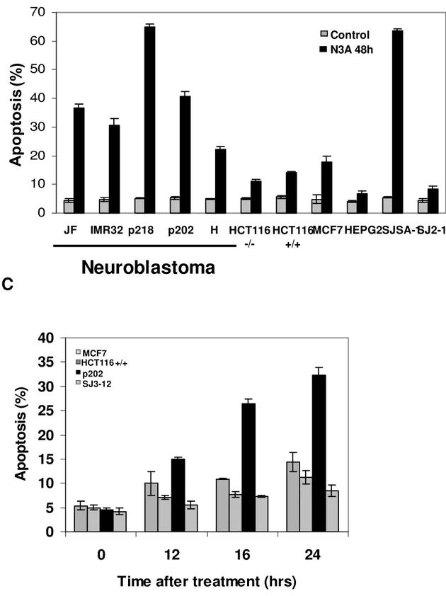
We determined the ability of the 9-cell panel to undergo apoptosis after treatment with Nutlin-3a. Apoptosis induction was tested in five neuroblastoma lines (JF, IMR32) and primary tumor cultures (p202, p218, H) and four non-neuroblastoma human solid tumors including colorectal (HCT116), breast (MCF7) and osteosarcoma (SJSA-1). All cell lines have been reported to express wild type p53.
24
All proliferating cells were incubated with Nutlin 3A 10 uM for 24 and 48 hours and the TdT-positive fraction was measured by flow cytometry. At 24 hours all the neuroblastoma cell lines showed a significant percentage of apoptosis (range: 28-37%) compared with other solid tumor (range: 7-14%) (Figure 5a).
At 48 hours, the overall apoptotic index in neuroblastoma cell lines did not change (range: 30-40%), except for the primary tumor cell line p218 (66%). A wide range of TdT positive cells was observed from as high as 69% (SJSA-1) to 10% (MCF7) in the non-neuroblastoma group at 48 hours. SJSA-1 line differs from the rest of the panel by the level of MDM2 expression; the gene is amplified 25 fold in this cell line, leading to correspondingly high expression of MDM2 protein. No differences in apoptotic response were noticed between the two lines HCT 116 wild type (HCT 116+/+) and null (HCT 116 -/-) at 24 and 48 hours (Figure 5b).
The elevated levels of apoptosis, exclusive to neuroblastoma cell lines, prompted us to evaluate earlier time points. The apoptotic fraction was measured in selected neuroblastoma (p202) or non-neuroblastoma (MCM7, HCT 116+/+) cells after 12, 16, and 24 hours of treatment. A p53 mutant neuroblastoma line (SJ3-12), which lacks part of the DNA binding domain, was used as control. The results showed a significant increase in the apoptotic response in p202 line after 16 hours of treatment compared to non-neuroblastoma lines, where the apoptotic index overall does not change at 12,16, and 24 hours after treatment. No apoptotic effect was detected in the p53 mutant line at each time point (Figure 5c).
Our data suggest that cancer cell lines can differ significantly in their kinetics of apoptotic response, possibly reflecting cells ability to undergo p53-dependent apoptosis, and neuroblastoma responds particularly rapidly to MDM2 inhibition.
Nutlin 3A-mediated Apoptosis Occurs Through Rapid p53 Stabilization
Next, we compare p53 stabilization in response to nutlin treatment in two neuroblastoma cell lines (IMR32 and the primary culture p202) and two non-neuroblastoma lines (SJSA-1, osteosarcoma and HCT116, colon carcinoma). The cells were exposed to the treatment with Nutlin 3A for 2, 4, 8, 16, and 24 hours. Western blot analysis showed a very rapid increase in p53 level that was elevated to a similarly high level in both neuroblastoma lines after 2-4 hours exposure to Nutlin 3A, consistently with
25
our previously published data Barbieri et al (85). In contrast, in the other two solid tumor lines p53 levels began to increase at 8 hours after treatment (Figure 6).
Surprisingly, p53 rapid stabilization in response to MDM2 inhibition is followed by equally rapid degradation in neuroblastoma lines, as shown by the dramatic decrease in p53 levels after 16 hours of treatment, that reach near the basal level after 24 hours. This modulation of p53 levels appears to be unique to neuroblastoma, as compared to the other tumor cell lines tested, where p53 stabilization gradual increase from 24 to 48 hours after treatment.
No significant effect in p53 stabilization was present in SJ3-12 after MDM2 inhibition. The p53 target, p21, was tested in one selected neuroblastoma line (IMR32) and is significantly elevated 3 hours after Nutlin 3A addition, indicating p53 transcriptional activity.
The rapid p53 stabilization above a critical threshold in response to Nutlin A appears to trigger the rapid apoptosis induction of neuroblastoma cells. Moreover, the unique profile of p53 degradation, despite MDM2 inhibition, suggests that p53 in the cell lines tested may be degraded in an Mdm2-independent fashion.
Nutlin 3a Potentiates the Antitumor Effects of Conventional Chemotherapeutic Agents in Neuroblastoma
Next, we tested the effects of the combined treatment of Nutlin and cisplatin on cell growth viability and apoptosis to determine if inhibition of the p53-MDM2 interaction might potentiate the antitumor effect of conventional chemotherapy in neuroblastoma. Both N-type (neuronal) IMR32 and JF, and S-type (stromal) MYCN3, were simultaneously treated with increasing low dose of cisplatin and fixed low dose (2 umol/L) of active or inactive enantiomer of Nutlin 3 for 72 hours. MTT analysis showed that treatment with Nutlin 3a plus cisplatin significantly reduced cell viability in all cell lines compared with treatment with cisplatin alone. The Nutlin 3b control showed no effect. The matched p53 wild-type HCT116+ and p53 null HCT116- cell lines were also tested to confirm the expected p53-dependent activity of Nutlin. Because MYCN expression can increase MDM2 expression, two MYCN-inducible cell lines, Tet21 and MYCN3, were tested to determine if changes in MYCN level altered the response to Nutlin. The IC50 did not change significantly upon MYCN induction, suggesting that the effect of Nutlin was sufficiently potent at these doses to overcome the 2- to 3-fold
26
increase in MDM2 seen upon MYCN induction in these cell lines. To assess the contribution of low dose of Nutlin 3a to apoptosis induced by chemotherapy, we did TdT assays after treatment with Nutlin 3a or Nutlin 3b plus chemotherapy. IMR32 cells were incubated with increasing doses of cisplatin or etoposide for 24 hours. TdT analysis shows that the addition of low-dose Nutlin 3a consistently increases apoptosis in combination with etoposide or cisplatin (Figure 7a). Thus, our data show that the addition of Nutlin 3a to chemotherapy both decreases cell viability as measured by MTT assay and increases sensitivity to apoptosis due to genotoxic chemotherapy. Because combined cisplatin and etoposide is commonly used for induction therapy in neuroblastoma, we further tested this combination plus Nutlin 3a. Although the addition of etoposide to cisplatin increased apoptosis at 24 hours, a dramatic increase was noted upon addition of Nutlin 3a. It should be emphasized that the doses of cisplatin and etoposide used in these studies induced minimal apoptosis as single agents (Figure 7b).
Nutlin 3a Induces Profound Changes in Gene Expression Profiles Exclusively in a p53-Dependent Manner
Our previous results demonstrated an efficient induction of apoptosis by Nutlin 3a in neuroblastoma cells expressing wild-type p53. To explore the molecular mechanism of action of Nutlin 3a in more detail, we conducted an oligonucleotide microarray experiment using a primary neuroblastoma line harboring wild-type p53, p202 (Figure 8). P202 cells were exposed to the treatment with Nutlin 3a or the inactive enantiomer Nutlin 3b for 8, 16 and 24 hours. Biological duplicates of the experiment were performed. We hybridized cDNA prepared from p202 cells untreated or treated with 10 uM of Nutlin 3a at different time points to Affymetrix hgu133a chips containing approximately 22,000 probe sets mapping to 15,000 known human genes.
First, we explored the global topology of gene expression by using the significance analysis of microarrays (SAM) procedure to assess the statistical significance of differential expression of individual genes. We then compared the effects of Nultin treatment by clustering analysis of samples.
An analysis using ANOVA showed that 796 genes were significantly influenced by the Nutlin treatment compared to untreated sample (p < 0.05). No changes in gene expression were detected in Nutlin 3b treated p53 cells. Figure 9 shows heat maps of
27
the main pathways affected by MDM2 inhibition. Based on our data, we conclude that in our system the biological effect of Nultin 3a is entirely p53 dependent.
Cooperative Induction of Apoptosis Through p53 Signaling and mTOR Inhibition in Neuroblastoma.
We sought to identify additional pathways to potentiate the p53 response. Mammalian target of rapamycin (mTOR) signaling pathway is active in neuroblastoma and mTOR inhibition has antiproliferative effects in vivo in neuroblastoma. It was recently demonstrated that two p53 target genes, Sestrin1 and Sestrin2, inhibit mTOR activity, linking genotoxic stress, p53 and the mTOR signaling pathway.
Since Sestrin 1 and Sestrin 2 genes appeared to be significantly affected by Nutlin treatment, we investigate the role of Sestrin1 and Sestrin2 in p53-mediated apoptosis in neuroblastoma and the interaction of mTOR and p53 pathways after their simultaneous blockade using the mTOR inhibitor, Temsirolimus and the MDM2 inhibitor, Nutlin 3a.
We show here that Sestrin1 and Sestrin2 genes are significantly upregulated in response to Nutlin in different neuroblastoma lines (SY5Y, MYCN non amplified, and IMR32, MYCN amplified) (Figures 10a and 10b). With MTT and Tunnel assays we demonstrate a p53-dependent synergistic effect of combined Nutlin 3a and Temsirolimus treatment on cell growth and apoptosis (Figures 11a and 11b).
Flow cytometric analysis of the phospho-S6 ribosomal protein, read out for the blockade of the mTOR signaling, demonstrates a profound dephosphorylation of S6 in vitro when low dose Nutlin 3a is combined with Temsirolimus. Cooperative dephosphorylation of phospho S6 by Temsirolimus and Nutlin-3 suggests that mTOR inhibition and p53 activation strongly disrupt mTOR-mediated translational control, resulting in imbalanced expression of pro- and antiapoptotic proteins and unstable mitochondrial membrane potential (Figure 11c).
Additional in vivo studies suggest that mTOR inhibition reduces tumor burden and phospho-S6 of neuroblastoma xenografts in nude mice. We show here that the mTOR inhibitor, Temsirolimus, has profound effects on the growth of established neuroblastoma xenografts. To investigate the therapeutic effects of mTOR inhibition on neuroblastoma growth in vivo, nude mice carrying SH-SY5Y xenografts were treated
28
with CCI-779 20 mg/Kg/day for 12 days. Tumor growth was significantly inhibited after treatment with CCI-779 compared with untreated controls (Figures 12a and 12b).
All these data support our hypothesis that MDM2 inhibition and Sestrin1 and Sestrin2 activation enhance the apoptotic response to mTOR blockade in neuroblastoma. As many pathway components are frequently affected in neuroblastoma, effective targeted therapies need to be developed that synergize through inhibition of multiple targets.
These findings suggest that Nutlin 3a actively enhances mTOR signaling. Thus, a combination strategy aimed at activating p53 signaling and inhibiting mTOR signaling
29
4. DISCUSSION AND CONCLUSIONS
Neuroblastoma remains a major therapeutic challenge in pediatric oncology despite decades of intensive research and therapeutic trials. This aggressive embryonal malignancy of neural crest origin has a peak age of onset of 22 months and accounts for approximately 11% of all pediatric cancers and 15% of all pediatric cancer deaths.
With current treatment protocols, including high-dose chemotherapy with autologous stem cell transplant, radiation and surgery, about 80% of high-risk patients will go into remission. However, the majority of these patients relapses and succumbs to therapy-resistant tumors producing a long-term survival of less than 50%.
Studies using both cell culture and animal models of neuroblastoma are producing exciting new insights into its pathogenesis and molecular biology. This provides hope that rationally designed and targeted molecular therapeutics can be translated into less toxic and more effective treatments for neuroblastoma.
A key feature of neuroblastoma is that it is uniformly p53 wild-type at diagnosis with intact intrinsic and extrinsic apoptotic mechanisms, including mitochondrial mediated cytochrome-c release and caspase activation. While altered in vitro expression of mitochondrial associated pro- and anti-apoptotic Bcl-2 family members (e.g. Mcl-1 and the IAP (inhibitor of apoptosis) proteins (e.g. survivin)) are found in neuroblastoma cell lines, in vivo transgenic neuroblastoma and human xenograft models respond to genotoxic stress with robust wild-type p53-mediated responses, including stabilization of nuclear p53, increased p53 transcriptional activity, cell cycle arrest and apoptosis. Clinically, genotoxic chemotherapy induces a rapid reduction in tumor volume in more than 80% of high-risk patients. In those patients with chemo-resistant disease at diagnosis, mutant p53 is exceptionally rare, as more than 98% of de novo neuroblastomas have wild-type p53 by sequence analysis.
An important clinical correlation is the rate of p53 mutations found in human neuroblastoma at relapse or after major chemotherapy treatment. Available evidence from tumor cell lines derived from patient samples suggest that less than 15% of relapsed samples harbor mutant p53 (86). However, the upstream regulators of p53 appear to be frequently altered. In particular, suppression of the MDM2 inhibitor p14ARF through multiple mechanisms (i.e. deletions, epigenetic silencing, etc.), amplification of MDM2, and elevated expression of ARF inhibitors BMI-1 (87) and TWIST-1 (88) are all found in subsets of relapsed samples
30
This suggests that inhibition of p53 activation upstream plays a critical role in neuroblastoma tumorigenesis and that therapeutic reversal of this inhibition should, in principle, lead to p53 activation and tumor death in vivo. Several in vitro and in vivo studies of MYCN demonstrate that this oncogene promotes cellular proliferation, metastasis, and genomic instability while also activating p53 mediated apoptotic stress responses. A deeper understanding of the mechanisms regulating the balance between proliferation and apoptosis in MYC oncogene stressed that cells will aid the rational design of better-targeted molecular therapies.
As a central modulator of numerous essential cellular processes, p53 is positioned at the nexus of many upstream regulators. The primary inhibitor of p53 activity is MDM2, an E3 ligase, which in complex with MDM4 and the E2 ligase UBcH5a (UBE2D1), acts to both ubiquitinate p53 and represses its transcriptional activity. MDM2 is in turn inhibited by the tumor suppressor p14ARF, which can bind to and prevent MDM2-mediated ubiquitination of p53, and activate p53 responses.
In normal cells, tight feedback regulation of the ARF/MDM2/p53 axis controls p53 activity through a host of protein modifications (e.g. phosphorylation, acetylation, sumoylation) as well as ubiquitination and de-ubiquitination and limits the effects of p53 activation.
In cancer cells with intact p53 activity such as neuroblastoma, MDM2 inhibition and subsequent rapid increases in nuclear p53 levels potently ‘re-activate’ dormant apoptotic pathways and rapidly induce apoptotic cell death.
Detailed insight into control of p53 in normal and malignant cells has created an opportunity to develop rationally designed drugs such as Nutlin-3, which specifically blocks MDM2/p53 interactions with high affinity at low micromolar concentrations. Such novel therapeutic approaches exploit the intact apoptotic machinery found in neuroblastoma and potentially other p53 wild-type malignancies.
Clues to the mechanism of resistance to p53-mediated apoptosis come from the embryonal origins of neuroblastoma. Neuroblastoma arises from a subset of neural crest cells, which migrate from the central nervous system and are destined to form the enteric sympathetic nervous system and sympathetic ganglia. Rapid apoptosis within the ganglia micro-environment is a normal component of the final differentiation and modeling of the sympathetic system. This process is partially regulated in normal neural crest cells via PIK/AKT and transient MYCN signaling. In the well characterized MYCN-transgenic mouse model of neuroblastoma, targeted expression of MYCN within the
31
neural crest lineage leads to hyper-proliferative ‘rests’ of PHOX2b / Nestin-positive neuroblasts in the maturing ganglia (89). These neuroblasts proliferate and transform despite the presence of wild-type p53 and strong developmentally programmed differentiation and apoptotic stimuli within the micro-environment (90).
Our recent data from the compound transgenic mouse model demonstrate that this process is markedly restrained by Mdm2 haplo-insufficiency, suggesting that aberrant regulation of MDM2 by the MYCN transgene is an important component of overall suppression of p53 in neuroblastoma (91).
A similar requirement for two functional alleles of Mdm2 to elicit maximal C-Myc driven tumorigenesis is also demonstrated in the eu-Myc driven mouse lymphoma model (92). Thus, in both normal and transformed neuroblasts, MYCN and MDM2 work together to inhibit apoptosis. This process is likely retained within neuroblastoma tumor stem cells (which share many attributes with their neural crest precursors), further supporting the therapeutic concept of using small molecule inhibitors to disrupt the MDM2/p53 interaction.
In our study we demonstrate for the first time that MDM2 contributes to MYCN-driven tumorigenesis in neural crest–derived neuroblastoma in vivo. The clinical implications of these data are very relevant.
First, they strongly concur with two recent genetic analyses of neuroblastoma patient samples, suggesting that an activating single nucleotide polymorphism (SNP) in the human MDM2 promoter, SNP 309, correlates with a shorter time to relapse and a shorter time to death (52, 93). The SNP309 modifies an SP1 transcription factor binding site in the MDM2 promoter, increasing affinity for SP1 and subsequent MDM2 transcription and attenuated p53 responses. Thus, elevated MDM2 transcription seems to significantly alter the behavior of human neuroblastomas. Importantly, the incidence of homozygosity for the activating SNP in neuroblastoma patients is twice that of controls, suggesting that elevated MDM2 levels contribute to neuroblastoma tumorigenesis. Second, after the induction of chemotherapy in the context of minimal residual disease, suppression of MDM2 activity with small-molecule inhibitors may prevent tumor regrowth and relapse, which is the primary cause of death in neuroblastoma.
Since the majority of children with neuroblastoma succumb to chemoresistant relapse, we sought to determine the efficacy of targeting MDM2/p53 interaction in chemoresistant and sensitive cell lines, using the small molecule MDM2 inhibitor Nutlin-3.