UNIVERSITA’ DEGLI
STUDI DI PISA
SCUOLA DI DOTTORATO DI RICERCA
IN BIOLOGIA
CICLO XXI - 2006
PhD Thesis:
Detoxification mechanisms
in different taxa of autotrophic
protists under heavy metal stress
PhD student: Laura Fantozzi
Tutor: Prof. Fernando Dini
Co‐Tutors: Dr. Elisabetta Morelli
(expert in aquatic ecotoxicology)Dr. Romano Ferrara
(expert in biogeochemical cycle of mercury)Index
Index
ABSTRACT page 5
CHAPTER 1: INTRODUCTION page 6
1.1. Heavy metals in marine environment page 6
1.2. Biogeochemical cycle of mercury page 8
1.2.1. Abiotic and biotic processes of Dissolved Gaseous Mercury
production page 11
1.3. Toxicity of heavy metals page 14
1.3.1. Copper (Cu) page14
1.3.2. Zinc (Zn) page 15
1.3.3. Lead (Pb) page 15
1.3.4. Cadmium (Cd) page 16
1.3.5. Mercury (Hg) page 16
1.3.6. Effects of heavy metals on autotrophic protists page 17
1.4. Ecological monitoring of heavy metal pollution page 18
1.4.1. Sediment toxicity tests page 20
1.4.2. Biomarkers page 23
1.4.2.1. Phytochelatins page 25
CHAPTER 2: AIM OF THE STUDY page 29
CHAPTER 3: METHODOLOGY page 33
3.1. Laboratory cultures of marine autotrophic protists page 33
3.2. Culture conditions page 33
Index
3.4. Growth rate measurement page 35
3.5. Natural seawater page 36
3.6. EDTA-buffered artificial seawater page 36
3.7. Sediment sampling and elutriate preparation page 36
3.8. Incubation experiment page 38
3.8.1. Evaluation of phytochelatins in P. tricornutum as biomarker
of metal exposure in marine waters page 38
3.8.2. Comparison of PC response in different taxa of autotrophic
protists page 40
3.8.3. Evaluation of PC synthesis in P. tricornutum, T. weissflogii and S. costatum as biomarker of metal bioavailability in
sediments page 40
3.8.4. Evaluation of phytochelatin synthesis and DGM production
in T. weissflogii exposed to mercury page 41
3.9. Determination of phytochelatins page 43
3.10. Measurement of metal concentrations page 44
3.11. Dissolved Organic Carbon (DOC) measurement page 45
3.12. Determination of Dissolved Gaseous Mercury production page 45
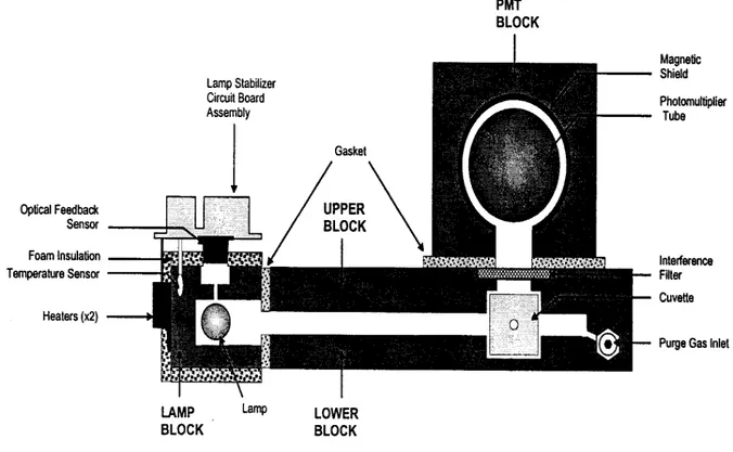
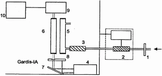
3.12.1. Experimental apparatus for DGM determination page 47
3.12.2. The atomic fluorescence spectrophotometer page 49
3.13. Determination of total dissolved mercury page 50
3.13.1. The atomic absorption spectrophotometer page 51
3.14. Determination of cellular and intracellular concentration of mercury page 52
3.15. Chemicals page 53
Index 4.1. Evaluation of phytochelatins in the diatom P. tricornutum as
biomarker of metal exposure in marine waters page 54
4.1.1. Exposure experiment of P. tricornutum in EDTA-buffered
artificial seawater page 55
4.1.2. Exposure experiment of P. tricornutum in natural seawater added with known amounts of heavy metals for evaluating
the applicability of the bioassay to natural seawater page 57 4.1.3. Application of the bioassay to polluted seawater page 59 4.2. Comparison of PC response in different taxa of autotrophic protists page 61 4.3. Evaluation of PC synthesis in P. tricornutum, T. weissflogii and
S. costatum as biomarker of metal bioavailability in sediments page 64
4.3.1. Short-term incubations page 65
4.3.2. Long-term incubations page 67
4.3.3. Effect of cellular density on PC production page 70 4.3.4. Application of the bioassay to a polluted area page 72 4.4. Evaluation of phytochelatins synthesis and Dissolved Gaseous
Mercury (DGM) production in T. weissflogii exposed to mercury page 76 4.4.1. Effect of mercury exposure on growth rate of T. weissflogii page 76 4.4.2. Two-day exposure of T. weissflogii to mercury page 77 4.4.3. Time course of non protein thiols pool and mercury
accumulation page 80
4.4.4. DGM production in T. weissflogii exposed to 5 nM mercury page 82
CHAPTER 5: CONCLUSIONS page 89
BIBLIOGRAPHY page 91
Abstract
ABSTRACT
For the comprehension of the processes of transport, transformation and accumulation of heavy metals in the marine environment an important contribute lies on the understanding of the diverse strategies developed during evolution by aquatic and terrestrial organisms in order to maintain an equilibrated relation with heavy metal ions present and available in the surrounding medium. The aim of the present study was to investigate the main mechanisms of detoxification acting in autotrophic protists to cope with heavy metal stress. Since a general response to metal stress in autotrophic organisms is represented by the synthesis of metal-binding peptides, named phytochelatins (PC), this study was focused on the induction of these peptides in different taxa of marine phytoplanktonic protists. Initially the induction of PC was investigated in cells of the marine diatom Phaeodactylum tricornutum exposed to environmentally relevant levels of dissolved Cd, Cu, Pb and Zn by performing short-term incubations both in EDTA-buffered artificial seawater and in natural seawater samples. Results showed that PC behave as a biomarker of exposure to the bioavailable metal fraction. Successively, experiments were carried out to examine the PC response in 5 taxa of autotrophic protists exposed to Cd, Cu and Pb. Results showed that the PC response depends on the particular species, the chemical form of the metal, the time of exposure and the metal concentration in the external medium. Successively, the research was devoted to develop new bioassays involving the presence of PC in phytoplankton as biomarkers of metal bioavailability in marine waters and sediments. The results obtained strongly support the feasibility of using this biochemical response in the assessment of toxicity of environmental systems. Besides the ability to synthesize metal binding peptides, the capability to produce Dissolved Gaseous Mercury (DGM) as another defence mechanism was investigated in
Thalassiosira weissflogii exposed to potentially toxic concentration of mercury. Mercury
exposure experiments needed a separate study given the chemical particularities of this metal in comparison to the other heavy metals and the peculiarity of the experimental methodology necessary for mercury determination. The results showed that the diatom responded to mercury exposure by synthesizing PC, besides to increase the intracellular pool of glutathione and γ-Glu-Cys. The time course of the non protein thiols pool and Hg intracellular concentration showed that PC, glutathione and γ-Glu-Cys represent a rapid cellular response to mercury exposure; however, at longer incubation times, their role in Hg detoxification seems to lose importance. At lower Hg concentration, at which the PC synthesis doesn’t seem to be involved, the occurrence of a process of reduction of the DGM production was investigated in the same diatom. The significant correlation between the cellular density in solution and the production of DGM, both in light and dark conditions, clearly showed that T.
weissflogii is capable to directly produce DGM. This finding has been confirmed by the
absence of DGM production in culture media containing formaldehyde-killed cells of T.
weissflogii. This approach is part of a wider study regarding the contribution of the eukaryotic
and prokaryotic microorganisms to the production of DGM in aquatic systems.
Keywords: heavy metals; autotrophic protists; detoxification mechanisms; phytochelatins; dissolved gaseous mercury
Introduction
CHAPTER 1 INTRODUCTION
1.1. Heavy metals in marine environment
Heavy metal pollution due to human activities represents a considerable concern for the modern world. Although human activities have always impacted on coastal areas, it is only within the last two centuries that the effects of industrialization, intensive agriculture and coastal engineering have seriously begun to threaten marine life (His et al., 1999). Forstner and Wittmann (1979) stated that compared with land systems, the relatively small biomass in aquatic environments generally occurs at a greater variety of trophic levels. This correlates to the particular sensitivity of aquatic systems with regard to pollution influences. Many substances pollute the marine environment, but non-biodegradable compounds are the most dangerous due to their innate ability to constantly remain with the ecosystem (Hernandez-Hernandez et al., 1990). In the last thirty years or so, heavy metals have become an increasingly common contaminant of sea and freshwater. Lakes and their sediments have long been recognized as common sinks for metals. Rivers, similarly, are capable of transporting large quantities of metals and constitute inputs for the marine environment. So the highest contamination levels of marine environment can be found in coastal and estuarine waters affected by the presence of anthropogenic activities and riverine inputs. Ober et al. (1987) affirmed that pollution of the marine ecosystem by heavy metals is a worldwide problem and the main sources of metal pollution are domestic/industrial sewage, industrial effluents, oil and chemical spills, combustion emissions, mining operations, metallurgical activities and non-hazardous landfill sites. According to Hernandez-Hernandez et al. (1990) the presence of metals in the marine environment is partly due to natural processes such as volcanic activity and erosion, but mostly results from industrial processes, with metals mainly entering the sea
Introduction
suspended in industrial wastes and in solid particles carried by winds, and eventually deposited in the sea (Jackson et al., 2005). Metals are usually present at low or very low concentrations in the oceans (Morel and Price, 2003). The concentrations of total dissolved cadmium, lead, copper and zinc measured in surface seawaters are in the range 0.01 - 0.2 nM, 0.05 - 0.4 nM, 1 - 5 nM and 1 - 40 nM, respectively (Millero and Sohn, 1992), but they can reach values 50-100 times higher in polluted and industrialized coastal areas. Sediments represent the major sink for contaminants in aquatic systems and they are the main location of the heavy elements in the hydrosphere; subsequently sediment re-suspension can act as source of contaminants for the overlying water column. Sediment disturbance, by way of natural factors, or human activities, can cause changes in the chemical properties of sediment, bringing to the mobilization of contaminants that could pose a threat to living organisms (Forstner and Salomons, 1991). Hence, the analysis of sediments yields useful information on the metal burden of natural waters.
Some metal ions are essential trace elements, but, essential or not, most heavy metals are toxic at higher concentrations. They appear to be dangerous pollutants since they have not only a short-term toxic effect on human and aquatic organisms, but also a dangerous mutagenous, embryo-toxic and gonadotoxic long-term effect. The hazard of toxic metals is due to the fact that the biologically available forms of heavy metals in the environment can be transformed and accumulated by various organisms. The fate and distribution of the heavy metals in the aqueous environment are determined by a large number of reactions occurring with dissolved inorganic and organic ligands as well as with natural heterogeneous compounds such as mineral surfaces and biological particles (Scarano and Morelli, 1999). The ability of some aquatic organisms to take up and store heavy metals and other chemical substances occurring in the environment is widely recognized. Thus heavy metals enter nutrition chains and potentially endanger living beings, becoming concentrated in fish and
Introduction
other edible organisms (known as bioaccumulation), particularly in near-shore areas (His et al., 1999). Overall, toxic effect of heavy metals occur as the result of a complex balance between the chemical speciation of the metal ion and the biology of the organism. There is now considerable evidence to suggest that the distribution and speciation of trace metals in the upper water column play an important role in the species composition and physiology of phytoplankton assemblages (Sunda 1994). The influence of metals and aquatic microorganisms is reciprocal: biological production in the oceans can strongly influence the oceanic chemistry of trace metals and, in turn, bioactive metals may affect the oceanic primary productivity.
Autotrophic microorganisms affect trace metal chemistry in natural and oceanic waters not only by surface reactions, but also by metal uptake and by production of extracellular organic matter capable to bind metal ions (Zutic et al., 1981; Seritti et al., 1986). Both the direct extracellular exudation products and the secondary products after biochemical modification have been demonstrated to have metal complexing properties (Imber et al., 1985; Zhou and Wangersky, 1985, 1989; Seritti et al., 1986). Thus, in natural waters, the production of the biogenic organic matter, by influencing the chemical forms of metal ions in solution, could affect the adsorption process on the cell surface (Scarano and Morelli, 1999).
1.2 Biogeochemical cycle of mercury
One significant aspect of the global biogeochemical cycling of mercury, that is different to other metals, is the volatility of this metal.
The natural sources of mercury emissions are weathering of rocks, windblown dust, volcanic activity, geysers, thermal fluids, degassing of the earth’s mantle, emanations from the oceans, transpiration and decay of vegetation and forest fires. The main transport pathway for
Introduction
mercury is through the atmosphere and for this reason mercury represents a powerful and dangerous pollutant that has become widespread throughout the world-wide environment. The volatilization of mercury may occur at any stage in the transport process. Volatile species are produced by chemical or biochemical reduction of Hg2+ to Hg0 and by the biomethylation of mercury to give dimethylmercury. Elemental mercury is a highly volatile chemical species and can be spread into the atmosphere even very far from the emission source (Schroeder et al., 1989).
The flux of mercury from the sea surface depends on the formation in the water column of volatile dissolved forms of mercury (90% elemental mercury), named Dissolved Gaseous Mercury (DGM), which pass from the water into the atmosphere due to their low water solubility (60µg/L at 25 °C) and high volatility (Henry coefficient < 0.3).
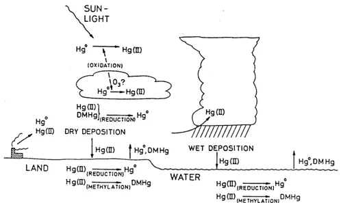
Mercury evasion from aquatic surfaces plays an important role in the complex biogeochemical cycle of this metal (Fig.1.1).
Fig. 1.1. Transports and transformations in the biogeochemical cycle of mercury (Lindqvist, 1994).
Introduction
Even if the regional and global atmospheric budgets need to be reassessed (Gustin et al., 2000), the oceanic evasion of the DGM into the atmosphere is a phenomenon comparable to terrestrial emissions (Lindberg et al., 1995; Gardfeldt et al., 2003). In particular, the mercury emission from the entire Mediterranean Sea has been estimated to be about 60 tons per year (Ferrara et al., 2000a); therefore the aquatic evasion represents the main natural source of this metal into the atmosphere in the Mediterranean area, higher than the emission from volcanoes (0.6-1.3 t/y; Ferrara et al., 2000b).
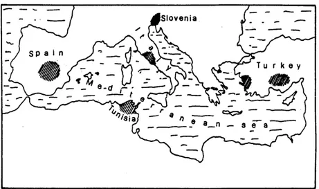
The Mediterranean basin is characterised by the presence of a noteworthy geochemical anomaly with large cinnabar deposits (Fig. 1.2) (Bernhard and Renzoni, 1977; Buffoni et al., 1982; Ferrara et al., 1997), by intense solar radiation and high environmental temperatures for many months of the year. These characteristics induce the formation of elemental mercury and its emission from soil and surface water through biological, chemical and photo-physical processes (Ferrara et al., 1997).
Introduction
1.2.1. Abiotic and biotic processes of Dissolved Gaseous Mercury production
The capability to produce Dissolved Gaseous Mercury (DGM), holds a noteworthy ecological importance in the context of the biogeochemical cycle of mercury. In aquatic systems the DGM formation and its volatilization to the atmosphere is a phenomenon comparable to terrestrial emissions (Lindberg et al., 1995; Gardfeldt et al., 2003); moreover, through DGM production, mercury is removed from the water column and is no longer available for methylation processes and subsequent bioaccumulation throughout food chains (Fitzgerald, 1993).
The dynamic of DGM formation results from several oxidation and reduction processes occurring simultaneously.
Literature records contrasting papers concerning the mechanisms of DGM formation: for a long period, authors have tried to establish if the production of elemental mercury can be defined “abiotic” or “biotic”. There is evidence for a photochemically enhanced abiotic production of DGM (Nriagu, 1994; Xiao et al., 1994), proved by the existence of a daily and seasonal behaviour of the DGM concentration in lakes, estuaries, riverine waters (Amyot et al., 1994, 1997; Krabbenhoft et al., 1998; Amyot et al., 2000; Zhang and Lindberg, 2000) and in seawater (Wangberg et al., 2001; Mason et al., 2001; Rolfhus and Fitzgerald, 2001; Lanzillotta and Ferrara, 2001; Lanzillotta et al., 2002). The occurrence of the highest DGM concentration values at the daily interval of maximum insolation and in summer supports this thesis. Photo-reduction may occur by photo-chemical reactions (Nriagu, 1994) in which organic substances take part (Matthiessen, 1998; Lanzillotta et al., 2004; O’Driscoll et al., 2004; Garcia et al., 2005), and/or by direct reactions of photolysis of the Hg2+ complexes to Hg0. The presence of Dissolved Organic Matter (DOM) may consistently play a role in the reaction rate of DGM formation (Costa and Liss, 1999, 2000); in particular, it has been identified a photo-sensitising role of the humic substances in marine photochemical reactions
Introduction
(Costa and Liss, 1999). However, Mason and Sullivan (1999) reported that mercury complexation by organic ligands reduces the concentration of Hg2+ available for the reduction processes. Besides an “abiotic” formation of DGM, a “biotic” Hg2+ reduction has been suggested to explain its presence in deep waters, especially close to the sediment (Kim and Fitzgerald, 1988). The importance of microorganisms on the direct formation of DGM under dark conditions has been suggested by some researches carried out in the Pacific Ocean; bacteria associated with deep marine sediments can produce Hg0 which is transported to the surface in the upwelling zones by the water currents (Kim and Fitzgerald, 1988).
The capability of microorganisms to reduce mercury compounds to volatile mercury is due to the unique characteristics of this metal. In fact to be detoxified by reduction, the redox potential of a given heavy-metal should be between that of the hydrogen/proton couple (-421 mV) and that of the oxygen/hydrogen couple (+808 mV) [calculed from Weast (1984) at 30 °C and pH 7.0], which is the physiological redox range for most aerobic cells. Thus, Hg2+ (+430 mV) and Cu2+ (-268 mV) may be reduced by the cell, but Zn2+ (-1.18 V) and Cd2+ (-824 mV) may not. If a cell chooses to detoxifies a compound by reduction, an efflux system should be present to export the reduced products. Only in the case of mercury do reducibility and a lower vapour pressure of the metallic reduction fit together; mercury is thus detoxified by reduction of Hg2+ to Hg2+ with diffusional loss of the Hg2+.
Nakamura et al. (2001) screened special mercury resistant bacteria in the mercury polluted sediments of the Minamata Bay (Japan), capable of volatilise mercury compounds at high rate, with the aim to develop a method for removing mercury by means of a biomass. Studies at high Hg2+ concentrations on the resistance to mercury of prokaryotic microorganisms have demonstrated that bacteria can convert both inorganic and organic mercury compounds to mercury vapour, using a plasmid-encoded enzymatic pathway, the mer gene; hence bacteria can play a role in the production of dissolved elemental mercury without the presence of solar
Introduction
radiation (Barkay et al., 1991; Barkay, 2001). An interesting review of the bacterial resistance to mercury is reported by Barkey et al. (2003). The existence of enzymatic reduction processes of mercury acting in bacteria has been widely reported (Barkay and Wagner-Dobler, 2005). DGM production in the dark has been examined by Rolfhus and Fitzgerald (2004) adding Hg2+ to dark and light-incubated coastal seawater samples from Long Island
Sound. The microbial reduction and oxidation of mercury in freshwaters lake were reported by Siciliano et al. (2002): authors concluded that H2O2 produced by solar radiation stimulates
mercury oxidase activity in lake water, which results in a decrease in DGM levels during the afternoon.
Literature reports data indicating that eukaryotic microorganisms can also reduce mercury, but only a few papers report on direct measurements of cellular generation of volatile mercury species (Ben-Bassat et al., 1972; Mason et al., 1995). Ben-Bassat and Mayer (1977, 1978) and Bentz (1977) found that the formation of Hg0 decreased as a function of the inhibition of photosynthesis in cultures of the green algae Chlorella. Moreover, the highest values of DGM concentration were observed together with the highest values of chlorophyll a, suggesting that phytoplankton may produce DGM (Vandal et al., 1991). It has been hypothesised that algae and diatoms are able to reduce Hg externally by cell surface enzymatic processes, like other metals (Jones et al., 1986). With refer to Cu2+ and Fe3+ and other metals, three mercury reduction path-ways were indentified for the diatom Thalassiosira weissflogii: reduction by organic compounds released into the medium, by cell wall components and via a plasmalemma enzymatic pathway (Price and Morel, 1990). Mason et al. (1995) found that small phytoplanktonic microorganisms (typically <3 µm diameter such as cyanobacteria) are the primary mercury reducers. Lanzillotta et al. (2004), studying the processes of DGM formation by phytoplanktonic microorganisms and by their organic compounds released into
Introduction
the marine environment, concluded that DGM formation derives mainly from a photochemical process acting on the biogenic organic matter.
1.3. Toxicity of heavy metals
Of the 90 naturally occurring elements, 21 are non-metals, 16 are light metals and the remaining 53 are heavy metals (Weast, 1984).
Most heavy metals are transition elements with incompletely filled d orbitals. These d orbitals provide heavy-metal cations with the ability to be redox-active. Hence a number of trace metals are used by living organisms to stabilize protein structures, facilitate electron transfer reactions and catalyze enzymatic reactions. For example, copper (Cu), zinc (Zn) and iron (Fe) are essential as constituents of the catalytic sites of several enzymes. Other metals, such as lead (Pb), mercury (Hg) and cadmium (Cd) may displace or substitute for essential trace metals and interfere with proper functioning of enzymes and associated cofactors (Nies, 1999).
The main biological characteristics of the most common heavy metals are described in the following paragraphs.
1.3.1. Copper (Cu)
The electrochemical potential of Cu2+/Cu+ is -268 mV, well within the physiological range. Copper easily interacts with radicals, best with molecular oxygen. Its radical character makes copper very toxic. Copper toxicity is based on the production of hydroperoxide radicals (Rodriguez Montelongo et al., 1993) and on interaction with the cell membrane (Suwalsky et al., 1998).
Introduction
Besides copper/zinc superoxide dismutase, the most important function of copper is in the cytocrome c oxidase and related enzymes, which are oxygen-dependent terminal oxidasees in the respiratory chain of many organisms (Nies, 1999).
1.3.2. Zinc (Zn)
Zinc occurs exclusively as the divalent cation Zn2+. With its completely filled d orbitals, the zinc cation is not able to undergo redox changes under biological conditions. Zinc is a component in such a variety of enzymes and DNA-binding proteins, such as zinc-finger proteins, which also exist in bacteria (Chou et al., 1998), that life seems not to be possible without this redox-inactive element. Zinc may be complexed by various cellular components and is transported by members of a variety of protein families (Nies, 1999).
1.3.3. Lead (Pb)
Pb appears ubiquitous in aquatic ecosystems and is bioaccumulated in aquatic organisms (Moriarty, 1990). It occurs in the environment in a wide range of physical and chemical forms that greatly influence its behaviour and its effects on the ecosystem. Most of the Pb in the environment is in the inorganic form and exists in several oxidation states (0, I, II, and IV). According to Nussey et al. (2000), Pb(II) is the most stable ionic species present in the environment and is thought to be the form in which most Pb is bioaccumulated by aquatic organisms (Jackson et al., 2005). Lead has been used in large amounts for 2500 years (Hong et al., 1994), recently as a fuel additive, although the toxicity of lead for animals and man has been well known for a long time (Johnson, 1998). Lead acts on the central nervous system, on blood pressure and on reproduction (Goyer, 1993). Pb resistance seems to be based predominantly on metal ion efflux (Nies, 1999).
Introduction
1.3.4. Cadmium (Cd)
A consistent amount of work has been done especially on cadmium toxicity in microorganisms; its effect regard the “thiol-binding and protein denaturation”, the “interaction with calcium metabolism and membrane damage” and the “interaction with zinc metabolism”. The main detoxification systems acting in plant and algae seems to be mediated by transport of glutathione/phytochelatin complexes by proteic transporters into the vacuoles (Nies, 1999).
1.3.5. Mercury (Hg)
Mercury is the most toxic of all the heavy metals. Both dissolved, inorganic Hg and MeHg, accumulate in phytoplankton (Andren et al., 1998) by passive diffusion across the membrane (Mason et al., 1995) or by facilitated diffusion transport (Watras et al., 1998). However, in contrast to MeHg, inorganic Hg is not biomagnified in the trophic transfer from phytoplankton to zooplankton. MeHg reaches its highest concentration in the tissues of fish at the top of aquatic food chain (WHO,1989). The affinity of Hg2+ to thiol groups is even stronger than the affinity of cadmium to sulphide.
Resistance to mercury is based on its unique peculiarities: its redox potential [its electrochemical potential of Hg2+/Hg0 at pH 7 is +430 mV] and the vapour pressure/melting/boiling point of metallic mercury, which is extraordinarily low for a metal [melting point -39 °C, boiling point 357 °C (Weast, 1984)]. Thus living cells such as bacteria are able to reduce Hg2+ to Hg0, which does not remain inside the cell with the potential of becoming oxidized again, but leaves the cell by passive diffusion (Silver, 1996; Silver and Phung, 1996). Once outside, however, metallic mercury may be oxidized again by other bacteria (Smith et al., 1998).
Introduction
1.3.6. Effect of heavy metals on autotrophic protists
As regards autotrophic protists, previous works have showed that toxic metals act on photosynthesis (Rosko and Rachlin, 1977), cell division (Rachlin et al., 1983), membrane permeability (Rachlin and Grosso, 1993) and cell motility (Anderson and Morel, 1978; Fennikoh et al., 1978).
To have any physiological or toxic effect, metal ions have to enter the cell. Surfaces of living particles are characterized by various sites capable to adsorb metal ions with high affinity (Scarano and Morelli, 1993). The binding of metals to the cell surface represents the initial mechanism of the metal ion uptake process of the microorganisms. When metal ions equilibrate on the outside of the cell, this rapid step is followed by a relatively slow uptake due to the membrane transport of the metal inside the cell. The adsorption process on cell surface can be interpreted in terms of surface coordination between the metal and one or more functional groups on the cell surface (Scarano and Morelli, 1993). The final equilibrium of this process depends on the chemical composition of the surrounding solution. Once inside the cell, metals affect cellular metabolism by forming coordination complexes with various biomolecules, including numerous enzymes that require specific metals as essential cofactors. However, when in excess, metal ions can interfere with numerous physiological processes, thus resulting very toxic to the cells. Hence, the intracellular concentration of heavy-metal ions has to be tightly controlled by living organisms.
Numerous species of algae, both macro- and micro-algae, are capable of sequestering significant quantities of either nutrient or toxic heavy metal ions from aqueous solutions. In order to cope with metal toxicity, the following mechanisms of detoxification have been observed in algae and autotrophic microorganisms (Gonzales-Davila, 1995):
1) the development of energy driven-efflux pumps that keep toxic element levels low in the interior of the cell.
Introduction
2) Oxidation state change by which a more toxic form of a metal can enzymatically and intracellularly be converted to a less toxic form.
3) Precipitation of insoluble metal complexes on the cell surface.
4) Complexing of metal ions with excreted metabolites (extracellular products), which can extracellularly mask a toxic metal.
5) Vaporization and elimination by means of converting a toxic metal to the volatile chemical species.
6) Binding of metal ions with proteins or polysaccharides in the interior of the cell, which may deactivate the metal ion’s toxicity.
7) Methylation of the element, which can enzymatically and intacellularly prevent a toxic element from reacting with a –SH group.
1.4. Ecological monitoring of heavy metal pollution
According to Robinson and Avenant-Oldewage (1997) the two factors which contribute principally to the damaging effect of metals as environmental pollutants are, firstly, their inadequate biological degradation to inert metals (as in the case of most organic pollutants), and secondly, the propensity of metals to accumulate and to remain largely in the aquatic environment.
The assessment of pollution effects may be more difficult on the ecosystem level than on the level of single species. There are a great deal of data on metal concentrations in various organisms exposed to experimental contamination or taken from areas subjected to anthropogenic inputs of metals, so as many are the studies that, taking solely metal concentrations in waters into account, have determined sub-lethal or acute toxicological parameters. However, these data do not permit the establishment of direct relations between metal pollution and toxicity of metals. The concentrations in living organisms are not related
Introduction
to the concentrations in water by a simple invariable function which takes no account of the chemistry of metals in seawater. When variations of the chemical composition of the external medium occur, numerous organisms are able to keep their internal chemical composition at a steady level, compatible with the normal development of their physiological functions. This ability varies from species to species. However, the ability to regulate is effective for moderate variations of concentrations in the environment. Moreover this ability seems to vary according to the physiological functions of the trace elements (Amiard-Triquet and Amiard, 1980). According to Robinson and Avenant-Oldewage (1997) and Hernandez-Hernandez et al. (1990), several factors affect the toxicity of pollutants to aquatic organisms and can be divided into biotic and abiotic factors. The former include physiological conditions, tolerance, growth and reproduction, species variations, inter- and intra-specific variation in life history stages, adaptive capabilities and behavioural responses; the latter could be represented by metal species in the water, the presence of other metals or pollutants, nature of dissolved organic matter, pH, temperature, alkalinity and hardness, metal interactions and dissolved oxygen and interactions between all them. The effect of two or more toxicants may be additive, antagonistic or even synergistic (Jackson et al., 2005). Hence, chemical analysis of individual toxic compounds are not adequate indicators of their associated biological and ecological effects and they should be combined with biological tools to assess the toxicity of the chemical that is biologically available (Martin-Diaz et al., 2004). Also, in addition to the common criteria on the individual level, such as mortality, reduced growth rates, metabolic activities and reproduction, further indications of stress may be of ecological significance. Toxicity measurement of wastewater, sediments and contaminated water bodies is a very important part of environmental pollution monitoring. Toxicity tests are desirable in water pollution evaluations because chemical and physical tests alone are not sufficient to assess potential effects on aquatic biota.
Introduction
The effect of heavy metals and other toxicants on various animal taxa and life stages of them have been extensively reported in the literature (MacDonald et al., 1988; McKenney and Neff, 1979; Ober et al., 1987; Stark, 1998; Jackson et al., 2005).
Hernadez-Hernandez et al. (1990) stated that with regard to metal bioaccumulation in marine organisms, several authors have proved their high accumulation ability in crustaceans, mollusks and fishes, which generally depends on their exposure time and the concentrations of metals in the water. In the processes of transformation and accumulation of contaminants, microorganisms participate primarily due to their considerable amount in the ecosystems and their great adaptability (Nelson and Colwell, 1975); in particular marine microalgae are promising indicator species for organic and inorganic pollutants and constitute important tools to monitor physiological changes in the presence of heavy metals (Torres et al., 2008).
1.4.1. Sediment toxicity tests
The majority of chemicals discharged into aquatic system eventually end up in sediments that may act both as sink and source of pollution. Many toxic and bioaccumulative pollutants are found only in trace amounts in water and often at elevated levels in sediments. In fact,the highest accumulation factors of all potential sample materials of the marine environment is exhibited by sediments and especially by the finest grain size fractions. Sediments near urban areas commonly contain high levels of contaminants, constituting a major environmental problem faced by many anthropogenically impacted aquatic environments.
The role of aquatic sediments as a sink and source of large quantities of contaminants has led to the development of a wide variety of bioassays for the toxicological assessment of sediments; they represent complementary approaches for characterizing the biological effects and hazards of contaminated sediments. These procedures range in complexity from short-term lethality tests that measure effects of individual contaminants on single species to
long-Introduction
term tests that determine the effects of chemical mixtures on the structure and function of communities.
The evaluated sediment phase may include whole sediment, suspended sediment, elutriates, or sediment extracts. Although whole-sediment testing seems the most realistic approach to evaluate the bioavailability of contaminants in sediments, porewaters and aqueous extracts are frequently used. Elutriate, consisting of seawater re-suspension of sediments, provides a measure of the amount of a substance that is exchanged between the sediment and the aqueous phase during sediment disturbance and gives information on the leaching capability of sediment-associated contaminant.
Elutriate Sediment Toxicity Test (ESTT) have been extensively considered the best method to replicate disturbance effects on the release of chemicals from the sediment (US-EPA, 1991). The standard elutriate test was jointly developed in the early 1970s by the U.S. Army Corps of Engineers and the U.S. Environmental Protection Agency to monitor the soluble release of contaminants into the water column during open-water disposal of dredged sediments. This approach is used in sediment studies to simulate processes that might disturb the sediment and bring contaminants into the water column. Toxicity testing of sediment elutriates is important because dissolved forms of pollutants are more bioavailable to aquatic biota for uptake and are the primary cause of adverse impacts in aquatic ecosystems.
In the past two decades a wide variety of toxicological tests, involving organisms at different trophic levels, including bacteria, phytoplankton, mollusks, crustaceans and fishes has been developed in order to assess the biological effects of contaminants in sediments (Cheung et al., 1997; Matthiessen et al., 1998; Geffard et al., 2003; Arizzi-Novelli et al., 2006).
The release, the bioavailability and the toxicity of contaminants in elutriates of estuarine sediments have been examined by studying concurrently their effects both on the
Introduction
embryogenesis and on the larval growth of the Crassostrea gigas larvae together with their bioaccumulation in those organisms (Mucha et al., 2004).
Several laboratory conditions for preparing and testing elutriates of sediments of industrial and urban contaminated areas of the Lagoon of Venice (Italy) have been assessed in experiments by using embryos of the sea urchin Paracentrotus lividus (Marin et al., 2001). The bioavailability of Cd, Cu, Zn, and Pb in the elutriates of two metal-contaminated sediments (Bidassoa and Dunkerque) has been studied by using the presence of metallothioneins in Crassostrea gigas larvae as a biomarker of metal exposure (Geffard et al., 2007); these authors concluded that the production of metallothioneins is a more sensitive indicator of heavy metal pollution than other physiological endpoints and could be proposed as an early biomarker of metal exposure in larvae.
Cheung et al. (1997) carried out Elutriate Sediment Toxicity Tests, using two microalgae
Skeletonema costatum (a diatom) and Dunaliella tertiolecta (a green alga), juvenile shrimp
(Metapenaeus ensis) and juvenile fish (Trachinotus obtaus), with the aim to study the feasibility of using different trophic organisms for evaluating the toxicity of dredged sediments arising in Hong Kong; these authors also employed two commercially available tests using bacteria (Microtox Test and Toxi-Chromotest) to test both the solid phase and elutriates of the sediments and concluded that bioassay tests using diatom on the sediment elutriate were correlated significantly (p < 0.05) with a number of physico-chemical properties of sediments and elutriates.
Bioassays with microorganisms are widely used as tools in estimating the potential risk of contaminated sediments. In particular, in the assessment of toxicity of sediment elutriates, bioassays based on the growth of phytoplanktonic microorganisms have been largely used (Pardos et al., 1998; Mucha et al., 2004; Tolun et al., 2001; Wong et al., 1999) because of their sensitivity to different contaminants. Davoren et al. (2005), using an integrated approach
Introduction
based on the use of a number of bioassays representing multiple trophic levels, concluded that the algal test was the most responsive to elutriates of estuarine sediments.
1.4.2. Biomarkers
The presence of chemical compounds in natural aquatic systems does not indicate, by itself, injurious effects to organisms (Wang et al., 1998), as bioavailability of these compounds should also be taken into account. The use of biomarkers can be an important tool for evaluating toxic effects of bioavailable contaminants.
A biomarker may be defined as a biochemical variation measured in tissue/body fluids of an organism that provides evidence of exposure and effects of one or more chemical pollutants (Phillips and Rainbow 1993; Depledge and Fossi 1994).
Biological monitoring or biomonitoring can be defined as the systematic use of biological responses to evaluate changes in the environment, with the intent of establishing a quality control program (Cairns and van der Schalie, 1980). Typically, biomarkers are considered quantitative measures of changes in the biological system that can be related to exposure to the toxic effects of environmental chemicals (WHO, 1993; Peakall and Walker, 1994). Although not explicitly contained in most definitions, the use of the term “biomarker” or “biomarker response” is often restricted to cellular, biochemical, molecular, or physiological changes that are measured in cells, body fluids, tissues, or organs within an organism that are indicative of xenobiotic exposure (Van Gestel and Van Brummelen, 1996; van der Oost et al., 2003; Lam and Gray, 2003). Often pollutant exposure at contaminated sites does not result in lethality to resident biota, but may produce more sub-lethal effects that may compromise the biochemical, physiological and reproductive functions of living organisms, and, ultimately, influence the long-term survival of populations. The advantages of biochemical biomarkers are that: 1) they represent a direct biological response to pollutant exposure; 2) they often
Introduction
respond to exposure at concentrations lower than that required for effects at the individual or population level; 3) they are often induced prior to effects at higher organizational levels. Thereby they constitute an early warning indication of the toxicity of a pollutant (MacFarlane et al., 2006; Torres et al., 2008).
In recent years a great variety of organisms have been employed in biomonitoring programs in order to assess the impact of pollutants on the aquatic environment. Bivalve mollusks, particularly mussels, have been elected as “sentinel” organisms in international environmental monitoring programs as part of the MUSSEL WATCH PROGRAM (Goldberg, 1975; Tavares et al., 1988; Claisse, 1989; Tripp et al., 1992; Tanabe, 1994). Many other organisms have also been used as regionally important tools in environmental programs, e.g., mangrove mussels in South Brazil (Torres et al., 2002), crabs in South Africa (Thawley et al., 2004), polychaetes in Spain and France (Gesteira and Dauvin, 2000), fish in Australia, Asia, and America (Edwards et al., 2001; Ueno et al., 2005; Carrasco-Letelier et al., 2006), respectively.
In addition to the massive use of marine animals in biomonitoring programs, photosynthetic organisms like algae have increasingly been used as biodetectors to monitor xenobiotics in marine environments (Jayasekera and Rossbach, 1996; Ali et al., 1999; Sánchez-Rodríguez et al., 2001; Barreiro et al., 2002; Conti and Cecchetti, 2003; Conti et al., 2007). Because of their natural and widespread occurrence along worldwide seashores, photosynthesizing organisms could be useful for a time-integrated picture of the ecosystem response to exposure to toxic compounds. Physiological changes, both in macroalgae (Sánchez-Rodríguez et al., 2001; Conti and Cecchetti, 2003) and microalgae (Rijstenbil et al., 1994; Tripathi et al., 2006), are important tools in the hazard of heavy metals in the aquatic environment (Torres et al., 2008). Algae have been suggested and used as potential bioindicators of aquatic pollution and its metabolic response to xenobiotic could point to important biomarkers (Witton and Kelly, 1995; Ali et al., 1999). Moreover, autotrophic microorganisms are particularly promising
Introduction
indicator species for organic and inorganic pollutants since they are typically the most abundant life forms in aquatic environments and occupy the base of the food chain. The study of physiological and biochemical alterations, as well as the identification and quantification of pollutants in basal-level trophic organisms are an essential diagnostic tool (Van Gestel and Van Brummelen, 1996, Handy et al., 2003).
The presence of metals in the plant kingdom induces the synthesis of several proteins, mainly phytochelatins (PC) (Cobbett and Goldsbrough, 2002; Perales-Vela et al., 2006), but also metallothionein (MTs) (Vasak, 2005), and heat shock proteins (HSPs) (Spijkerman et al., 2007).
Metallothioniens and phytochelatins are similar in many ways, including the high number of cysteine molecules, and the fact that both are responsible for the detoxification of heavy metals. In fact, PC were originally classified as class 3 MTs, until they were deemed sufficiently different in structure and synthesis pathway to be classified as PC. All MTs have three characteristics in common: they have low molecular weight (6–7 kDa), a large fraction of cysteine residues, and a high metal content with coordination of metal ions in metal-thiolate clusters. Metallothioneins, cysteine-rich and metal-binding proteins, are products of mRNA translation and this distinguishes them from PC, which are the product of an enzymatic synthesis (Grill et al., 1989; Cobbett and Goldsbrough, 2002).
The presence of intracellular PC constitutes an early and specific signal of metal stress in plants and autotrophic microorganisms (Cobbett, 2000; Kawakami et al., 2006a), so these compounds can be considered suitable biochemical indicators of metal exposure.
1.4.2.1. Phytochelatins
A widespread mechanism of defence developed by plants and autotrophic microorganisms against metal stress involves phytochelatins. The general structure has been determined to be
Introduction
(γ-Glu-Cys)n-Gly where chain length “n” ranges between 2 and 11 units (Rauser, 1995, Steffens, 1986, Cobbett and Goldsbrough, 2002). It is important to note that the glutammic acid residues are not bond with cysteine by means of an α-carboxyl group as in transcriptional amminoacids but with an γ-carboxyl group. The gamma-glutammyl linkages present, which cannot be prepared by ribosomes, lead to the search for an enzyme-mediated path for the production of PC. Grill et al., 1989 demonstrated that PC are synthesized by the enzyme “phytochelatin synthase” (PC), which is a γ-glutamylcysteine dipeptidyl transpeptidase. It catalyzes the transpeptidation of the γ-Glu–Cys moiety of glutathione (γECG) onto a second γECG molecule to form PC2 or onto a PC molecule to produce a n + 1 oligomer. The enzyme
was described as a tetramer of MW 95 000 with a Km for glutathione of 6.7 mM (Steffens, 1986 and Cobbett and Goldsbrough, 2002).
The general mechanism involved in PC biosynthesis is:
[γGlu–Cys]n-Gly + [γGlu–Cys]-Gly → [γGlu–Cys]n+1-Gly + Gly
Numerous physiological, biochemical and genetic studies have confirmed that glutathione (or, in some cases, related compounds) is the substrate for PC biosynthesis (Rauser, 1995, 1999; Zenk, 1996). Early studies with cell cultures demonstrated that induction of PC in the presence of Cd coincided with a transient decrease in levels of glutathione. Furthermore, the exposure of either cell cultures or intact plants to an inhibitor of glutathione biosynthesis, “buthionine sulfoximine”, conferred increased sensitivity to Cd with a corresponding inhibition of PC biosynthesis. This could be reversed by the addition of glutathione to the growth medium.
The dependence of phytochelatin synthase on heavy metals for activity has invariably been interpreted in terms of direct metal binding to the enzyme. Few investigators have considered
Introduction
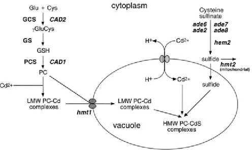
explicitly how heavy metals activate PC synthase but those that have considered it, have assumed that activation is consequent on the direct binding of metal ions to the enzyme (Zenk,1996; Cobbett, 2000). Indeed, in the most recent model for PC synthase action, it has been proposed that the strongly conserved N-terminal half of the enzyme is responsible for catalysis and that activation arises from the binding of metal ions to residues, possibly cysteine residues, within this domain. This provides a mechanism to autoregulate the biosynthesis of PC in which the product of the reaction chelates the activating metal, thereby terminating the reaction (Cobbett, 2000, Fig. 1.3).
Fig. 1.3. PC biosynthetic pathway (Cobbett, 2000).
In the last years a considerable interest has been attracted to PC as biochemical indicators of metal stress both in higher plants (Keltjens et al., 1998; Sneller et al., 1999; Sun et al., 2005) and in microalgae (Ahner et al., 1997; Knauer et al., 1998; Pawlik-Skowronska, 2000, Wei et al., 2003; Le Faucheur et al., 2005a), since these compounds constitute an early and specific signal of the actual intracellular metal concentration. This is supported by the finding that enhanced cellular PC have been measured in natural populations of marine and freshwater
Introduction
microalgae collected in polluted areas (Ahner et al., 1994; Ahner et al., 1997; Knauer et al., 1998).
The induction of phytochelatins in phytoplanktonic algae has been demonstrated, both in laboratory cultures (Gekeler et al., 1988) and in field studies (Ahner et al., 1997), even at very low concentrations of many heavy metals. Although a direct relationship between metal dose and PC production has been observed, the magnitude of the response, the degree of polymerization of PC synthesized and the relative level of individual peptides seem to depend on the organism analyzed (Ahner et al., 1995) as well as on the particular metal used as inducer (Ahner and Morel, 1995). Constitutive differences such as the size of the cellular glutathione pool, the rate of metal uptake or the capability to activate other mechanisms of metal tolerance, can account for the wide variability of PC production among species.
Aim of the study
CHAPTER 2 AIM OF THE STUDY
The research performed in the framework of the PhD course has been devoted to analyse the workings of known biological metal-resistance systems and their ecological significance with the aim to give a contribution to the comprehension of the processes of transport, transformation and accumulation of heavy metals in the marine environment.
Metal ions, regardless of whether they are biologically essential or not, may exert toxic effects to the living organisms when a critical concentration is reached. This concentration depends on the organism, on the metal and on the metal speciation. In aquatic systems, metal ions or compounds in solution are available for biota by adsorption on the surface of organisms and by translocation into the cells.
Many organisms can grow in contaminated environments since they have developed physiological adaptations to metal excess as defence mechanism. Among others, unicellular phytoplankton possess molecular mechanisms that allow them to discriminate non-essential heavy metals from the essential ones for their growth. In addition, they must maintain non-toxic concentrations of these ions inside their cells. In this way, two principal mechanisms have been identified, one which prevents the indiscriminate entrance of metal ions into the cell, i.e., exclusion, and the other which prevents bioavailability of these toxic ions once inside the cell, i.e., the formation of complexes and the metal volatilization.
On the basis of the simplicity of metal exposure and because the same cells both absorb and detoxify metal ions, unicellular phytoplankton represent suitable organisms for the purpose of surveilling and improving water quality, constituting sensitive indicators of the metal load of aquatic ecosystems. Moreover autotrophic microorganisms form the base of marine food web
Aim of the study
and it is obvious that, through the potential transfer along food chains, they might play a fundamental role in accumulation of heavy metals.
The goal of this research was to evaluate and to compare the ecological importance of the detoxification processes executed by different taxa of autotrophic protists exposed to environmentally relevant levels of dissolved Cd, Cu, Pb, Zn and Hg. In addition, a feature of this research was devoted to examine the detoxification mechanisms acting in these microorganisms, which can be suitable to investigate the quality of metal-polluted water systems.
Mercury experiments needed a separate study given the chemical particularities of this metal in comparison to the other heavy metals and the peculiarity of the experimental methodology necessary for mercury determination.
It is well known that a widespread mechanism of defence developed by plants and algae against metal stress involves intracellular metal-binding peptides. These molecules, called phytochelatins (PC), have glutathione (GSH) as biosynthetic precursor, and their main function in cells is to chelate metal ions in the cytoplasm, thereby reducing the concentration of the cytotoxic free metal ions. Five species of autotrophic protists: Phaeodactylum
tricornutum, Thalassiosira weissflogii and Skeletonema costatum (diatoms), Dunaliella tertiolecta, (green algae), Emiliania huxleyi (coccolitophore), were examined in order to
understand if this defence mechanism is conservative throughout the species.
Initially the research was focalized on the evaluation of the suitability to use the accumulation of PC as a biomarker of metal bioavailability in bioassays for the assessment of metal pollution in the marine environment. Results of preliminary laboratory experiments, carried out with P. tricornutum cells incubated in EDTA-buffered artificial seawater added with known amount of Cd, Pb and Cu metal ions, showed increasing cellular PC concentration with increasing free metal ions in the medium, indicating that PC behave as a biomarker of
Aim of the study
exposure to the bioavailable metal fraction. Comparative experiments carried out using controlled systems, such as laboratory cultures of the five species of eukaryotic microorganisms, showed that Phaeodactylum tricornutum, Thalassiosira weissflogii and
Skeletonema costatum were the more sensitive species with respect to the synthesis of PC. So,
these diatoms were used to develop a new bioassay involving the presence of PC as response to metal bioavailability in re-suspensions of marine sediments (elutriates) collected in a metal-polluted coastal area (Foce dello Scolmatore and Marina di Pisa). Elutriates of marine sediments were considered because they yields useful information on the metal burden of natural waters. In fact sediments represent the major sink for contaminants in aquatic systems and subsequently sediment re-suspension can act as source of contaminants for the overlying water column (Forstner and Salomons, 1991).
Successively, T. weissflogii was investigated in order to understand the relative importance of two defence mechanisms acting in this diatom when exposed to potentially toxic concentration of mercury. Besides the ability to synthesize metal binding peptides, its capability to produce dissolved gaseous mercury (DGM) was investigated. In the literature there are many reports concerning the synthesis of PC in eukaryotic microalgae in response to heavy metals, such as Cd and at lesser extent Cu, Pb or Zn, but only a very few studies report on the use of Hg as a PC inductor. As regards the DGM production in aquatic systems, the existence of enzymatic reduction processes of mercury acting in bacteria has been widely reported (Barkay et al., 1991; Barkay, 2001; Barkey et al., 2003; Barkay and Wagner-Dobler, 2005; Nakamura et al., 2001); only a few papers assumed the existence of DGM production by eukaryotic microorganisms, although this mechanism is poorly known (Ben-Bassat and Mayer, 1977,1978; Bentz, 1977; Mason et al., 1995). To our knowledge, no attempt has been made to follow and correlate both these Hg-induced responses.
Aim of the study
The ecological importance of the process of DGM production in the context of the biogeochemical cycle of this metal has been also discussed. In aquatic systems the DGM formation and its volatilization to the atmosphere is a phenomenon comparable to terrestrial emissions (Lindberg et al., 1998; Gardfeldt et al., 2003); moreover, through DGM production, mercury is removed from the water column and is no longer available for methylation processes and subsequent bioaccumulation throughout food chains.
Methodology
CHAPTER 3 METHODOLOGY
3.1. Laboratory cultures of marine autotrophic protists
The marine autotrophic protists: Phaeodactylum tricornutum (Bohlin), Thalassiosira
weissflogii (Grunow in Van Hemck) Fryxell & Haxle and Skeletonema costatum (Greville)
Cleve (diatoms), Dunaliella tertiolecta, (green algae), Emiliania huxleyi (coccolithophorid), were obtained from the Culture Collection of Algae and Protozoa, Dunstaffnage Marine Laboratory, UK.
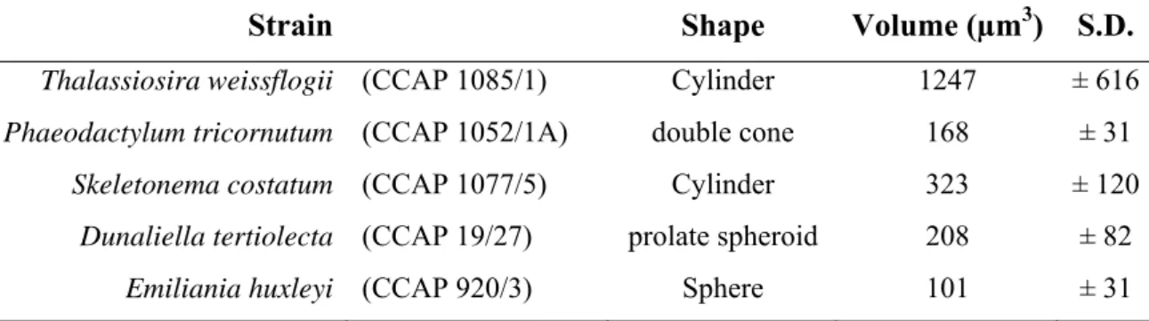
Cellular volumes were estimated from cell measurements and assumption of particular geometric shapes (Tab. 3.1).
Strain Shape Volume (µm3) S.D.
Thalassiosira weissflogii (CCAP 1085/1) Cylinder 1247 ± 616 Phaeodactylum tricornutum (CCAP 1052/1A) double cone 168 ± 31 Skeletonema costatum (CCAP 1077/5) Cylinder 323 ± 120 Dunaliella tertiolecta (CCAP 19/27) prolate spheroid 208 ± 82 Emiliania huxleyi (CCAP 920/3) Sphere 101 ± 31
Tab. 3.1. Strain, shape and cellular volume of the microorganisms examined.
3.2. Culture conditions
Stock cultures of P. tricornutum, T. weissflogii, S. costatum, D. tertiolecta and E. huxleyi, were grown in axenic conditions, in natural seawater enriched with f/2 medium (Guillard, 1975) at one–fifth the normal trace metal concentration (Tab. 3.2), at 21°C and fluorescent daylight (100 μmol photons × m-2 × s-1) in a 16:8 light-dark cycle. The culture medium was
Methodology
camera-box with laminar airflow. Exponential growth was maintained by inoculating weekly into a fresh sterilized medium.
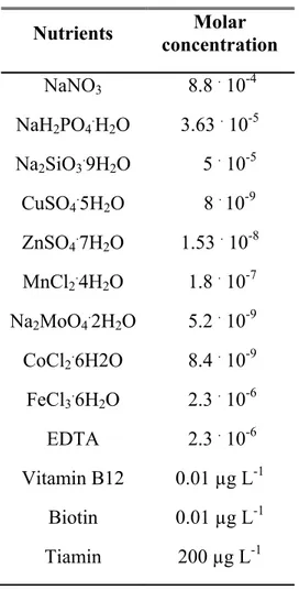
Nutrients Molar concentration NaNO3 8.8 . 10-4 NaH2PO4.H2O 3.63 . 10-5 Na2SiO3.9H2O 5 . 10-5 CuSO4.5H2O 8 . 10-9 ZnSO4.7H2O 1.53 . 10-8 MnCl2.4H2O 1.8 . 10-7 Na2MoO4.2H2O 5.2 . 10-9 CoCl2.6H2O 8.4 . 10-9 FeCl3.6H2O 2.3 . 10-6 EDTA 2.3 . 10-6 Vitamin B12 0.01 µg L-1 Biotin 0.01 µg L-1 Tiamin 200 µg L-1
Tab. 3.2. f/2 medium (Guillard, 1975) modified at one–fifth the normal trace metal concentration.
3.3. Cell density measurement
Cell density was measured, under an optical microscope (Zeiss), by means of two types of haemocytometers (depending on cellular density): the Thoma counting chamber and the Neubauer counting chamber.
Methodology
For as regards the Thoma counting chamber each grid consist of 16 fields containing 256 squares; the area of each square is 0.0025 mm2, corresponding to a total area of each grid of 0.64 mm2 and to a volume of 64 . 10-6 cm3 (chamber depth = 0.01 cm). Cell count consists of the total number of cells found in the 16 fields; cell density (cells mL-1) is obtained using the following proportion:
¾ Average counted cells : 64 . 10-6 cm3 = x* : 1 cm3 (*x = cell number)
For as regards the Neubauer counting chamber, the cell count consists of the total number of cells found in the 4 corner fields of the grid. The total area of the 4 corner fields is 0.04 cm2 and the volume is 4 . 10-4 cm3 (chamber depth = 0.01 cm). Cell density (cells mL-1) is obtained using the following proportion:
¾ Average counted cells : 4 . 10-4 cm3 = x* : 1 cm3 (*x = cell number)
3.4. Growth rate measurement
The growth rate of the cultures (µ), expressed as doublings . day-1, has been evaluated during the exponential phase of growth by counting cells at the time of the inoculum (t0) and after 6
days; the following equation has been used:
μ = 1/t log2 (N/N0)
t = days of growth
N0= number of cells at t = 0
Methodology 3.5. Natural seawater
Natural seawater, used throughout the research work, was collected 3 miles offshore from the Island of Capraia (Tyrrhenian Sea, Italy), filtered through 0.45 μm membrane filters (Sartorius) and stored in the dark at + 4 °C. To avoid any contamination, pretreatment and cleaning procedures for sampling bottles and other labware were performed following indications suggested by Mart (1976).
3.6. EDTA-buffered artificial seawater
EDTA-buffered artificial seawater was prepared by following the Aquil recipe (Price et al., 1991) omitting the micronutrient metal stock solution, but adding EDTA (10.0 μM) and calculated amounts of Cd, Pb or Cu. Free metal ion concentrations in these metal buffers were calculated by means of the MINEQL+ chemical equilibrium program (Westall et al., 1976). All solutions were allowed to equilibrate overnight before performing tests.
3.7. Sediment sampling and elutriate preparation
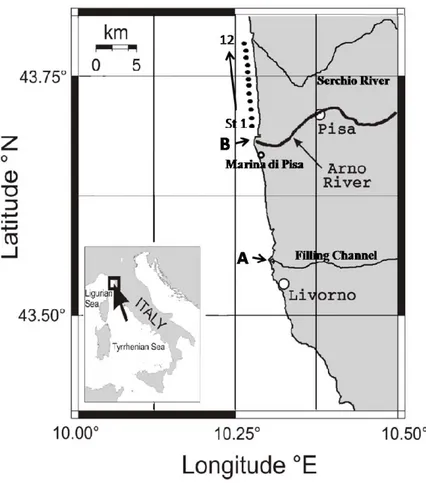
In the assessment of toxicity of sediment elutriates sediment samples were collected in a coastal area of the province of Pisa, Tuscany (Italy), affected by anthropogenic and industrial inputs. Two sediment samples, collected in the shore(line) on the northern side of the mouth of the Filling Channel of the Arno River (sediment A) and at Marina di Pisa (sediment B), were chosen for the development of the bioassay (Fig. 3.1).
In addition, sediment samples were collected approximately 500 m away from the shoreline, along a transect of twelve stations going from the northern side of the mouth of the Arno River (st 1) to the southern side of the mouth of the Serchio River (st 12), an area impacted by the plume of the Arno River (Vignudelli et al., 2004).
Methodology
Surface sandy sediment samples (5-10 cm) were collected with a stainless steel Van Veen grab. The sediments were placed in acid-cleaned (10% HNO3) polyethylene containers,
transported to the laboratory as soon as possible, homogenized with a blender and stored at 4°C in the dark until their use. The elutriation procedure was carried out within 2 weeks after collection. An aliquot of the wet sediment was dried at 60° C to allow the ratio dry weight: fresh weight to be determined.
Fig. 3.1. Map of the two sites (named A and B) in the polluted coastal area in the province of Pisa (Tuscany, Italy) where sediments were collected.
The elutriation procedure was carried out mixing sediment to natural seawater in 1:4 (w/v) ratio, based on the sediment dry weight. The mixture was shaken at 500 rpm for 24 h at room temperature by using a vertical Stirrer (Velp Scientific) and let to settle for 30 min. The
Methodology
aqueous fraction was centrifuged at 7000 rpm for 15 min and filtered (0.45 µm membrane filters). Elutriates were immediately used for bioassays or, alternatively, stored at -20° C.
3.8. Incubation experiments
3.8.1. Evaluation of phytochelatins in P. tricornutum as biomarker of metal exposure in marine waters
A first set of exposure experiments was carried out by using the marine diatom P.
tricornutum. The capability of this diatom to synthesize phytochelatins in response to metal
exposure has been widely studied by the research group working in the “Institute of Biophisics” of the CNR of Pisa (Morelli and Scarano, 2001; Morelli et al., 2002; Morelli and Scarano, 2004; Morelli and Fantozzi, 2008).
Before metal incubation experiments, a preculture was prepared by inoculating P.tricornutum from a stock culture on day 7 of growth (exponential phase) to provide an initial cell concentration of 5 - 7 × 10⁴ cells mL-1 in natural seawater enriched with NaNO3 and NaH2PO4
at a final concentration of 8.8 × 10⁻⁴ M and 3.6 × 10⁻⁵ M, respectively (equivalent to N and P concentrations in f/2 medium). In this medium, the growth rate of P. tricornutum was similar (10-15% lower) to that obtained in the maintenance medium. At the end of the logarithmic growth phase (approx. 2 × 10⁶ cells mL-1), calculated aliquots of the preculture were reduced to a volume of 10-20 mL by gentle filtration (1.2 μm membrane filters) and the resulting concentrated cell suspension was immediately transferred to the medium for PC induction experiments. The microorganisms were not let dry on the filter in order to avoid cell stress. Incubations were carried out both in EDTA-buffered artificial seawater and in natural seawater. Unless otherwise specified, 2 × 10⁸ cells were incubated in 200 mL medium, to
obtain a cell density of 1 × 10⁶ cells mL-1. PC induction experiments were carried out under continuous light conditions (100 μmol photons × m-2 × s-1), at 21 °C. Cell counts carried out
Methodology
after metal exposure showed that cell density was not appreciably changed during the incubation.
Before performing cell incubations, all the natural seawater samples were treated with NaNO3
and NaH2PO4 at a final concentration equivalent to that of the preculture. This procedure was
chosen to avoid the cells undergo significant changes in the culture medium. Metal enriched natural seawater samples were prepared by adding increasing amounts of Cd in the range 5 – 100 nM and of Pb or Cu in the range 25 – 200 nM. After 16 h equilibration and before cell addition, the electrochemically labile fraction of dissolved metal ions (Melab) was measured, at natural pH (8.2), by Anodic Stripping Voltammetry.
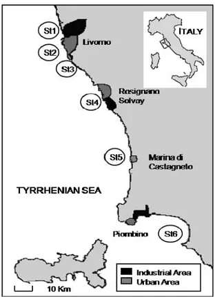
Seawater samples for field experiments were collected at six different stations, selected in contaminated areas in the province of the industrial city of Livorno, located in the Tyrrhenian coast of Tuscany (see map of Fig. 3.2).
Methodology
Tests were carried out by incubating 4 × 10⁸ cells in a 1-liter sample (4 × 10⁵ cells mL-1) for 6 hours.
At the end of the exposure, cells were collected and submitted to the phytochelatins assay.
3.8.2. Comparison of PC response in different taxa of autotrophic protists
In order to compare the PC response in different taxa of autotrophic protists, short-term exposure experiments (6 h) were carried out by incubating the protists in natural seawater enriched with known amounts of heavy metals and assaying for phytochelatins. For this purpose, precultures were prepared by inoculating P. tricornutum, S. costatum, T. weissflogii,
D. tertiolecta and Emiliania huxleyi from stock cultures on day 7 of growth (exponential
phase) to provide initial cell densities ranging from 3-5 × 10³ cells mL-1 for T. weissflogii, to 1-5 × 10⁴ cells mL-1 for the other species.
The preparation of the precultures and the conditions of exposure were carried out accordingly to the procedure described for P. tricornutum in the previous paragraph.
The cell densities were: 5 × 10⁵ cells mL-1 for P. tricornutum, 3 × 10⁵ cells mL-1 for S.
costatum, 4× 10⁴ cells mL-1 for T. weissflogii, 5 × 10⁵ cells mL-1 for D. tertiolecta and 2 × 10⁵
cells mL-1 for Emiliania huxleyi.
3.8.3. Evaluation of PC synthesis in P. tricornutum, T. weissflogii and S. costatum as biomarker of metal bioavailability in sediments
Two different experiments were carried out in order to evaluate the PC synthesis in P.
tricornutum, T. weissflogii and S. costatum as biomarker of metal bioavailability in sediments.
In short-term incubation experiments, a preculture was prepared by inoculating cells from a stock culture on day 7 of growth (exponential phase) to provide an initial cell concentration of approximately 5 × 10⁴ cells mL-1 for P. tricornutum and S. costatum and of 5 × 10³ cells mL
-Methodology 1 for T. weissflogii, in natural seawater enriched with the f/2 medium lacking the trace metal
stock solution. At the end of the logarithmic growth phase, calculated aliquots of the preculture were reduced to a volume ≤ 10 mL by gentle filtration (1.2 μm membrane filters) and the resulting concentrated cell suspension was immediately transferred to the medium (200 mL) for PC induction experiments. Incubations were carried out under continuous light conditions, at 21 °C, for 5 h, using as media the elutriates diluted with natural seawater at 0, 25 , 50, 75 and 100% concentration. Cell densities were: 5 × 10⁵ cells mL-1 for P.
tricornutum, 4 × 10⁴ cells mL-1 for T. weissflogii, and 3× 10⁵ cells mL-1 for S. costatum.
Long-term incubation experiments were carried out by inoculating cells from a stock culture (on day 7 of growth) in the elutriates properly diluted with natural seawater (from 0 to 100% concentration), at an initial cell density of 1 × 10⁴ cells mL-1 for P. tricornutum, 1 × 10³ cells mL-1 for T. weissflogii, and 5× 10³ cells mL-1 for S. costatum.
All media (100 mL) were enriched with the f/2 medium lacking the trace metal stock solution, and the cultures were let to grow during the exponential phase, for 6 days. Cell counts were performed at the 3rd and at the 6th day and the growth rate (expressed as doublings day-1) was calculated.
3.8.4. Evaluation of phytochelatins synthesis and DGM production in T. weissflogii exposed to mercury
All the mercury incubation experiments were carried out using, as culture medium, natural seawater enriched with the f/2 medium lacking the trace metal stock solution. Calculated volumes of the stock cultures of T. weissflogii were used as inoculum to obtain an initial cell density of 1 x 10³ cells mL-1.
In a first set of incubation experiments, designed to evaluate the effect of mercury on the growth rate of T. weissflogii, 100 mL culture media were spiked with HgCl to final