Brief Original Article
An unidentified cluster of infection in the Peruvian Amazon region
Angela Cornejo1, Cláudia Gomes2, Luis Suarez1, Sandra Martínez-Puchol2, Pershing Bustamante4, Maria J. Pons1, Joaquim Ruiz1, 2, Juana del Valle Mendoza1,3
1
Centro de Investigación de la Facultad de Ciencias de la Salud, Universidad Peruana de Ciencias Aplicadas (UPC), Lima, Peru
2 ISGlobal, Barcelona Centre for International Health Research (CRESIB), Hospital Clínic – Universitat de Barcelona, Barcelona, Spain
3
Instituto de Investigación Nutricional, Lima, Peru 4
Dirección Regional de Salud de Amazonas, Chachapoyas, Peru Abstract
Introduction: Bartonella bacilliformis is the etiological agent of Carrion’s disease, which is a neglected disease linked to people in low-socioeconomic populations in Andean valleys. An outbreak of B. bacilliformis was reported in a rural area of the Peruvian Amazon region. The aim of this study was to characterize this outbreak using molecular techniques.
Methodology: Fifty-three blood samples from patients diagnosed with Carrion’s disease were analyzed by molecular tools, using both a Bartonella-specific polymerase chain reaction (PCR) and an universal PCR, both based on 16S rRNA gene amplification. Additional water samples from the area were also analyzed.
Results: Unexpectedly, the samples were positive only when the universal PCR was used. Although environmental contamination cannot be ruled out, the results showed that Sphingomonas faeni was the possible causative agent of this outbreak, and that water was the most feasible infection source.
Conclusions: Diagnosis by clinical criteria or microscopy may lead to misdiagnosis. There is a need to include molecular tools in the routine diagnosis of febrile syndromes, including Carrion’s disease.
Key words:Bartonella spp.; Sphingomonas; diagnosis; outbreak; Peru. J Infect Dev Ctries 2015; 9(5):524-529. doi:10.3855/jidc.6235
(Received 09 November 2014 – Accepted 10 February 2015)
Copyright © 2015 Cornejo et al. This is an open-access article distributed under the Creative Commons Attribution License, which permits unrestricted use, distribution, and reproduction in any medium, provided the original work is properly cited.
Introduction
In northern Peru, the presence of different etiologic causes of febrile syndromes, including dengue, oropuche, mayaro, Plasmodium spp., or Bartonella bacilliformis, is well established [1-4]. Their correct diagnosis is necessary to enable administration of correct treatment. Nonetheless, in rural areas of low- and middle-income countries, correct diagnosis is a challenge, due to the presence of a series of common vague and undefined symptoms, especially in the early disease phases, and to the local diagnostic facilities.
Bartonella bacilliformis is the etiological agent of Carrion’s disease, which is a neglected disease linked to people in low-socioeconomic populations in Andean valleys. This bacterium is transmitted by the bite of a sandfly, a member of the genus Lutzomyia. No reservoir other than humans has been identified [2]. This illness has two well-differentiated phases.
The first is the acute so-called Oroya fever phase, characterized by fever and severe anemia (related to the fact that B. bacilliformis invade the red blood cells) in addition to a transitional immunosuppression [2]. Additionally, several symptoms and signs, including headache and jaundice, have also been described [1,5]. Regardless of its being considered the most lethal bacterial infection in the pre-antibiotic era, this illness may be successfully treated with a series of antimicrobial agents, including quinolones and macrolides, among others. However, it should be noted that the clinical cure has no direct correlation with microbiological eradication; asymptomatic carriers are frequent in endemic areas. The second phase of the disease, affecting semi-immune people, is characterized by the presence of endothelial proliferation resulting in warts (the so-called Peruvian warts) [2].
Since this illness mainly affects people in rural areas, one of the main problems is the lack of adequate technical and human resources for definitive diagnosis. Although some molecular diagnostic tools have been proposed [1,2,5], the diagnosis of Carrion’s disease in rural endemic areas is mostly restricted to clinical criteria or optical microscopy and it is frequently misdiagnosed by both [1,2].
In this context, the presence of an active outbreak of Oroya fever was reported on March 2013 in Rodríguez de Mendoza, a rural area in the northeast of Peru [6]. Diagnosis was primarily based on the sudden presence of ill people, mostly with fever and chills, and was confirmed by positive microscopy results. This outbreak was of special concern because Carrion's disease had never been reported in this area, but had been reported in some endemic areas are relatively near the affected population; it has been reported that current climatic alterations underlie an expansion of this illness to new areas [2,7].
The aim of this study was to characterize this outbreak using molecular techniques.
Methodology Patients and sampling
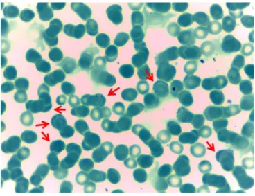
Fifty-three blood samples recovered in Rodríguez de Mendoza (Amazonas, Peru) were sent to the Instituto de Investigación Nutricional/Universidad Peruana de Ciencias Aplicadas to be included in the study. In all cases, patients were previously diagnosed with Carrion’s disease following clinical or microscopy criteria (Figure 1). Clinical and demographic data were recorded in a questionnaire.
Additionally, domestic water samples and water-well samples were also recovered and processed. Bacterial culture conditions
Microorganisms were cultured as previously described [1]. Briefly, 2 mL of the blood samples were grown in Columbia agar medium supplemented with 10% sheep blood and incubated at 28°C under anaerobic conditions. The plates were visually inspected at 24, 48, and 72 hours to detect contamination, and then every seven days for bacterial growth.
DNA extraction
The DNA was extracted from 200 µL of blood/water samples using a commercial extraction kit (High Pure Kit Preparation template, Roche Applied Science, Mannheim, Germany). Bacterial DNA
nuclease-free water and then processed or stored at -20°C until use.
PCR procedures
Two different 16S rRNA PCR approaches were used: one using specific primers for Bartonella genus, and the other using universal primers. In both cases, primers and conditions have been previously described [1]. All the products obtained were recovered and sequenced (Macrogen, Seoul, Korea).
Results
A demographic analysis showed that 52.8% of the subjects included in the study were men and ranged in age from 4 months to 85 years; the subjects were mostly adults (60.4%), and 22 (41.5%) reported engaging in agrarian activities. Additionally, 11 patients (20.7%) were not residents of the area, having reported a visit of less than one month in length (median, 2.4 days; range, 1-7 days) to the area. Nine (17%) patients, mostly outsiders (8/9) with a median stay in the area of 1.6 days (range, 1 to 3), reported engaging in aquatic activities, including in the river and thermal baths (Table 1).
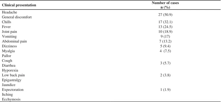
The review of the clinical data showed that the most frequent symptoms were headache and general discomfort (50.9%), followed by chills (32.1%) and fever (24.5%) (Table 2). Regarding treatment, 28 (52.8%) were treated with ciprofloxacin, while 12 did not receive antibiotic treatment; the remaining 8 patients were treated with amoxicillin plus clavulanic acid (4 patients), cotrimoxazole (2 patients), and
Figure 1. Giemsa-stained red blood cells. Arrows mark red blood cells with internal bodies, which leads to misidentification.
Table 1. Epidemiological characteristics No. Frequency (%) Age < 5 years 5 9.4 % 6–18 years 16 30.2 % > 18–55 years 24 45.3 % > 56 years 8 15.1 % Gender Male 28 52.8 % Female 25 47.2 % Ocupation1 Farmer 22 41.5 % Housewife 6 11.3% Not indicated2 7 13.2 %
Living in the area
Resident3 28 52.8 %
Non-resident 11 20.7 %
No data 14 26.4 %
Other
Water activities 9 17 %
1Only adults or minor developing economical activities; 2Four were not residents of the area; 3Patients reporting more than one month in the area
Table 2. Clinical presentation
Clinical presentation Number of cases
n (%) Headache 27 (50.9) General discomfort Chills 17 (32.1) Fever 13 (24.5) Joint pain 10 (18.9) Vomiting 9 (17) Abdominal pain 7 (13.2) Dizziness 5 (9.4) Myalgia 4 (7.5) Pallor 3 (5.7) Cough Diarrhea Hyporexia
Low back pain 2 (3.8)
Epigastralgy 1 (1.9) Jaundice Expectoration Itching Ecchymosis
All samples were analyzed by microscopy, microbiological and molecular techniques. Despite 29 (54.7%) cases being initially reported as positive by microscopy techniques, no amplified products were obtained with the 16S rRNA Bartonella-specific primers, showing that microscopy leads to a misidentification of Bartonella infections. Meanwhile, correct amplification of 1,503 bp was obtained in all the cases using the 16S rRNA universal primers. Twenty-six (49%) of these amplified products were randomly recovered and sequenced. Unexpectedly, the sequencing showed the presence of Sphingomonas faeni, suggesting that this microorganism was the causative microorganism of this outbreak.
To determine the presence of B. bacilliformis, all samples were cultured under non-aerobic conditions. Bacterial growth was only detected in three samples. The DNA was extracted from the three growing microorganisms, and the different 16S rRNA PCR approaches were performed. Amplified product was obtained only with 16S rRNA universal primers. Analysis of the sequences showed the presence of Staphylococcus epidermidis, which was considered a contaminant.
Domestic water samples and water-well samples were collected. DNA extraction followed by PCR amplification using the 16S rRNA universal primers was performed; the results showed the presence of different microorganisms, including previous uncultured aquatic microorganisms related to GenBank access numbers gb|GU758935.1 and GU758935.1 from water well, and HM238175 from both domestic and well samples, the last one belonging to a member of the Sphingomonas genus. Discussion
The present report shows the presence of a misdiagnosed B. bacilliformis outbreak. The results showed the presence of S. faeni, an environmental microorganism that to the best of our knowledge, has never been described as a causative agent of an infectious outbreak. Although it cannot be ruled out, environmental PCR contamination was unlikely because PCR amplifications and DNA extractions were performed more than once, at different times, in two different settings. Moreover, negative controls were used, and no other Sphingomonas spp. was detected in any of the other laboratory samples analyzed prior, during, or after this study.
In a previous report, the symptoms significantly associated with Oroya fever were chills, joint pain,
With the exception of chills, these symptoms were absent or had a minimal presence, being then non-suggestive of Carrion’s disease. The fact that this outbreak was first associated with B. bacilliformis was probably due to the unexpected amount of cases, the proximity of endemic areas and, especially, due to the presence of positive blood thin smears (Figure 1).
It has been shown that Sphingomonas spp. possess virulence factors showing a degree of identity with Brucella intracellular survival factors [8]. Moreover, Sphingomonas spp. possess the ability to penetrate within epithelial cells in vitro [9], and some reports have shown the presence of intracellular Gram-negative microorganisms during Sphingomonas spp. infections [10]. However, to the best of our knowledge, no data about the ability of S. faeni or other Sphingomonas spp. to invade erythrocytes exists in the literature; the possible concomitant presence of other pathogens able to invade erythrocytes cannot be ruled out. A possibility is the presence of hemoplasma, which are wall-less erythrocytic bacteria unable to be cultured in vitro, classified within the genus Mycoplasma, which have been described in human and animal infections [11]. However, an in silico analysis showed that the 16S rRNA universal primers used were able to amplify a fragment of 1,433 bp of the 16S rRNA gene of hemoplasma species (i.e., Candidatus Mycoplasma haemominutum, GeneBank access NC_021007.1). Similarly, the primers used were also able to detect other well-known intraerythrocytic bacteria such as Anaplasma spp.
Bacterial growth was only detected in three samples, but the growing microorganism was S. epidermidis, which was considered to be a sample contamination. These low positivity rates may have been found because the samples were directly plate-cultured but no hemoculture was done, and because of the specific growth conditions cultured in order to detect the presence of B. bacilliformis [1]. Sphingomonas is a strictly aerobic microorganism commonly distributed both in hospitals and the natural environment in soil and water [12-14]. Several Sphingomonas spp. infections in humans have been reported, mostly limited to sporadic case reports or intra-hospital outbreaks, and mostly related to Sphingomonas paucimobilis [13,15]. To our knowledge, this is the first infectious Sphingomonas faeni outbreak described, and the first Sphingomonas spp. outbreak described in a non-hospital environment.
Although S. faeni has low clinical virulence, it has a close relationship with S. paucimobilis, which is able
fact, together with the possible acquisition of virulence factors, as has been proposed for other member of the genus [8], may allow this microorganism to infect healthy people, causing an outbreak. Moreover, the specific socio-sanitary conditions of the area should be considered, including nutritional status, which may enhance the possibility of infections by low-virulence microorganisms [17]. In this sense, malnutrition and anemia among the infant population of the Amazonas region is about 32.8% and 56.7%, respectively [18].
Regarding the focus of infection, all probes seemed to be from the water. Sphingomonas is an environmental microorganism, and in this outbreak, almost 17% of the patients were reported to have engaged in aquatic activities; moreover, another Sphingomonas spp. (coincident with that recorded as GeneBank HM238175.1) was also detected in both domestic and well water sources.
Unfortunately, we were not able to recover water from the thermal baths. Nevertheless, the environmental nature of S. faeni together with the presence of other Sphingomonas spp. in the potable water suggest an association between the water consumed and participation in aquatic activities with this outbreak.
Conclusions
Though sample contamination or the presence of a non-detect microorganisms cannot be ruled out, our findings strongly suggests the emergence of S. faeni as the causative agent of a community-acquired outbreak, probably associated with water. To our knowledge, this is the first report of a Sphingomonas spp. extra-hospital outbreak, as well as the first description of S. faeni as a causative infectious agent.
This outbreak was mistakenly attributed to B. bacilliformis, demonstrating that diagnosis of febrile syndromes by clinical criteria or microscopy may lead to misdiagnosis. Training of health personnel and the development of new diagnostic tools able to be implemented in endemic rural areas are urgently needed to overcome erroneous diagnoses and to avoid inappropriate treatments.
Acknowledgements
This study was funded by the Instituto de Salud Carlos III (ISCIII, Spain) (grant number: FI12/00561, which included FEDER funds), by the Spanish Network for the Research in Infectious Diseases (REIPI RD12/0015) and Generalitat de Catalunya, Departament d’Universitats, Recerca i Societat de la Informació (2014 SGR 26) (JR) and by internal funds of the Universidad Peruana de Ciencias Aplicadas (UPC), Lima-Peru (JdV).
JR has a fellowship from the program I3, of the ISCIII (grant number: CES11/012). CG has a PhD fellowship of the ISCIII (FI12/00561). MJP has a postdoctoral fellowship from CONCYTEC.
We thank Donna Pringle for language and idiomatic corrections.
Authors’ contributions
CG, JR, and JdV designed the study. AC, CG, SMP, and MJP performed the experiments. LS and PB gathered clinical and epidemiological data. JR and JdV analyzed the data. CG, JR, and JdV wrote the manuscript. All the authors read and approved the final manuscript.
References
1. del Valle Mendoza J, Silva Caso W, Tinco Valdez C, del Valle LJ, Casabona Oré V, Champin Michelena D, Bazán Mayra J, Zavaleta Gavidea V, Vargas M, Ruiz J (2014) Diagnosis of Carrion’s disease by direct blood PCR in thin blood smear negative samples. PloS One 9: e92283.
2. Sanchez Clemente N, Ugarte-Gil CA, Solórzano N, Maguiña C, Pachas P, Blazes D, Bailey R, Mabey D, Moore D (2012) Bartonella bacilliformis: a systematic review of the literature to guide the research agenda for elimination. PLoS Negl Trop Dis 6: e1819.
3. Ministerio del Salud del Peru (2005) Perfil etiológico del síndrome febril en áreas de alto riesgo de transmisión de enfermedades infecciosas de impacto en salud pública en el Perú, 2000-2001. Rev Peru Med Exp Salud Publica 22: 165-174.
4. Troyes L, Fuentes L, Troyes M, Canelo L, Garcia M, Amaya E, Tapia R, Cespedes M (2006) Etiología del síndrome febril agudo en la provincia de Jaén, Perú 2004-2005. Rev Peru Med Exp Salud Publica 23: 5-11.
5. Huarcaya E, Maguiña C, Torres R, Rupay J, Fuentes L (2004) Bartonelosis (Carrion's Disease) in the pediatric population of Peru: an overview and update. Braz J Infect Dis. 8: 331-339. 6. International Society for Infectious Diseases, ProMED-mail
(2013) Bartonellosis – Peru: (Amazonas). Available: http://www.promedmail.org/direct.php?id=20130304.156988 8. Accessed 24 September 2014.
7. Maroli M, Feliciangeli MD, Bichaud L, Charrel RN, Gradoni L (2013) Phlebotomine sandflies and the spreading of leishmaniases and other diseases of public health concern. Med Vet Entomol 27: 123-147.
8. Saeb ATM, David SK, Al-Brahim H (2014) In silico detection of virulence gene homologues in the human pathogen Sphingomonas spp. Evol Bioinform 10: 229-238. 9. Ammendolia MG, Bertuccini L, Minelli F, Meschini S,
Baldassarri L (2004) A Sphingomonas bacterium interacting with epithelial cells. Res Microbiol 155: 636-646.
10. Souto A, Guinda M, Mera A, Pardo F (2012) Artritis séptica por Sphingomonas paucimobilis en un paciente inmunocompetente. Reumatol Clin 8: 378-379
11. Tasker S, Peters IR, Mumford AD, Day MJ, Gruffydd-Jones TJ, Day S, Pretorius AM, Birtles RJ, Helps CR, Neimark H (2010) Investigation of human haemotropic Mycoplasma infections using a novel generic haemoplasma qPCR assay on blood samples and blood smears. J Med Microbiol 59: 1285-1292.
12. Brossi MJ, Mendes LW, Germano MG Lima AB, Tsai SM (2014) Assessment of bacterial bph gene in Amazonian dark earth and their adjacent soils. PloS One 9: e99597.
13. Meric M, Willke A, Kolayli F, Yavuz S, Vahaboglu H (2009) Water-borne Sphingomonas paucimobilis epidemic in an intensive care unit. J Infect 58: 253-255.
14. Takeuchi M, Hamana K, Hiraishi A (2001) Proposal of the genus Sphingomonas sensu stricto and three new genera, Sphingobium, Novosphingobium and Sphingopyxis, on the basis of phylogenetic and chemotaxonomic analyses. Int J Syst Evol Microbiol 51: 1405-1417.
15. Bayram N, Devrim I, Apa H, Gülfidan G, Türkyılmaz HN, Günay I (2013) Sphingomonas paucimobilis infections in children: 24 case reports. Mediterr J Hematol Infect Dis 5: e2013040.
16. Busse HJ, Denner EB, Buczolits S, Salkinoja-Salonen M, Bennasar A, Kämpfer P (2003) Sphingomonas aurantiaca sp. nov., Sphingomonas aerolata sp. nov and Sphingomonas faeni sp. nov., air- and dustborne and Antarctic, orange-pigmented, psychrotolerant bacteria, and emended description of the genus Sphingomonas. Int J Syst Evol Microbiol 53: 1253-1260.
17. Katona P, Katona-Apte J (2008) The interaction between nutrition and infection. Clin Infect Dis 46: 1582-1588. 18. Pan American Health Organization (2009) Estrategia de
cooperación técnica OPS/OMS, Perú 2010-2014. Lima (Peru): 48p.
Corresponding author
Joaquim Ruiz
CRESIB, Edifici CEK, C/Rosselló149-153 08036-Barcelona, Spain
Phone: +342275400 ext 4547 Fax: +34932279853 Email: [email protected] Juana del Valle-Mendoza
Universidad Peruana de Ciencias Aplicadas-UPC Av. San Marcos cuadra 2, Cedros de Villa Lima, Peru
Phone +5113133333 ext 2704 Fax: +5113496025
Email: [email protected]