Research Article
First Description of Macrolide-Resistant
Mycoplasma
pneumoniae in Adults with Community-Acquired
Pneumonia in Italy
Daniela Loconsole,
1Anna Lisa De Robertis,
1Rosanna Mallamaci,
2Anna Sallustio,
1Anna Morea,
1Rosa Prato
,
3Michele Quarto
,
1Domenico Martinelli
,
3and Maria Chironna
11Department of Biomedical Sciences and Human Oncology-Hygiene Section, University of Bari, Piazza G. Cesare 11, 70124 Bari, Italy 2Department of Biosciences, Biotechnology and Biopharmaceutics, University of Bari, Via Orabona 4, 70124 Bari, Italy
3Department of Medical and Surgical Sciences, University of Foggia, Viale Pinto 1, 71122 Foggia, Italy
Correspondence should be addressed to Maria Chironna; [email protected] Received 5 January 2019; Accepted 3 March 2019; Published 17 March 2019 Academic Editor: Haruki Komatsu
Copyright © 2019 Daniela Loconsole et al. This is an open access article distributed under the Creative Commons Attribution License, which permits unrestricted use, distribution, and reproduction in any medium, provided the original work is properly cited.
Background. Mycoplasma pneumoniae is a common cause of community-acquired pneumonia (CAP). This cross-sectional study
aimed to determine the prevalence of macrolide-resistant M. pneumoniae strains in a convenience series of 234 adult hospitalised and nonhospitalised subjects with a diagnosis of CAP in January 2013 to April 2015 in South Italy. Methods. Respiratory samples were subjected to real-time PCR. In M. pneumoniae-positive samples, domain V of 23S rRNA was sequenced to detect resistance-conferring point mutations. P1 major adhesion protein typing and multiple loci variable-number tandem repeat analysis (MLVA) were also performed. Results. Of the 234 samples, 15 (6.4%) were positive for M. pneumoniae. Three of these had a macrolide-resistant genotype: two and one had A2063G and A2064G mutations, respectively. Fourteen of the 15 strains were subtyped: half had subtype 1 and half had subtype 2. Eight strains underwent MLVA profiling: one each had the J, A, and Z type. The remainder was unclassifiable. Conclusions. This novel discovery of macrolide-resistant M. pneumoniae strains in adults with CAP in Italy suggests that there may be increasing circulation of these strains in the population. To facilitate rapid optimization of the antibiotic strategy in Italy, macrolide resistance should be monitored by a surveillance system that is based on molecular methods.
1. Introduction
Mycoplasma pneumoniae is a leading cause of upper and
lower respiratory tract infections in all age groups. It is also a frequent cause of community-acquired pneumonia (CAP).
M. pneumoniae infections occur endemically and
epidemi-cally worldwide. However, the majority of M. pneumoniae infections are self-limited, especially in adults, and can be treated with antibiotic therapy without pathogen-specific diagnostic testing [1].
In Europe, M. pneumoniae may be responsible for approx-imately 11% of CAP cases [2] and, in Italy, up to 17% of the hospitalised adult cases of CAP [3]. Macrolides are the first choice of antibiotics for the treatment of CAP caused by M. pneumoniae in both adults and children [4].
However, the inappropriate or overuse of such drugs has led to the emergence of macrolide-resistant M. pneumoniae strains in several countries [4]. In Italy, macrolide-resistant
M. pneumoniae strains started emerging in children in 2010
[5]. Moreover, a recent study reported the case of a family in Italy in which a clonal macrolide-resistant M. pneumoniae strain was transmitted from the index pediatric case to adult family members [6]. However, the prevalence of macrolide-resistant M. pneumoniae strains in adult Italians with CAP has not been reported.
This cross-sectional study examined the prevalence of macrolide-resistant M. pneumoniae strains in M.
pneumo-niae-positive specimens from hospitalised and
nonhospi-talised adults with pneumonia in South Italy (Puglia) in
Volume 2019, Article ID 7168949, 5 pages https://doi.org/10.1155/2019/7168949
January 2013 to April 2015. Multiple-locus variable-number tandem repeat analysis (MLVA) and P1 adhesion protein typing were also performed.
2. Materials and Methods
2.1. Study Design, Setting, and Participants. Between January
2013 and April 2015, ten general practitioners and pneumol-ogists working in 12 Divisions of Pulmonary and Respiratory Disease across the Puglia region obtained clinical samples from a convenience series of hospitalised and nonhospi-talised Puglia-resident adults (≥18 years) who were diagnosed with CAP. In particular, patients were diagnosed with CAP in presence of at least two clinical criteria among new cough or sputum production, fever>38.0∘C or hypothermia<36.1∘C, chest pain, dyspnoea, tachypnoea, new altered mental status, abnormal lung examination, respiratory failure, leucocytosis (white cell count>10 × 109/L or >15% bands) or leucopenia (white cell count<4.5 × 10⁹/L), C reactive protein value >3 times the upper limit of normal, and hypoxaemia with a partial oxygen pressure<60mm Hg while the patient was breathing room air and in presence of evidence on chest radiography consistent with pneumonia [7]. The general practitioners and pneumologists were from all six provinces in the Puglia region.
2.2. Samples Collection. Nasopharyngeal swabs and/or
spu-tum and/or bronchoalveolar lavage were obtained from all subjects diagnosed with CAP. All clinical samples were collected within 24 h of the clinical onset of symptoms.
2.3. DNA Extraction and Polymerase Chain Reaction Ampli-fication. All the collected samples were subjected to
real-time PCR analysis for the molecular detection of M.
pneumoniae and other common agents of CAP, namely, Legionella spp., Chlamydia pneumoniae, Streptococcus pneu-moniae, Haemophilus influenzae, Moraxella catarrhalis, and Staphylococcus aureus. When multiple biological samples
were available from the same patient, only the sample that was positive for the detected pathogens was used for analysis. For the real-time PCR, total nucleic acid was extracted from each respiratory specimen by using the MagnaPure LC automated extraction system (Roche Diagnostics, Milan, Italy). A com-mercial real-time PCR kit (FTD Bacterial pneumoniae CAP, Arrow Diagnostics, Genoa, Italy) was then used.
2.4. Identification of Macrolide Resistance Genotype and Sub-typing. M. pneumoniae-positive samples were tested for the
presence of macrolide resistance by PCR and direct amplicon sequencing in the M. pneumoniae 23S rRNA gene to detect single nucleotide polymorphisms that are known to associate with resistance to macrolides. The details have been described elsewhere [5]. The detected M. pneumoniae strains were also subtyped molecularly on the basis of their point mutations within the gene encoding the P1 major adhesion protein [8]. Strains were classified as type 1 or type2.
2.5. Multiple-Locus Variable Number Tandem Repeat Analysis (MLVA). MLVA was performed on nucleic acid extracts from
clinical specimens as described previously [9]. Interpreta-tion of profiles and MLVA type assignment were achieved according to guidelines reported recently [10]. All tests were performed at the Laboratory of Molecular Epidemiology of the Hygiene Unit of Azienda Ospedaliero-Universitaria Policlinico, Bari, Italy.
2.6. Ethical Approval. All procedures performed in the
study were in accordance with the ethical standards of the institutional and national research committee and with the 1964 Helsinki declaration and its later amendments or comparable ethical standards. Ethical approval was obtained from the Institutional Review Board at the Apulian Regional Observatory for Epidemiology. Informed written consent was obtained from all individuals who provided the specimens.
3. Results
In total, 234 hospitalised and nonhospitalised adult patients with CAP who resided in Puglia during the study period provided specimens. Of these 234 specimens, 15 [6.4%, 95% confidence interval (CI): 3.9–10.3] were positive for M.
pneumoniae on real-time PCR. S. pneumoniae was detected
in 43% of the patients, Haemophilus influenzae in 17%,
Chlamydia pneumoniae in 1%, Legionella pneumophila in
1%, and Moraxella catarrhalis in 5%. The 15 patients ranged between 18 and 87 years of age and a third were hospitalised. The patients with M. pneumoniae infection did not show coinfections with other pathogens.
Of the 15 M. pneumoniae-positive strains, three were macrolide-resistant. Two were from male patients who were 32 and 37 years of age. They had the same 23S rRNA mutation, namely, A2063G. One patient was hospitalised. A link between these two patients was not detected. The third case was a 31-year-old man who was not hospitalised and who had the A2064G mutation (Table 1). Whether these patients had received macrolide treatment before sampling was not known.
P1 typing was performed on 14 of the specimens to differentiate between the strains on the basis of the genetic sequence that encodes the P1 adhesion molecule. One sample was not available for P1 typing. Seven strains were type 1 strains while the remaining seven strains were type 2 strains. Eight M. pneumoniae strains were characterized by MLVA. The remaining 7 strains were not characterized due to insufficiency of clinical samples. Multiple MLVA types were detected. In three cases (patients Nos. 4, 7, and 10 of Table 1), the MLVA types were J, A, and Z: all three types have been identified previously. In the other five cases, the strains were untypeable when using the scheme proposed by Degrang`e et
al. [9].
4. Discussion
The increasing resistance of M. pneumoniae strains to mac-rolides has become a worrisome health problem in the last few decades.
In the present study, the overall prevalence of M.
pneumo-niae in adult Italian patients with CAP was 6.4%.
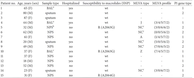
Table 1: Findings of the 15 patients living in the Puglia region of Italy whose swab, sputum, or BAL samples were positive for M. pneumoniae in January 2013 to April 2015.
Patient no. Age, years (sex) Sample type Hospitalized Susceptibility to macrolides (SNP) MLVA type MLVA profile P1 gene type
1 65 (F) BALa yes wt - - 1 2 80 (M) sputum no wt - - 2 3 87 (F) sputum no wt - - -4 44 (M) BALa yes wt J (3/4/5/7/2) 1 5 32 (M) NPSb no R (A2063G) NCc (3/0/6/6/2) 2 6 62 (M) NPS no wt NCc (0/0/5/6/2) 2 7 61 (F) NPS no wt A (1/4/5/7/2) 1 8 20 (M) sputum yes wt NCc (5/0/5/6/2) 2 9 49 (M) NPS no wt NCc (7/0/6/5/2) 2
10 37 (F) BALc yes R (A2063G) Z (7/4/5/7/2) 1
11 37 (F) NPS no wt - - 1 12 18 (M) NPS yes wt - - 1 13 52 (M) NPS no wt - - 2 14 23 (F) sputum no wt NCc (3/0/6/7/2) 1 15 31 (F) NPS no R (A2064G) - - 2 aBronchoalveolar Lavage. bNasopharyngeal swab.
cNot classifiable using the scheme proposed by Degrang`e et al. (2009).
MLVA: multiple-locus variable-number tandem repeat analysis; R: resistant; SNP: single nucleotide polymorphism; wt: wild type.
In Italy, macrolide-resistant M. pneumoniae was first reported in pediatric patients in an outbreak of M.
pneumo-niae in 2010 [5] and only sporadically in Italian adults [6].
At present, there is relatively little information in the literature on macrolide-resistant M. pneumoniae in adults in Europe. Macrolide-resistant M. pneumoniae at very low level was detected in a study conducted on a population of wide age range (15–65-year-old patients) in England and Wales [11].
Recently, a German study showed that, in 2011–2012, the 3.1% of adults with CAP had macrolide-resistant M.
pneumoniae strains [12]. Our novel findings thus suggest that
there is a spread in adults, although at low level, of macrolide resistance of M. pneumoniae also in Italy at present.
In our study, the most frequent mutation in domain V of the 23S rRNA gene was the A2063G mutation: two of the three patients with macrolide-resistant M. pneumoniae had this mutation. The remaining patient bore the A20164G mutation. All of these mutations are known to confer strong resistance to macrolides [13]. This presence of macrolide resistance may be the consequence of the frequent use of macrolides in Italy, especially for CAP in home-treated patients; this may have placed selective pressure on M.
pneumoniae strains [14, 15]. We also observed that the M. pneumoniae strains we detected were equally likely to be
P1 gene type 1 and type 2. By contrast, in the outbreak of M. pneumoniae in 2010 among Italian children, the P1 subtype predominated [5]. This disparity may reflect the fact that the latter cases were detected in an epidemic (where one subtype may be more likely to predominate), whereas our cases were not epidemiologically linked. No correlation emerged between macrolide resistance and other molecular characteristics of MP strains (P types and MLVA profiles).
MLVA typing of our M. pneumoniae strains revealed the J, A, and Z type [9] in one patient each. The remaining five strains that were tested were not classifiable. This indicates the heterogeneity of the MLVA profiles of the circulating strains. As is known, locus MPN1 is localized in the variable hsdS gene that changes constantly under the environmental pressure and variations in this locus do not necessarily represent emerging of a new the strain [15]. If the MPN1 allele profile is excluded from the classification, as was proposed by some authors [15], the most frequent profile in our samples was 4/5/7/2, which was detected in three cases. This MLVA profile frequency is in accordance with the frequency of MLVA profiles reported by other studies on adults with CAP due to M. pneumoniae [15, 16]. Nevertheless, we believe that the MPN1 allele sequence helps to assess the relationships between M. pneumoniae strains and we still recommend the method of Degrang`e et al. [9]. On the other hand, a more discriminative scheme is needed for epidemiological purposes [17]. Whole-genome sequencing of M. pneumoniae could help in the future to improve such analyses.
European guidelines indicate that macrolides should be the first choice treatment of M. pneumoniae infections in both children and adults [18]. However, if the fever persists 48–72 h after starting this first-line drug, it is recommended to change treatment to second-line antibiotics [19]. It is important to preserve the efficacy of macrolides for M.
pneumoniae infections and other antibiotics while we wait
for the development of new antibiotic agents that have an acceptable safety profile in all age groups. The best way to do this is to take into account the evidence that shows that macrolide-resistant M. pneumoniae are circulating and to advance antibiotic stewardship initiatives. These initiatives are particularly important in countries such as Italy, which
is one of the European countries that have serious problems with antibiotic resistance [20].
The study has some limitations. First, the number of CAP cases positive for M. pneumoniae was relatively low; this may be due to the fact that samples were collected during an interepidemic phase. Further studies should be undertaken during the epidemic phase to estimate the actual prevalence of macrolide-resistant M. pneumoniae strains. Second, the results of our study should be interpreted with caution due to possible selection bias caused by the sampling method.
Finally, our study shows that strict surveillance and monitoring are needed to manage the emerging resistance to first-line therapy for M. pneumoniae in adults in Italy.
5. Conclusions
We found macrolide-resistant M. pneumoniae strains in hospitalised and nonhospitalised adult patients with CAP. In all cases, the mutations identified in domain V of the 23S rRNA gene conferred strong resistance to macrolides. Char-acterization of M. pneumoniae strains by P1 major adhesion protein gene typing and MLVA revealed the circulation of very heterogeneous strains. To facilitate rapid optimization of the antibiotic strategy in Italy, macrolide resistance should be monitored by a surveillance system that is based on molecular methods.
Abbreviations
M. pneumoniae: Mycoplasma pneumoniae
CAP: Community-acquired pneumonia MLVA: Multiple-locus variable-number
tandem repeat analysis.
Data Availability
The data used to support the findings of this study are available from the corresponding author upon request.
Disclosure
An earlier version of this manuscript was presented in the European Scientific Conference on Applied Infectious Disease Epidemiology (ESCAIDE), Malta 21-23 November 2018.
Conflicts of Interest
The authors declare no conflicts of interest.
Authors’ Contributions
Daniela Loconsole and Anna Lisa De Robertis contributed equally to this work as first authors. Maria Chironna, Rosa Prato, and Michele Quarto designed the study. Daniela Loconsole and Rosanna Mallamaci performed data collection and management. Anna Lisa De Robertis, Anna Sallustio, and Anna Morea designed and performed the sample testing. Domenico Martinelli analyzed and interpreted the results.
Daniela Loconsole and Maria Chironna wrote and edited the manuscript. All authors reviewed the manuscript and approved the final version of the manuscript.
Acknowledgments
The authors are greatly indebted to Domenico Gatti, Vita Nuzzolese, Daniele Casulli, and Donata Anna Pepe for their invaluable help in laboratory testing the nasopharyngeal swabs for M. pneumoniae and sequence analysis.
References
[1] M. H. Diaz, A. J. Benitez, and J. M. Winchell, “Investigations of Mycoplasma pneumoniae infections in the United States: trends in molecular typing and macrolide resistance from 2006 to 2013,” Journal of Clinical Microbiology, vol. 53, no. 1, pp. 124– 130, 2006.
[2] M. Woodhead, “Community-acquired pneumonia in Europe: causative pathogens and resistance patterns,” European
Respira-tory Journal, vol. 20, Supplement 36, pp. 20S–27s, 2002.
[3] F. Meloni, E. Paschetto, P. Mangiarotti et al., “Acute Chlamy-dia pneumoniae and Mycoplasma pneumoniae infections in community-acquired pneumonia and exacerbations of COPD or asthma: Therapeutic considerations,” Journal of
Chemother-apy, vol. 16, no. 1, pp. 70–76, 2004.
[4] B. Cao, J. X. Qu, Y. D. Yin et al., “Overview of antimicrobial options for Mycoplasma pneumoniae pneumonia: focus on macrolide resistance,” The Clinical Respiratory Journal, vol. 4, pp. 419–429, 2015.
[5] M. Chironna, A. Sallustio, S. Esposito et al., “Emergence of macrolide-resistant strains during an outbreak of Mycoplasma pneumoniae infections in children,” Journal of Antimicrobial
Chemotherapy, vol. 66, no. 4, pp. 734–737, 2011.
[6] M. Chironna, D. Loconsole, A. L. De Robertis et al., “Clonal spread of a unique strain of macrolide-resistant mycoplasma pneumoniae within a single family in Italy,” Medicine (United
States), vol. 95, no. 11, Article ID e3160, 2016.
[7] A. W. Chow, C. B. Hall, J. O. Klein, R. B. Kammer, R. D. Meyer, and J. S. Remington, “Evaluation of new anti-infective drugs for the treatment of respiratory tract infections,” Clinical Infectious
Diseases, vol. 15, no. Supplement 1, pp. S62–S88, 1992.
[8] E. B. M. Spuesens, T. Hoogenboezem, M. Sluijter, N. G. Hartwig, A. M. C. van Rossum, and C. Vink, “Macrolide resistance deter-mination and molecular typing of Mycoplasma pneumoniae by pyrosequencing,” Journal of Microbiological Methods, vol. 82, no. 3, pp. 214–222, 2010.
[9] S. D´egrange, C. Cazanave, A. Charron, H. Renaudin, and C. B´eb´ear, “Development of multiple-locus variable-number tandem-repeat analysis for molecular typing of Mycoplasma pneumoniae,” Journal of Clinical Microbiology, vol. 47, no. 4, pp. 914–923, 2009.
[10] V. Chalker, S. Pereyre, R. Dumke et al., “International Mycoplas-ma pneumoniae typing study: interpretation of M. pneumo-niae multilocus variable-number tandem-repeat analysis,” New
Microbes and New Infections, vol. 7, pp. 37–40, 2015.
[11] R. J. Brown, L. Macfarlane-Smith, S. Phillips, and V. J. Vicki, “Detection of macrolide resistant Mycoplasma pneumoniae in England, September 2014 to September 2015,” Eurosurveillance, vol. 20, no. 48, Article ID 30078, 2015.
[12] R. Dumke, C. Schnee, M. W. Pletz et al., “Mycoplasma pneu-moniae and chlamydia spp. Infection in community-acquired pneumonia, Germany, 2011–2012,” Emerging Infectious Diseases, vol. 21, no. 3, pp. 426–434, 2015.
[13] M. Morozumi, T. Takahashi, and K. Ubukata, “Macrolide-resistant Mycoplasma pneumoniae: characteristics of isolates and clinical aspects of community-acquired pneumonia,”
Jour-nal of Infection and Chemotherapy, vol. 16, no. 2, pp. 78–86, 2010.
[14] N. Adriaenssens, S. Coenen, A. Versporten et al., “European Surveillance of Antimicrobial Consumption (ESAC): Outpa-tient macrolide, lincosamide and streptogramin (MLS) use in Europe (1997-2009),” Journal of Antimicrobial Chemotherapy, vol. 66, no. 6, pp. vi37–vi45, 2011.
[15] H. Sun, G. Xue, C. Yan et al., “Multiple-locus variable-number tandem-repeat analysis of mycoplasma pneumoniae clinical specimens and proposal for amendment of MLVA nomencla-ture,” PLoS ONE, vol. 8, no. 5, p. e64607, 2013.
[16] M. H. Diaz, A. J. Benitez, K. E. Cross et al., “Molecular detec-tion and characterizadetec-tion of mycoplasma pneumoniae among patients hospitalized with community-acquired pneumonia in the United States,” Open Forum Infectious Diseases, vol. 2, no. 3, Article ID ofv106, 2015.
[17] K. B. Waites, L. Xiao, Y. Liu, M. F. Balish, and T. P. Atkin-son, “Mycoplasma pneumoniae from the respiratory tract and beyond,” Clinical Microbiology Reviews, vol. 30, no. 3, pp. 747– 809, 2017.
[18] M. Woodhead, F. Blasi, S. Ewig et al., “Guidelines for the management of adult lower respiratory tract infections–full version,” Clinical Microbiology and Infection, vol. 6, supplement 6, pp. E1–59, Nov 2011.
[19] T. Yamazaki and T. Kenri, “Epidemiology of Mycoplasma pneumoniae infections in Japan and therapeutic strategies for macrolide-resistant M. pneumoniae,” Frontiers in Microbiology, vol. 7, no. 693, 2016.
[20] ECDC. Antimicrobial resistance surveillance in Europe 2015. http://ecdc.europa.eu/en/publications/Publications/antimicro-bial-resistance-europe-2015.pdf.
Stem Cells
International
Hindawi www.hindawi.com Volume 2018 Hindawi www.hindawi.com Volume 2018 INFLAMMATIONEndocrinology
International Journal ofHindawi www.hindawi.com Volume 2018 Hindawi www.hindawi.com Volume 2018
Disease Markers
Hindawi www.hindawi.com Volume 2018 BioMed Research InternationalOncology
Journal of Hindawi www.hindawi.com Volume 2013 Hindawi www.hindawi.com Volume 2018Oxidative Medicine and Cellular Longevity
Hindawi
www.hindawi.com Volume 2018
PPAR Research
Hindawi Publishing Corporation
http://www.hindawi.com Volume 2013 Hindawi www.hindawi.com
The Scientific
World Journal
Volume 2018 Immunology Research Hindawi www.hindawi.com Volume 2018 Journal ofObesity
Journal of Hindawi www.hindawi.com Volume 2018 Hindawi www.hindawi.com Volume 2018 Computational and Mathematical Methods in Medicine Hindawi www.hindawi.com Volume 2018Behavioural
Neurology
Ophthalmology
Journal of Hindawi www.hindawi.com Volume 2018Diabetes Research
Journal ofHindawi
www.hindawi.com Volume 2018
Hindawi
www.hindawi.com Volume 2018
Research and Treatment
AIDS
Hindawi
www.hindawi.com Volume 2018
Gastroenterology Research and Practice
Hindawi www.hindawi.com Volume 2018