UNIVERSITY OF SIENA
Department of Biotechnology, Chemistry and Pharmacy
PHD SCHOOL IN BIOCHEMISTRY AND MOLECULAR BIOLOGY BiBiM 2.0
(Cycle XXXIII)
Coordinator of PhD school: Prof. Lorenza Trabalzini
SNAI1 target genes in myoblasts
S.S.D. BIO/11
Tutors:
Prof. Maurizio Orlandini
Prof. Federico Galvagni
PhD Candidate:
Ines Elia
Academic year: 2021
Digitallysigned by ELIA
SUMMARY 1. ABSTRACT ... 1 2. INTRODUCTION ... 3 2.1 Snai superfamily ... 3 2.2 Snai functions ... 5 2.3 Snai regulation ... 8 2.4 FGF21 ... 11
2.5 ER stress and ATF3 ... 12
2.6 Skeletal muscle regeneration ... 15
2.7 Transcriptional regulation of skeletal muscle regeneration ... 17
2.8 Animal models of muscle injury ... 19
3. MATERIALS AND METHODS ... 21
3.1 Animals ... 21
3.2 Genotyping ... 21
3.3 In vivo assay ... 22
3.4 X-Gal Staining ... 22
3.5 Muscle embedding and cryosectioning ... 22
3.6 Hematoxylin and Eosin staining ... 23
3.7 Tissue Immunostaining ... 23
3.8 Cell culture, differentiation and treatment ... 23
3.9 Fgf21 plasmid construction ... 24
3.10 Site directed mutagenesis ... 25
3.11 Atf3 plasmid construction ... 26
3.12 Expression vectors ... 28
3.13 Bioinformatic analysis ... 28
3.14 Plasmid DNA preparation ... 28
3.15 Transfection ... 29
3.16 Dual-luciferase reporter assay ... 29
3.17 C2C12 transfection by electroporation ... 29
3.18 ChIP-qPCR ... 30
3.19 RNA extraction and RT-qPCR ... 32
3.20 Western blot analysis ... 33
3.22 Lentiviral particle production ... 36
3.23 C2C12 transduction ... 37
3.24 Immunofluorescence ... 38
3.25 Statistical analysis ... 38
4. AIM OF THE THESIS ... 39
5. RESULTS ... 40
5.1 SNAI1 and SNAI2 are upregulated during early stages of skeletal muscle regeneration ... 40
5.2 Snai1 and Snai2 are expressed in myoblasts during muscle regeneration ... 41
5.3 Transcriptome analysis of myoblasts silenced for SNAI1 ... 44
5.4 Analysis of FGF21 expression in C2C12 cells and during muscle regeneration ... 48
5.5 Analysis of ATF3 expression during muscle regeneration ... 51
5.6 Fgf21 and Atf3 are downregulated in C2C12 cells overexpressing SNAI1 ... 52
5.7 Involvement of SNAI1 transcription factor in the regulation of Fgf21 expression ... 53
5.8 Cloning of Atf3 promoter region ... 57
5.9 Involvement of SNAI1 in the regulation of Atf3 expression ... 58
5.10 Analysis of Fgf21 expression in C2C12 cells in response to thapsigargin treatment ... 59
5.11 Involvement of ATF3 in the Fgf21 expression ... 60
5.12 SNAI1 impairs proliferation in C2C12 cells ... 61
5.13 Fgf21 and Atf3 are repressed by the transcription factor SNAI1 ... 63
6. DISCUSSION ... 64
7. REFERENCES ... 68
1. ABSTRACT
SNAI proteins are zinc finger transcription factors that act as transcriptional repressors through a conserved domain (SNAG domain) located in the N-terminus of the protein. These factors bind to a palindromic sequence of the E-box group (CANNTG) in the regulatory regions of their target genes. The role of SNAI11 and SNAI2 is well known in the epithelial mesenchymal transition, where they act as regulators increasing the capacity of tumor cells to metastasize. Less is known about their role as mediators in tissue homeostasis and differentiation. Recent studies have showed SNAI1 and SNAI2 as repressors of muscle differentiation, with the function of maintaining myoblasts in an undifferentiated state during the proliferative phase.
In this study, we explored the function of SNAI1 and SNAI2 in myogenesis both in vitro and in vivo.
In vitro, we analyzed the expression of SNAI1 and SNAI2 in proliferating murine myoblasts, at various time points after inducing their differentiation. To evaluate their expression during myogenesis in vivo, we induced skeletal muscle regeneration by injecting the myotoxic agent Bupivacaine in the tibialis anterior muscles of wild-type and transgenic mice. We demonstrated that SNAI1 and SNAI2 are upregulated in proliferating myoblasts both in vitro and in vivo.
Through the analysis of the transcriptome in C2C12 myoblasts silenced for the expression of SNAI1, we have identified several target genes, among which Fgf21 and Atf3. FGF21 is a growth factor involved in muscle differentiation as well as in glucose and lipid metabolism. In muscle differentiation, FGF21 expression is increased during myogenic differentiation and its knockdown impairs myogenic differentiation in C2C12 cells. ATF3 is a transcription factor that induces endoplasmic reticulum stress (ER-stress), phenomenon behind numerous physiological processes, including muscle differentiation and metabolism regulation. Recent studies have showed that ATF3 is able to regulate chemokine mRNA expression in C2C12 myotubes and it attenuates
i
nflammation of skeletal muscle upon muscle-damaging eccentric exercise.Herein, we analyzed the direct involvement of SNAI1 in the regulation of Fgf21 and Atf3. For this purpose, several Fgf21 and Atf3 promoter deletion mutants, cloned in front of the reporter gene for luciferase, were generated in order to progressively exclude the possible binding sites for SNAI1. We used the Dual-Luciferase Reporter Assay System and ChIP-qPCR analysis to demonstrate that SNAI1 directly binds to the promoter region of Fgf21 and Atf3, leading to the activation of Fgf21 and Atf3 expression in mouse C2C12 myoblasts.
Finally, we generated a SNAI1 knockout C2C12 cell line, using the CRISPR-Cas9 genome editing technique and we confirmed that SNAI1 acts as repressor of Fgf21 and Atf3 in proliferating myoblasts.
2. INTRODUCTION 2.1 Snai superfamily
The first member of the Snai superfamily, Snai1, has been initially described in Drosophila melanogaster (Grau et al., 1984; Nüsslein-Volhard et al., 1984), where it drives the development of the mesoderm (Leptin et al., 1991). Later on, Snai1 homologues have been found in many species within the Animalia kingdom. More than 50 Snai family members have been described, three of which in mammals: Snai1 (also called Snail), Snai2 (Slug) and Snai3 (Smuc). They constitute a superfamily that groups two independent families, Snail and Scratch, originated by the duplication of an ancestral gene and by independent duplication events, that led to a different number of family members in each group (Manzanares et al., 2001; Nieto et al., 2002) (Figure 1).
Figure 1. Proposed evolutionary history of the Snai superfamily. The duplication of a unique Snail gene in the metazoic ancestor gave rise to two genes: Snail and Scratch. Independent duplication events in Protostomes and Deuterostomes gave rise to a different number of family members in each group (Nieto et al., 2002).
Snai family members encode zinc-finger transcription factors with have a conserved structure composed of a highly preserved C-terminal region, containing four-six zinc fingers and a divergent N-terminal region. These C2H2-type zinc fingers are sequence-specific DNA-binding motifs, structurally composed of 2 β-strands followed by an α-helix, the amino-terminal part which binds to the major groove of the DNA (Buorlay et al., 1987; Nieto et al., 2002). Through these domains, SNAI factors recognize and bind to an E-box, 5’CANNTG-3’, a consensus sequence containing a core of six bases. This consensus motif is identical to the core binding site of basic helix-loop-helix (bHLH) transcription factors, such as MRFs (myogenic regulatory factors), which indicates that
SNAI proteins might compete with bHLH for the same binding sequences (Kataoka et al., 2000; Braun et al., 1991; Mauhin et al., 1993).
Recently, Soleimani et al. have demonstrated that SNAIl-HDAC1/2 repressive complex binds and excludes MyoD from its targets. Notably, SNAIl binds E-box motifs that are G/C rich in their central dinucleotides and such sites are almost exclusively associated with genes expressed during differentiation. In ChIP-seq experiments, SNAI1/HDAC1/2 complex preferentially binds to G/C-rich E-boxes in myoblasts. Importantly, these sites are not enriched for MyoD in myoblasts (Figure 2). However, during differentiation, removal of SNAI1 and SNAI2 by miR-30a and miR-206 respectively results in MyoD occupancy on G/C-rich differentiation-specific E-boxes (Soleimani et al., 2012).
Figure 2. SNAIl/HDAC1/2 complex preferentially binds to G/C-rich E-boxes in myoblasts and excludes MyoD from its targets. During myoblasts differentiation, removal of SNAI1 and SNAI2 by miR-30a and miR-206, respectively, results in MyoD occupancy on G/C-rich differentiation-specific E-boxes (Soleimani et al., 2012).
SNAI family members act as transcriptional repressors (Kataoka et al., 2000; Batlle et al., 2000; Bolos et al., 2003). Their repressor capacity is dependent on both the zinc finger DNA-binding domain and the SNAG domain (Snai1/Gfi), a conserved short sequence (7-9 amino acids) localized in the N-terminal region of the protein (Nakayama et al., 1998; Batlle et al., 2000). The N-terminal regulatory domain is necessary for transcriptional repression and it mediates the repression by recruitment of chromatin-modifying enzymes (Peinado et al., 2004). The serine-proline-rich domain in the central region of SNAI proteins is highly divergent between SNAI members. SNAI2 protein
contains the so-called SLUG domain in this region and its function is elusive. By contrast, SNAI1 protein has two functional domains in the central region: a regulatory domain containing a Nuclear Export Signal (NES) and a destruction box domain characterized by the DSGXXS amino acid sequence, which is recognized by the β-Trcp factor (Peinado et al., 2007) (Figure 3). The phosphorylation on serine residues in both regions is involved in subcellular location of SNAI, protein stability and repressor activity. Thus, a mechanism based on phosphorylation can control the activity of these factors (Dominguez et al., 2003; Zhou et al., 2004).
Figure 3. Main structural domains found in mammalian SNAI1 and SNAI2. SNAI factors are transcriptional repressor, characterized by the presence of a conserved domain (SNAG), a Serine/Proline rich domain and a C-terminal region containing 4-6 zinc fingers. In the central region of SNAI1 are present two different functional domain: destruction box (DB) and nuclear export signal (NES); by contrast, in SNAI2 is present the SLUG domain (Peinado et al., 2007).
If studies on SNAI1 and SNAI2 family members are abundant, not so much is known yet about SNAI3. The SNAI3 protein contains five DNA-binding zinc finger domains in its C-terminal region, which bind the same E-box sequence recognized by the SNAI1 and SNAI2 proteins, acting as a transcriptional repressor (Kataoka et al., 2000).
2.2 Snai functions
The functions of the Snai family as mesodermal determinants are essential during embryonic development; in fact, the expression of Snai1 gene is important in the formation and morphogenesis of mesoderm (Barrallo-Gimeno et al., 2005; Leptin et al., 1991). SNAI members induce the conversion of epithelial cells into migratory mesenchymal cells (epithelial–mesenchymal transitions,
EMTs). They upregulate the mesenchymal markers, such as metalloprotease, fibronectin and vitronectin, and they downregulate the epithelial markers, such as E-cadherin, occludins and cytokeratins (Barrallo-Gimeno et al., 2005; Cano et al., 2000). EMT process is crucial for the formation of many different tissues and organs during embryogenesis, such as the mesoderm in amniotes, the neural crest in all vertebrates, as well as the heart cushions and the palate (Nieto et al., 2002). Although Snai is required in all processes of EMT that have been studied, this does not necessarily mean that the induction of EMT is the prevalent role of Snai genes. One EMT-independent role of all Snai superfamily members is the protection of cells from death, acting as potent survival factors. SNAI-expressing cells survive to the loss of survival factors or to direct apoptosis and are resistant to DNA damage (Barrallo-Gimeno et al., 2005; Kajita et al., 2004; Martinez-Alvarez et al., 2004; Vega et al., 2004).
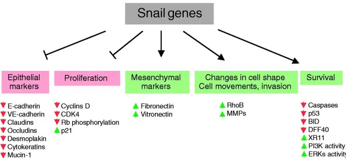
EMT is also well known to play an essential role in pathological processes, such as tumor progression, that occur concomitantly with the cellular acquisition of migratory and invasive properties following the downregulation of the E-cadherin, a cell-to-cell adhesion protein encoded by the CDH1 gene. E-cadherin is currently thought to be a suppressor of invasion during carcinoma progression, in fact the functional loss of this protein is one of the hallmarks of EMTs (Peinado et al., 2004; Nieto et al., 2002; Cano et al., 2000). SNAI1 is a strong repressor of transcription of the E-cadherin gene, directly binding its promoter (Herranz et al., 2008; Peinado et al., 2007; Thiery et al., 2006). Deregulation of SNAI1 has been observed in a variety of tumors including breast cancer (Phillips et al., 2014), gastric carcinoma (Yang et al., 2016), colorectal cancer (Jägle et al., 2017) and prostate cancer (Osorio et al., 2016). Furthermore, SNAI1 directly suppresses the gene expression of proteins representing other adhesive complexes, such as claudins and occludins, integral membrane proteins localized at tight junctions (Ikenouchi et al., 2003). SNAI1 factor represses also other epithelial markers, such as MUC1 and cytokeratin 18 (Guaita et al., 2002). In addition, SNAI1 is involved in the decrease of proliferation. It impairs the transition from early to late G1 by maintaining low levels of Cyclins D and can block the G1/S transition by maintaining high levels of p21 (Vega et al., 2004). SNAI1 is also involved in the resistance to apoptosis, reducing the levels of p53 (Kajita et al, 2004) (Figure 4). Other SNAI1 targets are vitamin D3 receptor (Palmer et al, 2004) and the β-subunit of the Na+/K+ ATPase (Espineda et al., 2004).
Figure 4. Downstream targets of Snai1. Snai1 gene expression induces the loss of epithelial markers and the gain of mesenchymal markers, as well as inducing changes in cell shape and changes related to morphology and to the acquisition of motility and invasive properties. The Snai1 genes also regulate cell proliferation and cell death. The molecules and processes shown in red are downregulated or impaired by Snai1, and those in green are upregulated or promoted by Snai1 (upregulation might be due to the Snai1 mediated repression of a repressor. However, their role as activators cannot be excluded) (Barrallo-Gimeno et al., 2005).
Furthermore, SNAI1 and SNAI2 are expressed in myoblasts, since they are widely expressed in mesodermal cells. They are expressed in proliferating myoblasts and rapidly turned off as muscle differentiation proceeds. A molecular switch involving various actors, including MRFs and SNAI1/2, regulates transition from proliferating myoblasts to terminally differentiated myotubes (Soleimani et al., 2012).
MRFs, such as myogenic differentiation antigen (MyoD), myogenic factor 5 (Myf5), myogenin (MyoG) and myogenic regulatory factor 4 (MRF4), are basic helix-loop-helix (bHLH) transcription factors that regulate myogenesis (Singh et al., 2013). Myf5 and MyoD are required in myoblasts to establish their myogenic identity and act upstream of MyoG and MRF4, which instead drive myogenic differentiation (Jiménez-Amilburu et al., 2013). SNAI1-HDAC1/2 repressive complex binds to multiple differentiation-specific genes under growth condition, allowing the exclusion of MyoD from these sites, therefore the block of differentiation. At the onset of differentiation, SNAI1/2 must be removed to allow the access of MyoD to differentiation genes sequences. Thus, a dynamic switch from a repressive to an activating complex on muscle-specific genes during differentiation (Soleimani et al., 2012). In addition, SNAI1/2 are targets of microRNAs, such as miR-30a and miR206, which are MRFs targets (Sweetman et al., 2008), when cells receive a differentiation signal
from a molecular cascade initiated by growth factors, MRFs activate the miRNAs that prevent Snai mRNA translation. As SNAI proteins turn over, MyoD gains access to differentiation-specific E-boxes (Soleimani et al., 2012).
SNAI1 and SNAI2 functions have been studied extensively during vertebrate embryogenesis and tumor progression, while the role of SNAI3 still needs to be studied (Bradley et al., 2013). SNAI3, also known as SMUC (Snail-related transcription factor of muscle cells), is highly expressed in the developing embryos and in the adult skeletal muscle and thymus. It is expressed in terminal T-cell and myeloid lineages, where it has been largely studied. It has been shown that this transcriptional repressor competes with MyoD for binding on a muscle-specific gene (Kataoka et al., 2000), this means that it probably works as a regulator of muscle differentiation processes with mechanisms similar to the other Snai family members. However, still a lot needs to be studied about SNAI3 and its function during myogenesis needs to be clarified.
2.3 Snai regulation
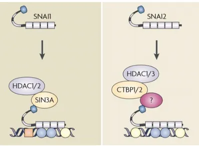
The modification of chromatin structure has emerged as an essential regulatory event promoted by SNAI proteins during EMT. The N-terminal regulatory domain of these factors is necessary for both transcriptional repression and repression by recruitment of chromatin-modifying enzymes (Peinado et al., 2004). SNAI can recruit numerous chromatin enzymes, including members of the histone-deacetylase family (HDACs) and of the lysine-specific histone demethylase (LSDs) to the E-cadherin promoter. These enzymes are essential to generate heterochromatin and promote DNA methyltransferase (DNMT)-mediated DNA methylation at the promoter region (Lin et al., 2014). SNAI factors repression of CDH1 involves the direct recruitment of a repressor complex formed by co-repressors and HDACs (Peinado et al., 2007) (Figure 5).
Snai activity is regulated both transcriptionally and post-transcriptionally. Snai genes are expressed in all EMT processes studied (Nieto et al., 2002). EMT can be triggered by different signaling molecules, such as by fibroblast growth factor (FGF), bone morphogenetic proteins (BMPs), epidermal growth factor (EGF), hepatocyte growth factor (HGF), transforming growth factor β (TGFβ), WNTs and Notch. These signaling molecules have been shown to induce Snai genes in different cellular contexts (Barrallo-Gimeno et al., 2005; De Craene et al., 2005) (Figure 6).
Figure 5. Schematic view of the main repression mechanism of SNAI factors, representing the main corepressor complexes or proposed interactions in the repressor mechanism of each transcription factor (Peinado et al., 2007).
Figure 6. Transcriptionally and post-transcriptionally regulation of Snail genes. Numerous signaling pathways induce the epithelial to mesenchymal transition (EMT), and all have been shown to activate the expression of Snail genes (Barrallo-Gimeno et al., 2005).
Snai promoter presents a functional 5′-CACCTG-3′ E-box that acts as a regulative negative element; in fact, SNAI factors bind to this element creating a negative loop that controls its own expression (Peirò et al., 2006).
SNAI contains a serine-proline rich domain, and the phosphorylation of which is involved in the regulation of SNAI, in particular in the protein degradation and subcellular localization (Dominguez et al., 2003; Zhou et al., 2004). SNAI is regulated by glycogen synthase kinase 3 beta (GSK-3β). GSK‐3β‐mediated phosphorylation of SNAI factors controls their turnover and sub-cellular localization during EMT (Kim et al., 2012). SNAI factors contain two GSK-3β phosphorylation motifs, separated by two proline residues. The phosphorylation of the second motif regulates their subcellular localization by induction of a conformational change that makes the NES domain more accessible to the transport proteins¸ allowing the nuclear export of these factors. Thus, the exportins, such as CRM1, which control the translocation of protein from the nucleus to the cytoplasm, are involved in exporting phosphorylated SNAI and in its inactivation as a transcription factor (Zhou et al., 2004; De Craene et al., 2005; Dominguez et al., 2003).
The first GSK-3β phosphorylation motif overlapped with the destruction box, DSGXXS, recognized by the β-Trcp factor, determines the degradation of SNAI. The phosphorylation of these two motifs by GSK-3β is required for the binding of β-Trcp in the ubiquitination of SNAI and the protein degradation in the proteasome. (Zhou et al., 2004).
Many upstream signaling pathways regulate the function of SNAI modulating the activity of GSK-3β. Oncogenic signals, such as PI(3)K/Akt, MAPK and Wnt, cause the inhibition of GSK-3β, the resulting increase of nuclear SNAI level and the initiation of EMT programs (Wu et al., 2012; Yook et al., 2005; Zhou et al., 2004) (Figure 7).
Figure 7. A model proposed to illustrate the inhibition of SNAI1 by GSK-3β. GSK-3β is a multi-tasking kinase involved in the Akt, Wnt and Hedgehog pathways, it is present in the nucleus and in the cytoplasm. It can phosphorylate several nuclear transcription factors, such as c-Myc and p53. GSK-3β binds and phosphorylates SNAI1 (at level of the motif 2) and thereby induces its nuclear export. Subsequent phosphorylation by GSK-3β (at level of the motif 1) results in the association of SNAI1 with β-Trcp and thus leads to the degradation of SNAI1 (Zhou et al., 2004).
The p21-activated kinase (PAK1) is also able to phosphorylate SNAI. PAK1-induced phosphorylation determines the nuclear localization of SNAI, thus, its activity as a transcription factor (Yang et al., 2005).
Furthermore, the small C-terminal domain phosphatase (SCP) is a specific phosphatase for SNAI. SCP interacts and colocalizes with SNAI in the nucleus, inducing SNAI dephosphorylation and stabilization, with a resulting increase of SNAI activity (Wu et al., 2009).
2.4 FGF21
Fibroblast Growth Factor 21 (FGF21) is an endocrine hormone expressed in numerous tissues including liver, brown adipose tissue, white adipose tissue and pancreas (Markan et al., 2014). Although the liver is generally considered the main site of FGF21 production, FGF21 could be considered a myokine since many studies indicate that the skeletal muscle may be a relevant source of FGF21 production, especially in response to insulin stimulation. In the skeletal muscle, expression and release of FGF21 are essential because they are related to myogenic differentiation, in both rodent and human cell models of myogenesis (Izumiya et al., 2008; Ribas et al., 2014). Recent studies have demonstrated that MyoD binds directly to the promoter region of Fgf21, leading to the activation of Fgf21 expression in mouse C2C12 myoblasts. In this cellular context, FGF21 activates expression of the early myogenic genes, promotes cell cycle exit and enhances myogenic differentiation of C2C12 cells. Even if the mechanism involved in these processes is not so clear, it is known that FGF21 not only regulates myogenesis, but also myofiber type transformation, promoting aerobic myofiber formation (Liu et al., 2016).
FGF21 is also a pivotal modulator involved in the regulation of several physiological processes, such as glucose and lipid metabolism, sensitivity to insulin and cardioprotection (Liu et al., 2016). As a member of the FGF family, FGF21 was initially identified as critical regulator of lipid metabolism and energy homeostasis. It stimulates glucose uptake and fatty acid oxidation (Guridi et al., 2015) and suppresses the accumulation of lipids in muscles (Wang et al., 2016). Several studies have reported that FGF21 is involved in the improvement of insulin sensitivity: long acting of this growth
factor improves liver metabolism and insulin signaling without side effects (Camporez et al., 2015). Moreover, it has been shown that in the cardiovascular system FGF21 released by cardiomyocytes could protect cardiac cells from hypertrophic injury (Planavila et al., 2013). Other studies show that FGF21 regulates the expression of genes involved in antioxidant pathways in heart tissue, preventing the induction of pro-oxidative pathways, and protects against cardiac apoptosis (Planavila et al., 2015; Zhang et al., 2015).
FGF21 expression is not only correlated with physiological processes, but also with metabolic disease and it is strongly induced in animal and human subjects with metabolic diseases, but little is known about the molecular mechanism of this induction (Wan et al., 2014; Itoh et al., 2014).
2.5 ER stress and ATF3
The development of skeletal muscles is an elaborated process that involves myoblasts proliferation and myofibers differentiation, which are mainly controlled by MyoD-Myf5 and MyoG-MRF4 transcription factors, respectively (Singh et al., 2013; Jiménez-Amilburu et al., 2013). In addition, numerous studies have shown that endoplasmic reticulum (ER) stress plays an essential part in the regulation of the skeletal muscle development (Chen et al., 2006; Nakanishi et al., 2015; Wei et al., 2016); in particular, ER stress occurs transiently during differentiation and myofiber formation, although its cause remains unknown (Nakanishi et al., 2015). ER stress plays a critical role both in physiological processes, such as in metabolic homeostasis and myogenesis, and in pathological processes, such as in the contribution of triggering insulin resistance, obesity, and type 2 diabetes (Wan et al., 2014; Cao et al., 2013).
ER is a specialized organelle required for its crucial role in the synthesis, assembly, folding, routing and degradation of a large numbers of proteins and in Ca2+ storage. ER stress is a compensatory process that aims to restore ER homeostasis in order to preserve cellular functions and survival (Kaufman et al., 1999). A variety of agents causes ER stress, e.g., ER redox imbalance or disruption of ER Ca2+ homeostasis, resulting in accumulation of misfolded or unfolded proteins in the ER lumen (Malhotra et al., 2007; Wang et al., 2016). This triggers an adaptive response called unfolded protein response (UPR). The UPR is mediated by three ER transmembrane sensors: protein kinase R (PKR)-like endoplasmic reticulum kinase (PERK), inositol-requiring protein 1α (IRE1α) and activating transcription factor 6 (ATF6) (Edagawa et al., 2014). Initially, the activation of these stress sensors results in a transient repression of protein synthesis, followed by a transcriptional modification that promotes the correct protein folding and the degradation of misfolded proteins and enhances ER folding capacity, thereafter the inhibition of protein synthesis is relieved. In case of prolonged or
strong ER stress, apoptotic pathways are activated (Schaap et al., 2013). Evidence suggests that ER stress induced UPR pathways may regulate various aspects of myogenesis. Specifically, levels of ER stress-related proteins, such as ATF6, CHOP and GRP74, and the activity of caspase-12 are increased in myoblasts undergoing apoptosis during myogenic differentiation (Afroze et al., 2019; Nakanishi et al., 2005). Heightened ER stress seems to be essential for proper progression of myogenesis because the inhibition of ATF6 or caspase-12 reduces the formation of multinucleated myotubes. An increase in caspase-12 activity is also observed during embryonic development of skeletal muscle, suggesting that the ATF6 arm of the UPR is required for the removal of a subpopulation of myoblasts that may not be able to sustain cellular stress (Afroze et al., 2019; Nakanishi et al., 2005; Nakanishi et al., 2007). ER stressors, such as tunicamycin and thapsigargin, increase cell death in C2C12 myoblast cultures after induction of differentiation. However, the surviving myoblasts more efficiently differentiate into functional myotubes, further suggesting that ER stress is a mechanism to remove differentiation-incompetent myoblasts during myogenesis (Nakanishi et al., 2007).
It has been reported that the phosphorylation of PERK and eIF2α and levels of CHOP are transiently increased in a subset of myoblasts, after incubation in differentiation medium. CHOP inhibits myogenic differentiation repressing the expression of transcription factor MyoD, which could be a mechanism to prevent premature differentiation of myoblasts (Alter et al., 2011). IRE1α has an endonuclease activity that mediates the unconventional splicing of XBP1 mRNA. Spliced XBP1 (sXBP1) is a powerful transcription factor that induces UPR target genes. In myogenic cells, the expression of Xbp1 is regulated by MyoD and myogenin. Xbp1 is able to inhibit myotube formation through upregulation of Mist1 (Blais et al., 2005; Acosta-Alvear et al., 2007). Furthermore, Afroze et al. found that PERK is required for the survival of satellite cells and the regeneration of myofibers upon injury. Altogether, these studies suggest that the UPR plays an important role in satellite cells homeostasis and myogenesis both in vitro and in vivo (Afroze et al., 2019).
Several signals that cause the UPR pathway induce also the activating transcription factor 3, ATF3 (Edagawa et al., 2014; Xu et al., 2012; Gjymishka et al., 2009). ATF3 is a member of the ATF/cyclic adenosine mono-phosphate response element binding (CREB) family of basic-region leucine zipper (bZIP) proteins (Hai et al., 1989); it is considered to be a regulatory factor of gene transcription. The ATF3 gene consists of four exons that encode a 181-amino acid protein with a molecular weight of 22 kDa (Hai et al., 1989). ATF3 has been demonstrated to be a transcriptional repressor by forming a homodimer. In addition, the transcription factor cooperates with other ATF/CERB family proteins or CCAAT/enhancer-binding protein (C/EBP) family proteins to form heterodimers producing inhibitory or stimulatory effects in a cell- and promoter-dependent context (Chen et al., 1994; Hai et al., 1991). ATF3 is induced in response to ER stress by a mechanism requiring PERK pathway.
Transcriptional regulator ATF4 is necessary to allow an increased expression of ATF3 protein in early response to stress (Schmitz et al., 2018; Jiang et al.¸ 2004). In fact, ATF3 levels are dramatically induced in many different tissues in response to a variety of cellular stressors; thus, while ATF3 expression is maintained at low levels in normal quiescent cells, it is induced by several stress conditions (Yang et al., 2016). A strong body of evidence shows that ATF3 is an adaptive-response gene and that its expression is increased by numerous signals, including those triggered by genotoxic agents, cytokines, cell death-inducing agents and physiological stress. Indeed, overwhelming evidence indicates that ATF3 plays an important role in metabolic regulation, immune response and oncogenesis (Hui-Chen et al., 2020; Lu et al., 2006; Hashimoto et al., 2002; Hai et al., 1999). Recent studies showed that ATF3 is a negative regulator of some inflammatory genes in skeletal muscle. ATF3 is able to regulate chemokine mRNA expression in C2C12 myotubes and to attenuate
i
nflammation of skeletal muscle upon muscle-damaging eccentric exercise (Fernández-Verdejo et al., 2017).2.6 Skeletal muscle regeneration
Skeletal muscle is an excitable, contractile tissue responsible for maintaining posture and moving the orbits, together with the appendicular and axial skeletons. It attaches to bones and the orbits through tendons. Excitable tissue responds to stimuli through electrical signals. Contractile tissue is able to generate tension of force. Skeletal muscle tissue is also extensible and elastic. Extensible tissue can be stretched, and elastic tissue is able to return to its original shape following distortion. Skeletal muscle is a type of striated muscle tissue, accounting for ∼40% of adult human body weight. Skeletal muscles consist of myofibers, neurons, vasculature networks and connective tissues, of which the structural and functional element of skeletal muscle is the myofiber. During development, myofibers are formed by fusion of mesoderm progenitors called myoblasts. In neonatal/juvenile stages, the number of myofibers remains constant, but each myofiber grows in size by fusion of satellite cells, a population of postnatal muscle stem cells. Each myofiber is surrounded by the endomysium. Bundles of myofibers are surrounded by the perimysium, while the entire muscle is contained within the epimysium. Each myofiber is anchored at its extremities to tendons or tendon-like fascia at the myotendinous junctions (MTJs) (Yin et al., 2013; Tidball et al., 1986). Myofibers are composed of actin and myosin myofibrils repeated as a sarcomere, which is the basic functional unit of skeletal muscle. Responding to the signals from motor neurons, myofibers depolarize and release calcium from the sarcoplasmic reticulum (SR). This drives the movement of actin and myosin filaments relative to one another and leads to sarcomere shortening and muscle contraction (Decary et al., 1997; Schmalbruch et al., 1991) (Figure 8).
Figure 8. Skeletal muscle structure and satellite cells niche.
Individual muscle cell fibers are surrounded by delicate connective tissue called endomysium. Skeletal muscle fibers are aligned in bundles called fascicles and these fascicles are surrounded by a stronger sheath of connective tissue called the perimysium. The fascicles are finally packaged in a stronger connective tissue encasement called the epimysium. Satellite cells are small mononuclear cells located between the plasmalemma of the myofibers and the basal membrane (Meiliana et al. 2015, adapted from The Company of Biologist, Ltd).
Skeletal muscle is a tissue that is able to regenerate after injury. Responding to injury, skeletal muscle undergoes a highly orchestrated degeneration and regenerative process that takes place at the tissue, cellular and molecular levels (Yin et al., 2013; Seale et al., 2000). The initial event of muscle degeneration is necrosis of the muscle fibers. This event is generally triggered by disruption of the myofiber sarcolemma resulting in increased myofiber permeability. The disruption of myofiber integrity is proved by increased serum levels of muscle proteins, such as MCK (Muscle Creatine Kinase, usually restricted to the myofiber cytosol). It has been hypothesized that increased Ca2+ influx after sarcolemma or sarcoplasmic reticulum damage results in a loss of Ca2+ homeostasis and increased Ca2+ dependent proteolysis that drives tissue degeneration resulting in focal or total autolysis depending on the extent of the injury. The early phase of muscle injury is usually accompanied by the activation of mononucleated cells, mainly inflammatory cells and myogenic cells (Chargè et al., 2004; Rappolee et al, 1992). Neutrophils are the first immune cells to invade the injured muscle, with a significant increase in their number being observed as early as 1–6 h after myotoxin or exercise-induced muscle damage. After neutrophil infiltration and 48 h post injury, macrophages become the predominant inflammatory cell type within the site of injury (Tidball et al., 2017; Seale et al., 2000). Macrophages infiltrate the injured site, through phagocytosis remove cellular debris and may affect other aspects of muscle regeneration by activating myogenic cells. Following proliferation, myogenic cells differentiate and fuse to existing damaged fibers or fuse with one another to form myofibers de novo. Newly formed myofibers have small caliber and centrally located myonuclei. At the end of muscle regeneration, newly formed myofibers increase in size, and myonuclei move to the periphery of the muscle fiber (Yin et al., 2013; Chargè et al., 2004).
This process, in many but not all aspects, recapitulates embryonic myogenesis. Skeletal myogenesis begins in the somites where multipotent mesodermal cells commit to the myogenic lineage. These mononucleated myoblasts then fuse and form multinucleated cells (myotubes) that, ultimately, develop into mature myofibers. During the course of muscle development, some myoblasts fail to differentiate and remains associated with the surface of the developing myofiber as quiescent muscle satellite cells in fully developed mature skeletal tissue (Yin et al., 2013; Chargè et al., 2004). Satellite cells activation may result from the ligation of the integrin molecule VL4 (4integrin a4b1) on PMNL (infiltrating polymorphonuclear leukocytes) and VCAM1 (vascular cell adhesion molecule-1) on resident satellite cells. HGF (Hepatocyte growth factor) is also postulated to activate satellite cells through its receptor c-Met, expressed in quiescent satellite cells. HGF may be produced by undamaged myofibers in response to physiological stimuli or to the damage to the basal lamina or extracellular matrix. Several growth factors have been implicated in the proliferation of satellite cells,
including PDGF (platelet derived growth factor), LIF (leukemia inhibitory factor), IL-6 (interleukin-6), FGF (fibroblast growth factor), IGF-1 (insulin-like growth factor-1) (Seale et al., 2000).
2.7 Transcriptional regulation of skeletal muscle regeneration
Discovery of the myogenic regulatory factor family of transcription factors MYF5, MYOD, Myogenin and MRF4 was a seminal step in understanding specification of the skeletal muscle lineage and control of myogenic differentiation during development. These factors are also involved in specification of the muscle satellite cell lineage, which becomes the resident stem cell compartment in adult skeletal muscle. While MYF5, MYOD, Myogenin and MRF4 have minor roles in mature muscle, they play a crucial role in directing satellite cells function to regenerate skeletal muscle, linking the genetic control of developmental and regenerative myogenesis. MRFs present highly related proteins structure. These class II basic helix-loop-helix (bHLH) transcription factors contain three conserved domains: the amino terminal transactivation domain with a histidine/cysteine-rich zone, the central region with the bHLH motif including the α-helical basic domain and Helix I and II and another transactivation domain in the carboxyl terminal containing Helix III . The basic domain directs DNA binding to the E-box consensus sequence CANNTG, but only specific ‘private’ sequences are associated with activating transcription (e.g. CAGGTG for MYOD), and E-box accessibility is epigenetically controlled (Zammit et al., 2017).
In intact muscle, satellite cells are sublaminar and mitotically quiescent (G0phase). Quiescent satellite cells are characterized by the expression of Pax7 but not MyoD or Myogenin. Upon exposure to signals from a damaged environment, satellite cells exit their quiescent state and start to proliferate (satellite cells activation). Proliferating satellite cells and their progeny are often referred as myogenic precursor cells (MPC) or adult myoblasts. Unlike quiescent satellite cells, myogenic precursor cells are characterized by the rapid expression of myogenic transcription factors MyoD and Myf5. Of note, the presence of MyoD, Desmin, and Myogenin in satellite cells was observed as early as 12 h after injury, which is before any noticeable sign of satellite cells proliferation. This early expression of MyoD is proposed to be related to a subpopulation of committed satellite cells, which are poised to differentiate without proliferation. In contrast, the majority of satellite cells express either MyoD or Myf5 by 24 h following injury and subsequently express both factors by 48 h (Yin et al., 2013). The ability of satellite cells to upregulate either MyoD or Myf5 suggests these two transcription factors may have different functions in adult myogenesis. MyoD−/− mutant mice display markedly reduced muscle mass. This atrophy phenotype is reportedly due to delayed myogenic differentiation. Similarly, muscle regeneration is also impaired in MyoD−/− mice, resulting in an increased number of myoblasts within the damaged area. These MyoD−/− myoblasts persist for prolonged periods of time,
fail to differentiate and do not fuse into myotubes. Expression of MyoD is an important determinant of myogenic differentiation, and in the absence of MyoD, activated myoblasts have a propensity for proliferation and self-renewal (Yin et al., 2013; Chargè et al., 2004).
Compared to the MyoD−/− mice, Myf5−/− mutant mice show a myofiber hypertrophy phenotype, and the proliferation of Myf5−/− myoblasts is compromised. Together, these results implicate a distinct role for Myf5 in adult myoblasts proliferation, while MyoD is essential for differentiation. Together, the aforementioned observations suggest the hypothesis that satellite cells enter different myogenic programs depending on whether Myf5 or MyoD expression predominates. Predominance of MyoD expression would drive the program toward early differentiation, as exemplified by the behavior of Myf5−/− myoblasts. In contrast, predominance of Myf5 expression would direct the program into enhanced proliferation and delayed differentiation, as shown by the behavior of MyoD−/− myoblasts. MyoD expression peaks in mid G1, whereas Myf5 expression is maximal at the G0 and G2 phases of the cell cycle (Ustanina et al., 2009; Yin et al., 2013; Chargè et al., 2004). After limited rounds of proliferation, the majority of satellite cells enters the myogenic differentiation program and it begins to fuse to damaged myofibers or fuse to each other forming new myofibers. The initiation of terminal differentiation starts with the expression of Myogenin and Myf6 (also called Mrf4). In this hypothesis, myogenic differentiation is an irreversible procedure and is driven by the sequential expression of key transcription factors (master regulators), which are destined to transduce gene expression signals to their target genes. The expression of these genes is essential for the proper formation, morphology, and function of skeletal muscle; thus they are regulated by multiple mechanisms. MyoD also induces the expression of p21 and subsequent permanent cell cycle arrest. The terminal differentiation ends with the expression of specific muscle proteins such as the Muscle Creatine Kinase (MCK) and the Myosin Heavy Chain (MHC) (Yablonka-Reuveni et al., 2011; Chang et al., 2014) (Figure 9).
Figure 9. Transcription regulation of skeletal muscle regeneration. During the initial stages of injury, Pax7+ satellite cells are activated, proliferate and begin to express MyoD, initiating transcription of muscle-specific genes necessary for early differentiation. As myogenesis proceeds, some activated satellite cells return to quiescence and renew the satellite cell reserve population, while others exit the cell cycle to undergo further differentiation. Those post-mitotic myocytes display a shift in gene expression that enables their fusion to form multinucleated myotubes that are able to undergo terminal differentiation (Tidball et al., 2014).
2.8 Animal models of muscle injury
Although the degenerative and regenerative phases of the muscle regeneration process are similar among several muscle types and after different causes of injuries, the kinetics and amplitude of each phase may be dependent on the extent of the injury, the muscle injured, or the animal model. To study the process of muscle regeneration in a controlled and reproducible way, it has therefore been necessary to develop animal models of muscle injury. The use of myotoxins, such as Bupivacaine (Marcaine), cardiotoxin (CTX), and notexin (NTX) is perhaps the easiest and most reproducible way to induce muscle regeneration (Chargé et al., 2004). These toxins have a wide range of biological activities that are not entirely understood. For example, NTX is a phospholipase A2 neurotoxin peptide extracted from snake venoms that blocks neuromuscular transmission by inhibition of acetylcholine release; CTX, also a peptide isolated from snake venoms, is a protein kinase C-specific inhibitor that induces the depolarization and contraction of muscular cells, leading to the disruption of membrane organization and the lysis of various cell types. Bupivacaine is a local anesthetic drug that induces Ca2+ release from the Sarcoplasmic Reticulum (SR) and simultaneously inhibits Ca2+ reuptake into the SR, resulting in persistently elevated intracellular Ca2+ concentrations. It also has a Ca2+ sensitizing effect on the contractile proteins. These mechanisms result in increased intracellular Ca2+ concentrations and contribute to pronounced skeletal muscle toxicity. Muscle fiber necrosis is extremely rapid after Bupivacaine induced injury. Injection of the drug into small skeletal muscles of
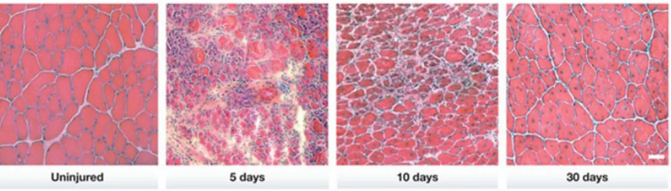
rat or mouse leads to immediate and massive myonecrosis followed by phagocytosis of necrotic debris and a rapid, complete regeneration of muscle fibers 3-4 weeks after injection (Zink et al., 2002). In our laboratory, 25 μl of Bupivacaine injected in adult mouse tibialis anterior muscle induced muscle degeneration leading to a wound coagulum with mononuclear cell infiltration within 1 day of injection. Inflammatory response and mononuclear cell proliferation were active the most within 1– 4 days of injection. Myogenic cell differentiation and new myotube formation were observed ∼5–6 days post injection. By 10 days post injection, the overall architecture of the muscle was restored, although most regenerated myofibers were smaller and displayed central myonuclei. The return to a morphologically and histochemical normal mature muscle was seen at ∼3–4 weeks post injection (Figure 10).
Figure 10. Overview of tissue histology during mouse skeletal muscle regeneration. A time course of histological changes in regenerating skeletal muscle. H&E staining of uninjured TA muscles and regenerating TA muscles at 5, 10 and 30 days after injury. Regenerating muscles are reduced to mostly mononuclear cells at day 5 but are able to re-establish multinucleated myofibers by day 10. Notably, the nuclei of uninjured myofibers are located at the periphery, whereas those of regenerating muscle fibers are centrally located. Scale bar, 50 μm, (Rudnicki et al., 2013).
3. MATERIALS AND METHODS 3.1 Animals
In vivo experiments on wild-type mice, were performed in collaboration with Prof. Libero Vitiello from the University of Padua. In vivo experiments on transgenic mice, were performed in collaboration with the Medical Doctor Stephen J. Weiss from the University of Michigan. Snai1flox/flox mice have been generated in Stephen J. Weiss laboratory as described in Rowe et al., 2009. The Snai1+/LacZ embryonic stem cells (ESCs) were generated by the International Knockout Mouse Consortium (EUCOMM/KOMP) with the details described at the Consortium website. Snai1+/LacZ mice were bred and maintained on a C57/B6 background. Mice carrying Snai1 fl/fl alleles were bred with Tamoxifen-inducible CAG-Cre/Esr1* mice (Jackson Laboratory: 004453) to generate Snai1fl/fl; CAG-Cre + conditional knockout mice (Yongshun et al., 2014).
Snai2+/LacZ mice were obtained from T. Gridley (Jiang et al, 1998; Grande et al., 2015). Snai1+/YFP mice were obtained from Robert Weinberg’s lab (Ye et al., 2015).
Snai1flox/flox, Snai1+/LacZ, Snai2+/LacZ and Snai1+/YFP mice were bred and maintained on a C57BL/6 genetic background. All mouse work was performed with IACUC approval and in accordance with a protocol approved by University of Michigan Institutional Animal Care & Use Committee.
3.2 Genotyping
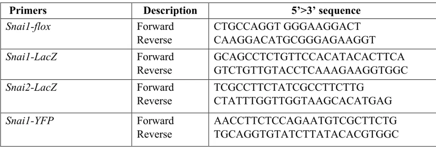
Transgenic mice tails were cut (~2 mm) and digested at 95 °C for 30 min in 50 µl of buffer 1 (10 N NaOH and 0.5 M EDTA, pH 12.0). An equal amount of buffer 2 (40 mM Tris-HCl, pH 5.0) was added to neutralize buffer 1. The mixtures were immediately vortexed and centrifuged at 12.000 xg for 5 min. The supernatants containing the tail genomic DNA were collected and stored at −20 °C for further use. For genotyping, 1 µl of extracted genomic DNA was used as a template in 20 µl of PCR reaction mixture containing 10 µl 2× GoTaq GreenMaster Mix (Promega), 2.5 µl forward/reverse primers 10 µM and 8.5 µl H2O. PCR primers for amplifying the indicated mice transgenes are listed in the Table 1.
Table 1. List of oligonucleotides used for the genotyping.
Primers Description 5’>3’ sequence
Snai1-flox Forward Reverse CTGCCAGGT GGGAAGGACT CAAGGACATGCGGGAGAAGGT Snai1-LacZ Forward Reverse GCAGCCTCTGTTCCACATACACTTCA GTCTGTTGTACCTCAAAGAAGGTGGC Snai2-LacZ Forward Reverse TCGCCTTCTATCGCCTTCTTG CTATTTGGTTGGTAAGCACATGAG Snai1-YFP Forward Reverse AACCTTCTCCAGAATGTCGCTTCTG TGCAGGTGTATCTTATACACGTGGC 3.3 In vivo assay
For the in vivo analysis, three-months mice received a pre-emptive dose of the analgesic Carprofen and then were anaesthetized with Isoflurane. Skin adjacent to the tibial anterior muscle was shaved, wiped clear of debris with sterile water, and sterilized with alternating scrubs of Iodine/Betadine and alcohol three times. 25 µl of Bupivacaine 0.5% were injected into the tibialis anterior muscles to induce acute skeletal muscle regeneration through a single intramuscular injection. Following Bupivacaine injection, mice were sacrificed at different time points and the tibialis anterior muscles were dissected, frozen and processed for further analysis.
3.4 X-Gal Staining
To detect βGal/LacZ activity, the dissected tibial anterior muscles were fixed in fixative solution (4% formaldehyde, 0.5% glutaraldehyde, 1.25 mM EGTA, 2 mM MgCl2, 0.1M sodium phosphate, pH 7.4), washed in rinse buffer (2 mM MgCl2, 0.2% deoxycholate, 0.2% NP-40. 0.1 M sodium phosphate, pH 7.4) and incubated overnight in X-gal staining buffer (2 mM MgCl2, 0.2% deoxycholate, 0.2% NP-40, 1 mg ml−1 X-gal, 5 mM potassium ferrocyanide, 0.1 M sodium phosphate, pH 7.4). After being stained, whole-mount tissues were washed with phosphate buffered saline (PBS), transferred to 70% alcohol and then visualized under a Leica dissecting microscope.
3.5 Muscle embedding and cryosectioning
After dissection, skeletal tibialis anterior muscles were embedded with minimum amount of Tissue-Tek O.C.T (Sakura Finetek USA). The embedded muscles were frozen by placing them into the cooled 2-methylbutane for 5 min and then the muscle samples were transferred to a −80 °C freezer
for storage. Before cryosectioning, the cryostat with the blade was pre-cooled to −22 ± 2 °C. Samples were placed in cryostat for at least 20 min for thermal equilibration, attached on the round metallic holders of the cryostat with Tissue-Tek O.C.T. 10 μm-thick sections were made and collected on room temperature positive charged microscope slides and then stored at −80 °C. These slides were further processed for Hematoxylin and Eosin staining or immunostaining.
3.6 Hematoxylin and Eosin staining
The slides were brought from the −80 °C freezer to room temperature and incubated with hematoxylin solution in a staining jar for 10 min to stain the nuclei. Slides were transferred to a staining jar with running water and then to a staining jar with Eosin solution for 3 min. Successively, the slides were transferred into staining jars with 70% ethanol for 20 sec, 90% ethanol for 20 sec, 100% ethanol for 1 min and xylene for 3 min. Finally, the slides were mounted with xylene-based mounting media and covered with cover slides. Clips were used to press the slides to squeeze bubbles. Hematoxylin and Eosin-stained images were captured with Leica DMI 6000B microscope.
3.7 Tissue Immunostaining
Sections were blocked with 0.5% normal goat serum (Jackson ImmunoResearch Laboratories) in PBST (PBS+0.3% Triton-X100) for one hour at room temperature. Sections were incubated with the anti-GFP primary antibody (Rockland) at 4 °C overnight. After three washes with PBS, sections were incubated with secondary antibody (Biotium) and DAPI for two hours at room temperature, washed three times with PBS and mounted in Prolong gold Antifade reagent (Invitrogen). Immunostained samples were imaged using Zeiss LSM700 confocal microscope.
3.8 Cell culture, differentiation and treatment
The C2C12 myoblasts, an immortalized mouse myoblast cell line established by Yaffe and Saxel (Yaffe et al., 1977) (www.atcc.org), were used as in vitro model for mammalian muscle differentiation. The cells were cultured in Dulbecco’s modified Eagle’s medium (DMEM) High Glucose medium (EuroClone), supplemented with 2 mM L-Glutamine, 100 µg/ml streptomycin, 100 U/ml penicillin and 20% (v/v) fetal bovine serum (FBS), at 37°C in humidified atmosphere
containing 5% (v/v) CO2. To induce myotube differentiation, the C2C12 were cultured in DMEM High Glucose, supplemented with 2 mM L-Glutamine, 100 µg/ml streptomycin, 100 U/ml penicillin and 2% (v/v) horse serum (HS).
The primary mouse satellite cells were cultured in DMEM High Glucose supplemented with 2 mM L-Glutamine, 100 µg/ml streptomycin, 100 U/ml penicillin 20% (v/v) fetal bovine serum, 10% (v/v) horse serum and 1% chicken embryo extract (CEE) at 37°C in humidified atmosphere containing 5% (v/v) CO2.
To induce ER stress, we used Thapsigargin 0.2 µM diluted in dimethyl sulfoxide (DMSO). The control cells were treated only with DMSO.
Lenti-X 293T cell line, a subclone of the transformed human embryonic kidney cell line HEK 293, was used to produce lentivirus particles, since they are highly transfectable and able to support high levels of viral protein expression. Lenti-X 293T cells were grown in the DMEM High Glucose supplemented with 2 mM L-Glutamine, 100 µg/ml streptomycin, 100 U/ml penicillin and 10% (v/v) fetal bovine serum (FBS), at 37°C in humidified atmosphere containing 5% (v/v) CO2.
3.9 Fgf21 plasmid construction
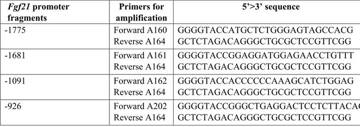
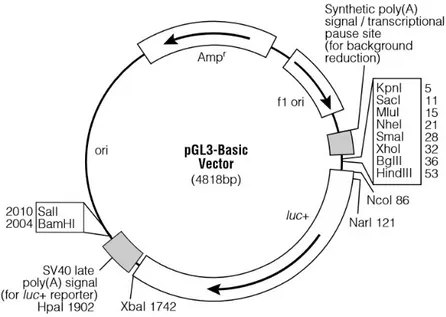
Based on the mouse Fgf21 promoter sequence, specific primers were designed (Table 2) to amplify a full-length promoter, spanning from -1775 bp respect to the transcription start site to +166 bp, and progressively shorter promoter fragments excluding the putative binding sites for SNAI1, previously identified by bioinformatic analysis. In total, four promoter fragments were amplified by PCR from Mus musculus genome using the Q5® High-Fidelity DNA Polymerase (BioLabs), cloned into the pGL3-Basic vector (Promega) (Figure 11) and named as follows: -1775, -1681, -1091 and -926.
Table 2. List of oligonucleotides used for the amplification of the Fgf21 promoter region.
Fgf21 promoter fragments Primers for amplification 5’>3’ sequence -1775 Forward A160 Reverse A164 GGGGTACCATGCTCTGGGAGTAGCCACG GCTCTAGACAGGGCTGCGCTCCGTTCGG -1681 Forward A161 Reverse A164 GGGGTACCGGAGGATGGAGAACCTGTTT GCTCTAGACAGGGCTGCGCTCCGTTCGG -1091 Forward A162 Reverse A164 GGGGTACCACCCCCCAAAGCATCTGGAG GCTCTAGACAGGGCTGCGCTCCGTTCGG -926 Forward A202 Reverse A164 GGGGTACCGGGCTGAGGACTCCTCTTACAC GCTCTAGACAGGGCTGCGCTCCGTTCGG
Figure 11. pGL3-Basic vector circle map. Graphic representation of the pGL3-Basic vector used for cloning.Additional description: luc+ (cDNA encoding the modified firefly luciferase); Ampr (gene conferring ampicillin resistance in E. coli); f1 ori (origin of replication derived from filamentous phage); ori (origin of replication in E. coli).
The four promoter fragments and the pGL3-Basic vector were digested with KpnI-HF/XbaI and KpnI-HF/NheI-HF restriction enzymes (NEB), respectively and then ligated by using the T4 DNA ligase (NEB). The ligation reaction was precipitated by adding 0.1 volumes of Sodium Acetate (3M, pH 5.2) and 2.5 volumes of 95% (v/v) ethanol. The DNA was resuspended in 5 µl of deionized sterile water and transformed into DH5α E. coli stain.
3.10 Site directed mutagenesis
The point-mutations on the -926 Fgf21 promoter E-boxes were introduced by using the QuickChange XL Site-Directed Mutagenesis kit (Agilent), according to manufacturer’s instructions. The primers used for PCR amplification are listed in Table 3.
Table 3. List of oligonucleotides used for the mutagenesis of the -926 Fgf21 promoter region. Fgf21 promoter point-mutants Primers fo amplification 5’>3’ sequence -926 m1 Forward A210 Reverse A211 GAACACAATTCCAGCAAGCTTGGCTCCTCAGCC GGCTGAGCAGCCAAGCTTGCTGGAATTGTCTTC -926 m2 Forward A218 Reverse A219 GACAGCCTTAGTGTCTTCTAGACTGGGGATTCAACACAGG CCTGTGTTGAATCCCCAGTCTAGAAGACACTAAGGCTGTC -926 m3 Forward A208 Reverse A209 TCAGGAGTGGGGAGGATCCGTGGGCGGGCCTGT ACAGGCCCGCCCACGGATCCTCCCCACTCCTGA
The primers were designed in order to obtain point-mutations of the E-boxes 1, 2, and 3, surrounding the TSS (see Figure 25), in the construct carrying the -926 Fgf21 promoter. To easily identify mutated clones, E-boxes were mutated by inserting the restriction sites for the enzymes HindIII (A210-A211), XbaI (A218-A219) or BamHI (A208-A209). After PCR amplification, the DpnI endonuclease was used to digest the methylated parental DNA template allowing the selection of the mutation-containing PCR-synthesized plasmids. The nicked vector DNA mutation-containing the desired mutations was then transformed into XL10-Gold Ultracompetent Cells.
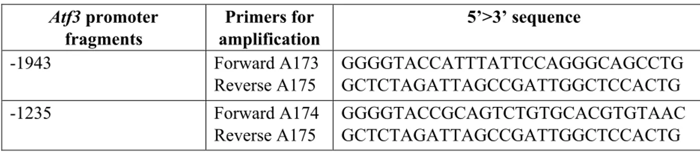
3.11 Atf3 plasmid construction
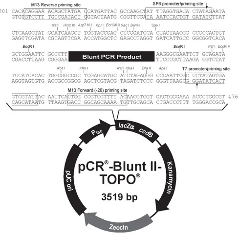
The full length of Atf3 promoter (-1943) and a deletion fragment excluding putative binding sites for SNAI1 (-1235) were generated by PCR amplification of Mus musculus genome. Considering the difficulty in cloning Atf3 promoter directly into the pGL3-Basic vector, the -1943 and -1235 fragments were cloned using the pCR®-Blunt II-TOPO® system (Invitrogen), within which successively was cloned the fragment Luc+-SV40 late poly(A) signal, derived from the pGL3-Basic vector.
Table 4. List of oligonucleotides used for the amplification of the Atf3 promoter region.
Atf3 promoter fragments Primers for amplification 5’>3’ sequence -1943 Forward A173 Reverse A175 GGGGTACCATTTATTCCAGGGCAGCCTG GCTCTAGATTAGCCGATTGGCTCCACTG -1235 Forward A174 Reverse A175 GGGGTACCGCAGTCTGTGCACGTGTAAC GCTCTAGATTAGCCGATTGGCTCCACTG
After the amplification, the PCR products were cloned into pCR®-Blunt II-TOPO® (Figure 12), using the Zero Blunt® TOPO® PCR Cloning kit (Invitrogen). The plasmid constructs were digested with NotI-HF restriction enzyme (NEB), purified by means of QIAquick PCR purification kit (QIAGEN), and blunt ends were generated by means of Large (Klenow) Fragment (NEB).
At the same time, the fragment Luc+-SV40 late poly(A) signal was obtained by digesting the pGL3Basic vector with XhoI and BamHI (NEB) restriction enzymes, separated by agarose gel electrophoresis and extracted from the 1% agarose gel by means of QIAquick gel extraction kit (QIAGEN). Successively, blunt ends were obtained as above. Then, the fragment Luc+-SV40 late poly(A) signal was inserted into the pCR®-Blunt II-TOPO® promoter constructs by ligase reaction with T4 DNA ligase (NEB). Subsequently, DNA was precipitated as described above and used to transform the DH5α E. coli strain.
Figure 12. pCR-Blunt II-TOPO vector. Graphic representation of the pCR-Blunt II-TOPO vector, where Atf3 promoter has been cloned adding the fragment Luc+-SV40 late poly(A) signal, derived from pGL3-Basic vector.
3.12 Expression vectors
The pCMV6 expression vectors for murine ATF3 and SNAI1 (OriGene) were used in experiments of co-transfection and pCMV6-Entry vector was used as control (Figure 13).
Figure 13. pCMV6-Entry vector. A mammalian vector with C-terminal Myc- DDK Tag, containing cDNA clones. In this vector, a TrueORF sequence is fused with a MYC/DDK tag at its carboxy terminus. The antibiotic selection marker for E. coli is kanamycin (25µg/ml), and neomycin (G418) for mammalian cells. The small dual tags facilitate the detection and purification of the ORF product with anti-Myc or anti-DDK antibody.
3.13 Bioinformatic analysis
The GC content of the Atf3 promoter sequence was assessed with the GC Content Calculator (https://www.biologicscorp.com/tools/GCContent/). GC content is usually calculated as a percentage value and sometimes called G+C ratio or GC-ratio. GC-content percentage is calculated as Count (G + C) / Count (A + T + G + C) * 100% in a defined window of nucleotides.
3.14 Plasmid DNA preparation
For the mini preparation of plasmid DNA, the NucleoSpin® Plasmid (Macherey-Nagel) kit was used according to manufacturer’s instructions. The protocol provides essentially three steps: cell lysis, plasmid DNA binding to a silica resin and washing step following by the plasmid DNA elution. For a greater plasmid production, the midi preparation of plasmid DNA was used the NucleoSpin® Xtra plasmid purification kit (Macherey-Nagel), according to manufacturer’s instructions.
3.15 Transfection
C2C12 cells were transfected using Attractene Transfection Reagent (Qiagen), a non-liposomal lipid that ensures highly efficient DNA transfection of all adherent eukaryotic cells.
C2C12 cells were seeded at a density of 9x10⁴ cells/well in 6-well culture plate and grown 24 hours. Before proceeding with transfection, the media was replaced with fresh media and 0.8 µg of luciferase reporter vector, 0.4 µg of the pCMV6 expression vectors (for SNAI1, ATF3 or empty vector), 0.025 µg of Renilla luciferase control vector (pNL1.1 TK[Nluc/TK]; Promega) and 4.5 µl of Attractene were added into 100 µl of DMEM and incubated for 15 minutes at room temperature. The transfection complexes were added to the cells, that were incubated with the transfection complexes under their normal growth condition. After 24 hours, the growth medium was removed from the cultured cells and replaced with 2 ml of differentiation medium when necessary. The day after, the cells were assayed for the expression of the transfected gene.
3.16 Dual-luciferase reporter assay
Following transfection, the cells were harvested in Passive Lysis Buffer (Promega) and the luciferase activity of the samples was measured by the Nano Dual-luciferase report™ assay system (Promega), following the manufacturer’s instructions. Luciferase activity was normalized to the Renilla luciferase internal control.
3.17 C2C12 transfection by electroporation
C2C12 transfection by electroporation was used to overexpress SNAI1 in order to analyze Fgf21 and Atf3 expression and to perform ChIP assay. C2C12 cells were trypsinized and 2.0 x 10⁶ cells/point were centrifuged three times for 5 minutes, at 190 xg with DMEM/F12 supplemented with 2.5% (v/v) FBS and 0.25% (w/v) bovine serum albumin (BSA). Then, the cells were resuspended in 200 𝜇l of the same DMEM/F12 used for washing, and 10 𝜇g of DNA (pcMV6-entry vector or pcMV6- SNAI1) were added. Each mix was put into an electroporation cuvette (Gene Pulser electroporation cuvette, Gap Width 0.2 cm, Bio-Rad). Electroporation was performed using the following parameters: 290 V, 1.000 μF, 200 Ω. After the electroporation, the cuvette content was resuspended with pre-warmed complete C2C12 medium and plated in six-wells dishes. 24 hours later the medium was changed, and after other 24 or 48 hours the cells were harvested for the analyses.
3.18 ChIP-qPCR
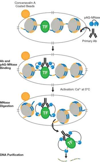
For the ChIP-qPCR analysis, we used the CUT&RUN Assay Kit (Cell Signaling) following the manufacturer instructions. Cleavage Under Target & Release Using Nuclease (CUT&RUN) is a new technology that can be used for chromatin profiling.
Briefly, to isolate the protein-DNA complex of interest, C2C12 cells transfected with pCMV6 or pCMV6-SNAI1 were first trypsinized, centrifuged and the pellet was resuspended in wash buffer containing spermidine and protease inhibitor cocktail. The input samples were collected at this time while the other cells were bound to Concanavalin A-coated magnetic beads. Cell membranes were permeabilized with digitonin to facilitate the entry of the primary antibody (FLAG M2, Sigma Aldrich) into the nuclei. At this point, the primary antibody could bind the transfected FLAG-tagged SNAI1. The pAG-MNase enzyme, a fusion of Protein A and Protein G to Micrococcal Nuclease, was then added to the reaction mixture, where it bound the primary antibody heavy chain, targeting the enzyme to the chromatin region of interest. The pAG-MNase was activated with the addition of Ca2+ in order to initiate DNA digestion around the target protein on the chromatin. The digestion products were about 200 bp in size. This allowed the cleaved chromatin complex to diffuse away from the genomic chromatin, out of the nuclei, into the sample supernatant (Figure 14). Then the digestion was stopped with the Stop Buffer containing digitonin, RNAse A and Spike-in DNA. The Sample Normalization Spike-In DNA is fragmented genomic DNA from the yeast S. cerevisiae that facilitate normalization between samples and between experiments during qPCR analysis.
At the end, input and enriched chromatin samples were collected with the phenol/chloroform extraction followed by ethanol precipitation. The purified, enriched DNA was quantified by qPCR with QuantiTec SYBR Green PCR Master Mix (Qiagen), following the manufacturer instructions, and results were analyzed using the Percent Input Method (Haring et al., 2007). qPCR amplification reaction of Spike-In DNA was performed for sample normalization and were analyzed using the Percent Input Method (Haring et al., 2007). Signals obtained from each immunoprecipitation were expressed as a percent of the total input chromatin.
Figure 14. Schematic representation of the CUT&RUN technology. CUT&RUN works by using the DNA cutting activity of a Protein A fused micrococcal nuclease (MNase) to specifically isolate DNA that is bound by a protein of interest. First, nuclei from tissue or cell culture are isolated using Concavalin A-coated magnetic beads. Nuclei are then incubated with a primary antibody against the protein of interest. The Protein A fused MNase is then added and Protein A binds Immunoglobulin G (IgG) thus targeting MNase to antibody bound proteins. Once MNase has been localized to target sites, the nuclease is briefly activated to digest the DNA around the target protein. This targeted digestion is controlled by the addition of calcium, which MNase requires for its nuclease activity and is chelated from the reaction up until this point. After MNase digestion, fragments are released from nuclei by a short incubation at 37 °C. These short DNA fragments can then be purified for subsequent analysis.
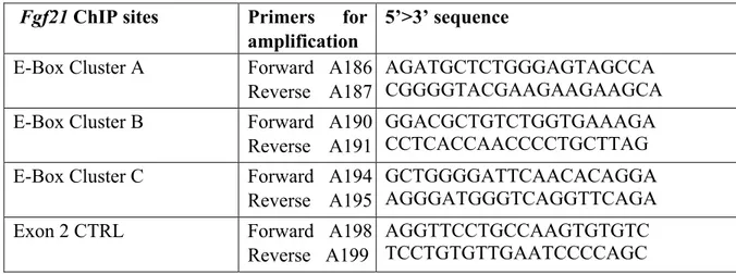
Table 5. List of oligonucleotides used for the Fgf21 ChIP. Fgf21 ChIP sites Primers for
amplification
5’>3’ sequence E-Box Cluster A Forward A186
Reverse A187
AGATGCTCTGGGAGTAGCCA CGGGGTACGAAGAAGAAGCA E-Box Cluster B Forward A190
Reverse A191
GGACGCTGTCTGGTGAAAGA CCTCACCAACCCCTGCTTAG E-Box Cluster C Forward A194
Reverse A195
GCTGGGGATTCAACACAGGA AGGGATGGGTCAGGTTCAGA
Exon 2 CTRL Forward A198
Reverse A199
AGGTTCCTGCCAAGTGTGTC TCCTGTGTTGAATCCCCAGC Table 6. List of oligonucleotides used for the Atf3 ChIP.
Atf3 ChIP sites Primers for amplification 5’>3’ sequence -1873 Forward A325 Reverse A326 AAAAGATGGGGCAGGTAGGAG GGCACAACCCCGAAGAAAG -1257 Forward A321 Reverse A322 CTTTACACCTCAGCGTCCTG GACTGCGGCCCAGGAAT -607 Forward A332 Reverse A333 TACGTTAACCCACAGCTGCTA CTCCGATGAATCCACACCGT
Exon 1 CTRL Forward A329
Reverse A330
CATCCATCACTTCTTGTCCCG GCCTCTACGCGGACTTAGG
3.19 RNA extraction and RT-qPCR
Total RNA was extracted from C2C12 cells by using RNeasy® Plus Mini Kit (Qiagen), according to manufacturer’s instruction.
Total RNA was quantified through QIAxpert by measuring the UV/VIS absorption spectrum and used to evaluate the gene expression by means of RT-qPCR (Reverse Transcription quantitative Polymerase Chain Reaction). In our experiments, the kit Brilliant III Ultra-Fast SYBR Green RT-qPCR Master Mix (Agilent Technologies) was used. Primers for RT-RT-qPCR are shown in Table 7.