1
DIPARTIMENTO DI SCIENZE DELLA VITA DOTTORATO DI RICERCA IN SCIENZE DELLA VITA
XXXIII CICLO
Investigation of the protective mechanism of Neisserial Heparin
Binding Antigen (NHBA) induced antibodies
Settore Scientifico Disciplinare: BIO/10
Relatore: Prof. Luca Bini Correlatore: Dr. Brunella Brunelli
(Dip.to di Scienze della Vita) (GSK Vaccines s.r.l.)
Coordinatore: Prof. Massimo Valoti
Tesi di Silvia Principato
2
INDEX OF CONTENTS
1. ABSTRACT ... 4
2. ABBREVIATIONS ... 6
3. INTRODUCTION AND BACKGROUND ... 8
3.1 Neisseria meningitidis and Meningococcal Disease ... 8
3.2 Meningococcal vaccines and Reverse Vaccinology ... 13
3.3 4CMenB ... 15
3.4 Neisserial Heparin Binding Antigen ... 16
3.5 Biological functions of NHBA ... 20
4. AIM OF THE STUDY ... 23
5. MATERIALS AND METHODS ... 24
5.1 Bacterial Strains ... 24
5.2 Generation of NGH38 NHBA-Over Expressing strain (NGH38OE) ... 24
5.3 Generation of luciferase reporter strains ... 25
5.4 Human monoclonal Antibodies (HumAbs) ... 26
5.5 Hexabody generation ... 26
5.6 Animal Polyclonal sera ... 26
5.7 Sources of complement ... 27
5.8 Serum Bactericidal Assay ... 27
5.9 Flow cytometry for protein surface exposure detection ... 28
5.10 Human C3 deposition assay ... 29
5.11 Human C9 deposition assay ... 30
5.12 Factor H deposition assay ... 30
5.13 Confocal Microscopy ... 31
5.14 Transmission Electron Microscopy ... 32
5.15 Enzyme-Linked Immunosorbent Assay ... 33
5.16 Surface Plasmon Resonance ... 33
5.17 In vivo infection model ... 34
6. RESULTS ... 37
3 6.2. Factor H interaction... 38 6.3. Vitronectin interaction ... 40 6.4. Antigen Density ... 46 6.4.1. Strain generation ... 46 6.4.2. Strain characterization ... 47
6.5. Antibody with enhanced C1q deposition ... 57
6.6. Meningococcal infection in vivo model ... 60
7. DISCUSSION AND CONCLUSIONS ... 66
8. ETHICAL STATEMENTS ... 72
9. BIBLIOGRAPHY ... 73
4
1. ABSTRACT
Neisserial Heparin Binding Antigen (NHBA) is one of the three main protein antigens of the Bexsero vaccine against Neisseria meningitidis serogroup B (MenB). It is a surface-exposed lipoprotein ubiquitously expressed by MenB strains but sparsely distributed on the bacterial surface. NHBA binds heparin and heparan sulfates through an arginine-rich region, it is cleaved by meningococcal and human protease, and its expression is upregulated at 32°C. Recent evidences suggest that NHBA plays a key role in bacterial adherence through its arginine-rich region and is able to affect endothelial permeability. Moreover, NHBA has a direct impact on DNA-dependent biofilm formation.
NHBA induces bactericidal antibodies in humans and confers protective immunity in the infant rat animal model. Anti-NHBA antibodies (either polyclonal or monoclonal) from mice and humans are functional, being able to induce complement-mediated bacterial killing, in the presence of rabbit complement (rSBA). However bactericidal activity is not measurable when human serum is used as a source of complement (hSBA).
The aim of this study was to further elucidate the functional properties that determine the mechanism of protection of anti-NHBA antibodies.
For this purpose, the role of negative regulators of the complement system, such as factor H and vitronectin, have been investigated. The effects of antigen density have also been explored through an NHBA overexpressing strain, used to characterize a panel of anti-NHBA monoclonal antibodies isolated from Bexsero immunized adults. In vivo properties of these anti-NHBA antibodies, both polyclonal and monoclonal, have been evaluated through the Infant Rat meningococcal infection model.
5
Non-specific downregulation of complement-mediated killing due to human factor H was found to result in an underestimation of anti-NHBA antibodies functionality in hSBA. By using the NHBA overexpressing strain, the relevance of antigen density on bactericidal activity was elucidated. Our investigations also highlighted a novel and specific interaction of NHBA with human vitronectin.
Multiple interactions with complement regulators were demonstrated to interfere with the in
vitro measurement of the bactericidal activity mediated by anti-NHBA antibodies in the
presence of human complement. Interestingly NHBA, as the Neisseria Opc and NhhA, interacts with the extracellular matrix component vitronectin. These findings further support the important role played by NHBA in pathogenesis and immunity.
6
2. ABBREVIATIONS
• BSA = Bovine Serum Albumin
• CDC = Complement Dependent Control
• CEACAM = carcinoembryonic antigen-related cell-adhesion molecule • CFU = Colony Forming Units
• CIC = Complement Independent Control • CPS = Capsular Polysaccharide
• CREN = Contact Regulatory Element of Neisseria • CSF = Cerebrospinal fluid
• ECM = Extracellular Matrix • eDNA = extracellular DNA • fHbp = factor H binding protein
• FMS = Fulminant Meningococcal Sepsis • GNA = Genome-derived Neisserial Antigen • hK1 = human Kallicrein 1
• hLf = human Lactoferrin
• hSBA = Serum Bactericidal Assay with human complement • HSPGs = heparan sulfate proteoglycans
• humAbs = human monoclonal Antibodies • IDP = Intrinsically Disordered Protein • IMD = Invasive Meningococcal Disease • IP = intraperitoneally
• KD = Dissociation Constant • LOS = Lipooligosaccharide
7 • mAb = monoclonal Antibody
• MATS = Molecular Antigen Typing System • MenB = Neisseria meningitidis serogroup B • MLST = Multi Locus Sequence Type
• NadA = Neisseria adhesin A
• NHBA = Neisserial Heparin Binding Antigen • NhhA = Neisseria Hia/Hsf homologue • NMR = Nuclear Magnetic Resonance • NS = Negative Staining
• OD = Optical Density
• OMVs = Outer Membrane Vesicles • Opa = Opacity associated protein • ORFs = Open Reading Frames • PE = Protein E
• PilA = Pilin A
• PKa = plasma Kallicrein • Por A = Porin A
• Por B = Porin B
• ROS = Reactive Oxygen Species
• rSBA = rabbit Serum Bactericidal Assay • RT = Room Temperature
• RU = Resonance Unit
• SPR = Surface Plasmon Resonance • ST = Sequence Type
8
3. INTRODUCTION AND BACKGROUND
3.1 Neisseria meningitidis and Meningococcal Disease
Neisseria meningitidis, also known as meningococcus, is a Gram-negative diplococcus,
member of the bacterial family of Neisseriaceae. It was identified in late 1800 by Weichselbaum from the cerebrospinal fluid (CSF) of a patient with meningitis (Weichselbaum 1887). This aerobic diplococcus has the peculiar “coffee-bean” shape (Figure 1) and can exist either as encapsulated or unencapsulated. The human nasopharynx is considered the natural biological niche colonized by N. meningitidis (Kiefer 1896). It has been estimated that up to 10-25% of the population is meningococcus-asymptomatic carrier in the nasopharynx tract (Stephens 2009).
Exposure and acquisition of meningococci are generally mediated by aerosol droplets or close direct contact with carriers (Nelson 1996). Colonization of the nasopharynx is usually asymptomatic, but very rarely in susceptible individuals it may result in penetration of the mucosal epithelium causing local inflammation. The passage into the bloodstream and survival of Neisseria, mostly mediated by the capsule, can lead to different clinical manifestations (McGee et al. 1983).
In some patients with low bacteremia degree, meningococci can be spontaneously cleared, manifesting a transient meningococcemia characterized by a short febrile flu-like episode (Sullivan and LaScolea 1987). If not completely cleared, bacteria can lead in very few hours to fulminant meningococcal sepsis (FMS) or meningitis, as already reported in 1919 by Herrick “no other infection so quickly slays” Herrick (1919).
9
Figure 1. Immuno-gold labelling and transmission electron microscopy images of Neisseria meningitidis shows
the typical “coffee-bean” shape of the diplococcus. Analysis of the strain was performed with anti-NHBA monoclonal antibody. Scale bar: 500nm.
Invasiveness of meningococcus can be influenced by multiple virulence factors (Figure 2):
i) capsular polysaccharide expression is essential for the survival of the organism in the blood, providing resistance to complement-mediated killing by antibodies and inhibiting phagocytosis (Uria et al. 2008);
ii) expression of surface adhesive proteins mediates meningococcus adhesion to host cells. Piliated meningococci better attach to human nasopharyngeal cells (Stephens and McGee 1981) than meningococci negative for pili expression. In
10
addition, pili are involved in facilitating DNA uptake by meningococci (Proft and Baker 2009).
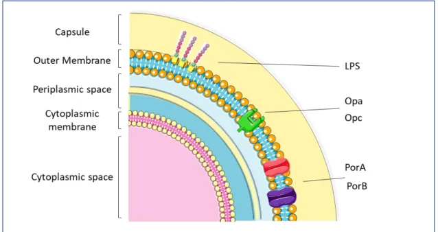
iii) outer membrane proteins including porins PorA and B, adhesion molecules Opa and Opc interact with multiple members of the CEACAM (carcinoembryonic antigen-related adhesion molecule) family (Virji et al. 1996) and with cell-surface associated HSPGs (heparan sulfate proteoglycans) (Virji et al. 1999);
iv) endotoxin (lipooligosaccharide, LOS) (Jennings et al. 1983, Gamian et al. 1992) plays a pivotal role in the adherence of the meningococcus (Kahler and Stephens 1998) and in activation of the innate immune system (Plant et al. 2006)
Figure 2. Schematic representation of the main neisserial virulence factors and their localization in the bacterial
compartments. Image adapted from Rosenstein NE, 2001
At least 13 distinct meningococcal serogroups have been identified based on the capsular polysaccharide (Branham 1953) but only six are considered to cause life-threatening disease
11
(A, B, C, W-135, X, Y) (Rouphael and Stephens 2012, Stephens et al. 2007, Boisier et al. 2007, Frasch 1989, Jarvis and Vedros 1987).
Molecular genetic typing systems are considered the gold standard for molecular epidemiological typing and identification of clonal groups. Based on detection of polymorphisms in seven house-keeping genes, MLST (Multi-locus Sequence Typing) has shown that the majority of disease-associated clinical isolates group into a few sequence types (ST) enabling the definition of hyper-virulent invasive meningococcal lineages (1, 4, ST-5, ST-8, ST-11, ST-32, ST41/44 and ST-269) (Yazdankhah et al. 2004, Maiden et al. 1998, Maiden 2008, Caugant 2008).
It is estimated that 1.2 million cases of meningococcal infection occur every year, with a death rate of ~135,000 worldwide. A few clonal complexes that can emerge and spread worldwide (Maiden et al. 1998) cause the great majority of disease cases.
The Epidemiology of meningococcal infection can be sporadic, hyper sporadic or epidemic with variable incidence patterns. Incidence can vary from 1/100,000 in Europe and North America, to 10-1,000 cases/100,000 in an area of sub-Saharian Africa (Jafri et al. 2013), renamed the “meningitis belt” by Lapeyssonnie in 1963 because it is characterized by periodic large epidemics of predominantly meningococcal meningitis (Lapeyssonnie 1963). Serogroup A has been associated with the largest and most severe meningococcal outbreaks in sub-Saharan Africa (Hart and Cuevas 1997) and is considered responsible for most of the meningococcal diseases in the early twentieth century but is now more uncommon in the US and Europe.
12
Serogroup B is usually associated with a lower incidence of cases if compared to serogroup A or C, but protracted MenB outbreaks can cause significant morbidity and mortality. Nowadays, MenB is the major cause of endemic meningococcal disease in developed countries, causing 30–40% of the disease in the US and up to 80% in Europe.
Although the geographic distribution is variable, age distribution is clear and definite: meningococcus is a common cause of bacterial meningitis in children and teenagers in the USA (Harrison et al. 1999), prevalently affecting children less than 2 years of age (Rosenstein et al. 1999, Kaplan et al. 2006).
Half of the cases in infants are due to serogroup B, while serogroup C is prevalently observed in adolescents and serogroups B and Y in older adults. Even though high incidence occurs among infants and adolescents, sporadic cases are seen in adults older than 18 years (Rouphael and Stephens 2012).
The case-fatality ratio of the meningococcal infection disease is 10% to 15%, increasing up to 40% in septicemia cases (CDC 2015). The most severe clinical forms of invasive meningococcal diseases (IMD) are meningitis (30-60%) and septicemia (20-30%), usually associated with severe manifestations as Purpura Fulminans (petechial or purpuric rash) (Pace and Pollard 2012).
Identification and diagnosis of meningococcal disease can be difficult due to similarities with some viral infections such as influenza, but the onset of symptoms may be fulminant. A delay in appropriate therapeutic treatment and rapid progression of the disease from bacteremia and/or meningitis to life-threatening syndrome can occur within the first few hours after the initial stages.
13
3.2 Meningococcal vaccines and Reverse Vaccinology
Given the rapid progression of the disease and difficulties in diagnosis (Rosenstein et al. 2001, Thompson et al. 2006), meningococcal vaccines are a fundamental need to successfully prevent the spread and control meningococcal disease. So far, no broadly protective vaccine is available to provide protection against all serogroups of Neisseria meningitidis although different meningococcal vaccines have been developed against the diverse serogroups. Vaccines against serogroups A, C, Y and W135 were developed in the 1960s by using purified capsular polysaccharide (CPS) as antigen. Subsequently a more effective approach was introduced, in which CPS components were conjugated to carrier proteins such as CRM197, a non-toxic mutant of the diphtheria toxin (Costantino et al. 2011).
When conjugated to a carrier protein, capsule polysaccharides show a significantly improved immunogenicity in young infants (Granoff and Pollard 2007). Formulations of monovalent, bivalent and tetravalent polysaccharide conjugative vaccines are available and effective against meningococcal serogroups A, C, Y and W-135 (Zahlanie et al. 2014) (http://www.who.int/ith/vaccines/meningococcal/en/).
Due to its similarity with human α(2->8)N-acetyl neuraminic acid, a polysialic acid present on the surface of human cells, group B polysaccharide capsule is poorly immunogenic and unsafe for potential induction of autoimmunity (Hayrinen et al. 1995); hence, a polysaccharidic approach could not be evaluated for vaccines against serogroups B.
First vaccine approach relied in the use of Outer membrane vesicles (OMVs) preparations. Surface-exposed proteins contained in the OMVs were demonstrated to be immunogenic in humans in Cuba, Norway and New Zealand, with a valid vaccine efficacy against the respective homologous strain but poor protection against heterologous strains (Rosenstein et al. 2001)
14
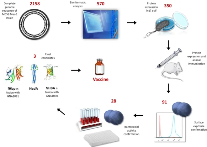
To overcome these limitations, a multicomponent recombinant protein-based vaccine was needed. The Neisseria meningitidis serogroup B (virulent strain MC58) genome was sequenced and analysed for the identification of promising vaccine antigens (Tettelin et al. 2000). This novel approach, named “reverse vaccinology” led to the identification of 570 Open Reading Frames (ORFs) by bioinformatic analysis (Pizza et al. 2000).
Amplification, cloning and expression of the corresponding genes in Escherichia coli allowed the identification of about 600 antigens, 320 of which were used for mice immunization to evaluate their ability to induce functional antibodies (Pizza et al. 2000) (Figure 3).
15
3.3 4CMenB
Among valid candidates, three protein antigens were selected for the final formulation: Genome-derived Neisseria Antigens (GNA) 2132 (Neisseria Heparin Binding Antigen, or NHBA as we will discuss later), GNA1870 (factor H binding protein, or fHbp) and GNA1994 (Neisseria adhesin A, or NadA). FHbp is present in variant 1.1, NHBA is present in peptidic variant p2 and NadA is present in variant 3. Two additional antigens, GNA2091 and GNA1030, were also selected for their ability to induce protective immunity. In order to facilitate large-scale manufacturing of the vaccine, four of the selected antigens were combined as two fusion proteins. The best performing combinations in terms of production and immunogenicity were NHBA fused with GNA1030 and GNA2091 with fHbp. These two fusion proteins were formulated with NadA in combination with Outer Membrane Vescicles (OMVs) from the
New-Zealand epidemic strain (NZ98/254) (Giuliani et al. 2006) (Figure 4). This novel vaccine named
4CMenB (4 Components vaccine against MenB), was licensed with the trade name of Bexsero and was approved in 2013 in Europe for infants and in 2015 in the US for the age group 10-25 years. Bexsero is now approved in over 40 countries worldwide.
(https://www.ema.europa.eu/en/documents/overview/bexsero-epar-summary-public_en.pdf ).
Figure 4. 4CmenB formulation includes two fusion proteins NHBA-GNA1030 and GNA2091-fHbp, NadA and OMVs
16
During bioinformatic analysis of the meningococcal genome, GNA2132 was predicted to be a surface lipoprotein with some similarities with transferrin-binding proteins. The gene was ubiquitously expressed in all the MenB strains tested and in the related N. lactamica, N.
gonorrhoeae, N. polysaccharea, and N. flavescens (Pizza et al. 2000, Jacobsson et al. 2006,
Bambini et al. 2009) (Muzzi et al. 2013, Lucidarme et al. 2009) .
Gene sequences from genetically diverse group B strains revealed the existence of more than 400 protein sub-variants that have some association with clonal complexes and sequence types (Comanducci et al. 2002, Muzzi et al. 2013).
3.4 Neisserial Heparin Binding Antigen
Only scarce information about the function of this protein were available until 2010, when Serruto et al. reported heparin binding ability of NHBA. The protein exerts its ability to bind heparin and heparin-like molecules through an arginine-rich region (-RSARSRRS-), highly conserved among different Nm strains (Serruto et al. 2010). The ability to bind heparin is considered a virulence factor due to its correlation with increased serum resistance of the bacteria (Rostand and Esko 1997, Schneider et al. 2006, Duensing et al. 1999, Menozzi et al. 2002). On the basis of this evidence, it was suggested to rename GNA2132 as Neisserial Heparin Binding Antigen (NHBA) (Serruto et al. 2010).
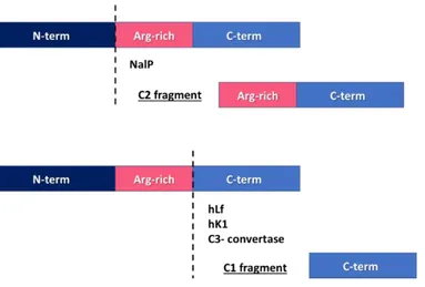
NHBA is a 60 kDa protein of about 450 residues, constituted by an extremely variable N-terminal domain (residues 1-230), an arginine-rich region (residues 235–245) and a highly conserved C-terminal domain (residues 305–426) (Pizza et al. 2000).
NHBA is target of cleavage by NalP, a bacterial phase variable autotransporter protein with serine protease activity (Turner et al. 2002, van Ulsen et al. 2003). The cleavage site is
17
upstream of the Arg-rich region and the C-term fragment generated, that still contains the heparin binding site, is named C2 (Serruto et al. 2010).
Interestingly, NHBA is also target of a human protease known for its ability to cleave the Arg-rich motif of bacterial surface proteins (Hendrixson et al. 2003, Qiu et al. 1998), human Lactoferrin (hLf). The cleavage mediated by hLf occurs immediately downstream of the Arg-rich region. The C-term fragment generated does not contain the heparin binding site any longer and is named C1 (Serruto et al. 2010). Both the cleaved fragments can be released into the culture medium but the ability to bind heparin is limited to the C2 fragment, suggesting a possible protective role of the hLf, avoiding the release of a functional fragment. On the other hand, bacterial proteases such as NalP can release an active fragment and it is reasonable to consider that this fragment might interact with secreted proteoglycans in order to exert its own biological role.
Recently, it has been demonstrated that another human protease present in saliva is able to process NHBA, human Kallikrein 1 (hK1). The fragments generated by Hk1 cleavage are identical to that generated by hLf. Interestingly, plasma Kallicrein (Pka) was also able to cleave NHBA (Pantano et al. 2019).
A schematic representation of the NHBA multiple cleavage sites is reported in Figure 5. A study, aimed at verifying whether the two NHBA fragments, C1 and C2, were involved in the alteration of endothelial permeability, demonstrated that only the C2 fragment is able to increases microvasculature endothelial permeability, to rapidly accumulate in the mitochondria of the cells and to enhance production of oxygen radicals. On the contrary, C1 does not reach mitochondria, is unable to trigger ROS production and does not alter endothelial permeability (Casellato et al. 2014). Moreover, alteration of endothelial permeability by the toxic C2 fragment can be associated with vascular leakage, a clinical
18
feature mostly related to Purpura fulminans lesions associated with meningococcemia (Coureuil et al. 2009).
Increased expression of NHBA, as well as increased aggregation, biofilm formation and cellular adherence, have been observed at 32°C versus 37°C. In fact, 32°C is a condition more similar to the nasopharynx environment encountered during initial colonization by N. meningitidis (Lappann et al. 2016) compared to 37°C, as commonly used in in vitro experiments to investigate pathogen-host interactions. These observations suggested that the effect of the C2 fragment on epithelial cells of the naso-pharynx should be investigated. Surprisingly, a study aimed to evaluate the effect of C2 on a cell line resembling the morphological features of human respiratory tract epithelium revealed no influence of the C2 fragment on epithelium permeability, but cleavage ability of epithelial cells was detected. Incubating both the C2 fragment and full-length NHBA with Normal Human Bronchial Epithelial, a smaller fragment was found lacking the Arg-rich region motif, consequently abrogating the toxic effect of the C2 fragment. C3-convertase of the alternative complement pathway was demonstrated to be responsible for this inactivating cleavage (Di Fede et al. 2018).
Figure 5. NHBA processing. NHBA can be cleaved by bacterial NalP releasing a toxic C2 fragment that still includes
the Arg-rich domain. Downstream from the Arg-rich motif, NHBA can be cleaved by human Lactoferrin (hFl), human Kallikrein 1 (Hk1) and C3-convertase releasing an inactivated C1 fragment.
19
The C-term, initially solved by Nuclear Magnetic Resonance (NMR) spectroscopy, is constituted by a β-barrel with 8 anti-parallel strands stabilized by a hydrogen bond network that links the β-strands (Esposito et al. 2011). Interestingly, this region of NHBA shares significant homology with, factor H binding protein (fHbp), another protein included in the 4CMenB vaccine, suggesting an ancestral precursor. The common architecture comprises a C-terminal domain consisting of 8 anti-parallel β-strands. The N-term of NHBA comprises regions possibly intrinsically unfolded, rich in small hydrophilic and hydrophobic amino acids and various low complexity sequences (e.g. polyglycine and polyserine motifs) (Esposito et al. 2011). Since intrinsically disordered proteins (IDPs) are predicted to lack a stable secondary structure, crystallization of the N-term of NHBA is challenging.
Two human monoclonal antibodies (humAbs 12E1 and 10C3) isolated from Bexsero vaccinees (Giuliani et al. 2018), targeting N-term of NHBA were used in the attempt to characterize the three-dimensional structure of NHBA. Fab fragments from these human mAbs were cloned and then used for binding, crystallization and co-crystallization studies (Maritan et al. 2017). More recently, an unnoticed NHBA fragment with 2-stranded ß-hairpin packing against the C-terminal domain was observed, with the putative role of stabilizing the barrel. Due to the Fab fragment of the humAb 5H2, the presence of a large cross-reactive conformational epitope on NHBA was observed (Maritan et al. 2018). Currently available NHBA 3D structures are reported in Figure 6.
20
Figure 6. Currently available 3D structures of NHBA from the RCSB Protein Databank: a. three-dimensional
structure of the C-terminal region solved by nuclear magnetic resonance (Esposito et al. 2011) (PDB code 2LFU);
b. Crystal structure of the C-terminal domain of NHBA (Maritan et al. 2018) (PDB code 6CUJ); c. human Fab 5H2
bound to NHBA (Maritan et al. 2018) (PDB code 5O1R); The antibody is shown in green. The structures were prepared with PyMOL (http://www.pymol.org).
3.5 Biological functions of NHBA
Many functions have been associated with NHBA, suggesting its key role as a virulence factor in multiple steps of meningococcal colonization, infection and survival.
As mentioned above, the name Neisserial Heparin Binding Antigen was assigned to the GNA2132 after the discovery of its ability to bind heparin and heparan sulphates (Serruto et al. 2010). This ability is commonly associated with increased bacterial survival and serum resistance (Chen et al. 1995, Menozzi et al. 2002, Duensing et al. 1999, Rostand and Esko 1997, Dubreuil et al. 2004). It has been proposed that binding of heparin could be an efficient strategy used by pathogens to recruit several human heparin binding proteins to their surfaces, bypassing the need to synthesize individual receptors for each of these proteins (Duensing et al. 1999).
21
The ability of NHBA to bind heparin or heparin-like molecules present on the cell surface and in the extracellular matrix suggested that it may also be involved in bacterial adhesion to the epithelium. In fact, NHBA was confirmed to bind epithelial cells through its Arg-rich region via direct interaction with heparan sulphate proteoglycans (HSPGs) present on the cell surface and in the extracellular matrix (ECM) (Vacca et al. 2016).
It is noteworthy that, in some N. meningitidis strains belonging to a hypervirulent clonal complex, a 150-bp region known as a Contact Regulatory Element of Neisseria (CREN) is present upstream of the NHBA coding sequence. This regulatory element in the promoter region, specific for pathogenic Neisseria species, is involved in the induction of the downstream-associated genes when bacteria are in contact with target eukaryotic cells. It is considered essential for effective meningococcal adhesion to epithelial cells (Deghmane et al. 2003, Deghmane et al. 2002).
It has been demonstrated that NHBA plays a key role in biofilm formation, highly structured microbial communities (Costerton et al. 1995). Characterized by the irreversible binding to surfaces, interfaces or other bacterial cells, provides the pathogen a protective niche enhancing resistance to antimicrobial agents, antibodies and host defence molecules (Leid et al. 2005, Jensen et al. 2010). In vitro biofilm formation is a typical feature of unencapsulated meningococci and is mainly dependent on the presence of eDNA released from bacterial autolysis (Lappann et al. 2010). Moreover, the lack of capsule could be considered as an advantage since unencapsulated cells bind to the epithelium more efficiently than capsulated cells (Stephens et al. 1983, Hammerschmidt et al. 1996, Virji et al. 1992). Since NHBA can bind heparin and heparan sulphates, that are negatively charged molecules, its ability to bind extracellular DNA (eDNA) and take part in biofilm formation was also investigated. Indeed, it was confirmed that NHBA can bind DNA and participates in eDNA-dependent biofilm
22
formation. In fact, inactivation of the nhba gene drastically affects the initiation of this type of biofilm formation. On the contrary, it does not influence eDNA-independent initiation of biofilm formation that occurs in some meningococcal strains (Arenas et al. 2013).
As mentioned above, NHBA has been recently associated with vascular leakage. Endothelium permeability can be affected by the C2 fragment (the C-term fragment that still includes the Arg-rich region. Figure 5) generated by the NalP- mediated cleave of NHBA. By localizing in the mitochondria, C2 induces phosphorylation and degradation of the adherents-junction protein VE-cadherin in a ROS-dependent manner.
Immunological studies of sera from mice immunized with GNA2132 have demonstrated that they bind to the surface of live N. meningitidis cells and elicit deposition of human C3b and iC3b. When the sera were incubated in the presence of baby rabbit complement, they showed high bactericidal activity, specific for the antigen. Moreover, anti-GNA2132 sera showed complete passive protection in the Infant Rat infection model of meningococcal bacteraemia (Welsch et al. 2003) and opsonophagocytic activity (Plested and Granoff 2008). Furthermore, anti-NHBA antibodies were able to inhibit meningococcal adhesion to epithelial cells (Vacca et al. 2016).
23
4. AIM OF THE STUDY
NHBA induces bactericidal antibodies in humans and confers protective immunity in the in
vivo animal model. Anti-NHBA antibodies (polyclonal and monoclonal) from mice and humans
are functional, being able to induce complement-mediated bacterial killing, in the presence of rabbit complement. However bactericidal activity is not measurable when human serum is used as a source of complement (hSBA).
The aim of this study was to elucidate further the functional properties of anti-NHBA antibodies. To this end, a number of aspects have been examined.
1. The possible effects of standard Serum Bactericidal Assay read-out on low-expression antigen testing, given the scarce distribution of NHBA on the bacterial surface in in vitro conditions.
2. To analyze the mechanism of action of these antibodies, the influence of complement negative regulators, such as Factor H and Vitronectin, was evaluated.
3. The role of antigen density in the activation of the complement cascade was considered, enabling an elucidation of the antibody-mediated killing mechanism in a high-NHBA-expression environment through the generation of an NHBA overexpressing strain.
4. The in vivo model for meningococcal infection has been used to analyze the immunological properties of polyclonal and monoclonal anti-NHBA sera.
24
5. MATERIALS AND METHODS
5.1 Bacterial Strains
NGH38 is a WT strain isolated in 1988 from a carrier in Norway and belongs to ST- 36. It expresses NHBA variant p2, fHbp variant 2.24. It does not express NadA and mismatches for PorA from OMVnz. It is considered the MATS-NHBA reference strain (Medini et al. 2015). 2996 is a WT invasive strain isolated in 1975 in the United Kingdom and belongs to ST-540. It expresses NHBA variant p20 and fHbp variant 2. It expresses NadA variant 3.8 and it mismatches for PorA from OMVnz.
Variants are named according to the classification reported on the Neisseria Multi Locus Sequence Type (PubMLST) database (https://pubmlst.org/neisseria/).
5.2 Generation of NGH38 NHBA-Over Expressing strain (NGH38OE)
With the aim of generating a meningococcal strain that overexpresses NHBA, a stabilized version of the PorA promoter was used to drive NHBA expression (Delany I). The plasmid for the generation of the mutant consisted of a 459 bp region (NMB2133) upstream of nhba amplified with KAPA Hi-Fi polymerase (KAPA Biosystems, Sigma), using primers AR_nmb2133_mc58_iPCR_Fwd/AR_nmb2133_mc58_ iPCR_Rev from a template vector containing the NEIS2110 gene (Serruto et al. 2010)), the PPorAst2 promoter fused to the intergenic region upstream of nhba, which included also the Contact Regulatory Element of
Neisseria (CREN) (Deghmane et al. 2003), and part of the NHBA coding sequence. These
regions were subcloned in a different plasmid (Redsted et al., paper under submission) and amplified with KAPA Hi-Fi polymerase (KAPA Biosystems, Sigma) using primers
25
AR_constructs_vPCR_Fwd/AR_constructs_vPCR_Rev (see Table 7). The two PCR products were mixed and cloned with the PIPE method (Klock and Lesley 2009). The XmaI-linearized vector was then used to transform the NGH38 strain, according to a previously reported procedure (Masignani et al. 2003), taking advantage of the NEIS2110 and N-terminus nhba regions as homologous recombination sites and selecting the mutated clone using kanamycin.
Table 7. Primers used for NGH38 OE strain generation
5.3 Generation of luciferase reporter strains
With the aim of generating N. meningitidis mutant lux reporter strains, a series of plasmids containing the bacterial luciferase expression under the control of the promoters under investigation was generated. Bacterial luciferase transcriptional fusions of the promoter under study at a chromosomal location between the two converging ORFs NMB1074 and NMB1075, flanked on both sides with transcriptional terminators, plasmid pSL-LuxFla was constructed for allelic exchange in N. meningitidis 2996 strains. Following a previously reported procedure (Fagnocchi et al. 2013), the promoterless luxCDABE operon and cat cassette were subcloned from pSB1075 into pBluescript II as an EcoRI-BamHI fragment and then cloned as a 6.5-kb XhoI-BamHI fragment into pSL-furlacZ, replacing a 4.7 kb fragment containing an erythromycin cassette and the fur-lacZ fusion, generating pSL-LuxFla. Either the nhba or the nadA promoters were cloned as a 250-bp Xho-KpnI fragment upstream of the luxCDABE operon, generating the pSLPnhba-lux and the pSLPnadA-lux, respectively. The pSL-LuxFla, pSLPnhba-lux and pSLPnadA-lux plasmids were used for transformation of the 2996 strain, generating the
26
isogenic recombinant mutants 2996-lux, 2996-Pnhba-lux and 2996-PnadA-lux, respectively, for the in vivo reporter analysis.
5.4 Human monoclonal Antibodies (HumAbs)
Anti-NHBA human monoclonal Antibodies (humAbs) were selected from a panel of recently isolated humAbs (Giuliani et al. 2018). Peripheral Blood Mononuclear Cells (PBMCs) from three adult subjects immunized with the 4CMenB vaccine were collected 8 days after the second dose of vaccine. Plasma blasts were isolated as single cells and used to obtain monoclonal antibodies (mAbs) as previously described by Beernink and co-workers (Beernink et al. 2015).
5.5 Hexabody generation
The VH regions of 3 selected anti-NHBA mAbs were cloned in a vector containing an IgG1 constant region carrying 2 point-mutations, namely E345R and S440Y. Transfection were performed in Expi 293 expression system following manufacturer instructions (de Jong et al. 2016).
5.6 Animal Polyclonal sera
To prepare antisera, 20 µg of NadA, NHBA-GNA1030, or GNA2091-fHbp antigen or a combination of 20 μg each of NHBA-GNA1030, GNA2091-fHbp, and NadA with or without 10 μg of deoxycholate-extracted OMVs derived from the NZ98/254 strain were used to immunize 6-week-old CD1 female mice (Charles River Laboratories International, Inc, Wilmington, MA,
27
USA). Five to ten mice per group were used. The antigens were administered intraperitoneally (i.p.), together with aluminum hydroxide (3 mg/ml), on days 0, 21 and 35.
5.7 Sources of complement
Pooled baby rabbit sera (Cedarlane, Burlington, Canada) or human serum obtained from volunteer donors under informed consent, were used as a complement source for rSBA or hSBA respectively.
5.8 Serum Bactericidal Assay
The Serum Bactericidal assay (SBA) was performed in 96-well plates (Thermo Scientific, Waltham, MA, USA). From frozen glycerol stocks, bacteria were seeded and grown overnight on chocolate agar plates at 37°C in 5% CO2. The day after 10-15 colonies were inoculated in Müller-Hinton broth containing 0.25% glucose to reach OD600 of 0.05 to 0.06 and incubated at 37°C with shaking until the OD600 reached 0.25. Bacteria were diluted 10,000-fold in Dulbecco’s phosphate buffered saline (DPBS), 1% (w/v) Bovine Serum Albumin (BSA), 0.1% glucose (w/v) and added to a reaction mix with a serial two-fold dilution of serum and complement. The plate was incubated for 1 hour at 37°C on a shaker; 7 μl of mix were spotted on Müller-Hinton agar plates that were incubated overnight at 37°C, and the Colony Forming Units (CFU) were counted the day after. Bactericidal titers were defined as the serum dilution resulting in 50% decrease in CFU compared to the negative controls, the Complement Dependents Control (CDC) and the Complement Independent Control (CIC). Figure 8 shows a schematic representation of the Serum Bactericidal Assay plate layout.
28
Figure 8. Schematic representation of the Serum Bactericidal Assay plate layout.
5.9 Flow cytometry for protein surface exposure detection
From frozen glycerol stocks, bacteria were seeded and grown overnight on chocolate agar plates at 37°C in 5% CO2. 10-15 colonies were inoculated in Müller-Hinton broth containing 0.25% glucose to reach OD600 of 0.05 to 0.06 and incubated at 37°C 5% CO2 with shaking until an OD600 of 0.25 or 0.5 was obtained. Bacterial cells were centrifuged at 3500 rpm for 10 min. The supernatant was discarded, and the pellet suspended in 5ml PBS 1% BSA (w/v).
The sample were sequentially incubated with mouse primary antibody (1:100, 1:200 and 1:400) for 1 hour at room temperature (RT). Murine monoclonal Antibody against Neisseria
meningitidis serogroup B capsule (Remel Microbiology Product, Thermo Scientific, Waltham,
MA, USA) was used as positive control. Bacterial cells were centrifuged at 3500 rpm for 10 min.
29
The binding was detected by using either anti-mouse or anti-human (whole-molecule) FITC-conjugated antibodies (Sigma-Aldrich, Saint Louis, Missouri, USA) at a 1:100 dilution in PBS 1% BSA (w/v) with 30-minutes incubation at RT. After the secondary antibody incubation step bacteria were fixed with formaldehyde and plated to check bacterial inactivation. The samples were analyzed with a Canto II cell counter (FACSCanto II, BD Biosciences, San Jose, CA, USA) and histograms generated using Flowjo 8.60 software (BD Biosciences, San Jose, CA, USA).
5.10 Human C3 deposition assay
From frozen glycerol stocks, bacteria were seeded and grown overnight on chocolate agar plates at 37°C in 5% CO2. 10-15 colonies were inoculated in Müller-Hinton broth containing 0.25% glucose to reach OD600 of 0.05 to 0.06 and incubated at 37°C 5% CO2 with shaking until an OD600 of 0.25 or 0.5 was obtained. Bacterial cells were centrifuged at 3500rpm for 10 min. The supernatant was discarded, and the pellet suspended in PBS 1% BSA (w/v).
50 μl of bacterial suspension was incubated with 25 μl of human complement were added resulting in a concentration of 10 mM MgCl2, 1.5 mM CaCl2 and 5 U/ml heparin: i.e 10 l of salt and 10 l of heparin in 1 ml of plasma) for 60 minutes at 37˚C with 5% CO2 with gentle shaking. Bacterial cells were centrifuged 3500 rpm for 10 min, the supernatant was discarded, the pellet was washed in PBS and sequentially incubated with anti-Human C3_FITC antibody (1:100) (Cederlane, Burlington, Canada) 30 minutes at room temperature (RT). After incubation the bacteria were fixed with formaldehyde and plated to check bacterial inactivation. The samples were analyzed with a Canto II cell counter (FACS Canto II, BD Biosciences, San Jose, CA, USA) and histograms were generated using Flowjo 8.60 software (BD Biosciences, San Jose, CA, USA).
30
5.11 Human C9 deposition assay
From frozen glycerol stocks, bacteria were seeded and grown overnight on chocolate agar plates at 37°C in 5% CO2. 10-15 colonies were inoculated in Müller-Hinton broth containing 0.25% glucose to reach OD600 of 0.05 to 0.06 and incubated at 37°C 5% CO2 with shaking until OD600 of 0.25 or 0.5 was obtained. Bacterial cells were centrifuged at 3500 rpm for 10 min, the supernatant was discarded, and the pellet was suspended in PBS 1% BSA (w/v).
50 μl of bacterial suspension was incubated with 25 μl of human complement were added resulting in a concentration of 10 mM MgCl2, 1.5 mM CaCl2 and 5U/ml Heparin: i.e 10ul of salt and 10 l of heparin in 1 ml of plasma) for 60 minutes at 37˚C with 5% CO2 with gentle shaking. Bacterial cells were centrifuged at 3500 rpm for 10 min, the supernatant was discarded, the pellet was washed in PBS and sequentially incubated with mouse anti-Human C9 (1:200) (Thermo Scientific, Waltham, MA, USA) 30 minutes at room temperature (RT).
The binding was detected using anti-mouse (whole-molecule) FITC-conjugated antibodies (Sigma-Aldrich, Saint Louis, Missouri, USA) at a 1:100 dilution in PBS 1% BSA (w/v) with 30-minutes incubation at RT. After the secondary antibody incubation bacteria were washed in PBS, fixed with formaldehyde and plated to check bacterial inactivation. Samples were analyzed with a Canto II cell counter (FACS Canto II, BD Biosciences, San Jose, CA, USA) and histograms generated using Flowjo 8.60 software (BD Biosciences, San Jose, CA, USA).
5.12 Factor H deposition assay
From frozen glycerol stocks, bacteria were seeded and grown overnight on chocolate agar plates at 37°C in 5% CO2. 10-15 colonies were inoculated in Müller-Hinton broth containing 0,25% glucose to reach OD600 of 0.05 to 0.06 and incubated at 37°C 5% CO2 with shaking until OD600 of 0.25 or 0.5. Bacterial cells were centrifuged 3500 rpm for 10 min, the supernatant was discarded, and the pellet was suspended in PBS 1% BSA (w/v).
31
50 μl of bacterial suspension was incubated with 25 μl of human complement or with human factor H (50 μg/ml) (Calbiochem, San Diego, CA, USA).
Bacterial cells were centrifuged at 3500rpm for 10 min, the supernatant was discarded, the pellet was washed in PBS and sequentially incubated with goat anti-human fH (1:100) (Calbiochem, San Diego, CA, USA) for 30 minutes at RT. The binding was detected using a donkey anti-goat FITC-conjugated antibody (Jackson Immuno Research Europe Ltd, Cambridge House, St. Thomas' Place, UK) at a 1:100 dilution in PBS 1% BSA (w/v) with 30-minutes incubation at RT. After the secondary antibody incubation bacteria were washed in PBS, fixed with formaldehyde and plated to check bacterial inactivation. The samples were analyzed with a Canto II cell counter (FACS Canto II, BD Biosciences, San Jose, CA, USA) and histograms generated using Flowjo 8.60 software (BD Biosciences, San Jose, CA, USA).
5.13 Confocal Microscopy
From frozen glycerol stocks, bacteria were seeded and grown overnight on chocolate agar plates at 37°C in 5% CO2. 10-15 colonies were inoculated in Müller-Hinton broth containing 0.25% glucose to reach OD600 of 0.05 to 0.06 and incubated at 37°C 5% CO2 with shaking until OD600 of 0.25 or 0.5. 1 ml of bacterial growth at OD600 of 0.25 or 0.5 was incubated with FM4-64FX (Thermo Fisher Scientific Inc., Waltham, MA USA) and then fixed with 4% Formaldehyde (Carlo Erba Reagents S.r.l., Milano, Italy). 150 μl were used for each staining condition and spotted on a glass slide (Polysine Corning® microscope slides, Sigma-Aldrich, Saint Louis, Missouri, USA). Staining was performed on the slide with 100 μl of mouse polyclonal antibodies diluted 1:250 and incubated for 1 h at RT. After several wash steps, the slides were incubated in 100 μl of rabbit anti-mouse antibody Alexa fluor 488 conjugated (Life technologies, Carlsbad, California, USA), diluted 1:1000 for 30 minutes at RT. The samples were dried under vacuum, mounted using ProLong gold antifade reagent with
diamidino-2-32
phenylindole (DAPI; Invitrogen-Thermo Fisher Scientific Inc., Waltham, MA USA) and analyzed by confocal microscopy using a Zeiss LSM 710 confocal microscope (Zeiss, Oberkochen, Germany).
5.14 Transmission Electron Microscopy
From frozen glycerol stocks, bacteria were seeded and grown overnight on chocolate agar plates at 37°C in 5% CO2. 10-15 colonies were inoculated in Müller-Hinton broth containing 0.25% glucose to reach OD600 of 0.05 to 0.06 and incubated at 37°C 5% CO2 with shaking until OD600 of 0.25 or 0.5.
For Negative Staining (NS) a 5 μl aliquot of a formaldehyde (Carlo Erba Reagents S.r.l., Milano, Italy) fixed bacterial culture with a final concentration of 1 OD/ml was loaded for 30 seconds onto a glow discharged copper 300-square mesh grid. After blotting the excess, the grid was negatively stained using NanoW (Nanoprobes, Yaphank, NY, USA) for 30 seconds. The samples were analyzed using a Tecnai G2 spirit and TEM FEI Tecnai G2 spirit microscope operating at 100kV and equipped with an 2k × 2k CCD Emsis Veleta camera (Emsis, Germany). The images were acquired using a Veleta CCD camera.
For Immunogold (Ig) a 5 μl aliquot of a formaldehyde (Carlo Erba Reagents S.r.l., Milano, Italy) fixed bacterial culture with a final concentration of 1 OD/ml was adsorbed on a 300-mesh formvar/carbon coated nickel grid (Agar Scientific Ltd, United Kingdom), blocked in PBS with 0.5% bovine serum albumin for 1 h and incubated with primary antibody (diluted 1:50 in in PBS with 0.5% bovine serum albumin) for 1 h. Grids were washed and incubated with 5- or 10-nm gold-labeled secondary antibody (Sigma-Aldrich, Inc. Merck, Germany) (diluted 1:20 in PBS with 0.5% bovine serum albumin) for 1 h. After washing with distilled water, the grids were negatively stained and observed using a TEM FEI Tecnai G2 spirit microscope operating at
33
100kV and equipped with an 2k × 2k CCD Emsis Veleta camera (Emsis, Germany). The images were acquired and processed using iTem (OSIS, Olympus, Shinjuku, Tokyo, Japan) software.
5.15 Enzyme-Linked Immunosorbent Assay
Vitronectin-coated 96 well plates (R&D Systems, Inc. Minneapolis, USA) were used for a direct binding assay. NHBA, NHBA-GNA1030 and GNA1030 serial two-fold dilutions were incubated for 2 h at 37°C. After 3 washes (PBS + Tween 0.05%), anti-NHBA-GNA1030, anti-NHBA and anti-GNA1030 (diluted 1:200 in PBS + Tween 0.05% + BSA 1%) mouse sera (100 l/well) were incubated for 1 h at 37°C. After 3 washes a goat anti-mouse IgG-ALP conjugated secondary antibody (Sigma Aldrich.Inc. Merck, Germany) was added (100 l/well) and incubated 1 h at 37°C. The signal was detected using a PNPP substrate (50 l/well) after incubating for 30 min at room temperature and reading the plate at 405nm on a Spectramax reader (Molecular Devices, California, USA).
5.16 Surface Plasmon Resonance
For Surface plasmon resonance (SPR) analysis human Vitronectin (Life Technologies, Carlsbad, CA, USA) was immobilized on the surface of a CM5 sensor chip (see Figure 9 for the chip characteristics) using an amine coupling procedure. The proteins were immobilized in the flow cells 2 while flow cells 1 were used as reference.
34
The recombinant NHBA-GNA1030, NHBA p2 and GNA1030 proteins were diluted in HBS-EP+ buffer (10 mM HEPES, 150 mM NaCl, 3 mM EDTA, 0.005 % P20 surfactant, pH 7.4) to reach a final concentration of 300 nM and injected onto the chip. Finally, the chip was regenerated by injection of 3 M Magnesium chloride.
The final read out of the analysis is the sensorgram, a plot of response (measured in resonance
units [RU]) against time, that shows the progress of the interaction. The response is directly
proportional to the concentration of biomolecules on the surface.
To assess the affinity of the binding, maintaining the same chip set up, five increasing concentrations (6.25 nM, 12.5 nM, 25 nM, 50 nM, 100 nM) of each protein were injected for 60 seconds each on the surface of the CM5 sensor chip. After the last injection, dissociation of the protein was followed for 120 seconds. After each analysis cycle, the chip was regenerated injecting 3 M Magnesium chloride. This analysis enabled the kinetic binding parameters to be measured: the ligand-analyte association (Kon) (described by the ascendant tract of the curve, 60 seconds, 30μl/min), the ligand-analyte dissociation (Koff) (described by the descendant tract of the curve during buffer injection into the system, 240-300 seconds of injection time) and the equilibrium dissociation constant (Kd) (the lower the Kd value, the higher is the binding affinity of the ligand for the analyte).
5.17 In vivo infection model
The ability of anti-NHBA antibodies to confer passive protection against N. meningitidis bacteremia was tested using the Infant Rat infection model, intraperitoneal (i.p.) challenging 5-7 days Wistar Han rat pups (Charles River Laboratories International, Inc, Wilmington, MA, USA) as previously described (Moe et al. 1999).
35
Bacteria from frozen glycerol stocks were seeded and grown overnight on chocolate agar plates at 37°C in 5% CO2. 10-15 colonies were inoculated in Müller-Hinton broth containing 0.25% glucose to reach OD600 0.05-0.06 and incubated at 37°C 5% CO2 with shaking until OD600 0.25 was reached.
For passive protection experiments, animals were treated i.p. at time zero with 100 μl of different dilutions of test or control antisera. Three hours later, the same animals were i.p. challenged with 104 or 106 CFU dose of N. meningitidis strains, in a final volume of 100 μl. Eighteen hours after the bacterial challenge, blood samples were obtained by cheek puncture and collected in Eppendorf tubes containing 25 U of heparin (Eparina Vister, Teva Italia S.r.l., Milano). 100 μl of collected blood were plated on Agar Columbia (5% blood) plates and incubated O/N at 37°C in 5% CO2 and the day after the CFUs were counted. Rats were considered infected when >10 CFUs were counted. Counts above the threshold were verified for positivity by examining plates carrying 10- and 100-fold dilutions of blood. Workflow of the experimental protocol is schematically represented in Figure 10.
Figure 10. Workflow of the Infant Rat challenge protocol
In vivo imaging of the bioluminescence of the strains was monitored in infant rats i.p. infected.
36
zero, 3 h and 20 h post-challenge, using an IVIS 100 system (Xenogen Corp., Alameda, CA, USA) according to the manufacturer’s instructions. Analysis was performed using Living Image 3.1 software (Xenogen Corp., Alameda, CA, USA). Quantification was performed using the photons emitted per second by each rat. Rats infected with the 2996 wild-type strain under the same conditions of acquisition were used for background signal subtraction. Blood samples were collected at 20 h post infection and plated to allow CFU counting, as for standard Infant Rat challenge.
37
6. RESULTS
6.1. Serum Bactericidal assay read out
The Serum Bactericidal Assay (SBA) is considered the gold standard for correlate of protection of meningococcal vaccines (Frasch et al. 2009). This assay, commonly used to investigate the ability of anti-meningococcal antibodies to elicit complement deposition and bacterial killing, has a classical read out of 7 μl reaction drop on Mueller-Hinton Agar. The colony count is performed the next day, after an O/N incubation at 37 °C 5% CO2. In this experiment the same reaction (Rabbit Anti-NHBA-GNA1030 serum against the Nm strain NGH38 WT, at OD600 = 0.25, in the presence of the same Human complement) was used to evaluate the effect of changing the bacterial dilution (1:10000 vs 1:15000), the medium (MHA vs Agar Chocolate) or the plating mode (7 μl reaction drop spot vs whole reaction volume using spread-plate method of isolation) (Figure 11).
Figure 11. In the image are reported the variables combined to test SBA read out variability: bacterial dilution
(1:10000 or 1:15000); medium (Mueller Hinton Agar or Agar Chocolate); plate condition (7 μl spot or whole plating);
High variability of hSBA results was observed when the bacterial dilution, the plating mode or the medium ere changed. The best combination to achieve maximum killing was showed to be 1:10000 bacterial dilution, on Agar Chocolate and plating the whole volume using
spread-38
plate method of isolation. By combining these conditions, a rabbit anti-NHBA-GNA1030 serum tested against the Nm strain NGH38 WT showed the bactericidal titer of 32 in the presence of human complement, compared to the negative titer obtained with the standard read out (Figure 12).
Figure 12. The graph reports the hSBA titers obtained with standard read out (1:10000 bacterial dilution, 7 μl
spot on MHA) and with the new combination (1:10000 bacterial dilution, whole volume with spread-plate method, on Agar Chocolate).
6.2. Factor H interaction
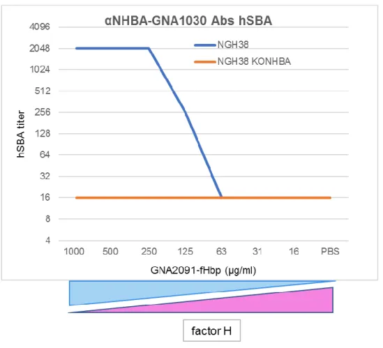
The factor H negative regulation of the Alternative Pathway was investigated to evaluate the impact of this down-regulation on the bactericidal activity of anti-NHBA-GNA1030 antibodies in the serum bactericidal assay. Factor H was sequestered from the reaction by the addition of fHbp at scalar concentrations. At a high sequestrator concentration, the amount of factor H available in the serum was dramatically decreased and no down regulation of the Alternative Pathway took place. As a direct effect, the anti-NHBA-GNA1030 antibodies showed strong
Standard Read Out New Combination h SB A tite r
39
bactericidal activity in the presence of human complement, directly proportional to the concentration of fHbp added to the reaction (Figure 13).
To demonstrate that removal of factor H did not make the bacteria susceptible to killing in absence of Ab, anti-NHBA-GNA1030 antibodies were tested against NHBA Knock-Out strain, and no killing was still observed in the presence of different fHbp amounts.
Figure 13. hSBA titers obtained by testing the anti-NHBA-GNA1030 serum against NGH38 WT (blue) and
NGH39KONHBA (orange) in presence of scalar concentrations of GNA2091-fHbp added to the reaction.
On the contrary, when human Factor H was added to rabbit SBA, a decrease of the titers was observed, mimicking the results obtained for hSBA. This effect was observed non-specifically testing anti-NadA serum, anti-NHBA serum or anti-fHbp serum, but the impact was more
40
evident in the case of a poorly expressed antigen on the bacterial surface, as for NHBA. In fact, for fHbp and NadA, which are highly expressed on the surface, the titers moved from very high to high (65536 to 16384) and from high to medium (4096 to 256), respectively. For anti-NHBA Abs, the titer decreased from medium to negative (1024 to <16) (Table 14). This underlines the significant impact of unspecific down regulation of factor H in the in vitro testing of anti-meningococcal antibodies, especially if they are raised against poorly expressed antigens.
Table 14. The table reports the rSBA and hSBA titers obtained testing NadA, NHBA-GNA1030 and
anti-GNA2091-fHbp sera against the BZ83 strain. 40 μl of Human factor H was added to rSBA.
6.3. Vitronectin interaction
To investigate whether Vitronectin and NHBA interact, an ELISA-based assay was performed. Serial two-fold dilutions of NHBA-GNA1030, NHBA or GNA1030 were alternatively incubated on Vitronectin-coated 96-well commercial ELISA plates. Anti-NHBA-GNA1030, anti-NHBA and anti GNA1030 sera were used for primary incubation and the signal was detected through ALP-conjugated secondary antibodies (Figure 15).
41
Figure 15. Schematic representation of the ELISA-based assay. Vitronectin coated on the plate in light blue;
NHBA-GNA1030 in blue; NHBA in purple; GNA1030 in grey; Primary antibodies are shown in grey; the asterisk represents an ALP-conjugated secondary antibody.
The results showed a specific and dose dependent NHBA-vitronectin binding: anti-GNA1030 serum gave negative results; a low signal was detected with a serum directed to NHBA alone, and the highest signal was observed when a serum to NHBA-GNA1030 was incubated (Figure 16).
42
Figure 16. Signals detected at OD405nm after incubation of NHBA-GNA1030 (blue), NHBA (purple) and GNA1030
(grey) scalar concentrations on Vitronectin coated plates, in presence of different antisera. a. anti-NHBA-GNA1030 serum diluted at 1:2000; b. anti-NHBA serum diluted at 1:2000; c. anti-anti-NHBA-GNA1030 serum diluted at 1:2000.
To characterize protein–protein interactions, Surface Plasmon Resonance analysis was performed with NHBA and vitronectin. This technique, measuring real-time quantitative binding affinities and kinetics, confirmed the NHBA-vitronectin binding previously observed in ELISA-based assay. Human Vitronectin was immobilized on the surface of the CM5 sensor chip
b.
43
(Figure 8) using an amine coupling procedure. The positive control PE (light blue) with an RU of 75.85and the negative control PilA (yellow) with RU of 0.65 are shown, a low binding between NHBA and Vitronectin was detected with an RU of 12.25 (Figure 17 and Table 18).
Figure 17. Time dependent SPR signal (RU) of PE (light blue), PilA (yellow), NHBA (pink), GNA1030 (red) and HBS
(green).
Table 18. Mean of reponses (RU) of the tested samples (at 300 nM concentration) on the ligand Vitronectin (20
44
Rmax is the maximum binding capacity of the immobilized ligand and depends directly on the molecular weight of the protein-protein complex that is formed on the surface of the sensor chip after the analyte molecule injection. The binding of the positive control PE to vitronectin showed a Rmax of 108.7 RU with a measured affinity of 1.3e-8 M. The binding between Vitronectin and NHBA was of a lower magnitude with a Rmax value of 19.5 RU, but the affinity was comparable to that of PE (3.6e-8 M) (Figure 19 and Table 20).
PE
45 NHBA
Figure 19. The time dependence of vitronectin binding to PE, PilA and NHBA.
Table 20. Affinity constants (Kd) measured for PE, PilA and NHBA on SPR vitronectin chip. The values considered
negative are shown in grey and those considered positive in green.
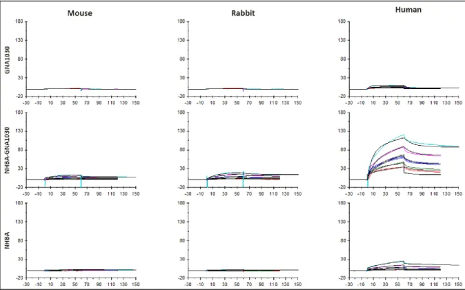
A comparison of NHBA binding to vitronectin from different origins (mouse, rabbit and human) by SPR revealed an increased binding for human vitronectin (Figure 21). Although the SPR GNA1030 signal was negative, the signal of NHBA-GNA1030 was higher with respect to NHBA alone, suggesting that the conformation of NHBA in the context of the fusion could expose crucial epitopes relevant for the binding.
46
Figure 21. SPR signals (RU) of GNA1030, NHBA-GNA1030 and NHBA binding to vitronectin from mouse, rabbit
and human origin.
6.4. Antigen Density
6.4.1. Strain generation
Given the scarce distribution of NHBA on the bacterial surface, an NHBA overexpressing strain was generated in the NGH38 WT strain background with the aim of studying the mechanism of protection mediated by antibodies raised against this antigen. A stabilized version of the PorA promoter was fused with the Contact Regulatory Element of Neisseria and used for constitutive overexpression of NHBA (Figure 22).
47 6.4.2. Strain characterization
Growth kinetic evaluation showed no major differences between the NGH38 WT and NGH38OENHBA strains (Figure 23).
Figure 23. The OD600nm dependence for NGH38 WT (blue) and NGH38OE NHBA (red) on time of growth.
Characterization of the strains by FACS analysis showed no differences among NGH38 WT, NGH38 OE and NGH38KONHBA regard to the capsule level of expression. However, a slight decrease of the fHbp signal was detected for the NHBAOE strain. The analysis of NHBA protein expression showed a clear increase on the intact bacterial surface of NGH38OE (Figure 24).
48
Figure 24. Flow cytometry analysis of anti-fHbp (dilution 1:200), anti-NHBA (dilution 1:200) and anti-capsule
(dilution 1:800) sera are reported for NGH38 WT (blue), NGH38 OE NHBA (red) and NGH38KO NHBA (green). The negative control of only bacteria detected with the FITC-conjugated secondary antibody is shown in grey.
Given the slight decrease of fHbp exposure on the overexpressing strain, factor H deposition on the bacterial surface was investigated to evaluate if NHBA overexpression could have some way impacted Factor H biding on the bacterial surface.
Purified human Factor H and human complement were used to investigate the ability of NGH38 WT, NGH39OE and NGH38KO NHBA to bind Factor H, showing no major differences between the two sources used. Overlays of signals showed no major differences between the strains in the ability of binding human Factor H (Figure 25 and 26).
49
Figure 25. Factor H deposition flow cytometer analysis performed on NGH38 WT, NGH38 OE NHBA and NGH38KO
NHBA. A comparison of bacteria incubated with human complement (in red) and purified human Facotr H (in blue). In grey the negative control of only bacteria detected with the FITC-conjugated secondary antibody.
Figure 26. Factor H deposition flow cytometer analysis performed on NGH38 WT (blue), NGH38 OE NHBA (red)
and NGH38KO NHBA (green). A comparison of bacteria incubated with purified human Facotr H (left figure) and human complement (right figure). In grey the negative control of only bacteria detected with the FITC-conjugated secondary antibody.
Complement C3 deposition assay was performed to detect C3b deposition on bacterial cell surfaces, so activation of the complement cascade. This ability was evaluated for NGH38 WT,
50
NGH39OE and NGH38KO NHBA, showing a great increase of complement deposition for the NGH38OE strain mediated by NHBA antibodies, but no differences were detected if anti-fHbp serum was used (Figure 27).
Figure 27. C3 deposition flow cytometer analysis performed on NGH38 WT (blue), NGH38 OE NHBA (red) and
NGH38KO NHBA (green). A comparison of bacteria incubated with purified fHbp serum (left panel) and anti-NHBA serum (right panel). In grey the negative control of only bacteria detected with the FITC-conjugated secondary antibody.
Furthermore, Complement C9 deposition assay was performed to detect C9 deposition on bacterial cell surfaces, with the aim of studying the end of the complement cascade, thereby the formation of the Membrane Attack Complement (MAC) for bacterial lysis. This ability was evaluated for NGH38 WT, NGH39OE and NGH38KO NHBA, showing great increase of complement deposition for the NGH38OE strain mediated by anti-NHBA antibodies, but no differences were detected if anti-fHbp serum was used (Figure 28).
51
Figure 28. C9 deposition flow cytometer analysis of NGH38 WT (blue), NGH38 OE NHBA (red) and NGH38KO
NHBA (green). A comparison of bacteria incubated with purified anti-fHbp serum (left figure) and anti-NHBA serum (right figure). In grey the negative control of only bacteria detected with the FITC-conjugated secondary antibody.
To evaluate the distribution of NHBA on the bacterial surface a confocal microscopy analysis was performed, comparing the NGH38 WT and NHBA OE strains. The scarce and punctiform distribution of NHBA on the bacterial surface was confirmed for the WT strain, being slightly more intense in the septum of the diplococcus (Figure 29).
52
Figure 29. Confocal microscopy images of the NGH38 WT strain incubated with anti-NHBA serum and the
secondary antibody Alexa fluor 488 conjugated.
For the Over Expressing strain, the distribution was found to be more homogeneous confirming that the amount of NHBA expressed on the bacterial surface is, as expected, high (Figure 30).
53
Figure 30. Confocal microscopy images of the NGH38 OE NHBA strain incubated with anti-NHBA serum and the
secondary antibody Alexa fluor 488 conjugated.
To evaluate if NHBA expression changes during bacterial growth, a time dependence study was undertaken. No major differences were observed for NHBA distribution on NGH38 WT and NGH38OE for the three time points evaluated (Early log phase OD600 = 0.25; Mid log phase OD600 = 0.5; Stationary phase OD600 = 1) (Figures 31 and 32).
54
Figure 31. Confocal microscopy images of the NGH38 WT strain (at OD600 =0,25-0,1-1) incubated with anti-NHBA
55
Figure 32. Confocal microscopy images of the NGH38 OE NHBA strain (at OD600 = 0.25-0.1-1) incubated with
anti-NHBA serum and the secondary antibody Alexa fluor 488 conjugated.
The impact of antigen density on bactericidal activity of anti-NHBA antibodies was evaluated by Serum Bactericidal Assay. Rabbit and mouse anti-NHBA sera were tested in presence of human complement against NGH38 WT showing negative titers, comparable to the negative control of Pre-Immune serum. However, they showed high hSBA titers when tested against
56
the NGH38OE strain, highlighting the importance of the antigen expression during the in vitro testing (Table 33).
Table 33. The hSBA titers of rabbit and mouse anti-NHBA-GNA1030 sera against NGH38 WT and NGH38OE NHBA
are reported. Preimmune serum was used as a negative control. The values considered negative are shown in grey and those considered positive in green.
To explore the ability of NHBA present in Bexsero to induce functional antibodies in humans, a panel of anti-NHBA monoclonal antibodies, isolated from Bexsero vaccinees (Giuliani et al. 2018) mapping different regions of NHBA and with different range of affinity, was selected to verify their ability to induce bactericidal killing in the functional assay. Although no bactericidal activity was detected in the presence of human complement against the NGH38 WT strain, high hSBA titers were detected when mAbs were tested against the NGH38OE strain. It is noteworthy that the mAb 12E1, with non-detectable affinity, also showed lower bactericidal titers when compared with the other mAbs (Table 34).
Table 34. The hSBA titers of anti-NHBA human monoclonal antibodies against the NGH38OE NHBA strain are