Università degli Studi di Ferrara
DOTTORATO DI RICERCA IN
"Farmacologia e Oncologia Molecolare"
CICLO XXIVCOORDINATORE Prof. Antonio Cuneo
ADENOSINE RECEPTORS IN RESPIRATORY
DISORDERS AS CHRONIC OBSTRUCTIVE
PULMONARY DISEASE AND MALIGNANT
PLEURAL MESOTHELIOMA
Settore Scientifico Disciplinare BIO/14
Dottorando Tutore Dott. Targa Martina Prof. Varani Katia
3 TABLE OF CONTENTS INTRODUCTION ... 4 Adenosine ... 5 G protein-coupled receptors ... 7 Adenosine receptors ... 8
Adenosine receptors in the respiratory system ... 12
Adenosine receptors in cancer ... 14
AIM OF THE THESIS ... 16
ADENOSINE RECEPTORS IN CHRONIC OBSTRUCTIVE PULMONARY DYSEASE PATIENTS ... 19
Chronic obstructive pulmonary disease ... 20
Materials and methods ... 25
Results ... 36
Discussion ... 65
ADENOSINE RECEPTORS IN MALIGNANT PLEURAL MESOTHELIOMA PATIENTS ... 69
Malignant pleural mesothelioma ... 70
Materials and methods ... 73
Results ... 80 Discussion ... 93 GENERAL CONCLUSION ... 96 REFERENCES ... 100 CURRICULUM VITAE ... 128 LIST OF PUBBLICATIONS ... 129 MEETINGS ... 130 ACKNOWLEDGEMENTS ... 131
4
5
Adenosine
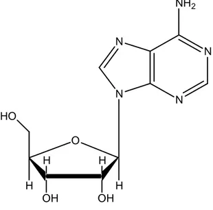
The endogenous nucleoside adenosine is composed of a molecule of adenine attached to a ribose sugar molecule (ribofuranose) via a β-N9-glycosidic bond (Figure 1).
N N N N NH2 O OH OH H H H H HO
Figure 1 – Chemical structure of adenosine
Adenosine plays a central role as a structural element of nucleic acids and in the energy metabolism of all living organism. The physiological effects of adenosine were first described in the cardiovascular system and gastrointestinal tract [1]. Adenosine, acting on specific membrane receptors, produces a number of physiological and pathophysiological effects in the central nervous system (CNS) [2, 3], in the cardiovascular system [4], as an endogenous pain modular [5], in the immune system [6, 7], cell growth and apoptosis [8]. Under normal conditions, adenosine is continuously formed intracellularly as well as extracellularly. The intracellular production is mediated either by an intracellular 5'-nucleotidase, which dephosphorylates or by hydrolysis of S-adenosyl-homocysteine [9]. Adenosine generated intracellularly is transported into the extracellular space mainly via specific bi-directional transporters through facilitated diffusion that efficiently evens out the intra- and extracellular levels of adenosine. The dephosphorylation of extracellular AMP to adenosine, mediated by ecto-5'-nucleotidase, is the last step in the enzymatic chain that catalyzes the breakdown of extracellular adenine nucleotides, such as ATP, to adenosine. Ectonucleotidases include ectonucleoside triphosphate diphosphohydrolase which can
6 hydrolyze ATP or ADP, ectonucleotide pyrophosphatase/phosphodiesterases, alkaline phosphatases and 5'-nucleotidases [10]. When adenosine levels in the extracellular space are high, adenosine is transported into cells by means of transporters. It is then phosphorylated to AMP by adenosine kinase or degraded to inosine by adenosine deaminase. Adenosine deaminase, but not adenosine kinase, is also present in the extracellular space [9]. Another potential source of extracellular adenosine is cAMP, which can be released from neurons and converted by extracellular phosphodiesterases into AMP and thereafter by an ecto-5'-nucleotidase to adenosine. The transport of adenosine by facilitated diffusion is equilibrative and bidirectional, meaning that the net transport of adenosine either into or out of the cell depends upon the adenosine concentration gradient in both sides of the membrane. Inhibition of adenosine transport can, therefore, inhibit either adenosine release or adenosine uptake, depending upon the intra- and extracellular levels of adenosine [11]. However, since the extracellular formation of adenosine from released adenine nucleotides constitutes a second source of adenosine, which is not affected by transport inhibition, the transport inhibitors usually cause an increase in the extracellular adenosine levels. Under hypoxic and ischemic conditions there is a marked increase in cytoplasmatic adenosine leading to an intense release of adenosine, which is inhibited by adenosine uptake inhibitors [12].
Excitatory aminoacid-mediated release of adenosine is certainly involved; however, of greater importance is probably the fact that whenever intracellular levels of adenine nucleotides fall as a result of excessive energy use, the intracellular levels of adenosine will rise dramatically [9]. For example, following hypoxia there is a decrease of intracellular ATP, accompanied by an accumulation of 5'-AMP and subsequently adenosine. The nucleoside is thereafter transported into the extracellular space via the transporters. Furthermore, when the intracellular level of adenosine is very high, adenosine simply diffuses out of cells. Direct release of intracellular adenine nucleotides, such as ATP, that is thereafter converted extracellularly by ecto-ATPase and ecto-ATP-diphosphohydrolase (ecto-apyrase) to AMP and dephosphorylated by ecto-5'-nucleotidase to adenosine, should also be considered [12]. Adenosine is neither stored nor released as a classical neurotransmitter since it does not accumulate in synaptic vesicles, being released from the cytoplasm into the extracellular space through a nucleoside transporter. The adenosine transporters also mediate adenosine reuptake, the direction of the transport being dependent upon the concentration gradient at both sides of the membrane [9].
7
G protein-coupled receptors
Considering the overall protein structure, adenosine receptors (ARs) display the topology typical of an important class of transmembrane proteins is the superfamily of G protein-coupled receptors (GPCRs) which are known as seven transmembrane (7TM) or heptahelical receptors. Sequence comparison between the different GPCRs revealed the existence of different receptor families sharing no sequence similarity even if specific fingerprints exist in all GPCR classes. They constitute a prominent superfamily targeted by many drugs. Up to 50% of all modern-day medicines act on GPCRs [13]. This makes GPCRs of great interest to both pharmaceutical and academic research, which is focused on drug discovery and the function and malfunction of various human systems. GPCRs play a vital role in signal transduction and may be activated by a wide variety of ligands, including photons, amines, hormones, neurotransmitters and proteins. GPCRs are single polypeptide chains having seven hydrophobic transmembrane-spanning segments that couple in the presence of an activator to an intracellular effectors molecule through a trimeric G protein complex [14]. The latter protein name originates from its interaction with guanine nucleotides. The class of guanine nucleotide binding proteins (G proteins) initiate some of the important signalling pathways in the cell. The members of the GPCR superfamily share two major structural and functional similarities. The first principal feature are the setup by seven membrane-spanning α-helices (TM1-7) connected by alternating intracellular (IL1, IL2, and IL3) and extracellular loop domains (EL1, EL2, and EL3). The orientation of the N and C terminus is also conserved across all GPCRs. The N-terminal tail is exposed to the extracellular environment and the C-terminal tail is located in the cytosol of the cell and thought to maintain an interaction with cytosolic G proteins. The family A of receptors, which comprises the well characterized rhodopsin/β2-adrenergic receptors, contains 90% of all GPCRs and is by far the largest and the most studied [15]. The high degree of conservation among these key residues suggests that they have an essential role for either the structural or functional integrity of the receptors. Only for class A, crystal structures of four GPCRs are known providing detailed molecular information on these receptors. G proteins transmit extracellular signals from GPCRs to downstream effectors proteins, which then cause further rapid changes in intracellular responses through signalling molecules such as cAMP, cGMP, inositol phosphates, diacylglycerol, arachidonic acid and cytosolic ions [16].
8
Adenosine receptors
The ARs are members of the superfamily of GPCRs belonging to the subfamily of rhodopsin-like receptors and thus, show the typical heptahelical structure. The adenosine receptor subtypes in a tissue or isolated cells are characterized by their G protein coupling preference. Biological functions of extracellular adenosine are mediated by four different adenosine receptor subtypes; including the A1 and A3AR subtypes, which couple to a Gi
protein that inhibit the intracellular adenylate cyclase (AC) and thus leading to a decrease of cAMP, and the A2A and A2BAR, which couple to a Gs protein that stimulate cAMP
production in brain slices at low (0.1-1 µM) and high (≥ 10 µM) adenosine concentration, respectively [17]. The four ARs have been cloned from several mammalian species, including human. There is extensive sequence similarity between species for the A1, A2A
and A2BARs , whereas A3ARs are more variable. Each ARs has different but overlapping
functions. Each of them is unique in pharmacological profile, tissue distribution, binding partners and on coupling to other second messenger systems, activation of K+ channels (A1), or phospholipase C (all subtypes) has been described [9]. Generally, the A2BARs
requires higher concentration of adenosine than other subtypes to be significantly activated. In particular, all of the ARs subtypes can also be characterized according to the potency of the natural agonist adenosine: the A1 and A2A subtypes are high-affinity receptors activated
by adenosine in nanomolar concentrations, while the A2B and A3ARs are low-affinity
subtypes that require high micromolar concentrations for activation. Based on the extensive roles of the ARs in both physiologic and pathophysiologic events, these subtypes are becoming important drug targets in the treatment of several diseases because they have a role in controlling physiological processes [18].
A1 adenosine receptors
The adenosine A1ARs are widely expressed throughout the body, having its highest
expression in the brain, spinal cord, atria and adipose tissue [19]. Adenosine, via A1ARs,
reduces heart rate, glomerular filtration rate, and renin release in the kidney, induces bronchoconstriction and inhibits lipolysis [20]. A1ARs can be coupled to different pertussis
toxin-sensitive G proteins, which mediate inhibition of adenylate cyclase and regulate calcium and potassium channels, as well as inositol phosphate metabolism [9]. A1ARs and
9 A2AARs are primarily responsible for the central effects of adenosine [21]. In addition to
their postsynaptic locations in different brain regions, A1ARs can be found pre-synaptically
and modulate neurotransmitter release. Pre-synaptic A1ARs are the prototype of GPCRs,
the stimulation of which decreases the probability of neurotransmitter release. The main mechanism of A1AR-mediated inhibition of exocytosis is a direct inhibitory effect on
voltage-dependent Ca2+ channels [22]. A1AR displays two different affinities for agonist,
which have classically been attributed to a different coupling to heterotrimeric G proteins. According to this two independent site model, coupled receptor–G protein complexes display high affinity for agonists and uncoupled receptors display low affinity. The reported cluster-arranged cooperative model predicts that the high- and low-affinity sites are a consequence of the negative cooperativity of agonist binding and do not seem to be related to the content of G protein-coupled or –uncoupled receptors [9]. Like other GPCR members, A1AR expression is regulated in response to agonist or antagonist stimulation.
Desensitization of A1ARs has been described in intact animals and in cell cultures.
Prolonged administration of A1AR agonists to animals leads to functional desensitization of
A1ARs in guinea pig heart, rat adipocytes, rat atrial muscle, and rat brain [23]. The reduced
functional response is attributable to a net loss of A1ARs or down-regulation, a decrease in
the proportion of A1ARs displaying the high-affinity state for agonists, and a decrease in
the content of Gi proteins. The loss of binding sites on the cell membrane owing to internalization of A1ARs is a slower event. Ser/Thr phosphorylation seems to be related to
short-term clustering and desensitization, as well as long-term internalization of A1ARs
[24].
A2 A adenosine receptors
The A2AARs exists in a wide variety of organs including major peripheral tissues (liver,
heart, lung, and the immune system) and the CNS [25]. In the developing rat brain, the expression of A2AAR is transiently regulated in various areas (e.g., the striatum, cortex, and
hippocampus), perhaps implying a role of adenosine in neuronal development. Soon after neurogenesis, the A2AAR is highly expressed by striatal neurons and co-localizes with the
D2 dopamine receptor in GABAergic striatopallidal neurons [26]. In addition to the intense expression in the striatum, low levels of A2AARs are found in many brain regions (e.g.,
cortex and hippocampus) and it has been suggested that adenosine via A2AAR regulates
10 The regulation of A2AAR gene expression is therefore likely to play an important role in
neuronal development, basal ganglia activity, and many other peripheral functions. In the CNS, l-DOPA enhanced the gene expression of the striatal A2AAR in 6-OHDA-lesioned
rats [27]. Treatment with an antagonist of the NMDA receptor (memantine) was also reported to elevate the transcript level of striatal A2AARs [28]. Like other GPCRs it can also
interact with other G proteins if the receptor is very over-expressed, but the evidence for such coupling in vivo is not compelling. In striatum the A2AARs interacts with Golf
proteins [29]. Most effects are probably due to activation of adenylyl cyclase and production of cAMP. The A2AAR can recruit β-arrestin via a GRK-2 dependent mechanism,
influenced by activation of cytokine receptors, which cause reduced desensitization of the A2AAR [30]. The cAMP responsive element-binding protein (CREB) is critical for many
forms of neuronal plasticity as well as other neuronal functions, phosphorylation of CREB at Ser133 by protein kinase A (PKA) activates CREB and turns on genes with cAMP responsive elements (CRE sites) in their promoters. One important feature of CREB is that it is a point of convergence for the cAMP/PKA and MAPK pathways [31]. The stimulation of A2AARs counteracts the inhibition of neurite outgrowth due to MAPK blockade. The
A2AAR activation alone also stimulates the Ras/Raf-1/MEK/ERK signalling through
PKA-dependent and PKA-inPKA-dependent pathways via Src- and Sos- mediated mechanisms, respectively [32]. Interestingly, phosphorylation/activation of CREB has been shown to compete with nuclear factor-κB (NF-κB) p65 for an important co-factor, CBP. Phosphorylated CREB was therefore proposed to mediate the anti-inflammatory effect of A2AARs and inhibition of NF-κB by A2AAR activation during in vivo acute inflammation
[2]. An interesting observation is that A2AARs activation facilitates adenosine transporters
via a protein kinase C (PKC)-dependent pathway in the hippocampus, and reduces the level of extracellular adenosine available for A1AR activation. In addition, PKC was shown to
play a key role in mediating the enhancement of noradrenaline release by A2AARs in rat tail
artery [33]. Activation of multiple signalling pathways by A2AARs appears to contribute to
their complex functions in various tissues.
A2 B adenosine receptors
A2BAR mRNA was originally detected in a limited number of rat tissues by Northern blot
analysis, with the highest levels found in cecum, bowel, and bladder, followed by brain, spinal cord, lung, epididymis, vas deferens, and pituitary. The use of more sensitive reverse
11 transcriptase-polymerase chain reaction techniques revealed a ubiquitous distribution of A2BARs [34]. mRNA encoding A2BAR was detected at various levels in different rat tissues
studied, with the highest levels in the proximal colon and lowest in the liver. In situ hybridization of A2BARs showed widespread and uniform distribution of A2BAR mRNA
throughout the brain [35]. In brain, functional A2BARs are found in neurons, glial cells, in
astrocytes and in different glioma cell lines [36]. The expression of A2BARs in glial cells,
which represent a majority of the brain cell population, can explain the original observation that slices from all brain areas examined showed an adenosine-stimulated cAMP response. Functional A2BARs have been found in fibroblasts and various vascular beds, hematopoietic
cells, mast cells, myocardial cells, intestinal epithelial and muscle cells, retinal pigment epithelium, endothelium, and neurosecretory. The activation of A2BARs can also increase
phospholipase C in human mast cells and in mouse bone marrow-derived mast cells. In addition, A2BAR activation elevates inositol triphosphate levels, indicating this receptor can
couple also to Gq-proteins. A2BARs have been implicated in the regulation of mast cell
secretion, gene expression, intestinal function, neurosecretion, vascular tone and in particular asthma [37].
A3 adenosine receptors
The A3AR is the only adenosine subtype which was cloned before its pharmacological
identification. It was originally isolated as an orphan receptor from rat testis, having 40% sequence homology with canine A1 and A2AARs and was identical with the A3AR later
cloned from rat striatum [38]. Homologs of the rat striatal A3ARs have been cloned from
sheep and human, revealing large interspecies differences in A3AR structure. For example,
the rat A3AR presents only 74% sequence homology with sheep and human A3AR, while
there is 85% homology between sheep and human A3AR. The A3AR has been mapped on
human chromosome 1p21-p13 [39] and consists of 318 aminoacid residues. The A3AR is a
G-protein-coupled receptor characterized by its C-terminal portion facing the intracellular compartment and 7 TM spanning domains. In contrast to other adenosine receptors, the C-terminal region presents multiple serine and threonine residues, which may serve as potential sites of phosphorylation that are important for rapid receptor desensitization upon agonist application [40]. Phosphorylation leads to a decrease of the number of receptors in the high-affinity state and a decrease of agonist potency to inhibit adenylyl cyclase activity. At the same time, the receptor is reversibly internalized in an agonist-dependent fashion
12 [41]. The A3AR has widely distributed its mRNA being expressed in testis, lung, kidneys,
placenta, heart, brain, spleen, liver, uterus, bladder, jejunum, proximal colon and eye of rat, sheep and humans. The classical pathways associated with A3AR activation are the
inhibition of adenylyl cyclase activity, through the coupling with Gi proteins, and the stimulation of phospholipase C, inositol triphosphate and intracellular Ca2+, via Gq proteins [9].
Adenosine receptors in the respiratory system
Increased adenosine levels have been found in the lungs of individuals with asthma or COPD, and ARs are known to be expressed on most, if not all, inflammatory and stromal cell types involved in the pathogenesis of these diseases [42]. A1ARs are responsible for
many effects induced by adenosine. In particular, this signalling nucleoside has been implicated in the regulation of asthma and chronic obstructive pulmonary disease (COPD); adenosine levels are elevated in the asthmatic lungs to an extent that can be directly correlated with the degree of inflammatory insult [43]. A1AR expression is also increased in
the epithelium and airway smooth muscle of human asthmatics. The involvement of A1ARs
in asthma was provided by studies on allergic rabbit models, where the adenosine-induced acute bronchoconstrictor response was attenuated by pretreatment with A1AR antagonists.
In particular, activation of A1ARs in human airway epithelial cells causes an increase in the
expression of the MUC2 gene, which is responsible for mucus hyper secretion. Moreover, activation of A1ARs is known to produce pro-inflammatory effects on various types of
human cells. The non-selective AR antagonists theophylline and doxofylline have been launched as bronchodilators for the treatment of various respiratory disorders [44]. Likewise, it has been recently reported that A1AR inhibits transendothelial and
transepithelial polymorphonuclear cell migration in a murine model of lipopolysaccharide (LPS)-induced lung injury, presumably by reducing the release of chemotactic cytokines into the alveolar space. In addition, A1AR is involved in decreasing microvascular
permeability and leukocyte transmigration in endothelial cells, suggesting also a protective and anti-inflammatory role for A1AR [45].
Pharmacological treatment of allergic rats with an A2A agonist has been shown to result in
diminished pulmonary inflammation. Moreover, a recent study in an ADA-deficient model has demonstrated that genetic removal of A2A leads to enhanced pulmonary inflammation,
13 invariant natural killer T cells and natural killer cells can reduce pulmonary inflammation in mice with sickle-cell anemia, improving baseline pulmonary function and preventing hypoxia-reoxygenation-induced exacerbation of pulmonary injury [47]. These findings further confirm the involvement of A2AARs in the anti-inflammatory networks of the lung.
Recently, A2BARs have been implicated in the mediation of several pro-inflammatory
effects of adenosine in inflammatory cells of the lung. A2BARs have been reported to
mediate degranulation and activation of canine mastocytoma and human mast cells line, thereby potentially playing a role in allergic and inflammatory disorders [48]. Adenosine constricts the airways of asthmatic patients through the release of histamine and leukotrienes from sensitized mast cells; although the receptor involved seems to be the A3ARs in rats and the A2BARs in humans. Accordingly, A2BARs antagonists potently
inhibit the activation and degranulation of HMCs induced by adenosine. In addition to mast cells, functional A2BARs have been found in bronchial smooth muscle cells and lung
fibroblasts. In these cells, adenosine, through stimulation of the A2B subtype, increases the
release of various inflammatory cytokines, lending weight to evidence that A2BARs play a
key role in the inflammatory response associated with asthma [49]. Furthermore, it has been reported that, through A2BARs activation, adenosine-differentiated dendritic cells express
high levels of angiogenic, pro-inflammatory, immune suppressor and tolerogenic factors, including vascular endothelium grow factor (VEGF), interleukin 8 8), interleukin 6 (IL-6), interleukin 10 (IL-10) and ciclooxigenase 2 [50]. Moreover, using ADA knockout animals, it has been shown that dendritic cells with a pro-angiogenic phenotype are highly abundant in vivo under conditions associated with elevated levels of extracellular adenosine. The first evidence for the involvement of A2BARs in asthma was provided by
studies concerning the selectivity of enprofylline, a methylxanthine structurally related to theophylline [50], and further support came from research demonstrating the presence of A2BARs on various type of cells involved in cytokine release in asthmatic disease, such as
smooth muscle cells, lung fibroblasts, endothelial cells, bronchial epithelium and mast cells. Expression of A2BARs has also been found in the mast cells and macrophages of patients
affected by COPD. In another study, activation of A2BARs in the HMC-1 mast cell line
provoked an increase in IL-8 release in vitro [51].
Particular relevance to the presence of A3ARs in the respiratory system has been reported.
A3ARs are expressed in human neutrophils where, together with A2A, they are involved in
the reduction of superoxide anion generation; they have also been implicated in the suppression of tumor necrosis factor α (TNF-α) release induced by endotoxin from human
14 monocytes [52]. In neutrophils, however, A3ARs also play a role in chemotaxis, in
conjunction with P2Y receptors. Moreover, A3ARs activation seems to inhibit
degranulation and superoxide anion production in human eosinophils [53]. Indeed, transcript levels for the A3 subtype are elevated in the lungs of asthma and COPD patients,
where expression is localized to eosinophilic infiltrates. Similar evidence has also been observed in the lungs of ADA knockout mice exhibiting adenosine-mediated lung disease [54]. Treatment of ADA knockout mice with 3-propyl-6-ethyl-5-[(ethylthio)carbonyl]-2phenyl-4-propyl-3-pyridine carboxylate (MRS 1523), a selective A3 antagonist, prevented
airway eosinophilia and mucus production. Nevertheless, these findings contrast sharply with the results of experiments performed in human eosinophils ex vivo, where chemotaxis was reduced by A3AR activation, suggesting that significant differences exist between the
impact of A3ARs signalling on eosinophil migration ex vivo and in the whole animal. More
recently, the involvement of the A3AR in a bleomycin model of pulmonary inflammation
and fibrosis has been explored. Results demonstrated that A3AR knockout mice exhibit
enhanced pulmonary inflammation that involves an increase in eosinophils. Accordingly, a selective upregulation of eosinophil-related chemokines and cytokines was found in the lungs of A3AR knockout mice exposed to bleomycin, thereby suggesting that the A3AR
performs anti-inflammatory functions in the bleomycin model [53].
Nonetheless, the role of the A3ARs in the human lung, and indeed in asthma, still remains
to be clarified. In general, receptor knockouts have provided significant new insights into adenosine’s control of complex physiological (i.e., cognition) and pathological (i.e., neuroinflammation) phenomena, suggesting that further studies in these animal models would help to clarify the role of A3ARs in inflammatory lung disease [55].
Adenosine receptors in cancer
Adenosine, which is released to the microenvironment by metabolically active tumor cells, fulfils a multitude of functions in regulating tumor cell proliferation [56]. At micromolar (µM) concentrations it directly induces an anti-proliferative effect toward various tumor cell types. Indirectly, it affects tumor development via its capability to affect cytokine release, cell migration, angiogenesis, and chemotaxis [57]. Moreover, adenosine induces activation or suppression of T killer or natural killer cells that affect tumor cell development [58]. It is quite impossible to assess the effect of adenosine in vivo due to the rapid metabolization by ADA. The effect of adenosine and of the agonists/antagonists on tumor cells depends on
15 their extracellular concentrations and on the expression of different adenosine receptor subtypes. Upon receptor activation, various signal transduction pathways are generated, resulting in a direct inhibitory effect on tumor growth [59]. Other cell types, such as immunocytes or endothelial cells, may respond to receptor activation by the release of cytokines and mediators that indirectly affect tumor growth [60]. Interestingly, among the four receptor subtypes, the A3AR was found to mediate a potent antitumor effect [61]. The
specificity of this target results from the finding that this receptor is highly expressed in tumor cells, whereas low receptor expression is reported in normal cells [59]. A3ARs are
involved in the tumor growth, in the regulation of cell cycle and mediate both pro and anti-apoptotic effects closely associated with the level of receptor activation [62, 63]. A3ARs
involve the inhibition of telomerase activity and arrest the G0/Gi phase of the cell cycle leading to a cytostatic effect in Nb2-11C lymphoma cells [64]. The A3ARs reduce the
ability of prostate cancer cells to migrate in vitro and metastasize in vivo. In particular it has been reported that activation of the A3ARs in prostate cancer cells reduced
PKA-mediated stimulation of ERK1/2 leading to lower NADPH oxidase activity and cancer cell invasiveness [65]. A3AR density was upregulated in colon carcinoma tissues closely
correlated to the disease severity. In addition the alteration of A3ARs reflected a similar
behavior shown in lymphocytes or neutrophils derived from colon cancer patients suggesting that these receptors may represent an interesting biological marker [66]. Recently, it has been reported that A3AR selective agonists induce anti-inflammatory and
anti-cancer effect in xenograft animal model utilizing Hep-3B hepatocellular carcinoma cells [61]. In this model, the A3AR upregulation was present in inflammatory liver tissues
similarly to those previously found in other inflammatory conditions [68].
The A3AR agonist 2-chloro-N6-(3-iodobenzyl)adenosine-5′-N-methyl-uronamide (CF102)
inhibited tumor growth via de-regulation of the NF-kB signal transduction pathways, resulting in apoptosis of tumor cells [69]. Pharmacological studies demonstrated that A3AR
agonists inhibited the growth of melanoma cells while promoting the proliferation of bone marrow cells, reduced cell viability in human breast cancer cells and induced arrest of cell cycle progression in human lung cancer cells [70-72]. 2-chloro-N6 -(3-iodobenzyl)adenosine-5′-N-methyl-carboxamide (Cl-IB-MECA) enhanced apoptosis via the modulation of NF-kB signalling pathway in thyroid cancer cells and reduced the ability of prostate cancer cells to migrate in vitro and metastasize in vivo [67]. Moreover, preclinical and Phase I studies showed that A3AR agonists are safe and well tolerated in humans and
16
17 The environmental risk factor such noxious particles exposure, in combination with a genetic predisposition, results in two causes of morbidity and mortality worldwide namely chronic obstructive pulmonary disease (COPD) and malignant pleural mesothelioma (MPM). Oxidative stress provided by inhalation of exogenous particles, smokes of sigarette for COPD and asbestos for MPM, may lead to the activation of many intracellular pathways including kinases, transcription factors and epigenetic events that modulate the inflammatory response and cell cycling/proliferation [74]. COPD is the fourth leading cause of mortality worldwide [75]. COPD is a disease state characterized by airflow limitation that is not fully reversible. The airflow limitation is usually both progressive and associated with an abnormal inflammatory response of the lung to noxious particles or gases. The main cause of COPD is cigarette smoking [76]. Adenosine has been suggested to play a role in the pathogenesis of COPD [77]. The exact role of adenosine in the pathogenesis of COPD is unknown and probably complex because adenosine receptors in the lungs, in vitro and in animal models, have both pro- and anti-inflammatory effects and may also cause bronchoconstriction [78].
On this background, the aim of this study is to describe a detailed analysis, of A1, A2A, A2B
and A3ARs expression in peripheral lung parenchyma, the major site of airflow obstruction
in COPD using immunohistochemistry, radioligand binding and real time quantitative polymerase chain reaction (RT-QPCR). ARs were analyzed in age-matched smokers with normal lung function (control group) and COPD patients. Moreover, we have investigated whether changes in affinity and density of these receptors are correlated with clinical parameters such as forced expiratory volume in one second (FEV1)/forced vital capacity
(FVC) ratio. We have also investigated, using the in vitro model of human lung type 2 alveolar-like cells (A549 cells), the effect of pro-inflammatory cytokines on adenosine receptor subsets. COPD is characterized by an increased oxidative and nitrosative stress correlates with decreases in lung function and disease severity [79]. In addition, there is clear evidence for persistent inflammation in the lungs and airways of COPD patients which increases with disease severity. In COPD patients the inflammation and oxidative stress persist many years after smoking cessation [80]. Alveolar macrophages are considered to have a central role in the pathogenesis of COPD whereas the pathogenetic role of mast cells in COPD peripheral lung is more controversial [81]. For this reason the A2BAR expression in bronchoalveolar lavage (BAL) macrophages from COPD smokers and
age-matched with normal lung function was investigated. The effect of oxidative and nitrosative stress and of pro-inflammatory cytokines (TNF-α and IL-1β) on A1, A2A, A2B
18 and A3ARs expression and on their functionality in human leukaemic monocyte lymphoma
cell line (U937), before and after phorbol 12-myristate 13-acetate (PMA)-treatment, and in the human mast cell line (HMC-1) was evaluated.
Considerable evidence have highlighted that adenosine through the interaction with its receptors plays an important role in controlling tumorigenesis via its receptors. MPM is a highly aggressive neoplasm whose incidence is increasing due to asbestos exposure [82]. The capacity of asbestos to induce MPM has been linked to the release of TNF-α that promotes mesothelial cell survival via NF-kB pathway [82]. To date, there is no standard curative therapy for MPM that is largely unresponsive to conventional chemotherapy or radiotherapy [83]. As a consequence, more effective therapeutic strategies are needed for this fatal disease with the aim to identify new candidates for targeted therapies. A3ARs have
been involved in the regulation of the cell cycle and both pro- and anti-apoptotic effects was closely associated with the level of receptor activation [84]. A3ARs play a role in the
modulation of mitogen-activated protein kinase (MAPK) activity and in the regulation of extracellular signal-regulated kinases (ERK1/2). It has been accepted that A3ARs are highly
expressed in tumor cells showing an important role in the development of cancer [85]. The serine/threonine kinase Akt/PKB has a central role in cell signalling downstream of growth factors, cytokines, and other cellular stimuli. Aberrant loss or gain of Akt/PKB activation underlies the pathophysiological properties of a variety of complex diseases, including cancer [86, 87]. Therefore, the purpose of this thesis, is to describe the alteration of A3ARs
in human MPM in comparison with healthy mesothelial pleura (HMP). We also studied the alteration of A3ARs in healthy mesothelial cells (HMC) treated with crocidolite asbestos
and in malignant mesothelioma cells (MMC) respect to untreated HMC. We investigated the reduction of Akt phosphorylation and NF-kB activation in tumor cells, mediating by A3ARs. Furthermore, A3AR stimulation on proliferation and apoptosis in MMC and in
19
ADENOSINE RECEPTORS IN
CHRONIC OBSTRUCTIVE PULMONARY
DISEASE PATIENTS
20
Chronic obstructive pulmonary disease
COPD is a common inflammatory lung disease and a major cause of illness and death throughout the world. The Global Initiative for Chronic Obstructive Lung Disease defines COPD as “a pulmonary disease characterized by airflow limitation that is not fully reversible. The airflow limitation is usually progressive and associated with an abnormal inflammatory response of the lung to noxious particles or gases” [88]. The abnormal inflammatory response is a pathology of COPD which it shares with asthma. In COPD this inflammatory process leads to a co-presence of small airway diseases (obstructive bronchiolitis) with fibrosis and obstruction, parenchymal lung tissue destruction (emphysema), loss of lung elasticity and closure of small airways and chronic bronchitis, characterized by cough and mucous hypersecretion. As a consequence, the airways undergo structural changes with further loss of lung elasticity and airflow limitation. These pathological mechanisms manifest at different degrees in COPD patients [89]. Despite its high global prevalence, there is still a fundamental lack of knowledge about the cellular, molecular and genetic causes of COPD and no available therapy which may reduce the disease progression or mortality [90].
COPD affects > 10% of the world population over the age of 40 years and every year almost 3 million people die of this disease [91]. According the World Health Organization WHO, in 2007 COPD was the 4th cause of death worldwide and it is predicted to become the 3rd leading cause of death by 2030 [91]. COPD is the 13th cause of morbidity today and will become the 5th cause of morbidity by 2020 [92]. Therefore, the economic and social burden of the disease is immense today and will dramatically increase in the future, also considering that at present the disease is under-estimated, it is insufficiently recognized and is poorly diagnosed [92]. For an individual, of course, the disease may dramatically lower the quality of life. Paradoxically, despite its global impact and compared to asthma, research in COPD is less progressing and highly under-funded.
COPD is a complex, multi-factorial pathology and both environmental and host-depended factors are needed for the clinical manifestation of the disease. However, cigarette smoking is undoubtedly the major causative environmental risk factor for COPD. It accounts for approximately 90% of all cases and a dose-dependent relationship between tobacco consumption and the development and severity of COPD has been observed [93].
21 Important is the age at which a person started smoking, the numbers of packages of cigarettes smoked per year, and the current smoking status. Passive exposure to cigarette smoke is another risk factor that is increasing the total amount of inhaled particles into the lung. However, only 10-20% of the smokers develop clinical symptoms of COPD and susceptibility and other environmental factors are therefore crucial for the pathology of COPD [94]. Additional environmental risk factors are occupational dust and chemical exposure, indoor and outdoor air pollution, bacterial and viral infections, the socioeconomic status, and asthma [95]. Although there is no conclusive evidence, adults with asthma are found to have a twelvefold higher risk of acquiring COPD than subjects without asthma [96]. Like many other diseases, COPD is a polygenic disease and gene-environment interactions are critical for the development of this disorder. So far, the best investigated genetic cause for the development of COPD is the hereditary deficiency of the alpha-1 antitrypsin, an inhibitor of serine proteases. The lack of this protein is leading to the development of emphysema and decline in lung function due to digestion of the lung forming extracellular matrix and cell-cell interactions [97].
Pathological changes in COPD include chronic inflammatory processes and airway remodeling, both localized in the proximal and peripheral airways, in the lung parenchyma and in pulmonary vasculature [98]. The chronic inflammation of COPD is characterized by an accumulation of neutrophils, macrophages, B cells, lymphoid aggregates and CD4+ and CD8+ T cells particularly in the small airways [99] and the degree of inflammation increases with the disease severity as classified by the Global Initiative for Chronic Obstructive Lung Disease (GOLD) [100]. Neutrophils and activated macrophages release oxygen radicals, elastase, and cytokines that are essential to the pathogenesis of COPD, with effects on goblet cells and submucosal glands, and on the induction of emphysema and inflammation. Monocytes/ macrophages are important effector cells in COPD due to the release of reactive oxygen species, extracellular matrix proteins, lipid mediators, cytokines, chemokines and matrix metalloproteinases and their numbers increase with increasing severity [101-103].
Cigarette smoking is by far the most prominent cause for COPD. The inflammatory processes and the airway remodeling increase with disease severity and persist after cessation of smoking. Therefore, it is assumed that after a certain threshold of disease severity is passed, simply quitting smoking may not be sufficient to prevent disease progression. In recent years, investigations on COPD led to a major expansion of paradigms explaining the pathobiology of the disease and many different models are proposed today.
22 For example, COPD can be seen as a disease of accelerated lung aging, since many cellular pathological processes are due to the accumulation of reactive oxygen species [104, 105]. There is also a group of scientist that define COPD as an auto-immune disease which response to antigens released after smoke induced tissue or cell injuries [106]. Although other mechanisms that may be involved in COPD have been investigated and potential targets for a therapeutic approach proposed, an efficient cure for this disease is still not available today.
In COPD the predominant inflammatory cells are neutrophils, macrophages, and CD8 positive T-cells. Especially, macrophages seem to play a pivotal role in COPD [103]. COPD patients with emphysema show a 25-fold increase in the numbers of macrophages in the tissue and in the alveolar space when compared to normal smokers and there is a correlation between macrophage numbers in the airways and the severity of COPD. The key inflammatory mediators in COPD are IL-8, LTB4 and TNF-α [107] and the inflammation is localized in the peripheral airways, the lung parenchyma and in addition the pulmonary vessels are affected, the bronchioles are obstructed and present with fibrosis. In COPD, the typical inflammatory cascade is triggered by noxious air-borne particles, mainly by oxidants derived from cigarette smoke, that activate macrophages to release IL-8, TNF-α and matrix metalloproteinase. The release of these factors is promoted by the inactivation of histone deacetylase (HDAC) leading to the transcription of NF-кB inducible cytokines [108]. Oxidative stress is defined as an excess of reactive oxygen species (ROS) that cannot be neutralized by the antioxidant defense mechanisms and thus leading to cell damage. There is evidence that oxidative stress plays a major role in the pathogenesis of COPD [109]. On one side, inflammatory cells are able to generate ROS, but also cigarette smoke itself contains ROS at high concentrations. ROS activate NF-кB, which induces the transcription of multiple inflammatory genes leading to an inflammatory response in the lung [104, 110]. Interestingly, oxidative stress in COPD may be also linked to the poor response to corticosteroids in COPD patients. Oxidative stress impairs the translocation of the glucocorticoid receptors to the nucleus and the binding to its corresponding target DNA sequence [111, 112].
Adenosine receptors in COPD
Adenosine has been suggested to play a role in COPD [113]. Patients with COPD are significantly more responsive to AMP than healthy smoking volunteers and smokers have
23 significantly increased concentrations of adenosine in the airway lining fluid [114]. Adenosine is released during tissue hypoxia and inflammation, and all inflammatory cells express ARs [115]. Indeed, adenosine-based approaches are currently being developed for the treatment of various disorders where inflammatory modulation is a key component [116]. It has both pro- and anti-inflammatory features, which are mediated by ARs [117]. Activation of ARs can have different effects, with respect to the cell types involved, activation of A1AR on neutrophils promotes adherence to endothelial cells and chemotaxis,
indicating a pro-inflammatory response [117], whereas activation of A1AR on cells from
the monocytes/macrophage lineage inhibits the production of several pro-inflammatory cytokines (TNF-α, IL-8, and IL-6) and enhances the release of the anti-inflammatory cytokine IL-10, displaying an anti-inflammatory response [118]. Therefore, each receptor can be either beneficially and/or detrimentally implicated in the inflammatory process of COPD [119]. Since activation of ARs can influence the secretion of mediators from inflammatory cells, we related changes in adenosine and its receptors with changes in growth factors such as VEGF and chemokines (monocyte chemoattractant protein 1) related to remodeling and inflammation that may underlie lung function loss in COPD. The effect of adenosine on cytokine production by macrophages has attracted considerable attention, because macrophage-derived cytokines are crucial initiators and orchestrators of immune responses [113]. As TNF-α was one of the first cytokines to be discovered, a substantial body of information has accumulated regarding the ability of ARs activation to limit TNF-α production following macrophage activation [120, 121].
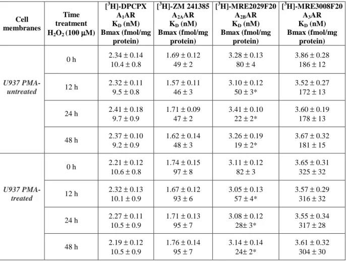
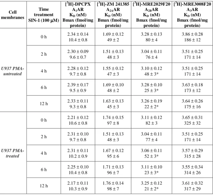
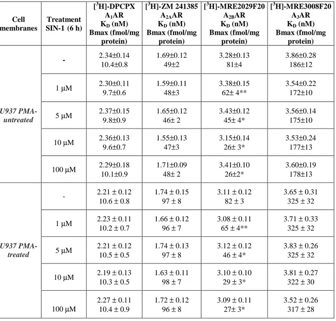
Until recently, the limited specificity of available selective agonists and antagonists has made it difficult to identify the expression of the different ARs. However, in the last few years significant advances in A1 and A2AARs pharmacology have been made through the
use of highly potent and selective agonist and/or antagonist radioligands such as [3 H]-1,3-dipropyl-8-cyclopentyl-xanthine ([3H]-DPCPX) and [3 H]-4-(2-[7-amino-2-(2-furyl)[1,2,4]triazolo[2,3-a][1,3,5]triazin-5-yl-amino]ethyl)phenol ([3H]-ZM 241385), respectively [122, 123]. More recently, the pharmacological characterization of a new, high-affinity, potent and selective radioligand ([3 H]-N-benzo[1,3[dioxol-5-yl-2-[5-(2,6- dioxo-1,3-dipropyl-2,3,6,7-tetrahydro-1H-purin-8-yl)-1-methyl-1H-pyrazol-3-yl-oxy]-acetamide, [3H]-MRE 2029F20) that is able to bind human A2BARs has allowed a better
characterization of this receptor subtypes in different human tissues [124]. In addition, the discovery of the new, high-affinity, and selective radioligand, [3 H]-5N-(4-methoxyphenylcarbamoyl) amino-8-propyl-2-(2-furyl) pyrazolo
[4,3-e]-1,2,4-triazolo[1,5-24 c]pyrimidine ([3H]-MRE 3008F20) that is able to bind the human A3AR with high affinity
has allowed its pharmacological characterization [66, 125].
Combined immunohistochemical and radioligand binding studies could be very useful to clarify the specific effects determined by differential expression of A1, A2A, A2B and
25
Materials and methods Subjects
We recruited thirty-four subjects undergoing lung resection for a solitary peripheral carcinoma, all subjects were recruited from the Section of Respiratory Diseases of the University Hospital of Ferrara, Italy. For the immunohistochemistry study six subjects were smokers with COPD and twelve were smokers with normal lung function (Table 1A). In addition, eight smokers with COPD and eight smokers with normal lung function were selected for the binding and RT-QPCR experiments (Table 1B). Seven smokers with COPD and six smokers with normal lung function were recruited for the western blot assays and immunocytochemistry experiments on the BAL from the Section of Respiratory Diseases of the University Hospital of Ferrara and the Section of Respiratory Diseases of the University Hospital of Katowicach, Poland (Table 1C). All former smokers had stopped smoking for more than one year. COPD and chronic bronchitis were respectively defined, according to international guidelines, as the presence of post-bronchodilator FEV1/FVC ratio <70% or
the presence of cough and sputum production for at least three months in each of two consecutive years. None of the study subjects had suffered a recent exacerbation, defined as increased dyspnea associated with a change in quality and quantity of sputum that would have led them to seek medical attention during the month previous the study. All subjects were free of acute upper respiratory tract infections and none had received glucocorticoids, theophylline, anti-oxidants or antibiotics within the preceding month. They were also nonatopic (i.e. they had negative skin tests for common allergen extracts) and had no past history of asthma or allergic rhinitis. All subjects were free from preoperative chemotherapy and/or radiotherapy. BAL was performed according to the local Ethics Committee Guidelines. Pulmonary function tests were performed as previously described [126] according to published guidelines. Predicted values for the different measures were calculated from the regression equations published by Quanjer [127]. The study was approved by the local Ethic Committee of the University Hospital of Ferrara and informed consent was obtained from each participant in accordance with the principles outlined in the Declaration of Helsinki.
26
Lung tissue processing
Two to four randomly selected tissue blocks were taken from the subpleural parenchyma of the lobe obtained at surgery, avoiding areas invaded by tumor. Tissue specimens were cut for immunohistochemical analysis and were placed on charged slides as previously reported [128]. Another piece of lung parenchyma was also taken and used in radioligand binding and RT-QPCR experiments.
Fiberoptic bronchoscopy, collection and processing of BAL
All subjects attended the bronchoscopy suite at 8.30 am after having fasted from midnight and were pre-treated with atropine (0.6 mg IV) and midazolam (5-10 mg IV). Oxygen (3 l/min) was administered via nasal prongs throughout the procedure and oxygen saturation was monitored with a digital oximeter. Using local anesthesia with lidocaine (4%) to the upper airways and larynx, a fiberoptic bronchoscope (Olympus BF10 Key-Med, UK) was passed through the nasal passages or mouth into the trachea. Further lidocaine (2%) was sprayed into the lower airways. BAL was performed from the right middlelobe using four successive aliquots of 60 ml of 0.9% NaCl. BALcells were spin (500 g, 10 min) and washed twice with Hanks' buffered salt solution (HBSS). Cell Cytospin (Cytospin II; Shandon, UK) slides were prepared with native pellet, and one of them stained with May-Grunwald Giemsa to determine the leukocyte differential cell count. A total of at least 500 cells (excluding epithelial cells) per slide was examined at x1000 magnification. Cell viabilitywas assessed using the trypan blue exclusion method. The remaining cell pellet was frozen at -80°C until its analysis [127].
Immunostaining for A1, A2A, A2B and A3ARs in lung sections
After deparaffinization and rehydration to expose the immunoreactive epitopes of ARs, the sections to be stained, immersed in citrate buffer 5 mM at pH 6.0, were incubated in a microwave oven (model NN S200W; Panasonic, Italy) on high power for 40 min or treated with trypsin. Endogenous peroxidase activity was blocked by incubating slides in 3% hydrogen peroxide (H2O2) in phosphate-buffered saline (PBS) followed by washing in PBS.
Non-specific labeling was blocked by coating with blocking serum (5% normal rabbit or goat serum) for 20 min at room temperature. After washing in PBS the sections were
27 incubated for 1 h at room temperature with goat or rabbit anti-human A1, A2A, A2B and
A3ARs at dilution of 1:100 of a 200 µg/ml solution. The primary antibodies were purchased
from Alpha Diagnostics (rabbit polyclonal anti-human A2BR code A2BR23-A) and Santa
Cruz Biotechnology (goat polyclonal anti-human A1R code sc-7500; goat polyclonal
anti-human A2AR code sc7502; rabbit polyclonal anti-human A3R code sc-13938). For the
negative control slides normal goat or rabbit non-specific immunoglobulins (Santa Cruz Biotechnology, CA) were used at the same protein concentration as the primary antibody. After repeated washing steps with PBS, the sections were subsequently incubated with anti-goat or anti-rabbit biotinylated antibody (Vector ABC Kit, Vector Laboratories, UK) for 30 min at room temperature. After further washing the sections were subsequently incubated with ABC reagent (Vector ABC Kit, Vector Laboratories, UK) for 30 min at room temperature. Slides were then incubated with chromogen-fast diaminobenzidine (DAB), with or without cobalt enhancement, for 1-5 min or 3-amino-9 ethylcarbazole (AEC) as chromogenic substances. After which they were counterstained in haematoxylin and mounted on permanent mounting medium [128].
Immunoperoxidase double staining in lung sections
Some sections were stained for A2A or A2BARs as described above except that sections
were stained for either CD68, tryptase or actin (smooth muscle specific) prior to counterstaining with haematoxylin. Non-specific labeling was again blocked by coating with blocking serum (5% normal horse serum) for 20 min at room temperature. After washing in PBS the sections were incubated for 1 h at room temperature with mouse anti-human CD68 (Dako, UK, 1:50 dilution of a 160 µg/ml solution) or with mouse anti-anti-human tryptase (Dako, UK, 1:150 dilution of a 105 µg/ml solution) or with mouse anti-human actin (smooth muscle specific) (Dako, UK, 1:50 dilution of a 70 µg/ml solution). For the negative control slides, normal mouse non-specific immunoglobulins (Santa Cruz Biotechnology, CA) were used at the same protein concentration as the primary antibody. After repeated washing steps with PBS, the sections were subsequently incubated with anti-mouse biotinylated antibody (Vector Alkaline Phosphatase Kit, Vector Laboratories, UK) for 30 min at room temperature. After further washing the sections were subsequently incubated with ABC reagent (Vector Alkaline Phosphatase Kit, Vector Laboratories, UK) for 30 min at room temperature. Slides were then incubated with chromogen fast red for
10-28 20 min, after which they were counterstained in haematoxylin and mounted on permanent mounting medium [128].
Immunocytochemical staining for A2BAR in BAL cytospins
BAL macrophage cytospins were allowed to warm at room temperature, were fixed in cold acetone at -20°C for 5 min, and air-dried for 10 min. Endogenous peroxidase activity was blocked by incubating sections with methanol containing 3% hydrogen peroxide (H2O2) for
1 h followed by washing in PBS. Immunostaining procedures were performed using the Vector ABC Kit (Vector Laboratories, UK). Non-specific labeling was blocked by coating with blocking serum (5% normal goat serum) for 20 min at room temperature. After washing in PBS the cytospins were incubated for 1 h at room temperature with rabbit anti-human A2BAR at a dilution of 1:100 of a 200 µg/ml solution (Alpha Diagnostics, TX, code
A2BR23-A). For the negative control slides normal rabbit non-specific immunoglobulins (Santa Cruz Biotechnology, CA) were used at the same protein concentration as the primary antibody. After repeated washing steps with PBS, the slides were subsequently incubated with anti-rabbit biotinylated antibody (Vector ABC Kit, Vector Laboratories, UK) for 30 min at room temperature. After further washing the sections were subsequently incubated with ABC reagent for 30 min at room temperature. Slides were then incubated with chromogen-fast diaminobenzidine (DAB) as chromogenic substance after which they were counterstained in haematoxylin and mounted on permanent mounting medium [128].
Saturation binding experiments for A1, A2A, A2B and A3ARs in peripheral lung
parenchyma
Peripheral lung parenchyma, was homogenized and filtered through two layers of gauze using a tris HCl 50 mM buffer pH 7.4. The homogenate was centrifuged at 40000 g for 10 min and the pellet was suspended in the same buffer described above containing 2 UI/ml ADA and incubated for 30 min at 37°C. After the incubation the suspension was centrifuged again at 40000 g for 10 min. The final pellet was suspended and used for radioligand binding assays [122-124]. Saturation binding experiments to A1ARs were
performed according by the method described previously using [3H]-1,3-dipropyl-8-cyclopentyl-xanthine ([3H]-DPCPX, specific activity 120 Ci/mmol; NEN-Perkin Elmer Life and Analytical Sciences, USA) as radioligand [122]. The lung parenchyma membranes
29 (100 µg of protein/assay) with 8 to 10 concentrations of the radioligand [3H]-DPCPX (0.01-30 nM) were incubated for 90 min at 25°C. Non specific binding was determined in the presence of DPCPX 1 µM. Saturation binding experiments to A2AARs were performed
according by the method described previously using [3H]-4-(2-[7-amino-2-(2-furyl)[1,2,4]triazolo[2,3-a] [1,3,5]triazin-5-ylamino] ethyl ([3H]-ZM 241385, specific activity 17 Ci/mmol; Tocris Cookson Ltd, UK) as radioligand [123]. The lung membranes (100 µg of protein/assay) were incubated for 60 min at 4°C with 8 to 10 concentrations of the radioligand [3H]-ZM 241385 (0.01-50 nM). Non specific binding was determined in the presence of ZM 241385 1 µM. Saturation binding experiments to A2BARs were performed
using [3H]-N-benzo[1,3[dioxol-5-yl-2-[5-(2,6-dioxo-1,3-dipropyl-2,3,6,7-tetrahydro-1H-purin-8-yl)-1-methyl-1H-pyrazol-3-yl-oxy]-acetamide ([3H]-MRE 2029F20, specific activity 123 Ci/mmol; Amersham International Chemical Laboratories, UK) as radioligand E5. The membranes (100 µg of protein/assay) with 8 to 10 concentrations of [3H]-MRE 2029F20 in the range 0.01-20 nM were incubated for 4°C at 60 min. Non specific binding was determined in the presence of MRE 2029F20 1 µM. Saturation binding experiments to A3ARs were performed using
[3H]-5N-(4-methoxyphenylcarbamoyl)amino-8-propyl-2-(2-furyl)pyrazolo[4,3-e]-1,2,4-triazolo [1,5-c]pyrimidine ([3H]-MRE 3008F20, specific activity 67 Ci/mmol; Amersham International Chemical Laboratories, UK) as radioligand [122]. The membranes (100 µg of protein/assay) with 8 to 10 concentrations in the range 0.01-50 nM of [3H]-MRE 3008F20 were incubated for 4°C at 150 min. Non specific binding was determined in the presence of MRE 3008F20 1 µM. In saturation binding experiments at the end of the incubation time, bound and free radioactivity were separated by filtering the assay mixture through Whatman GF/B glass fiber filters by use of a Brandel cell harvester. The filter bound radioactivity was counted using a 2500 TR liquid scintillation counter Packard with an efficiency of 58%.
Real time quantitative polymerase chain reaction on peripheral lung parenchyma
Total messenger RNA was extracted by the acid guanidinium thiocyanate phenol method. RT-QPCR was carried out using gene-specific double fluorescent labeled TaqMan MGB probe (minor groove binder) in a ABI Prism 7700 Sequence Detection System (Applied Biosystems, UK) [129]. For the RT-QPCR of mRNA of A1, A2A, A2B and A3AR-the
assays-on demand ™ Gene expressiassays-on Products NM 000674, NM 000675, NM 000676 and NM 000677 were used respectively. As an internal control for loading the human
30 glyceraldehyde-3-phosphate dehydrogenase (GAPDH) kit was used, and the fluorescent-labeled probe was VICTM (Applied Biosystems, UK). The mRNA content of each adenosine receptor was expressed as adenosine receptor mRNA/GAPDH mRNA. Similar results were obtained by using β-actin mRNA as internal control and the probe was fluorescent labeled with VICTM (Applied Biosystems, UK) [130].
Western blotting analysis of ARs expression in BAL macrophages
Whole cell proteins were extracted from human bronchoalveolar cell pellet (more of 95% cells were macrophages). Cells were suspended with mechanical disruption in RIPA lysis buffer with a protease inhibitors cocktail immediately frozen to –70° C and thawed after at least 60 min. Particulate matter was removed by centrifugation at 12000 g for 10 min at 4°C. Protein concentration was measured in the supernatant by the Bradford method according to the manufacturer’s instructions (Bio-Rad Laboratories, UK). An equal volume of Laemmli sample buffer 2X concentrate was added to the final volume of the sample. At least 50 µg/lane of whole-cell proteins were subjected to 10% SDS-polyacrylamide gel electrophoresis, and transferred to nitrocellulose filters (Hybond-ECL, GE Healthcare, UK) by blotting. Filters were blocked for 45 min at room temperature in Tris-buffered saline (TBS), 0.05% Tween 20, 5% non-fat dry milk. The filters were then incubated with rabbit anti-human A2BAR (Alpha Diagnostic, TX, code A2BAR23-A; dilution 1:1000) antibodies
for 1 h at room temperature in TBS, 0.05% Tween 20, 5% non-fat dry milk at dilution of 1:1000. Filters were washed three times in TBS, 0.5% Tween 20 and then incubated for 45 min at room temperature with secondary antibody conjugated to horseradish peroxidase (Dako, UK)in TBS, 0.05% Tween 20, 5% non-fat dry milk, at a dilution of 1:4000. After further three washes in TBS, 0.05% Tween 20, visualization of the immunocomplexes was performed using the ECL as recommended by the manufacturer (GE Healthcare, Chalfont St. Giles, UK). As an internal control we reprobed each filter with an anti-human actin antibody (Santa Cruz Biotechnology, CA). The 43kDa (actin) and 55kDa (A2BAR) bands
intensities were quantified using densitometry with Grab-It and VisionWorks LS software (UVP, Cambridge, UK) and expressed as the ratio with the corresponding β-actin optical density value of the same lane [129].
31
Cell culture and treatment conditions
A549 (American Type Culture Collection number CCL185), epithelial, human lung type 2 alveolar-like cells were grown in Dulbecco’s modified Eagle’s medium containing 10% fetal calf serum before incubation for 48 h in serum-free media [131]. At the onset of each experiment, cells were placed in fresh medium and then cultured in the presence of IL-1β (1 ng/ml) or TNF-α (10 ng/ml) (R&D Systems, UK). The IKK2 inhibitor (AS602868) was kindly provided by Dr Michel Dreano (Basle, Switzerland). Cells were treated with AS602868 for 0.5 h before stimulation with IL-1β or TNF-α for 6 h.
U937 cells were grown in RPMI 1640 medium containing 10% fetal calf serum, 100 IU/ml penicillin and 100 µg/mg streptomycin before incubation for 48 h in serum-free media. At the onset of each experiment, cells were placed in fresh medium and cultivated with or without the addition (in various combinations) of: a) IL-1β (1 ng/ml) or TNF-α (10 ng/ml) (R&D Systems, UK); b) hydrogen peroxide (H2O2) or SIN-1 at different concentrations
from 1 to 100 µM; c) N-acetylcysteine (NAC) 100 µM prior to the beginning of the experiments. Cell viability was assessed after the addition of the solutions by trypan blue staining and viability was at least 90% at the beginning and at the end of each experiment. For the membrane preparation the cell suspension was centrifuged at 1000 g for 10 min and the cell pellet was suspended in hypotonic buffer. The suspension was then homogenized, centrifuged at 40000 g for 30 min and the membrane pellet was frozen at −80°C until the use in saturation binding experiments. Analogous experiments were also performed in the same experimental conditions on phorbol 12-myristate 13-acetate (PMA)-transformed U937 cells. In addition, in the same cell lines the capability of NAC to reduce H2O2 or
SIN-1 effects was also investigated.
HMC-1 cells (Prof. Massimo Triggiani, University of Naples, Italy) were originally obtained from a patient with mast cell leukemia [132]. Cells were grown at 37°C with 5% CO2 in MEM without phenol red supplemented with 10% dialyzed fetal calf serum (FCS)
from Gibco BRL (Invitrogen, France). Experiments analogous to the U937 cells were also carried out in the same experimental conditions in HMC-1 cells. In addition the capability of NAC to reduce the H2O2 or SIN-1 effects was also investigated.
32
Saturation Binding Assays of ARs in A549, in untreated or PMA treated U937 and HMC-1 membranes
Saturation binding experiments to ARs were performed in U937 or HMC-1 membranes (60
µg of protein/assay) with 8 to 10 concentrations of the radioligands and were incubated. At the end of the incubation time, bound and free radioactivity were separated by filtering the assay mixture, and the filter bound radioactivity was counted using a 2500 TR liquid scintillation counter Packard. A detailed description of the methods utilized is as previously described (see: Saturation binding experiments for ARs in peripheral lung parenchyma). Analogous binding experiments were performed using A549 cells membranes to evaluate the presence and the effect of pro-inflammatory cytokines IL-1β (1 ng/ml) or TNF-α (10 ng/ml) with or without NF-κB inhibitor (AS602868) on A1, A2A, A2B and A3ARs density
and affinity.
Real time quantitative polymerase chain reaction on A549 and U937 cell lines
RNA extraction from A549 cells and from U937 cells was performed using an RNeasy Mini Kit according to the manufacturer’s instructions (Qiagen, UK). RT-QPCR assay was performed using specific primers for A1, A2A, A2B and A3 receptor mRNAs. Relative levels
of cDNAs were established using the ∆Ct methods against the housekeeping gene: β-actin for A549 (Ambion Ltd, UK), and GAPDH for U937 (Qiagen, UK). Thermal cycling conditions were 15 min at 95°C, followed by 45 cycles of 15 s at 94°C, 25 s at 60°C, 25 s at 72°C and 5 s at 86°C. After normalization the value of ∆Ct was subtracted from 45 (total number of RT-PCR cycles), thus higher ∆Ct levels indicate higher mRNA levels. Also relative levels of mRNAs were expressed as the ratio of the Ct value for the gene of interest Ct/housekeeping gene. A non-template control was run with every assay and all determinations were performed at least in duplicates to achieve reproducibility [131].
Western blotting for ARs in U937 cells and in A549 cells
Whole cell proteins were extracted from U937 and A549. A549 cells were suspended with mechanical disruption in RIPA lysis buffer with a protease inhibitors cocktail immediately frozen to –70° C and thawed after at least 60 min. Particulate matter was removed by
![TABLE 2 – Radioligand binding assay for adenosine receptors in A549 cell membranes A549 membranes [ 3 H]-DPCPXA1ARKD (nM) Bmax (fmol/mg protein) [ 3 H]-ZM 241385A2AARKD (nM) Bmax (fmol/mg protein) [ 3 H]-MRE2029F20A2BARKD (nM)Bmax (fmol/mg protein) [ 3](https://thumb-eu.123doks.com/thumbv2/123dokorg/4718463.45548/44.892.126.790.180.608/radioligand-binding-adenosine-receptors-membranes-membranes-dpcpxa-protein.webp)