ARTICLE OPEN ACCESS CLASS OF EVIDENCE
Antibodies to neurofascin, contactin-1, and
contactin-associated protein 1 in CIDP
Clinical relevance of IgG isotype
Andrea Cortese, MD, PhD, Raffaella Lombardi, MA, Chiara Briani, MD, Ilaria Callegari, MD, Luana Benedetti, MD, PhD, Fiore Manganelli, MD, Marco Luigetti, MD, PhD, Sergio Ferrari, MD,
Angelo M. Clerici, MD, Girolama Alessandra Marfia, MD, Andrea Rigamonti, MD, Marinella Carpo, MD, PhD, Raffaella Fazio, MD, Massimo Corbo, MD, Anna Mazzeo, MD, Fabio Giannini, MD,
Giuseppe Cosentino, MD, PhD, Elisabetta Zardini, MA, Riccardo Curr`o, MD, Matteo Gastaldi, MD, PhD, Elisa Vegezzi, MD, Enrico Alfonsi, MD, Angela Berardinelli, MD, Ludivine Kouton, MD, Constance Manso, MSc, Claudia Giannotta, MA, Pietro Doneddu, MD, Patrizia Dacci, MD, PhD, Laura Piccolo, MD, Marta Ruiz, MD, Alessandro Salvalaggio, MD, Chiara De Michelis, MD, Emanuele Spina, MD, Antonietta Topa, MD, Giulia Bisogni, MD, Angela Romano, MD, Sara Mariotto, MD, Giorgia Mataluni, MD, PhD, Federica Cerri, MD, PhD, Claudia Stancanelli, MD, Mario Sabatelli, MD, Angelo Schenone, MD, Enrico Marchioni, MD, Giuseppe Lauria, MD, Eduardo Nobile-Orazio, MD, J´erˆome Devaux, PhD,* and Diego Franciotta, MD*
Neurol Neuroimmunol Neuroinflamm 2020;7:e639. doi:10.1212/NXI.0000000000000639
Correspondence Dr. Cortese [email protected]
e639
Abstract
ObjectiveTo assess the prevalence and isotypes of anti-nodal/paranodal antibodies to nodal/para-nodal proteins in a large chronic inflammatory demyelinating polyradiculoneuropathy (CIDP) cohort, compare clinical features in seronegative vs seropositive patients, and gather evidence of their isotype-specific pathogenic role.
Methods
Antibodies to neurofascin-155 (Nfasc155), neurofascin-140/186 (Nfasc140/186), contactin-1 (CNTN1), and contactin-associated protein 1 (Caspr1) were detected with ELISA and/or cell-based assay. Antibody pathogenicity was tested by immunohistochemistry on skin biopsy, intraneural injection, and cell aggregation assay.
Results
Of 342 patients with CIDP, 19 (5.5%) had antibodies against Nfasc155 (n = 9), Nfasc140/ 186 and Nfasc155 (n = 1), CNTN1 (n = 3), and Caspr1 (n = 6). Antibodies were absent from healthy and disease controls, including neuropathies of different causes, and were mostly detected in patients with European Federation of Neurological Societies/ Peripheral Nerve Society (EFNS/PNS) definite CIDP (n = 18). Predominant antibody isotypes were immunoglobulin G (IgG)4 (n = 13), IgG3 (n = 2), IgG1 (n = 2), or
MORE ONLINE
Class of Evidence Criteria for rating therapeutic and diagnostic studies
NPub.org/coe
*These authors contributed equally to this work.
From the Department of Brain and Behavioral Sciences (A.C., I.C., G.C., R.C., E.V.), University of Pavia, Pavia, Italy; Department of Neuromuscular Disease (A.C.), UCL Queen Square Institute of Neurology, London, United Kingdom; Neuroalgology Unit (R.L., P.D., L.P., G.L.), IRCCS Fondazione Istituto Neurologico“Carlo Besta,” Milan, Italy; Department of Neurosciences (C.B., M.R., A.S.), University of Padova, Padova, Italy; IRCCS Mondino Foundation (I.C., G.C., E.Z., R.C., M.G., E.V., E.A., A.B., D.F.), Pavia, Italy; Department of Neuroscience (L.B., C.D.M., A.S.), Rehabilitation, Ophthalmology, Genetics, Maternal and Child Health (DiNOGMI), University of Genova, Genova, Italy; IRCCS Ospedale Policlinico San Martino (L.B., C.D.M., A.S.), Genova, Italy; Department of Neurosciences (F.M., E.S., A.T.), Odontostomatological and Reproductive Sciences, University of Naples“Federico II,” Naples, Italy; Fondazione Policlinico Universitario Agostino Gemelli-IRCCS. UOC Neurologia (M.L., A.R., M.S.), Rome, Italy; Universit`a Cattolica del Sacro Cuore (M.L., A.R., M.S.), Rome, Italy; Section of Neurology (S.F., S.M.), Department of Neuroscience, Biomedicine and Movement Sciences, University of Verona, Verona, Italy; Department of Neurology and Stroke Unit (A.M.C.), Ospedale di Circolo/Fondazione Macchi, Varese, Italy; Department of Systems Medicine (G.A.M., G.M.), University of Rome Tor Vergata, Rome, Italy; Neurological Department (A.R.), ASST Lecco; Ospedale Treviglio ASST Bergamo Ovest (M.C.), Italy; Department of Neurology (R.F., F.C.), San Raffaele Scientific Institute, Milan, Italy; Department of Neurorehabilitation Sciences (M.C.), Casa Cura Policlinico (CCP), Milan, Italy; Department of Clinical and Experimental Medicine (A.M.), University of Messina, Messina, Italy; Department of Medicine, Surgery and Neurosciences (F.G.), University of Siena, Italy; Referral Center for Neuromuscular Diseases and ALS (L.K., E.M.), AP-HM, Timone University Hospital, Marseille, France; Universit´e de Bordeaux (C.M.), Interdisciplinary Institute for Neuroscience, Bordeaux, France; Humanitas Clinical and Research Center (C.G., P.D., E.N.-O.), Milan University, Milan, Italy; IRCCS Centro Neurolesi“Bonino Pulejo” (C.S.), Messina, Italy; Department of Biomedical and Clinical Sciences “Luigi Sacco” (G.B., G.L.), University of Milan, Milan, Italy; and Institute for Neurosciences of Montpellier (J.D.), INSERM U1051, Montpellier University, Hopital Saint Eloi, Montpellier, France.
Go to Neurology.org/NN for full disclosures. Funding information is provided at the end of the article. The Article Processing Charge was funded by the MRC and Wellcome Trust.
This is an open access article distributed under the terms of the Creative Commons Attribution License 4.0 (CC BY), which permits unrestricted use, distribution, and reproduction in any medium, provided the original work is properly cited.
undetectable (n = 2). IgG4 antibody-associated phenotypes included onset before 30 years, severe neuropathy, subacute onset, tremor, sensory ataxia, and poor response to intravenous immunoglobulin (IVIG). Immunosuppressive treatments, including rituximab, cyclophosphamide, and methotrexate, proved effective if started early in IVIG-resistant IgG4-seropositive cases. Five patients with an IgG1, IgG3, or undetectable isotype showed clinical features indistinguishable from seronegative patients, including good response to IVIG. IgG4 autoantibodies were associated with morphological changes at paranodes in patients’ skin biopsies. We also provided preliminary evidence from a single patient about the pathogenicity of anti-Caspr1 IgG4, showing their ability to penetrate paranodal regions and disrupt the integrity of the Nfasc155/CNTN1/Caspr1 complex.
Conclusions
Ourfindings confirm previous data on the tight clinico-serological correlation between antibodies to nodal/paranodal proteins and CIDP. Despite the low prevalence, testing for their presence and isotype could ultimately be part of the diagnostic workup in suspected inflammatory demyelinating neuropathy to improve diagnostic accuracy and guide treatment.
Classification of evidence
This study provides Class III evidence that antibodies to nodal/paranodal proteins identify patients with CIDP (sensitivity 6%, specificity 100%).
Chronic inflammatory demyelinating polyradiculoneuropathy (CIDP), the most commonly acquired inflammatory neuropa-thy worldwide, is clinically heterogeneous. Proven treatments for CIDP include corticosteroids, plasma exchange, and in-travenous immunoglobulin (IVIG). The response rates to treatments were reported to be heterogeneous in subgroups of patients, and the availability of specific biomarkers could provide guidance for patient-tailored immunotherapeutic options.1 Antibodies to cell adhesion molecules of the paranodal complex, neurofascin-155 (Nfasc155), contactin-1 (CNTN1), and contactin-associated protein 1 (Caspr1), and to nodal neurofascin-140/186 (Nfasc140/186) have been identified in various percentages of patients with CIDP, with IgG4 being the predominant isotype of these antibodies.2–8 Moreover, IgG4-seropositive patients show specific clinical features and a poor response to IVIG.
The prevalence of anti-CNTN1 and Nfasc155 IgG4 has been well documented in cohorts of Japanese patients.2,5,6,9,10It has been shown that anti-CNTN1 IgG4 antibodies are pathogenic in animal models and have a function-blocking activity.11,12 More-over, anti-CNTN1 and Nfasc155 IgG4 are associated with spe-cific alterations of the paranodal axo-glial contacts in nerve biopsies,13,14suggesting that these antibodies induce conduction defects in patients by altering paranode integrity, hence the term paranodopathy. In Europe, the study of these antibodies has been so far limited to small cohorts of Spanish, French, and German
patients with CIDP,3,4,15,16and there is little information on the role and associated clinical features of antibodies of the IgG1-3 isotype. Moreover, the prevalence of anti-Caspr1 antibodies is still largely unknown, as well as their possible pathogenic role. Here, we assessed the prevalence and isotypes of antibodies against Nfasc155, Nfasc140/186, CNTN1, and Caspr1 in a large cohort of Italian patients affected by CIDP to better characterize their clinical associations. We also investigated skin biopsies from antibody-reactive patients for morpho-logical abnormalities and the pathogenicity of anti-Caspr1 antibodies using in vitro and in vivo models.
Methods
Patients and sera
The primary research question of this study is to test the frequency of antibodies to nodal/paranodal proteins in CIDP (Class III ev-idence). Sera from 342 patients fulfilling the diagnostic criteria for CIDP17(306 typical and 36 atypical) were collected in 11 Italian centers with specific expertise in neuromuscular disorders and were all tested by ELISA and cell-based assay (CBA) for antibodies to Nfasc155 and CNTN1 and by CBA only for antibodies to Nfasc140/186 and Caspr1 by 2 independent laboratories (Istituto di Ricovero e Cura a Carattere Scientifico [IRCCS] Mondino Foundation, National Neurological Institute of Pavia, Italy, and Institute for Neurosciences of Montpellier, France). As controls,
Glossary
Caspr1 = contactin-associated protein 1; CBA = cell-based assay; CIDP = chronic inflammatory demyelinating polyradiculoneuropathy; CNTN1 = contactin-1; EFNS/PNS = European Federation of Neurological Societies/Peripheral Nerve Society; GBS = Guillain-Barr´e syndrome; GFP = green fluorescent protein; HC = healthy control; IVIG = intravenous immunoglobulin; MMN = multifocal motor neuropathy; MRC = medical research council; Nfasc155 = neurofascin-155; ONLS = Overall Neuropathy Limitation Scale; PN = peripheral neuropathy.
samples from healthy controls (HCs, n = 60); patients with Guillain-Barr´e syndrome (GBS, n = 31), multifocal motor neu-ropathy (MMN, n = 13), inherited neuneu-ropathy (n = 18), other noninflammatory neuropathy (n = 52) including para-proteinemic neuropathy (n = 18) and toxic-metabolic neuropa-thy (n = 34), and patients with MS (n = 60) were tested. Methods for ELISA18and CBA6,10were previously reported and are available as supplementary methods, links.lww.com/NXI/ A165. Clinical information was collected from seropositive patients with CIDP and from 64 randomly selected seronegative patients with CIDP. Weakness was graded as mild if the medical research council (MRC) score of the weakest muscle considered of either proximal or distal muscle groups was <5 and >3, moderate if MRC = 3, or severe if MRC <3. Disability was graded using the Overall Neuropathy Limitation Scale (ONLS).19 Re-sponse to treatment was investigated post hoc and was defined as a persistent improvement of disability with an ONLS change after treatment≥1. Reactivity of sera to teased fibers from murine sciatic nerves was also evaluated as previously described.2 Immunohistochemistry on skin biopsy
Three patients with anti-Nfasc155 IgG4 antibodies, 1 patient with CNTN1 IgG3/IgG4 antibodies, 1 patient with anti-Caspr1 IgG4 antibodies, 1 patient with anti-Nfasc155 antibodies of an undetectable isotype, and 6 seronegative patients with CIDP underwent 3-mm punch skin biopsies at 1 distal site of the lower limb as per EFNS/PNS guidelines.20Nodal/internodal morphological analysis was performed by double immunofluo-rescence staining for myelin basic protein and Nfasc or myelin basic protein and Caspr1. Detailed methods are available in supplementary methods, links.lww.com/NXI/A165.
Purification of patients’ antibodies
For in vitro and in vivo studies, IgG1 and IgG4 were purified from the serum of 1 Caspr1-reactive patient and the plasma of a HC and their pathogenicity was tested by intraneural in-jection on murine sciatic nerve and cell aggregation assay. Methods for in vitro and in vivo studies are detailed in sup-plementary methods, links.lww.com/NXI/A164.
Statistics
Clinical data were presented as number (%) or mean (min-max). Categorical variables were compared in seronegative and sero-positive patients using theχ2test or Fisher exact test; unpaired 2-tailed Student t test was used for comparing continuous vari-ables. Analysis of clinical variables was performed using STATA. p values inferior to 0.05 were considered significant.
Standard protocol approvals, registrations, and patient consents
The study was approved by local institutional ethical com-mittees. Written informed consent was obtained from all patients (or guardians of patients) participating in the study. Data availability
Anonymized data from this study will be shared by request from any qualified investigator.
Results
Serologic findings
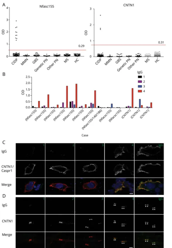
Of 342 sera from patients with CIDP tested, 10 (3%) were positive for antibodies to anti-Nfasc155 using ELISA and CBA (figure 1A). IgG isotypes were IgG4 in 7, IgG3 in 1, and undetectable in 2 patients (figure 1B). One of these latter patients showed IgG4 reactivity against Nfasc140/186 and was thus considered a Nfasc140/186-reactive patient. Three patients (1%) tested positive on ELISA and CBA for anti-CNTN1 antibodies (figure 1A). IgG isotypes were IgG4 in 2 patients and mixed IgG3/IgG4 in 1 patient (figure 1B). All sera reactive to Nfasc155 or CNTN1 by ELISA were also confirmed by CBA, and no nonspecific reactivities by ELISA only were identified. Six patients (2%) showed reactivity against Caspr1 on CBA (figure 1C). Three had an IgG4-predominant isotype, 2 had an IgG1 isotype, and 1 did not have a detectable isotype. Overall, 19 patients (6%) had antibodies against one of these 4 targets. None of the HCs and patients with GBS, MMN, hereditary neuropathy, other noninflammatory neuropathies, or MS tested positive, in-dicating that the presence of antibodies to nodal/paranodal proteins accurately identify patients with CIDP with sensi-tivity 6% and specificity 100%. Of note, none of the sera from 36 atypical CIDP cases tested positive.
Sera from patients with IgG4 anti-Nfasc155, anti-CNTN1, or anti-Caspr1 antibodies, but not anti-Nfasc155 of the IgG3 isotype or undetectable isotype, consistently showed re-activity against paranodes when tested on teasedfibers from murine sciatic nerves (figure 1D). Sera with anti-Nfasc140/ 186 IgG4 that co-reacted against Nfasc155 stained the nodes of Ranvier on teasedfibers.
Clinical features
Patients with anti-Nfasc155 IgG4 antibodies
Of patients with anti-Nfasc155 IgG4 antibodies, 4 were men and 3 were women, with an average age at onset of 31 ± 18 years (table e-1, links.lww.com/NXI/A164). The onset of neuropathy was subacute in 5 patients. One case was initially diagnosed with GBS.
All patients had symmetric 4-limb muscle weakness, which was mild to moderate in the upper limbs and proximal muscles of the lower limbs but severe in 5 patients in the lower limb distal muscles. Pinprick and position sense at the hallux were reduced or abolished in all patients. Tremor was also invariably present, and 6 had sensory ataxia. One patient reported neuropathic pain at onset, and 1 had dysphagia. One of them presented optic neuritis during steroid tapering. Brain MRI was thus performed but did not show evidence of disseminated inflammatory CNS disease. The neuropathy was moderately to severely disabling with an ONLS at onset of 5.8 ± 2.2. Two patients required a wheelchair 1 month after the disease onset. The patients received IVIG (n = 6), steroids (n = 7), azathioprine (n = 1), or rituximab (n = 1). Only 1 patient responded to IVIG and
Figure 1Reactivity to Nfasc155, CNTN1, and Caspr1 in CIDP by ELISA and CBA
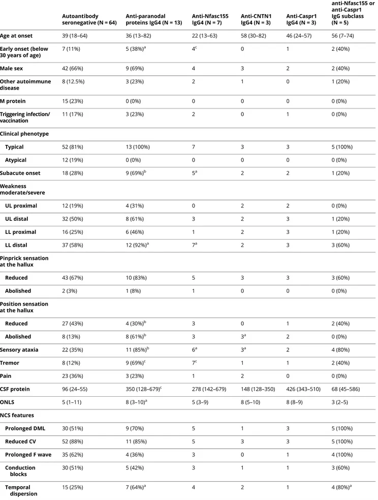
(A) Serum samples from patients with CIDP (n = 342), MMN (n = 13), GBS (n = 31), genetic PN (n = 18), other noninflammatory PN (n = 52), MS (n = 60) and from HCs (n = 60) were tested for autoantibodies to human Nfasc155 (left) and CNTN1 (right) by ELISA. OD are shown after subtraction of the baseline OD reading to bovine serum albumin. The red line represents the mean OD in HCs plus 3 standard deviations. (B) IgG isotype in Nfasc155- and CNTN1-positive patients. (C) The sera (here case 14) were tested on living HEK cells transfected with CNTN1 and Caspr1 (red) and then revealed with mouse antihuman IgG1, IgG2, IgG3, or IgG4 (green) as indicated. Nuclei were stained with DAPI (blue). (D) These are teased fibers from mouse sciatic nerves immunostained for CNTN1 (red) and the serum from case 14, then revealed with mouse antihuman IgG1, IgG2, IgG3, or IgG4 (green) as indicated. IgG1 and IgG4 from this patient reacted against Caspr1 and bound to the paranodal regions. Scale bars: 10μm. Caspr1 = contactin-associated protein 1; CBA = cell-based assay; CIDP = chronic inflammatory demyelinating polyradiculoneuropathy; CNTN1 = contactin-1; GBS = Guillain-Barr´e syndrome; HC = healthy control; HEK = human embryonic kidney; MMN = multifocal motor neuropathy; Nfasc155 = neurofascin-155; OD = Optical density; PN = peripheral neuropathy.
steroids (ONLS 4→ 2), 1 patient showed response to steroids (ONLS 6→ 2), and 1 patient unresponsive to IVIG and ste-roids had sustained response after rituximab (ONLS 9→ 2).
Patients with anti-CNTN1 IgG4 antibodies
Of patients with anti-CNTN1 IgG4 antibodies, 3 were men with a mean age at onset of 56 ± 26 years (table e-1, links.lww. com/NXI/A164). The onset of neuropathy was subacute in 2 patients. Strength was impaired in both proximal and distal muscles in the upper and lower limbs, often to a severe degree, and 2 of them required a wheelchair. Pinprick sensation and proprioception were either severely reduced or abolished, and all of them had sensory ataxia. One patient showed cranial nerve involvement and respiratory failure. One patient with anti-CNTN1 IgG3/IgG4 antibodies had a concurrent onset of CIDP and nephrotic syndrome because of membranous glo-merulonephritis. A kidney biopsy showed subepithelial deposits of immune complexes and complement deposition. CNTN1 patients had the highest disability at onset with an ONLS of 7.6 ± 2.5. One patient died of cardiac disease 6 months after the disease onset. The patients were treated with IVIG (n = 3), steroids (n = 3), plasma exchange (n = 2), and immunosuppressive drugs (n = 2). None of them had a sus-tained response to either IVIG or steroids. Cyclophosphamide was effective in 2 patients, leading to significant improvement of the neuropathy in 1 case and complete remission of the neuropathy together with regression of the nephrotic syn-drome in the other case, who did not require further treatment.
Patients with anti-Caspr1 IgG4 antibodies
Of seropositive patients with anti-Caspr1 IgG4 antibodies, 2 were men and 1 was a woman, with a mean age at onset of 43 ± 17 years (table e-1, links.lww.com/NXI/A164). The onset was subacute in 2 patients. Weakness was mild to moderate in proximal muscle groups and moderate to severe in distal muscle groups in both upper and lower limbs. Pinprick sen-sation was reduced, and position sense was abolished at the hallux in all of them. None of them reported neuropathic pain. One patient complained of dysphagia, and 1 patient had tremor. The disease was severe, with a mean ONLS of 8.3 ± 0.6. The patients were treated with IVIG (n = 3), steroids (n = 3), plasma exchange (n = 1), methotrexate (n = 1), and interferon-alfa (n = 1). A sustained response was observed only in 1 patient receiving steroids and methotrexate (ONLS 9→ 6) and 1 patient treated with interferon-alfa (ONLS 8→ 7).
Seropositive patients with an IgG1-3 isotype or undetectable IgG isotype
Six patients had IgG1 or IgG3 antibodies or an undetectable IgG isotype against Nfasc155 (n = 3) or Caspr1 (n = 3) (table e-1, links.lww.com/NXI/A164). One of these patients presented antibodies reacting to both Nfasc155 and Nfasc140/186. The clinical features of this patient, mainly consisting in a very early onset IVIG-responsive neuropathy, have been previously de-tailed.16We will thus focus here on the 5 other patients, 2 men and 3 women. In 2 of them, both reactive against Caspr1, the disease started in thefirst decade of life. The mode of onset was
subacute in 1 patient only. If present, weakness was graded as mild to moderate in upper limbs and proximal muscles of lower limbs. All 5 patients had weakness in lower limb distal muscles, which was severe in 2 of them. Pinprick sensation was reduced in 3 and position sense was reduced at the hallux in 2 patients, but none had absent proprioception. Sensory ataxia was reported in 4 patients, 2 with anti-Nfasc155 antibodies and 2 with anti-Caspr1 antibodies, whereas tremor was observed in 2 patients with anti-Caspr1 antibodies. Disability was usually low with a mean ONLS at onset of 3.2 ± 1.3. Only 1 case with anti-Caspr1 antibodies needed walking aids. The patients were treated with IVIG (n = 4) and steroids (n = 2). One patient with minimal disability did not require treatment. Three of 4 patients treated with IVIG had a good response.
Comparison of seropositive vs seronegative patients
We compared the clinical features and responses to treat-ments of patients with IgG4 antibodies against Nfasc155, CNTN1, or Caspr1 with those of 64 randomly selected patients with CIDP who were negative for antibodies against Nfasc155, Nfasc140/186, CNTN1, or Caspr1. We also compared seropositive patients with an IgG1, IgG3, or un-detectable IgG isotype with seronegative patients (table 1). Anti-Nfasc155 and anti-Caspr1 IgG4 seropositive patients had more frequently an early disease onset, before 30 years of age, as opposed to that of anti-CNTN1 IgG4 seropositive patients and seronegative patients. A subacute onset was reported in up to two-thirds of anti-Nfasc155, anti-CNTN1, and anti-Caspr1 IgG4 seropositive patients compared with 28% of seronegative patients. According to the EFNS/PNS criteria, CIDP pheno-type was typical in all IgG4-seropositive patients. Tremor was invariably present in patients with Nfasc155 IgG4 anti-bodies but was also observed in anti-CNTN1 and anti-Caspr1 IgG4 seropositive patients. Severe proprioceptive loss and sensory ataxia were significantly more frequent in IgG4-seropositive patients and were observed in all patients with anti-CNTN1 antibodies, 2 patients with anti-Caspr1 IgG4 antibodies, and 3 patients with anti-Nfasc155 IgG4 antibodies (table 1). In anti-CNTN1 and anti-Caspr1 IgG4 seropositive patients, weakness was usually graded as moderate to severe across proximal and distal muscle groups, whereas in patients with anti-Nfasc155 IgG4 antibodies, motor impairment showed a characteristic distal predominance in the lower limbs. A higher level of CSF total protein and the presence of tem-poral dispersion on nerve conduction studies were also char-acteristic features of patients with autoantibodies of the IgG4 isotype. IgG4-seropositive patients had a higher disability at onset: 5 patients needed a walking aid, and 5 patients required a wheelchair. In particular, patients with CNTN1 or anti-Caspr1 IgG4 antibodies showed the highest disability at onset. Patients with Nfasc155, CNTN1, or Caspr1 IgG4 anti-bodies had a lower response rate to IVIG compared with seronegative patients (8% vs 67%, p < 0.001), but there was no remarkable difference in the response to steroids or
Table 1Clinical features of patients with CIDP and antibodies to Nfasc155, CNTN1, and Caspr1 Autoantibody seronegative (N = 64) Anti-paranodal proteins IgG4 (N = 13) Anti-Nfasc155 IgG4 (N = 7) Anti-CNTN1 IgG4 (N = 3) Anti-Caspr1 IgG4 (N = 3) Anti-IgG1-3 or undetectable anti-Nfasc155 or anti-Caspr1 IgG subclass (N = 5) Age at onset 39 (18–64) 36 (13–82) 22 (13–63) 58 (30–82) 46 (24–57) 56 (7–74)
Early onset (below 30 years of age) 7 (11%) 5 (38%)a 4c 0 1 2 (40%) Male sex 42 (66%) 9 (69%) 4 3 2 2 (40%) Other autoimmune disease 8 (12.5%) 3 (23%) 2 1 0 1 (20%) M protein 15 (23%) 0 (0%) 0 0 0 0 (0%) Triggering infection/ vaccination 11 (17%) 3 (23%) 2 0 1 0 (0%) Clinical phenotype Typical 52 (81%) 13 (100%) 7 3 3 5 (100%) Atypical 12 (19%) 0 (0%) 0 0 0 0 (0%) Subacute onset 18 (28%) 9 (69%)b 5a 2 2 1 (20%) Weakness moderate/severe UL proximal 12 (19%) 4 (31%) 0 2 2 0 (0%) UL distal 32 (50%) 8 (61%) 3 2 3 1 (20%) LL proximal 16 (25%) 6 (46%) 1 2 3 1 (20%) LL distal 37 (58%) 12 (92%)a 7a 2 3 3 (60%) Pinprick sensation at the hallux Reduced 43 (67%) 10 (83%) 5 3 3 3 (60%) Abolished 2 (3%) 1 (8%) 1 0 0 0 (0%) Position sensation at the hallux Reduced 27 (43%) 4 (30%)b 3 0 1 2 (40%) Abolished 8 (13%) 8 (61%)b 3 3a 2 0 (0%) Sensory ataxia 22 (35%) 11 (85%)b 6a 3a 2 4 (80%) Tremor 8 (12%) 9 (69%)c 7c 1 1 2 (40%) Pain 23 (36%) 3 (23%) 1 2 0 0 (0%) CSF protein 96 (24–55) 350 (128–679)c 278 (142–679) 148 (128–350) 426 (343–510) 68 (45–586) ONLS 5 (1–11) 8 (3–10)a 5 (3–9) 8 (5–10) 8 (8–9) 3 (2–5) NCS features Prolonged DML 30 (51%) 9 (70%) 5 1 3 5 (100%) Reduced CV 52 (88%) 11 (85%) 5 3 3 5 (100%) Prolonged F wave 35 (62%) 4 (36%) 3 0 1 4 (100%) Conduction blocks 30 (51%) 5 (42%) 3 1 1 3 (60%) Temporal dispersion 15 (25%) 7 (64%)a 4 2 1 4 (80%)a Continued
immunosuppressive treatment. In particular, 1 patient with anti-Nfasc155 IgG4 antibodies was treated with rituximab, 1 patient with anti-Nfasc155 IgG4 antibodies was treated with azathio-prine, 2 patients with anti-CNTN1 IgG4 antibodies were treated with cyclophosphamide, and 1 patient with anti-Caspr1 IgG4 antibodies was treated with methotrexate. Four of them who were started on immunosuppressors within 1 year from disease onset showed long-lasting good response.
Patients with antibodies to Nfasc155, CNTN1, or Caspr1 of the IgG1, IgG3, or undetectable isotype did not show distinct clinical features or response to treatments compared with seronegative patients, except for more frequent temporal dispersion on nerve conduction studies.
Morphological changes of the nodes of Ranvier in skin biopsy of seropositive patients
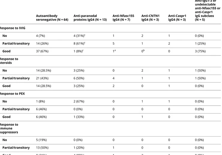
We evaluated skin biopsies from 3 patients with anti-Nfasc155 IgG4 antibodies, 1 patient with anti-CNTN1 IgG3/IgG4
antibodies, 1 patient with anti-Caspr1 IgG4 antibodies, 1 pa-tient with anti-Nfasc155 antibodies of an undetectable isotype, and 6 seronegative patients with CIDP. Analysis of myelinated fibers from patients with anti-Nfasc155 and anti-Caspr1 IgG4 showed elongation of the nodes of Ranvier and loss of para-nodal Nfasc155 and Caspr1 staining. Moderate elongation of the nodes of Ranvier and loss of Nfasc155 paranodal staining were also observed in myelinatedfibers of the patient with anti-CNTN1 IgG3/IgG4 antibodies. Contrarily, we did not observe similar changes in the patient with anti-Nfasc155 antibodies with an undetectable isotype, seronegative patients CIDP, or HCs (figure 2).
Pathogenic effects of anti-Caspr1 IgG4 and IgG1 antibodies
Anti-CNTN1 IgG4 antibodies were previously found to have function-blocking activity and to disrupt the interaction be-tween CNTN1 and its glial partner Nfasc155.12 We have previously shown that anti-CNTN1 IgG4 and anti-Nfasc155
Table 1 Clinical features of patients with CIDP and antibodies to Nfasc155, CNTN1, and Caspr1(continued)
Autoantibody seronegative (N = 64) Anti-paranodal proteins IgG4 (N = 13) Anti-Nfasc155 IgG4 (N = 7) Anti-CNTN1 IgG4 (N = 3) Anti-Caspr1 IgG4 (N = 3) Anti-IgG1-3 or undetectable anti-Nfasc155 or anti-Caspr1 IgG subclass (N = 5) Response to IVIG No 4 (7%) 4 (31%)c 1 2 1 0 (0%) Partial/transitory 14 (26%) 8 (61%)c 5 1 2 1 (25%) Good 37 (67%) 1 (8%)c 1a 0b 0 3 (75%) Response to steroids No 14 (28.5%) 3 (25%) 0 2 1 1 (50%) Partial/transitory 21 (43%) 6 (50%) 4 1 1 1 (50%) Good 14 (28.5%) 3 (25%) 2 0 1 0 (0%) Response to PEX No 1 (8%) 2 (67%) 0 1 1 0 (0%) Partial/transitory 6 (46%) 0 (0%) 0 0 0 0 (0%) Good 6 (46%) 1 (33%) 0 1 0 0 (0%) Response to immune suppressors No 5 (19%) 0 (0%) 0 0 0 0 (0%) Partial/transitory 13 (50%) 1 (20%) 1 0 0 0 (0%) Good 8 (31%) 4 (80%) 1 2 1 0 (0%)
Abbreviations: Caspr1 = contactin-associated protein 1; CIDP = chronic inflammatory demyelinating polyradiculoneuropathy; CNTN1 = contactin-1; CV = conduction velocity; DML = distal motor latency; IVIG = intravenous immunoglobulin; LL = lower limb; NCS = nerve conduction study; Nfasc155 = neurofascin-155; ONLS = Overall Neuropathy Limitation Scale; PEX = plasma exchange; UL = upper limb.
ap < 0.05. bp < 0.01. c
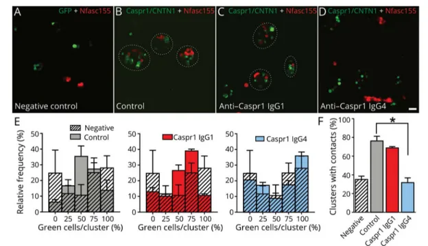
IgG4 antibodies are pathogenic and disrupt the paranodal regions.11,21We thus examined whether anti-Caspr1 antibodies also affect the integrity of the CNTN1/Caspr1/Nfasc155 complex. In particular, we purified IgG1 and IgG4 and tested the effects of the different antibody isotypes. First, we tested the impact of IgG4 and IgG1 fractions from Caspr1-positive patients on the interaction of CNTN1/Caspr1 with Nfasc155 by a cell aggregation assay (figure 3). Human embryonic kidney cells were transfected with Nfasc155 and mCherry and incubated for 2 hours with cells coexpressing CNTN1/Caspr1 and green fluorescent protein (GFP). The percentage of cell clusters showing contacts between red and green cells and the percent-age of green and red cells per cell clusters were quantified. As a negative control, Nfasc155-mCherry-transfected cells were incubated with cells expressing GFP only. In the absence of CNTN1/Caspr1, Nfasc155-expressing cells do not form signif-icant contact with GFP-expressing cells, and most clusters
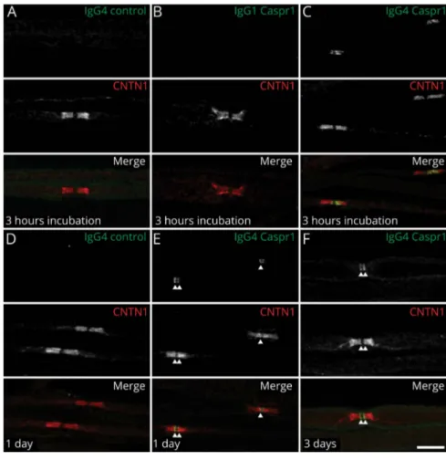
express only green or red cells. The expression of CNTN1/ Caspr1 significantly increases the number of clusters with con-tacts, and most cell clusters show an average of 50% of CNTN1/ Caspr1- and 50% of Nfasc155-expressing cells. The presence of 10 μg of IgG4 from a Caspr1-positive patient, but not IgG1, abolished the interaction between CNTN1/Caspr1- and Nfasc155-expressing cells to the level of negative controls, thus supporting a blocking function of anti-Caspr1 IgG4 antibodies. To further confirm these findings, mouse sciatic nerve segments were incubated in vitro with 10μg of IgG4 or IgG1 fractions from Caspr1-positive patients for 3 hours, and IgG deposition was monitored. IgG4 but not IgG1 antibodies were found to penetrate the paranodal regions (figure 4). These findings were replicated in vivo by performing intraneural injections. Again, only IgG4 antibodies were found to penetrate the paranodal regions. Of interest, the level of antibody penetration across the
Figure 2Morphological alterations of the nodes of Ranvier in patients with CIDP with IgG4 autoantibodies
We evaluated skin biopsies from 3 patients with IgG4 Nfasc155 antibodies, 1 patient with IgG3/IgG4 CNTN1 antibodies, 1 patient with IgG4 anti-Caspr1 antibodies, 1 patient with undetectable isotype IgG anti-Nfasc155, and 6 seronegative patients with CIDP. Analysis of myelinated fibers showed elongation of the nodes of Ranvier and loss of paranodal Nfasc155 staining in skin biopsies from patients with anti-Nfasc155 (C) and Caspr1 (E) IgG4. Moderate elongation of the nodes of Ranvier and loss of Nfasc155 paranodal staining were also observed in myelinated fibers of a CNTN1 IgG3/IgG4-positive patient (D). Contrarily, we did not observe similar changes in the patient with undetectable isotype IgG anti-Nfasc155 antibodies (F), in seronegative patients with CIDP (B), or in HCs (A). A complete loss of Caspr1 staining was observed in biopsies from patients with IgG4 antibodies to paranodal proteins (I, L, M), but not in an Nfasc155 seropositive patient with an undetectable isotype (N) and in seronegative CIDP (H) or healthy patients (G). Caspr1 = contactin-associated protein 1; CIDP = chronic inflammatory demyelinating polyradiculoneuropathy; CNTN1 = contactin-1; HC = healthy control; Nfasc155 = neurofascin-155.
paranodal region was similar at 1 or 3 days after intraneural injection, and IgG4 deposition was detected only at the border of the nodes of Ranvier (figure 4). No paranodes presented a complete invasion by IgG4 at 1 or 3 days postinjection.
Discussion
Antibodies to nodal/paranodal protein were found, albeit with a low frequency, in Italian patients with CIDP and are associ-ated with target- and isotype-specific clinical features, as previously reported. The prevalence of Nfasc155 and anti-CNTN1 antibodies was similar to that reported by previous studies in European patients but was lower compared with that in Japanese patients.5,10The discrepancy may be attributable to differences in inclusion criteria, ethnicities, and non-standardized laboratory techniques. The frequency of anti-Caspr1 IgG4 antibodies was equal to that of antibodies against CNTN1, confirming that Caspr1 may also represent a relevant target of the immune response in Caucasian patients with CIDP. None of the healthy or pathologic controls tested, in-cluding patients affected by other neuropathies, showed re-activity against Nfasc155, CNTN1, or Caspr1. These maximum levels of specificity entail that the tests perform very well for the diagnosis of such CIDP subtypes.
In addition, it is worth noting that the results of antibody testing by ELISA and CBA were concordant in all samples
across the 2 testing centers in Pavia and Montpellier, in-dicating that these techniques, although not broadly available, have a high reproducibility if performed in specialized centers. Ourfindings largely confirm previous observations by several groups showing a tight clinical serologic correlation of anti-bodies to nodal/paranodal proteins and clinical features of seropositive patients with CIDP.
The patients with anti-Nfasc155 IgG4 antibodies showed earlier onset, distal predominant lower limb weakness, and gait disturbance. Tremor was also present, although only in 1 case this was disabling. One patient with such antibodies developed bilateral optic neuritis during steroid tapering 18 months after the onset of the neuropathy.
The patients with anti-CNTN1 antibodies were older and showed a subacute or rapidly progressive severe sensory and motor neuropathy. Previous studies found that anti-CNTN1 antibodies were associated with either predominant motor or sensory impairment. Here, both sensory and motorfibers were equally severely affected, and the patients became wheelchair dependent few months after the disease onset. Notably, 1 pa-tient with anti-CNTN1 antibodies of IgG3 and IgG4 subclasses showed the contemporary occurrence of membranous glo-merulonephritis. This association had been reported in 3 cases with anti-CNTN1 antibodies,7,22,23and either a direct damage
Figure 3IgG4 to Caspr1 disrupt the interaction between Nfasc155 and CNTN1/Caspr1
(A–D) HEK cells transfected with CNTN1 and Caspr1 (green) or Nfasc155 (red) were incubated together for 2 hours in the presence of control IgG4 (B), anti-Caspr1 IgG1 (C), or anti-anti-Caspr1 IgG4 (D). As negative controls, HEK cells transfected with Nfasc155 (red) were incubated with cells transfected with GFP (A). Anti-Caspr1 IgG4, but not anti-Caspr1 IgG1, abrogated the aggregation of Nfasc155-transfected cells with CNTN1/Caspr1. Scale bar: 10μm. (E–F) The graphs represent the relative frequency of green cells per aggregates (n = 4 experiments for each condition). The percentage of cell clusters with contacts between green and red cells was quantified (F). The percentage of contacts was significantly decreased in the presence of anti-Caspr1 IgG4 (p < 0.005 by unpaired 2-tailed Student t tests and by one-way ANOVA, followed by Bonferroni post hoc tests). Bars represent mean and SEM. ANOVA = analysis of variance; Caspr1 = contactin-associated protein 1; CNTN1 = contactin-1; GFP = green fluorescent protein; HEK = human embryonic kidney; Nfasc155 = neurofascin-155.
or indirect damage, after the deposition of immune complexes, can be hypothesized. Of interest, a recent study showed that complement deposition may contribute to the pathophysiology of anti-CNTN1-associated neuropathy, particularly in patients with a predominance of the IgG3 subclass.24
Neuropathy in patients with anti-Caspr1 IgG4 antibodies was also highly debilitating. Pain did not seem to be a clinical feature associated with the presence of anti-Caspr1 antibodies in our series, as opposed to thefirst report of 2 patients with Caspr1-associated inflammatory neuropathy.8
As a novel observation of our study, we found that patients with anti-Nfasc155 or Caspr1 antibodies of the IgG1, IgG3, or un-detectable IgG isotype did not show clinical features distinct from seronegative patients. Most importantly, patients with anti-Nfasc155, CNTN1, or Caspr1 IgG4 antibodies, but not patients with antibodies to Nfasc155 or Caspr1 of IgG1, IgG3, or undetectable IgG isotype, showed a significantly lower re-sponse rate to IVIG compared with seronegative patients. Analysis of myelinatedfibers in the skin biopsy of patients with anti-Nfasc155, CNTN1, or Caspr1 IgG4 antibodies showed morphological changes of the nodes of Ranvier, including
elongation of the node and loss of neurofascin and Caspr1 staining at paranodes, which were absent or less prominent in seronegative CIDP cases or in the case with anti-Nfasc155 antibodies of an undetectable isotype. These data add to other recent histopathologic and neurophysiologic observations in patients with antibodies to nodal/paranodal proteins14,25and indicate that isotype determination appears to be crucial to correctly identify such patients and to guide treatment. The high frequency of anti-Caspr1 antibodies in our series prompted us to further investigate their pathogenic effects. Albeit the experimental setting was limited to the examination of the effects of anti-Caspr1 autoantibodies from a single pa-tient, we found that anti-Caspr1 IgG4, but not anti-Caspr1 IgG1, antibodies affect the interaction between the CNTN1/ Caspr1 and Nfasc155 complex using a cell aggregation assay. Thesefindings are in line with the previously reported patho-genic role of anti-CNTN1 IgG4 antibodies11,12 and suggest that IgG4 antibodies targeting the CNTN1/Caspr1 complex may have a function-blocking activity and disrupt the paranodal axo-glial contact. In keeping with this experimental evidence, our data also seem to indicate that anti-Caspr1 IgG4 antibodies can penetrate the paranodal regions. It is worth noting that the pathogenic mechanism of IgG4 antibodies in other diseases
Figure 4Anti-Caspr1 IgG4, but not IgG1, invades the paranodal regions
(A–C) Sciatic nerve fibers were incubated in vitro with purified control IgG4 (A), anti-Caspr1 IgG1 (B), or anti-Caspr1 IgG4 (C) for 3 hours and immunolabeled for IgG (green) and CNTN1 (red). (D–F) Sciatic nerves were fixed 1 or 3 days after intraneural injections of purified control IgG4 (D), anti-Caspr1 IgG4 (E–F), immunolabeled for CNTN1 (red), and human IgG (green). Note that only anti-Caspr1 IgG4 penetrated the paranodes. One or 3 days after injection, IgG4 antibodies were detected at the paranode borders (arrows). Images are representative of 3 independent experiments. Scale bar: 10μm. Caspr1 = con-tactin-associated protein 1; CNTN1 = contactin-1.
implicates the disruption of cell adhesion protein complexes too, by inhibiting protein-protein interaction, and that thera-pies aiming at downregulating the humoral immune response, such as rituximab, showed some efficacy in anti-Nfasc155- and anti-Caspr1 antibody-associated CIDP, as well as in other IgG4-related diseases, likely because of the depletion of the Nfasc155- or Caspr1-reactive B cells.26
Despite the limitation entailed by the retrospective data collection, in the present series, immunosuppressive treat-ment, including rituximab, cyclophosphamide, and metho-trexate, seemed effective in IVIg-resistant IgG4 seropositive patients if started early in the disease course. Of note, we did not detect a significant difference in the response to steroids between patients with anti-paranodal IgG4 antibodies and seronegative patients, confirming steroids as an effective therapeutic option in CIDP cases, independent from their serologic status for anti-Nfasc155, CNTN1, or Caspr1 antibodies. Low numbers of patients treated with plasma exchange, and the temporal overlap of plasma exchange cycles with other treatments (case 12), prevented us from drawingfirm conclusions on its role in patients with anti-paranodal IgG4 antibodies.
Although patients with antibodies against the nodal/ paranodal component often, albeit not always, show addi-tional clinical features, CIDP phenotype was typical in all IgG4 and non-IgG4 seropositive patients according to the EFNS/PNS criteria, whereas none of the sera from atypical CIDP cases or control sera showed the presence of these antibodies. Together, this observation challenges the view that the search for antibodies against the nodal/paranodal component may be limited to atypical CIDP cases.
Testing for the presence of antibodies against Nfasc155, CNTN1, and Caspr1, followed by IgG isotype determination in seropositive cases should be part of the diagnostic workup in inflammatory neuropathies to improve diagnostic accuracy and guide treatment. Moreover, knowledge of the mechanism underlying these CIDP subtypes might shed light on the pathophysiology and help further understanding of this complex and heterogeneous disease.
Study funding
This work was supported by the Italian Ministry of Health “Ricerca Corrente” 2017–2019 Grant (Grant code: RC1812C) to the IRCCS Mondino Foundation, GBS/CIDP Foundation Non-profit Grant 2017 Grant code: 501(c)(3) to Fondazione IRCCS Istituto Neurologico Carlo Besta, and Agence Nationale pour la Recherche (NECCIN; JD) and by the Association Française contre les Myopathies (grant#21532; JD), the Italian Ministry of Health “Ricerca Finalizzata 2016 (Grant code: RF-2016-02361887) to Humanitas Clinical and Research Center, IRCCS. Andrea Cortese is funded by MRC (MR/T001712/1) and received funding from Wellcome Trust (204841/Z/16/Z) and the Inherited Neuropathy Consortium (INC), which is a part of
the NIH Rare Diseases Clinical Research Network (RDCRN) (U54NS065712).
Disclosure
The authors report no disclosures. Go to Neurology.org/NN for full disclosures.
Publication history
Received by Neurology: Neuroimmunology & Neuroinflammation June 14, 2019. Accepted infinal form October 3, 2019.
AppendixAuthors
Name Location Role Contribution
Andrea Cortese, MD, PhD Department of Brain and Behavioral Sciences, University of Pavia, Pavia, Italy; Department of Neuromuscular Disease, UCL Queen Square Institute of Neurology, London, United Kingdom
Author Designed and conceptualized the study, major role in the acquisition of data, analyzed the data, and drafted the manuscript for intellectual content Raffaella Lombardi, MA Neuroalgology Unit, IRCCS Fondazione Istituto Neurologico “Carlo Besta,” Milan, Italy
Author Designed and conceptualized the study, major role in the acquisition of data, analyzed the data, and drafted the manuscript for intellectual content Chiara Briani, MD Department of Neurosciences, University of Padova, Padova, Italy
Author Major role in the acquisition of data, analyzed the data, and drafted the manuscript for intellectual content Ilaria Callegari, MD Neuroscience Consortium, University of Pavia, Monza Policlinico and Pavia Mondino, Italy
Author Major role in the acquisition of data, analyzed the data, and drafted the manuscript for intellectual content Luana Benedetti, MD, PhD Department of Neuroscience, Rehabilitation, Ophthalmology, Genetics, Maternal and Child Health (DiNOGMI), University of Genova, Genova, Italy; IRCCS Ospedale Policlinico San Martino, Genova, Italy
Author Major role in the acquisition of data and revised the manuscript for intellectual content Fiore Manganelli, MD Department of Neurosciences, Odontostomatological and Reproductive Sciences, University of Naples“Federico II,” Naples, Italy
Author Major role in the acquisition of data and revised the manuscript for intellectual content Marco Luigetti, MD, PhD Fondazione Policlinico Universitario Agostino Gemelli-IRCCS. UOC Neurologia, Universit`a Cattolica del Sacro Cuore, Roma, Italy
Author Major role in the acquisition of data and revised the manuscript for intellectual content
Appendix (continued)
Name Location Role Contribution
Sergio Ferrari, MD Section of Neurology, Department of Neuroscience, Biomedicine and Movement Sciences, University of Verona, Verona, Italy
Author Major role in the acquisition of data and revised the manuscript for intellectual content
Maurizio Clerici, MD
Department of Neurology and Stroke Unit, Ospedale di Circolo/Fondazione Macchi, Varese, Italy
Author Major role in the acquisition of data and revised the manuscript for intellectual content Girolama Alessandra Marfia, MD Department of Systems Medicine, University of Rome Tor Vergata, Rome, Italy
Author Major role in the acquisition of data and revised the manuscript for intellectual content Andrea Rigamonti, MD Neurological Department, ASST Lecco
Author Major role in the acquisition of data and revised the manuscript for intellectual content Marinella Carpo, MD, PhD Neurology Unit, Ospedale Treviglio, ASST Bergamo Ovest, Italy
Author Major role in the acquisition of data and revised the manuscript for intellectual content Raffaella Fazio, MD Department of Neurology, San Raffaele Scientific Institute, Milan, Italy
Author Major role in the acquisition of data and revised the manuscript for intellectual content Massimo Corbo, MD Department of Neurorehabilitation Sciences, Casa Cura Policlinico (CCP), Milan, Italy
Author Major role in the acquisition of data and revised the manuscript for intellectual content Anna Mazzeo, MD Department of Clinical and Experimental Medicine, University of Messina, Messina, Italy
Author Major role in the acquisition of data and revised the manuscript for intellectual content Fabio Giannini, MD Department of Medicine, Surgery and Neurosciences, University of Siena, Siena, Italy
Author Major role in the acquisition of data and revised the manuscript for intellectual content Giuseppe Cosentino, MD, PhD IRCCS Mondino Foundation, Department of Brain and Behavioral Sciences, University of Pavia, Pavia, Italy
Author Major role in the acquisition of data and revised the manuscript for intellectual content
Elisabetta Zardini, MA
IRCCS Mondino Foundation, Pavia, Italy
Author Major role in the acquisition of data and revised the manuscript for intellectual content Riccardo Curr`o, MD Neuroscience Consortium, University of Pavia, Monza Policlinico and Pavia Mondino, Italy
Author Major role in the acquisition of data, analyzed the data, and revised the manuscript for intellectual content
Appendix (continued)
Name Location Role Contribution
Matteo Gastaldi, MD, PhD
IRCCS Mondino Foundation, Pavia, Italy
Author Major role in the acquisition of data and revised the manuscript for intellectual content Elisa Vegezzi, MD Neuroscience Consortium, University of Pavia, Monza Policlinico and Pavia Mondino, Italy
Author Major role in the acquisition of data, analyzed the data, and revised the manuscript for intellectual content Enrico
Alfonsi, MD
IRCCS Mondino Foundation, Pavia, Italy
Author Major role in the acquisition of data and revised the manuscript for intellectual content Angela Berardinelli, MD IRCCS Mondino Foundation, Pavia, Italy
Author Major role in the acquisition of data and revised the manuscript for intellectual content Ludivine
Kouton, MD
Referral Center for Neuromuscular Diseases and ALS, Timone University Hospital, Marseille, France
Author Major role in the acquisition of data and revised the manuscript for intellectual content Constance Manso, MSc Universit´e de Bordeaux, Interdisciplinary Institute for Neuroscience, Bordeaux, France
Author Major role in the acquisition of data and revised the manuscript for intellectual content
Claudia Giannotta, MA
Humanitas Clinical and Research Center, IRCCS, Milan, Italy
Author Major role in the acquisition of data and revised the manuscript for intellectual content Pietro
Doneddu, MD
Humanitas Clinical and Research Center, IRCCS, Milan, Italy
Author Major role in the acquisition of data and revised the manuscript for intellectual content Patrizia Dacci, MD, PhD Neuroalgology Unit, IRCCS Fondazione Istituto Neurologico “Carlo Besta,” Milan, Italy
Author Major role in the acquisition of data and revised the manuscript for intellectual content Laura Piccolo, MD Neuroalgology Unit, IRCCS Fondazione Istituto Neurologico “Carlo Besta,” Milan, Italy
Author Major role in the acquisition of data and revised the manuscript for intellectual content Marta Ruiz, MD Department of Neurosciences, University of Padova, Padova, Italy
Author Major role in the acquisition of data and revised the manuscript for intellectual content Alessandro Salvalaggio, MD Department of Neurosciences, University of Padova, Padova, Italy
Author Major role in the acquisition of data and revised the manuscript for intellectual content
References
1. Doneddu PE, Cocito D, Manganelli F, et al. Atypical CIDP: diagnostic criteria, progression and treatment response. Data from the Italian CIDP database. J Neurol Neurosurg Psychiatry 2019;90:125–132.
2. Devaux JJ, Odaka M, Yuki N. Nodal proteins are target antigens in Guillain-Barr´e syndrome. J Peripher Nerv Syst 2012;17:62–71.
3. Querol L, Nogales-Gadea G, Rojas-Garcia R, et al. Antibodies to contactin-1 in chronic inflammatory demyelinating polyneuropathy. Ann Neurol 2013;73:370–380. 4. Querol L, Nogales-Gadea G, Rojas-Garcia R, et al. Neurofascin IgG4 antibodies in CIDP associate with disabling tremor and poor response to IVIg. Neurology 2014;82:879–886. 5. Ogata H, Yamasaki R, Hiwatashi A, et al. Characterization of IgG4 anti-neurofascin 155 antibody-positive polyneuropathy. Ann Clin Transl Neurol 2015;2:960–971. 6. Miura Y, Devaux JJ, Fukami Y, et al; CNTN1-CIDP Study Group. Contactin 1 IgG4
associates to chronic inflammatory demyelinating polyneuropathy with sensory ataxia. Brain 2015;138:1484–1491.
7. Doppler K, Werner C, Sommer C. Disruption of nodal architecture in skin biopsies of patients with demyelinating neuropathies. J Peripher Nerv Syst 2013;18:168–176. 8. Doppler K, Appeltshauser L, Villmann C, et al. Auto-antibodies to
contactin-associated protein 1 (Caspr) in two patients with painful inflammatory neuropathy. Brain 2016;139:2617–2630.
9. Kadoya M, Kaida K, Koike H, et al. IgG4 anti-neurofascin155 antibodies in chronic inflammatory demyelinating polyradiculoneuropathy: clinical significance and di-agnostic utility of a conventional assay. J Neuroimmunol 2016;301:16–22.
Appendix (continued)
Name Location Role Contribution
Chiara De Michelis, MD Department of Neuroscience, Rehabilitation, Ophthalmology, Genetics, Maternal and Child Health (DiNOGMI), University of Genova, IRCCS Ospedale Policlinico San Martino, Genova, Italy
Author Major role in the acquisition of data and revised the manuscript for intellectual content Emanuele Spina, MD Department of Neurosciences, Odontostomatological and Reproductive Sciences, University of Naples“Federico II,” Naples, Italy
Author Major role in the acquisition of data and revised the manuscript for intellectual content Antonietta Topa, MD Department of Neurosciences, Odontostomatological and Reproductive Sciences, University of Naples“Federico II,” Naples, Italy
Author Major role in the acquisition of data and revised the manuscript for intellectual content Giulia Bisogni, MD
Centro Clinico NEMO Adulti, Roma, Italy
Author Major role in the acquisition of data and revised the manuscript for intellectual content Angela Romano, MD Institute of Neurology, Universit`a Cattolica del Sacro Cuore, Rome, Italy
Author Major role in the acquisition of data and revised the manuscript for intellectual content Sara Mariotto, MD Section of Neurology, Department of Neuroscience, Biomedicine and Movement Sciences, University of Verona, Verona, Italy
Author Major role in the acquisition of data and revised the manuscript for intellectual content Giorgia Mataluni, MD, PhD Department of Systems Medicine, University of Rome Tor
Vergata, Rome, Italy
Author Major role in the acquisition of data and revised the manuscript for intellectual content Federica Cerri, MD, PhD Department of Neurology, San Raffaele Scientific Institute, Milan, Italy
Author Major role in the acquisition of data and revised the manuscript for intellectual content Claudia Stancanelli, MD IRCCS Centro Neurolesi “Bonino Pulejo,” Messina, Italy
Author Major role in the acquisition of data and revised the manuscript for intellectual content Mario Sabatelli Universit`a Cattolica del Sacro Cuore; Centro Clinico NEMO Adulti, Roma, Italy
Author Major role in the acquisition of data and revised the manuscript for intellectual content
Appendix (continued)
Name Location Role Contribution
Angelo Schenone, MD Department of Neuroscience, Rehabilitation, Ophthalmology, Genetics, Maternal and Child Health (DiNOGMI), University of Genova, IRCCS Ospedale Policlinico San Martino, Genova, Italy
Author Major role in the acquisition of data and revised the manuscript for intellectual content Enrico Marchioni, MD IRCCS Mondino Foundation, Pavia, Italy
Author Major role in the acquisition of data and revised the manuscript for intellectual content Giuseppe Lauria, MD Neuroalgology Unit, IRCCS Fondazione Istituto Neurologico “Carlo Besta,” Milan, Italy; Department of Biomedical and Clinical Sciences“Luigi Sacco,” University of Milan, Milan, Italy
Author Designed and conceptualized the study, major role in the acquisition of data, analyzed the data, and drafted the manuscript for intellectual content
Eduardo Nobile-Orazio, MD
Humanitas Clinical and Research Center, IRCCS, Milan, Italy
Author Major role in the acquisition of data and revised the manuscript for intellectual content J´erˆome Devaux, PhD Institute for Neurosciences of Montpellier University, Hopital Saint Eloi, Montpellier, France.
Author Designed and conceptualized the study, major role in the acquisition of data, analyzed the data, and drafted the manuscript for intellectual content Diego
Franciotta, MD
IRCCS Mondino Foundation, Pavia, Italy
Author Designed and conceptualized the study, major role in the acquisition of data, analyzed the data, and drafted the manuscript for intellectual content
10. Devaux JJ, Miura Y, Fukami Y, et al. Neurofascin-155 IgG4 in chronic inflammatory demyelinating polyneuropathy. Neurology 2016;86:800–807.
11. Manso C, Querol L, Mekaouche M, Illa I, Devaux JJ. Contactin-1 IgG4 antibodies cause paranode dismantling and conduction defects. Brain 2016;139:1700–1712. 12. Labasque M, Hivert B, Nogales-Gadea G, Querol L, Illa I, Faivre-Sarrailh C. Specific
contactin N-glycans are implicated in neurofascin binding and autoimmune targeting in peripheral neuropathies. J Biol Chem 2014;289:7907–7918.
13. Koike H, Kadoya M, Kaida KI, et al. Paranodal dissection in chronic inflammatory demyelinating polyneuropathy with neurofascin-155 and contactin-1 anti-bodies. J Neurol Neurosurg Psychiatry 2017;88:465–473.
14. Vallat JM, Yuki N, Sekiguchi K, et al. Paranodal lesions in chronic inflammatory demyelinating polyneuropathy associated with anti-neurofascin 155 antibodies. Neuromuscul Disord 2017;27:290–293.
15. Ng JK, Malotka J, Kawakami N, et al. Neurofascin as a target for autoantibodies in peripheral neuropathies. Neurology 2012;79:2241–2248.
16. Delmont E, Manso C, Querol L, et al. Autoantibodies to nodal isoforms of neurofascin in chronic inflammatory demyelinating polyneuropathy. Brain 2017;140:1851–1858. 17. Joint Task Force of the EFNS and the PNS. European Federation of Neurological Societies/ Peripheral Nerve Society guideline on management of chronic inflammatory demyelinating polyradiculoneuropathy: report of a joint task force of the European Federation of Neurological Societies and the Peripheral Nerve Society—first revision. J Peripher Nerv Syst 2010;15:1–9. 18. Cortese A, Devaux JJ, Zardini E, et al. Neurofascin-155 as a putative antigen in combined central and peripheral demyelination. Neurol Neuroimmunol Neuro-inflamm 2016;3:e238. doi: 10.1212/NXI.0000000000000238.
19. Graham RC, Hughes RA. A modified peripheral neuropathy scale: the overall neu-ropathy limitations scale. J Neurol Neurosurg Psychiatry 2006;77:973–976. 20. Lauria G, Hsieh ST, Johansson O, et al; European Federation of Neurological
Soci-eties; Peripheral Nerve Society. European Federation of Neurological Societies/ Peripheral Nerve Society Guideline on the use of skin biopsy in the diagnosis of small fiber neuropathy. Report of a joint task force of the European Federation of Neu-rological Societies and the Peripheral Nerve Society. Eur J Neurol 2010;17:903–912, e44–49.
21. Manso C, Querol L, Lleix`a C, et al. Anti-neurofascin-155 IgG4 antibodies prevent paranodal complex formation in vivo. J Clin Invest 2019;130:2222–2236. 22. Taieb G, Le Quintrec M, Pialot A, et al.“Neuro-renal syndrome” related to
anti-contactin-1 antibodies. Muscle Nerve 2019;59:E19–E21.
23. Hashimoto Y, Ogata H, Yamasaki R, et al. Chronic inflammatory demyelinating polyneuropathy with concurrent membranous Nephropathy: an anti-paranode and podocyte protein antibody study and literature Survey. Front Neurol 2018;9: 997.
24. Appeltshauser L, Weishaupt A, Sommer C, Doppler K. Complement deposition induced by binding of anti-contactin-1 auto-antibodies is modified by immunoglo-bulins. Exp Neurol 2017;287:84–90.
25. Uncini A, Vallat JM. Autoimmune nodo-paranodopathies of peripheral nerve: the concept is gaining ground. J Neurol Neurosurg Psychiatry 2018;89:627–635. 26. Querol L, Rojas-Garc´ıa R, Diaz-Manera J, et al. Rituximab in treatment-resistant CIDP
with antibodies against paranodal proteins. Neurol Neuroimmunol Neuroinflamm 2015;2:e149. doi: 10.1212/NXI.0000000000000149.
DOI 10.1212/NXI.0000000000000639
2020;7;
Neurol Neuroimmunol Neuroinflamm
Andrea Cortese, Raffaella Lombardi, Chiara Briani, et al.
Clinical relevance of IgG isotype
Antibodies to neurofascin, contactin-1, and contactin-associated protein 1 in CIDP:
This information is current as of November 21, 2019
Academy of Neurology.. All rights reserved. Online ISSN: 2332-7812.
Copyright © 2019 The Author(s). Published by Wolters Kluwer Health, Inc. on behalf of the American Published since April 2014, it is an open-access, online-only, continuous publication journal. Copyright
is an official journal of the American Academy of Neurology.
Services
Updated Information &
http://nn.neurology.org/content/7/1/e639.full.html
including high resolution figures, can be found at:
References
http://nn.neurology.org/content/7/1/e639.full.html##ref-list-1
This article cites 26 articles, 7 of which you can access for free at:
Citations
http://nn.neurology.org/content/7/1/e639.full.html##otherarticles
This article has been cited by 2 HighWire-hosted articles:
Subspecialty Collections
http://nn.neurology.org//cgi/collection/peripheral_neuropathy
Peripheral neuropathy
nating_polyneuropathy
http://nn.neurology.org//cgi/collection/chronic_inflammatory_demyeli
Chronic inflammatory demyelinating polyneuropathy
http://nn.neurology.org//cgi/collection/autoimmune_diseases
Autoimmune diseases
http://nn.neurology.org//cgi/collection/all_neuromuscular_disease
All Neuromuscular Disease
following collection(s):
This article, along with others on similar topics, appears in the
Permissions & Licensing
http://nn.neurology.org/misc/about.xhtml#permissions
its entirety can be found online at:
Information about reproducing this article in parts (figures,tables) or in
Reprints
http://nn.neurology.org/misc/addir.xhtml#reprintsus
Information about ordering reprints can be found online:
Academy of Neurology.. All rights reserved. Online ISSN: 2332-7812.
Copyright © 2019 The Author(s). Published by Wolters Kluwer Health, Inc. on behalf of the American Published since April 2014, it is an open-access, online-only, continuous publication journal. Copyright
is an official journal of the American Academy of Neurology.