68
Observation | Dermatol Pract Concept 2018;8(1):17DERMATOLOGY PRACTICAL & CONCEPTUAL
www.derm101.com
Learning Points
• Dermoscopy can help the clinical diagnosis of vulvar basal cell carcinoma showing linear and arborizing telangiecta-sia, pinkish background, blue ovoid nests, blue globules, white shiny structures and brown dots.
• Reflectance confocal microscopy can be used to confirm the clinical diagnosis of vulvar basal cell carcinoma and to identify its surgical margins.
Case Presentations
Case 1
An 82-year-old woman was referred to our clinic with com-plaints of itching and of bleeding from the vulva. She was
otherwise healthy and denied a personal history of skin can-cers, sexually transmitted diseases, and irradiation. A physical examination revealed a 3 x 2 cm ulcerated plaque involving the anterior vulvar commissure (Figure 1A). The rest of the genital and pelvic examination did not reveal any other pathological findings. Dermoscopic examination performed with Dermlite Foto System (DermLite, 3 Gen, San Juan Capistrano, CA, USA) coupled with a camera (Sony Cyber-Shot digital still camera 7.2 megapixels, Sony Corporation, Tokyo, Japan) and applying a disposable sterile transparent film (Visulin; Paul Hartmann AG, Heidenheim, Germany) on the tip of the instrument, showed the presence of reddish and well-focused arborizing vessels on a pinkish background associated with whitish homogeneous areas (Figure 1B,C). These features suggested the diagnosis of ulcerated BCC and
Dermoscopic and reflectance confocal microscopy
features of two cases of vulvar basal cell carcinoma
Elisa Cinotti, MD
1, Giulia Tonini
1, Jean Luc Perrot
2, Cyril Habougit
3, Stefano Luisi
4, Pietro Rubegni
11 Department of Medical, Surgical and Neuro-Sciences, Dermatology Unit, University of Siena, Siena, Italy 2 Dermatology Department, University Hospital of Saint Etienne, Saint Etienne, France
3 Pathology Department, University Hospital of Saint Etienne, Saint Etienne, France
4 Department of Molecular and Developmental Medicine, Obstetrics and Gynecology, University of Siena, Siena, Italy
Key words: basal cell carcinoma, dermoscopy, imaging, reflectance confocal microscopy, vulva
Citation: Cinotti E, Tonini G, Perrot JL, Habougit C, Luisi S, Rubegni P. Dermoscopic and reflectance confocal microscopic features of two cases of vulvar basal cell carcinoma. Dermatol Pract Concept. 2018;8(1):68-71. DOI: https://doi.org/10.5826/dpc.0801a17
Received: July 21, 2017; Accepted: September 14, 2017; Published: January 31, 2018
Copyright: ©2018 Cinotti et al. This is an open-access article distributed under the terms of the Creative Commons Attribution License, which permits unrestricted use, distribution, and reproduction in any medium, provided the original author and source are credited.
Funding: None.
Competing interests: The authors have no conflicts of interest to disclose. All authors have contributed significantly to this publication.
Corresponding author: Giulia Tonini, MD, Dermatology Section, University of Siena, S. Maria alle Scotte Hospital, 53100 Siena, Italy. Tel : 00 39 3292063153; Fax. 00 39 0577 44238. Email: [email protected]
Basal cell carcinoma (BCC) is the most common malignant skin cancer. Its genital localization is rare, and the diagnosis in this site could be challenging. Here, we report two patients with vulvar BCC and describe their clinical, dermoscopic and in vivo and ex vivo reflectance confocal microscopic (RCM) features. Dermoscopy and RCM can be useful tools for helping the clinical diagnosis of vulvar BCC and for identifying the correct surgical margins.
Observation | Dermatol Pract Concept 2018;8(1):17
69
control of the surgical margins (Figure 4D) that resulted in being tumor-free. The histopathologic examination showed a nodular BCC and confirmed that the margins were free of disease (Figure 5A and B). No relapse was observed during the maximum follow-up of 48 months.Discussion
Basal cell carcinoma (BCC) is the most common malignant skin cancer, and in almost 85% of cases it is located in head and neck areas [2]. Its genital localization is rare: vulvar BCC accounts for less than 1% of all BCCs and represents only 2-5% of all vulvar cancers [3-7]. Usually, a single vul-var lesion is observed, although bilateral, multifocal or dis-seminated forms are possible [5]. Vulvar BCC may present with superficial, nodular, infiltrative, vegetating, ulcerated and pedunculated lesions [5]. The tumor usually appears as a pink or flesh-colored lesion with a pearly and translucent sheen [7]. Pigmentation of vulvar BCC has been detected in only 3% of Caucasian patients and in up to 81% of Chinese supported the necessity of surgical treatment. The tumor was
excised with a 1 cm margin previously identified by the der-moscopic examination. A histopathologic examination of the lesion confirmed the diagnosis of ulcerated BCC and showed a nodular subtype (Figure 2). No relapse was observed during the maximum follow-up of six months.
Case 2
A 79-year-old woman presented with a history of an itchy lesion of the vulva. On clinical examination, an erythematous-whitish plaque with focal erosions was found on the right labium major. The periphery of the plaque had an irregular gray-blue pigmentation (Figure 3A). Dermoscopy performed with the high-definition dermoscope (DermLite; 3 Gen, San Juan Capistrano, CA, USA) coupled with Canon PowerShot G10 camera (Canon Inc., Tokyo, Japan) revealed fine linear telangiectasia, blue ovoid nests, a blue globule and white shiny structures on a pinkish background (Figure 3B, C). Areas of brown dots surrounded by a grayish pigmentation were also seen (Figure 3D). All these features were strongly suggestive of nodular BCC. For hygienic purposes, a dispos-able sterile transparent film (Visulin; Paul Hartmann AG, Heidenheim, Germany) was applied on the tip of the camera. In vivo RCM performed with Vivascope 3000 (Caliber, New York, NY, USA, distributed in Europe by MAVIG GmbH, Munchen, Germany) showed dark silhouettes correspond-ing to tumor islands and numerous medium reflective large cells corresponding to melanophages in the papillary dermis (Figure 4A-C). The lesion was excised with a 3 mm margin previously identified by the dermoscopic and in vivo RCM examination. Ex vivo RCM (VivaScope 2500, Generation 3, Caliber, New York, USA, distributed in Europe by MAVIG GmbH, Munich, Germany) with a horizontal scanning of the fresh surgical specimen without prior preparation (“en face” technique) [1] was also performed for the perioperative
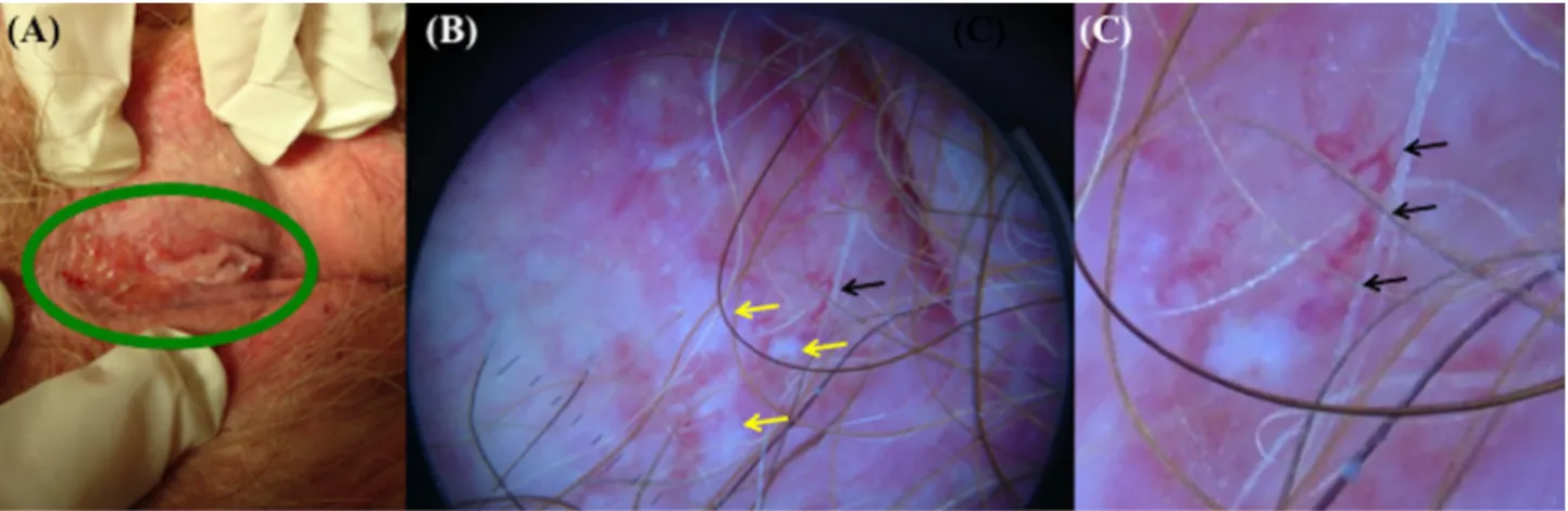
Figure 1. Clinical (A) and dermoscopic examination (B, C) of the first case of vulvar basal cell carcinoma. Clinical examination (A) shows
an ulcerated plaque (green circle) involving the anterior vulvar commisure. Dermoscopy (B, C) shows a reddish and well-focused arborizing vessel (black arrow) on a pinkish background associated with whitish homogeneous areas (yellow arrows). Magnified dermoscopic image (C) of arborizing vessel (black arrows). [Copyright: ©2018 Cinotti et al.]
Figure 2. Histopathologic examination shows a nodular basal cell
carcinoma, a superficial ulceration and enlarged vessels (hematoxy-lin and eosin, 5x). [Copyright: ©2018 Cinotti et al.]
70
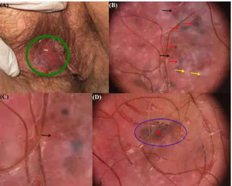
Observation | Dermatol Pract Concept 2018;8(1):17Figure 3. Clinical (A) and dermoscopic examination (B, C, D) of the second case of vulvar
basal cell carcinoma. Clinical examination (A) shows an erythematous-whitish plaque (green circle) with focal erosions and an irregular gray-blue pigmentation at the periphery (red arrow) on the right labium major. Dermoscopy (B, C, D) shows fine linear telangiectasia (black arrows), blue ovoid nests (red arrows), a blue globule (red dashed arrow), white shiny structures (yellow arrows) on a pinkish background and areas of brown dots (red asterisk) surrounded by a grayish pigmentation (blue circle). [Copyright: ©2018 Cinotti et al.]
Figure 4. In vivo (A,B,C) and ex vivo (D) reflectance confocal microscopy of the second
case of vulvar basal cell carcinoma. In vivo reflectance confocal microscopy (920 x 920 μm) reveals small dark silhouettes (red asterisks) in connection to the basal layers of the epidermis (A) and larger tumor islands with peripheral clefts (red asterisks) in the superficial dermis (B). In some areas, abundant melanophages (blue arrows) are also visible in the superficial dermis around tumor islands (C). Ex vivo reflectance confocal microscopy (single images of 750 x 750 μm) performed with the en face technique shows the tumor islands (red asterisks) (D).
patients [2]. The tumor is usually larger than 1 cm at presentation indicating a late diagnosis [2]. Subjective symptoms and clinical signs are often present for a prolonged period before the diagnosis. They include itching, irritation, discom-fort, palpable vulvar mass, pain, and bleeding [2,4,5]. The last sign is related to ulcerated lesions, which represent 28% of all vulvar BCCs [2,5]. Vulvar BCC can mimic inflammatory diseases such as eczema, psoriasis, and infec-tions (chronic candidiasis). It can also simulate Bowen disease, Paget disease, squamous cell carcinoma, leukoplakia, lichen ruber planus, lichen sclerosus, melanocytic nevus, melanoma, sebor-rheic keratosis, angioma, and other pig-mented and non-pigpig-mented tumors [6]. Therefore, the diagnosis is frequently delayed and it is usually performed after inappropriate treatment [2,3].
Dermoscopic features of vulvar BCC have been reported in only two cases, but RCM features have not been reported yet [6,8]. Non-invasive imag-ing techniques, such as dermoscopy and RCM, are of great interest since they orient the diagnosis of this rare tumor in this sensitive area. The dermoscopic features of our cases and of the previ-ously reported two patients with vulvar BCC [6,8] showed blue ovoid nests and telangiectasia as extragenital BCC. The previously published cases were both pigmented [6,8], whereas one of our cases was a non-pigmented BCC. In the case of pigmented BCC, ovoid nests allow for diagnosis of this tumor easily. Non-pigmented lesions can be more dif-ficult to identify because they can mimic inflammatory and infectious diseases. In the cases presented here, the presence of reddish and well-focused arborizing telangiectasia was a relevant clue to the diagnosis. Interestingly, both of our cases and one previously reported [6] showed prominent homogeneous whit-ish areas, which could be an additional clue for vulvar BCC. We hypothesize that these whitish areas could correlate with a peritumoral fibrosis that could
Observation | Dermatol Pract Concept 2018;8(1):17
71
confirm that the excision was complete. In vivo and ex vivo RCM have been reported to have a good diagnostic accuracy for cutaneous BCC [1,10], and our case suggests that they could also be used for genital BCC.In conclusion, vulvar BCC should be considered in the differential diagnosis of both pigmented and non-pigmented vulvar lesions. Dermoscopy and RCM can be useful tools for its diagnosis and treatment and for the identification of its surgical margins. Also, ex vivo RCM could be used in the perioperative setting of genital BCC. In our two cases, dermoscopic and RMC features were similar to extragenital BCC, except for the peculiar aspect of brown dots surrounded by grayish pigmentation that was observed on dermoscopic examination (Figure 2C).
References
1. Espinasse M, Cinotti E, Grivet D, et al. ‘En face’ ex vivo reflectance confocal microscopy to help the surgery of basal cell carcinoma of the eyelid. Clin Exp Ophthalmol. Epub 2017 Jan 31. 2. De Giorgi V, Salvini C, Massi D, et al. Vulvar basal cell carcinoma:
retrospective study and review of literature. Gynecol Oncol. 2005;97:192–194.
3. Fleury AC, Junkins-Hopkins JM, Diaz-Montes T. Vulvar bas-al cell carcinoma in a 20-year-old: Case report and review of the literature. Gynecol Oncol Case Rep. 2011; 2: 26-27.
4. Mulayim N, Foster Silver D, Tolgay Ocal I, et al. Vulvar basal cell carcinoma: two unusual presentations and review of the literature.
Gynecol Oncol. 2002; 85: 532-537.
5. Chokoeva AA, Tchernev G, Castelli E, et al. Vulvar cancer: a review for dermatologists. Wien Med Wochenschr. 2015;165:164-177.
6. Dobrosavljevic Vukojevic D, Djurisic I, Lukic S, et al. Derma-toscopy in vulvar basal cell carcinoma. J Eur Acad Dermatol
Venereol. 2017;31:e180-181.
7. Venkatesan A. Pigmented lesions of the vulva. Dermatol Clin. 2010;28:795-805.
8. De Giorgi V, Massi D, Mannone F et al. Dermoscopy in vulvar basal cell carcinoma. Arch Dermatol. 2007;143:426–427. 9. Bichakjian CK, Olencki T, Aasi SZ, et al. Basal Cell Skin Cancer,
Version 1.2016, NCCN Clinical Practice Guidelines in Oncology.
J Natl Compr Canc Netw. 2016;14(5):574-597.
10. Cinotti E, Jaffelin C, Charriere V, et al. Sensitivity of handheld reflectance confocal microscopy for the diagnosis of basal cell carcinoma: A series of 344 histologically proven lesions. J Am
Acad Dermatol. 2015;73:319-320.
be more visible on mucosa compared to the skin due to the absence of the stratum corneum and to the thinner epithe-lium.
In vivo RCM showed similar features of cutaneous BCC and allowed us to understand some dermoscopic findings better. One of our cases showed brown dots at dermoscopy that correlated with the superficial tumor islands in con-nection with the epidermis that were well visualized under RCM. The grayish pigmentation around the dots visible at dermoscopy correlated with the presence of pigmented melanophages under RCM. The presence of melanophages could be explained by the possible trauma in the genital area.
Vulvar BCC requires surgical margins wider than 4 mm because it is high risk for recurrence [9] and its extension can be difficult to identify by clinical examination due to the pink-ish color of the mucosa [6]. In fact, the erythema associated with BCC could be difficult to differentiate from the normal color of the mucosa. In our cases, both dermoscopy and RCM were used to identify the tumor margins before surgery. In vivo RCM examination allowed us to use narrow lateral sur-gical margins thanks to their precise identification. Moreover, the perioperative ex vivo RCM examination allowed us to
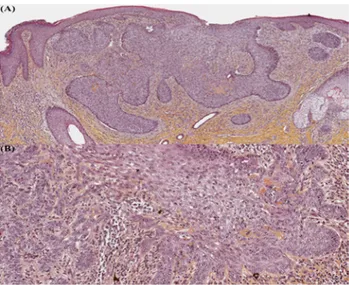
Figure 5. Histopathologic examination (A,B) of the second case of
vulvar basal cell carcinoma. Histopathologic examination shows a nodular basal cell carcinoma (A, hematoxylin and eosin, 2x) sur-rounded by an inflammatory reaction with melanophages (B, hema-toxylin and eosin, 10x). [Copyright: ©2018 Cinotti et al.]