The olfactory cleft localization of REAH seems to be more fre-quent than it was thought before.4,5Therefore, it is mandatory to think about this entity in all cases of diffuse nasal polyposis when, because of polypous tissue masses, it is not possible to distinguish where does the suspected mass come from: between the middle turbinate and the nasal septum or from the ostiomeatal complex proper. This is the main point to discover the real background of the disease. Moreover, misinterpretation of the REAH as chronic sinus inflammation and diffuse polyposis may result in an inap-propriate treatment.
In our case, the preoperative situation strongly suggested esthesineuroblastoma, not polyposis at all. Because of the opacity between middle turbinate and nasal septum, we thought also about REAH in particular because we knew that Athre and Ducic9found that REAH could be a locally aggressive process! They represent the patient with frontal sinus hamartoma with expansion to the posterior right orbital wall. The patient was cured with a frontal craniotomy and cranialization. This is exactly why we decided to remove the entire tumor, including both olfactory bulbs.
Regardless of the localization, the treatment of choice for hamar-tomas is complete local excision of the tumefaction. Conservative surgical resection seems to be curative, with no recurrences reported in variable follow-up periods.4,9Y12In comparison with this, our sur-gical treatment was really a radical one.
CONCLUSIONS
Hamartomas of the nasal cavity and paranasal sinuses are rare. These lesions are characterized by an abnormal overgrowth of tissue elements indigenous to a particular area of the body. Hamartomas do not have unlimited growth potential nor do they have metastatic potential. Ex-tensive progression of the disease to involve the orbit and intracranial vault is exceedingly rare but possible.
Conservative surgical resection of hamartomas is curative, and the literature describes no instances of recurrent, persistent, pro-gressive, or metastatic disease.
Our case denies some literature statements regarding the pro-gressiveness of this tumefaction because it was found to penetrate from the olfactory clefts to the endocranium. To our knowledge, our case is the first one reported with extension of the olfactory cleft REAH to the endocranium.
REFERENCES
1. Albrecht E. Uber Hamartome. Verh Dtsch Ges Pathol 1904;7:153Y157
2. Delbrouck C, Aguilar SF, Choufan G, et al. Respiratory epithelial adenomatoid hamartoma associated with nasal polyposis. Am J Otolaryngol 2004;25:282Y284
3. Wenig BM, Heffner DK. Respiratory epithelial adenomatoid hamartomas of the sinonasal tract and nasopharynx: a clinicopathologic study of 31 cases. Ann Otol Rhinol Laryngol 1995;104:639Y645
4. Lima NB, Jankowski R, Georgel T, et al. Respiratory epithelial adenomatoid hamartoma must be suspected on CT-scan enlargement of the olfactory clefts. Rhinology 2006;44:264Y269
5. Cao ZW, GU ZW, Yang J, et al. Respiratory epithelial adenomatoid hamartoma of bilateral olfactory clefts associated with nasal polyposis: three cases report and literature review. Auris Nasus Larynx 2010;37:352Y356
6. Liang J, O’Malley BW Jr, Feldman M, et al. A case of respiratory epithelial adenomatoid hamartoma. Am J Otolaryngol
2007;27:277Y279
7. Mortuaire G, Pasquesoone X, Leroy X, et al. Respiratory epithelial adenomatoid hamartoma of the sinonasal tract. Eur Arch Otorhinolaryngol 2007;264:451Y453
8. Di Carlo R, Rinaldi R, Ottaviano G, et al. Respiratory epithelial adenomatoid hamartoma of the maxillary sinus: case report. Acta Otorhinolaryngol Ital 2006;26:225Y227
9. Athre R, Ducic Y. Frontal sinus hamartomas. Am J Otolaryngol 2005;26:419Y421
10. Endo R, Matsuda H, Takahashi M, et al. Respiratory epithelial adenomatoid hamartoma in the nasal cavity. Acta Otolaryngol 2002;122:398Y400
11. Himi Y, Yoshizaki T, Sato K, et al. Respiratory epithelial adenomatoid hamartoma of the maxillary sinus. J Laryngol Otol 2002;116:317Y318
12. Metselaar RM, Stel HV, Van der Baan S. Respiratory epithelial adenomatoid hamartoma in the nasopharynx. J Laryngol Otol 2005;119:476Y478
Transient Cardiac Failure Due
to Takotsubo Cardiomyopathy
After Surgical Reduction
of Nasal Fracture
Matteo Brucoli, MD, Francesco Arcuri, MD, Mariangela Giarda, MD, Arnaldo Benech, MD, PhD
Introduction: Takotsubo syndrome, also known as ampulla car-diomyopathy, broken heart syndrome, idiopathic apical ballooning syndrome, and stress-induced myocardial stunning, has been first described by Japanese authors in 1996 and subsequently specified in 2001; it derives from the resemblance between the ancient round-bottomed, narrow-necked Japanese fishing pots used to trap octo-pus in Asia and the end-systolic appearance of the left ventricle on ventriculography.
Clinical Report: We introduce the case of a woman who was in-volved in a traffic car crash and, subsequently, was admitted to the Maxillo-Facial Unit of the Novara Major Hospital with a diagnosis of nasal fracture. She underwent general anesthesia for the reduction of the fracture; after surgery, she developed acute chest pain, elevated cardiac biomarkers, ischemic electrocardiogram changes, and tran-sient akinesis of the left ventricle without significant epicardial cor-onary artery disease. A diagnosis of takotsubo syndrome was made. Conclusions: This syndrome, which presents the same clinical features of a ventricular failure, is probably underdiagnosed, but after the introduction of sophisticated cardiac imaging and coro-nary intervention, more cases are identified and an unnecessary thrombolytic therapy can be spared. This reversible condition, which
From the Department of Maxillo-Facial Surgery, Azienda Ospedaliera Maggiore della Carita`, University of Piemonte Orientale BAmedeo Avogadro,[ Novara, Italy.
Received September 1, 2010.
Accepted for publication September 17, 2010.
Address correspondence and reprint requests to Francesco Arcuri, MD, S.C.D.U. di Chirurgia Maxillo-Facciale, Ospedale Maggiore della Carita`, Corso Mazzini 18, 28100 Novara, Italy; E-mail: [email protected] The authors report no conflicts of interest.
Copyright* 2011 by Mutaz B. Habal, MD ISSN: 1049-2275
DOI: 10.1097/SCS.0b013e31822ea720
The Journal of Craniofacial Surgery
&
Volume 22, Number 5, September 2011 Brief Clinical Studies* 2011 Mutaz B. Habal, MD
1907
is, to our knowledge, never mentioned after a craniomaxillofacial surgical procedure, should be considered in the diagnostic algorithm for all patients presenting with acute onset of chest pain, elevated cardiac biomarkers, and ischemic changes on the electrocardiogram after a general anesthesia.
Key Words: General anesthesia, takotsubo syndrome, nasal fracture
T
akotsubo syndrome, also known as ampulla cardiomyopathy, broken heart syndrome, idiopathic apical ballooning syndrome, and stress-induced myocardial stunning, has been first described by Japanese authors1in 1996 and subsequently specified in 20012; although most cases have been initially described in this country,3,4 nowadays there are reports from other parts of the world.5,6The name of this syndrome (tako means octopus and tsubo means pot) derives from the resemblance between the ancient round-bottomed, narrow-necked Japanese fishing pots used to trap octo-pus in Asia and the end-systolic appearance of the left ventricle on ventriculography.
This condition is characterized by a combination of the follow-ing: (1) acute psychologic/physical stress before the onset of chest pain, 2) disproportionately low release of cardiac enzymes with re-spect to the degree of the left ventricular dysfunction, 3) ischemic changes on the electrocardiogram (ECG), 4) no significant epicar-dial coronary artery disease, 5) transient akinesis or dyskinesia of the left ventricle, 6) apical ballooning with basal hyperkinesis on the left
ventriculogram, 7) rapid resolution of the cardiac dysfunc tion, and 8) absence of recent head trauma, intracranial bleeding, pheochromo-cytoma, myocarditis, and hypertrophic cardiomyopathy.7
We introduce the case of a takotsubo syndrome in a patient who underwent general anesthesia for the reduction of a nasal fracture.
CLINICAL REPORT
On April 27, 2010, a 57-year-old white woman (American Society of Anesthesiologists status II, 56 kg) was involved a traffic car crash and, subsequently, was admitted to the Maxillo-Facial Unit of the Novara Major Hospital with a diagnosis of nasal fracture (Fig. 1). She had a history of hypertension, hepatitis, and bronchial asthma. She had no known drug allergies, and results of the preoperative laboratory tests were within the reference ranges; preoperative blood pressure and heart rate were 140/80 mm Hg and 74 beats/min, respectively.
General anesthesia was induced with propofol, and after the ad-ministration of rocuronium, the trachea was intubated with a 7-mm endotracheal tube; anesthesia was maintained with sevoflurane and fentanyl. Intraoperative monitoring consisted of ECG, noninvasive blood pressure measurement, and pulse oximetry. After a successful surgical reduction of the nasal fracture, the patient was extubated, but 2 hours after the end of the surgical procedure, she presented acute chest pain and hypertension; both conditions were rapidly con-trolled by esmolol and 2 consecutive sublingual administrations of nitroglycerine. Serial 12-lead ECG recordings demonstrated evolv-ing anterolateral T wave inversion; postoperative cardiac enzymes FIGURE 1. Axial computed tomographic scan demonstrating
the nasal fracture.
FIGURE 2. A and B, A 12-lead ECG showing evolution of widespread ST/T wave changes throughout the anterior cheat leads without Q waves including dynamic ST elevation (A) and T wave inversion (B).
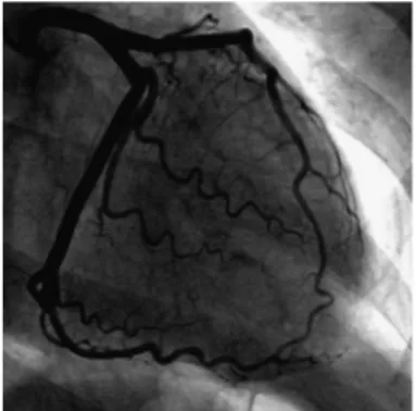
FIGURE 3. Diagnostic coronary angiogram demonstrating normal epicardial coronary arteries.
Brief Clinical Studies The Journal of Craniofacial Surgery
&
Volume 22, Number 5, September 20111908
* 2011 Mutaz B. Habal, MDshowed elevation of troponin I and creatine kinase MB (1.76 and 8 ng/mL, respectively; reference,G0.01 and G5.00 ng/mL, respectively; Fig. 2). A transthoracic echocardiogram demonstrated severely im-paired left ventricular systolic function with akinesia of all mid and apical segments, hyperkinetic basal contraction, and left ventricular ejection fraction of 35%.
Transfer to the cardiothoracic center took place with emergency diagnostic coronary angiography and cardiac magnetic resonance image demonstrating normal epicardial coronary arteries, apical akinesia with typical ballooning, and basal hyperkinesias without signs of ischemic lesions; diagnosis of takotsubo cardiomyopathy was made (Figs. 3Y5). On postoperative day 3, a test transesophageal echocardiography showed normal ventricular function with no seg-mental wall motion abnormality; on the same day, the troponin I level decreased to 0.04 ng/mL. The patient’s postoperative course was un-eventful, and on the postoperative day 5, she was discharged to home with aspirin, A-blocker, and angiotensin-converting enzyme inhibi-tor. The patient has been reviewed in the outpatient clinic and she is well; a subsequent echocardiograph showed a normal volume status with normal wall motion and no mitral regurgitation.
DISCUSSION
Takotsubo is a rare disorder; in a recent study, the annual inci-dence of this syndrome in a Western population is calculated to be 0.00006%.7Interestingly, in the Japanese population, the prevalence of this syndrome has been reported to be 1%.8There might be a genetic component that has not been yet discovered; the mean age at onset of this syndrome is 62 to 75 years, and it frequently occurs in postmenopausal women.
The pathophysiology of this condition is debatable; numerous hypotheses have been introduced: (1) catecholamine-mediated car-diotoxicity, (2) coronary vasospasm, (3) microvascular dysfunction, (4) left ventricular outflow tract obstruction, and (5) cardiac auto-nomic imbalance.9 Catecholamine mediated cardiotoxicity is the most accepted mechanism; patients characteristically present with a preceding history of psychologic/physical stress leading to increased sympathetic activity with a direct catecholamine toxic effect on the cardiac myocytes.10
Results are, however, conflicting; some studies report elevated catecholamine levels in takotsubo cardiomyopathy, whereas in others, catecholamine levels are normal; it is more likely that the pathogenesis of this syndrome is multifactorial.11The overall prog-nosis of this condition is favorable; however, fatal complications such as cardiogenic shock and tachyarrhythmias can occur. Although the presence of a psychologic or physical stress is accepted as one of the
main features of the takotsubo syndrome, some cases have been reported to occur without any triggering effect.3
We theorize that stress caused by superficial anesthesia, insuf-ficient analgesia, and surgical pain stimulus created a stressful event that caused a potentially life-threatening catecholamine release as basis of takotsubo syndrome in our patient. Because this condition leads to ventricular dysfunction with apical ballooning, the patients are potentially at a risk for ventricular thrombus formation and decisions regarding anticoagulation need to be discussed. Patients can also develop functional mitral regurgitation with hemodynamic failure; nowadays, no established guidelines exist.5
According to the literature, this syndrome has been frequently associated to emotional stress, sexual intercourse, and traumatic injury; it has been also described in patients with epileptic attacks, bronchial asthma exacerbations, dialysis, and during electrophysi-ological studies.
Wittstein et al12 described 19 patients with ventricular dys-function after sudden emotional stress who presented ECG, echo-cardiographic, and angiographic patterns similar to takotsubo cardiomyopathy; they identified a remarkable increase in catechol-amine levels in such patients.
Tsuchihashi et al2 described patients with transient apical bal-looning associated with delivery, intubation, tracheotomy, lung bi-opsy, orthopedic surgery, colonectomy, and cholecystectomy.
It is evident that this syndrome is undistinguishable from the myo-cardial ischemia, and proper diagnosis is possible with angiographic evidence. The course is related to the entity of the left ventricular dys-function, and signs of cardiac failure can be very severe, although the lesions are reversible.
The characteristic ECG features of takotsubo cardiomyopathy are nonspecific and include dynamic ST elevation (usually less than in acute anterior myocardial infarction) and/or T wave inversion typi-cally throughout the anterior leads13,14; our patient also had elevated levels of cardiac biomarkers.
Transthoracic echocardiography can identify regional wall ab-normalities such as akinesia of the apex and/or the midportion of the left ventricle. Typically, the area of dysfunction usually involves a larger territory than that supplied by 1 epicardial coronary artery.
Diagnostic coronary angiography needs to be performed in all patients to exclude obstructive epicardial CAD; ventriculography may demonstrate the apical ballooning that occurs in addition to hypercontraction of the basal segments.15
Cardiac magnetic resonance imaging provides information re-garding functional involvement, chamber dimensions, and presence of intramyocardial edema; it also allows the physicians to exclude infarction and inflammatory processes.16
FIGURE 4. A and B, Contrast left ventriculogram showing akinesis of the apical region and hyperkinesis of the basal segments at end-systole (A) and end-diastole (B).
FIGURE 5. Cardiac magnetic resonance image showing left ventricular apical akinesis and basal hyperkinesis at end-systole without signs of ischemic lesions.
The Journal of Craniofacial Surgery
&
Volume 22, Number 5, September 2011 Brief Clinical Studies* 2011 Mutaz B. Habal, MD
1909
CONCLUSIONS
This syndrome, which presents the same clinical features of a ven-tricular failure, is probably underdiagnosed, but after the introduc-tion of sophisticated cardiac imaging and coronary intervenintroduc-tion, more cases are identified, and an unnecessary thrombolytic therapy can be spared.
This reversible condition, which is, to our knowledge, never mentioned after a craniomaxillofacial surgical procedure, should be considered in the diagnostic algorithm for all patients presenting with acute onset of chest pain, elevated cardiac biomarkers, and ischemic changes on the ECG after a general anesthesia.
REFERENCES
1. Satoh H, Tateishi H, Uchida T. Takotsubo-type cardiomyopathy due to multivessel spasm. In: Kodama K, Haze K, Hon M, eds. Clinical Aspects of Myocardial Injury: From Ischemia to Heart Failure. Tokyo, Japan: Kagakuhyouronsya Co, 1990:56Y54 2. Tsuchihashi K, Ueshima K, Uchida T, et al. Transient left ventricular
apical ballooning without coronary artery stenosis: a novel heart syndrome mimicking acute myocardial infarction. Angina Pectoris-Myocardial Infarction Investigations in Japan. J Am Coll Cardiol 2001;38:11Y18
3. Kondo M, Matsuoka R, Araki M, et al. Assessment of clinical features in transient left ventricular apical ballooning. J Am Coll Cardiol 2003;41:737Y742
4. Dote K, Sato H, Tateishi H, et al. Myocardial stunning due to simultaneous multivessel coronary spasms: a review of 5 cases. J Cardiol 1991;21:203Y214
5. Kawai S, Kitabatake A, Tomoike H. Guidelines for diagnosis of takotsubo (ampulla) cardiomyopathy. Circ J 2007;71:990Y992 6. Gianni M, Dentali F, Grandi AM, et al. Apical ballooning syndrome
or takotsubo cardiomyopathy: a systematic review. Eur Heart J 2006;27:1523Y1529
7. Donohue D, Movahed MR. Clinical characteristics, demographics and prognosis of transient left ventricular apical ballooning syndrome. Heart Fail Rev 2005;10:311Y316
8. Akashi YJ, Musha H, Kida K, et al. Reversible ventricular dysfunction takotsubo cardiomyopathy. Eur J Heart Fail 2005;7:1171Y1176
9. Akashi YJ, Barbaro G, Sakurai T, et al. Cardiac autonomic imbalance in patients with reversible ventricular dysfunction takotsubo cardiomyopathy. QJM 2007;100:335Y343
10. Iqbal MB, Moon JC, Guttmann OP, et al. Stress, emotion and the heart: tako-tsubo cardiomyopathy. Postgrad Med J 2006;82:29 11. Dote K, Sato H, Tateishi H, et al. Myocardial stunning due to
simultaneous multivessel coronary spasms. A review of 5 cases. J Cardiol 1991;21:203Y214
12. Wittstein IS, Thiemann DR, Lima JA, et al. Neurohumoral features of myocardial stunning due to sudden emotional stress. N Engl J Med 2005;352:539Y548
13. Mitsuma W, Kodama M, Ito M, et al. Serial electrocardiographic findings in women with takotsubo cardiomyopathy. Am J Cardiol 2007;100:106Y109
14. Kurisu S, Sato H, Kawagoe T, et al. Tako-tsuboYlike left ventricular dysfunction with ST-segment elevation: a novel cardiac syndrome mimicking acute myocardial infarction. Am Heart J 2002;143: 448Y455
15. Klinceva M, Widimsk] P, Pesl L, et al. Prevalence of stress-induced myocardial stunning (tako-tsubo cardiomyopathy) among patients undergoing emergency coronary angiography for suspected acute myocardial infarction. Int J Cardiol 2007;120:411Y413 16. Mitchell JH, Hadden TB, Wilson JM, et al. Clinical features and
usefulness of cardiac magnetic resonance imaging in assessing
myocardial viability and prognosis in takotsubo cardiomyopathy (transient left ventricular apical ballooning syndrome). Am J Cardiol 2007;100:296Y301
Maxillary Cementoblastoma
in a Child
Fa´bio Wildson Gurgel Costa, DDS, MS,* Karuza Maria Alves Pereira, DDS, PhD, MS,. Marcelo Magalha˜es Dias, DDS,
MS,-Ma´rcia Cristina da Costa Miguel, DDS, PhD, MS,` Maria Adriana Skeff de Paula Miranda, DDS, MS,k Eduardo Costa Studart Soares, DDS, PhD, MSP
Abstract: Cementoblastoma is a rare benign tumor that almost al-ways occurs in the premolar or molar region and more commonly in the mandible than in the maxilla. We present a unique incisor maxillary cementoblastoma in an 11-year-old child not previously described. To our knowledge, only 2 maxillary cases, both related to canine teeth, were described in the international literature. Thus, the aim of this article was to discuss the clinical presentation, diagnosis, and subsequent treatment of a patient with a cementoblastoma in the anterior maxillary region.
Key Words: Cementoblastoma, odontogenic tumors, maxilla
C
ementoblastoma is a rare benign tumor and comprises less than 1% to 6.2% of all odontogenic tumors.1This lesion is more frequent in young patients, with about 50% of the cases arising in younger than 20 years.2 Approximately all benign cemento-blastomas are intimately associated and partially enclosed to 1 or more roots of a single posterior mandibular erupted permanent tooth.2,3However, a few articles concerning maxillary lesions have been published, most of them forthcoming in the posterior site.4We present a unique incisor maxillary cementoblastoma in a child not previously described. To date, only 2 maxillary cases, both related to canine teeth, were reported in the international litera-ture.4,5Therefore, the aim of this work was to discuss the clinical
From the *Division of Stomatology, School of Dentistry, Federal University of Ceara´ Sobral Campus;.Division of Oral Pathology, Federal University of Ceara´ Sobral Campus, Ceara´;-Division of Oral Rehabilitation, School of Dentistry, Catholic Faculty Rainha do Serta˜o, Quixada´;`Division of Oral Pathology, Federal University of Rio Grande do Norte, Natal; kDivision of Stomatology, School of Dentistry, Catholic Faculty Rainha do Serta˜o, Quixada´; andPDivision of Oral Surgery, School of Dentistry, Federal University of Ceara´, Ceara´, Brazil.
Received August 19, 2010.
Accepted for publication September 25, 2010.
Address correspondence and reprint requests to Fa´bio Wildson Gurgel Costa, DDS, MS, Coordena0a˜o do Curso de Odontologia da Universidade Federal do Ceara´ Campus Sobral; Av. Comte. Mauroce´lio Rocha Pontes, s/n.- Derby, CEP 62.041-040, Sobral-Ce, Brazil; E-mail: fwildson@ yahoo.com.br
The authors report no conflicts of interest. Copyright* 2011 by Mutaz B. Habal, MD ISSN: 1049-2275
DOI: 10.1097/SCS.0b013e31822ea6e8
Brief Clinical Studies The Journal of Craniofacial Surgery