Università degli Studi di Messina
Dipartimento di Scienze Matematiche, Informatiche, Fisiche e Scienze della Terra (MIFT)
Dottorato di Ricerca in Fisica XXXI CICLO
Biocompatible Nanoparticles: Synthesis,
Analysis and Applications
in Biological and Medical fields
PhD Student: Nancy RESTUCCIA
PhD Tutor:
Chiar.mo Prof. Lorenzo TORRISI
Referee: Prof. Mackova Anna
Prof. Ronca Sara
Settore Scientifico Disciplinare FIS/01 2017-2018
REFEREES:
Professor Mackova Anna
Nuclear Physics Institute (ASCR), 25068 Rez, Czech Republic
Mail: [email protected]
Professor Ronca Sara
Loughborough University, Department of Materials
Leicestershire, UK, LE11 3TU
I
INTRODUCTION
This Thesis reports the work I have done at the Department of Mathematics, Informatics, Physics and Earth Sciences (MIFT) of the University of Messina under the supervision of Prof. Lorenzo Torrisi, from 2015 to 2018. The study carried out in this period was focused on the use of metallic nanoparticles in the medical and biological fields.
In recent years, technological development has allowed us to prepare, manipulate and characterize more nano-sized materials. The nano prefix, which derives from the Greek nanos and means ‘dwarf’, describes materials, technologies or properties with dimensions on the nanoscale (10-9 m). Nanotechnology is an interdisciplinary
field that involves the study and application of natural sciences and engineering to develop tools at the nanoscale. The nanotechnology applied to life sciences has led to improve knowledge about cellular and molecular processes and factors that cause a variety of diseases. The understanding of novel disease pathways has acted as a catalyst for the development of nanomedicine. Advances in this field promise to increase the accuracy and specificity of medical diagnoses and treatments of diseases. Metallic nanoparticles (NP), based on Ti, Au, Ag, Bi and others, embedded in an insulating medium behave like ionizing radiation absorbing centers, being able to show certain absorption resonances bands at specific wavelengths, thank to the Surface Plasmon Resonance (SPR) absorption effect [1]. Their size distribution in a fluid changes the electronic and mass density and the material equivalent atomic number. In the present study the behaviour of metallic nanoparticles in therapy and in diagnostics was determined experimentally, in particular by calculating the ratio of the delivered dose to a given depth and the delivered dose to the surface in various media (water, adipose tissue and compact bone) in presence or not of the nanoparticles. The choice of gold, as starting material to prepare nanoparticles, is not accidental. In fact, gold is stable, not oxidable and biocompatible for humans and, more in general, also for other living beings. In the
II light of these results, the medicine demonstrates a particular interest to the gold nanoparticles for different aspects concerning prosthesis, interface adhesion, high Z contrast medium for X-ray images.
The anticancer potential of the gold nanoparticles is generated by the different advantageous physico-chemical properties. Numerous studies have demonstrated the safety and biocompatibility of gold both in vitro and vivo, suggesting that the gold nanoparticles can be administered safely with minimal inflammatory activation and few local or systemic side effects [2] enhancing the equivalent Z-value of the tissue at significant concentrations.
Fig. 1- . Au NPs with various shapes and size (a), bio-functionalized with various biomolecules (c), creates bioconjugaties. The subministration can take place in various ways, such as intratumoral injection, intravenously and by means of suitable drugs. The NPs have the ability to enhance radiation therapy of tumors (b), as well as serve as high Z imaging contrast agents (d).
Figure 1 shows Au NPs of various shapes and sizes (a), their functionalization with particular molecules (c) which allows the transfer to the tumor sites via or biological processes or use of drugs by localized injection into the tumor zone.
The so attached metallic NPs to the tumor have the ability to improve radiotherapy and imaging of localized tissues (b, d).
III The high atomic number of gold (Au, Z = 79) allows a high absorption and enhancement of ionizing radiation, as well as the X-ray attenuation and X-ray characteristic line fluorescence for better imaging applications [3].
The inclusion of metallic nanoparticles in a liquid, at different concentrations, modifies significantly the solution properties, especially in physical terms concerning the surface tension, density, viscosity, thermal and electric conduction, vapour pressure, and other parameters [4].
The wettability of the liquid changes introducing specific nanoparticles and the resulting adhesion of the liquid to a surface may be drastically modified. This aspect is of special interest in Biology and Medicine because it has been demonstrated that the wetting ability determines the protein absorption and the cellular response [5] and that it represents a key parameter for the success of prostheses implantation and functionality in human body [6].
The main goals of this thesis work are three: i) to synthesize various metallic nanoparticles (such as Au, Bi,…) by laser ablation in water; ii) to characterize the so obtained nanoparticles by means of different spectroscopy techiniques; iii) to apply them in some interesting biological and medical fields. In particular, the thesis consists of six chapters. The first two chapters are an overview of metallic nanoparticles, their properties, the synthesis methods and the characterization systems. The main chapters are the third, fourth and sixth.
The third shows our obtained results in modifying the materials by adding nanoparticles.
The fourth chapter shows the obtained results by exploiting the nanoparticles (Au, Ag and Bi) in the field of imaging.
Finally, in the last chapter we propose a theoretical study of a clinical case of breast cancer treated with radiotherapy; the behavior of Au nanoparticles in radiotherapy was determined by calculating in particular the ratio between the relased dose at the tumor depth and the released dose on surface.
INDEX
INTRODUCTION ... I
1. Nanoparticles and Biomaterials ... 1
1.1. Nanoparticles ... 2
1.2. Surface Plasmon Resonance ... 4
1.3 Biomaterials ... 10
1.3.1 Metallic Biomaterials ... 12
1.3.2 Polymeric Materials ... 14
1.3.3 Ceramic Biomaterial ... 17
2. Synthesis and Characterization of Nanoparticles ... 21
2.1. Synthesis of Nanoparticles ... 22
2.2. Nanofabrication by “Top Down” Methods. ... 24
2.2.1 Laser Ablation ... 24
2.2.2 Our set up for Laser Ablation ... 26
2.2.3 The Laser Ablation: Theoretical background ... 28
2.2.4 Temperature Dependence ... 29
2.2.5 Irradiation, Energy, Wavelength and Time Depedence ... 31
2.2.6 Photochemical effects of Laser ... 33
2.3. Nanofabrication by “Bottom-Up” Methods ... 35
2.4. Physical Characterization of Nanoparticles ... 39
2.5.1 Electron Microscopy: TEM and SEM ... 40
2.5.2 Optical Spectroscopy (UV-Vis Spectroscopy) ... 46
2.5.3 X-Ray Diffraction (XRD) ... 49
2.5.4 Raman Spectroscopy ... 52
3. Modification of the material using Metallic Nanoparticles ... 55
3.1. Wettability ... 56
3.2. Wettability of Biological Liquids with Metallic Nanoparticles ... 59
3.2.2 Methods of analysis ... 60
3.2.3 Results ... 63
3.3. Optical and mechanical properties: Static and dynamic characterization of biomedical polyethylene laser welding using biocompatible nano-particles. 72 3.3.1 Materials ... 73
3.3.2 Physical Characterization ... 74
3.3.3 Mechanical Characterization ... 75
3.3.4 Results ... 76
3.4. Gold Nanoparticles produced by Laser Ablation in water and in graphene oxide suspension ... 81
3.4.1 Experimental set up ... 82
3.4.2 Results ... 85
4. Use of Metallic Nanoparticles for Biological and Medical Diagnostics ... 95
4.1. Contrast Agents in X-ray imaging and computed Tomography ... 96
4.1.1 Contrast agents for CT ... 97
4.1.2 Gold nanoparticles as x-ray contrast agents ... 98
4.1.3 Contrast enhancement ... 100
4.1.4 Design of AuNPs as x-ray contrast agents ... 102
4.2. Laser-produced Au nanoparticles as X-ray contrast agents for diagnostic imaging ... 104
4.2.1 Used Materials ... 107
4.2.2 Mechanism of X-ray absorption in liquids and tissue ... 109
4.2.3 X-ray diagnostic enhanced by Au NPs ... 112
4.3. Comparison between Au and Ag Nanoparticles generated by laser in liquid as contrast medium. ... 116
4.3.1 Used Materials ... 117
4.3.2 Results ... 118
4.4. Laser-generated Bismuth Nanoparticles for application in Imaging and Radiotherapy. ... 120
4.4.1 Materials ... 121
4.4.2 Results ... 124
4.5. Comparing different types of Nanoparticles as X-ray contrast agents for diagnostics imaging ... 129
5. Radioterapy and Thermotherapy ... 134
5.1. Radiotherapy ... 135
5.1.1 Radiation with Photons ... 137
5.1.2 Radiotherapy with Electrons ... 138
5.1.3 Protontherapy ... 139
5.2. Different Types of radiotherapy ... 142
5.3. Radiotherapy Techniques and Criteria ... 144
5.3.1 Doses given to the Patient ... 145
5.4. Radiotherapy enhancement with Gold Nanoparticles ... 147
5.4.1 Properties of gold nanoparticles ... 148
5.4.2 Physics of Metal-Enhanced Radiotherapy ... 150
5.4.3 Therapy using Gold Nanoparticles in mice ... 151
5.5. Thermalterapy (Hyper-Thermia) ... 154
6. Theoretical Case Study: Radiotherapy with Gold Nanoparticles of Breast Cancer ... 158
6.1. Calculation of the Dose Ratio between the surface and the depth at which the tumor is located. ... 162
6.1.1 Protons ... 163
6.1.2 Electrons ... 167
6.1.3 Photons ... 171 CONCLUSIONS ... I REFERENCES ... I
2
1.1. Nanoparticles
In the last three decades nanoparticles have received increasing interest from scientific research. This is due to the dimension-dependent unique properties of nanoparticles, they have often been thought of as a distinct and intermediate state of matter between the individual atoms and the massive material [7].
The properties of the nanoparticles are presented as a consequence of the confinement of the electronic wave function and of an extremely high proportion of the surface of the atoms; both these factors are directly dependent on the nanoparticle size [8]. Indeed the possibility to check the properties, adjusting the nanoparticle size, has been the cause and the subject of so much research. Unlike bulk materials that have constant physical properties indifferent to mass, the nanoparticles offer unique opportunities for control when varying their diameter and they have alterable electronic, magnetic and optical properties. These effects arise because the energy levels for small particles are not continuous, as in bulk, but discrete due to the confinement of the wave function electron. The physical properties of the nanoparticles are therefore determined by theri size and by the relatively small physical dimension in which the wave function is confined (Fig. 1.1).
Fig. 1.1- The evolution of the band gap and the density of states as the number of atoms in a system grows (from right to left). EF is the Fermi energy level of the material and δ is the Kubo
3 The energy level of Fermi (EF) is the highest level of energy occupied by the system
in its ground state (lower energy). The band gap (Eg) of these systems is the energy
space between the highest unoccupied and the lowest occupied by the energy states. In these systems, from discrete atoms to bulk materials, the energy ranges are determined by the extent of the overlap between the electronic orbitals of the material. The individual atoms have well-known atomic orbitals. They can be combined into molecules to form molecular orbitals and also extensive band structures, such as metals or semiconductors. The value of Eg is proportional to EF
divided by the number of delocalized electrons in the extended band structure. For a bulk metal, the number of delocalized electrons in the band structure is equal to the number of atoms in the mass of the material. This results in a very low Eg value,
and therefore observed only at low temperature. Under a normal temperature, the delocalized metal electrons can easily be promoted to a higher energy state, and can move freely in the structure. This gives the material its electrically conductive nature. In traditional semiconductor materials the number of delocalized electrons is significantly lower than the number of atoms. This is evidenced by a higher value of Eg which is significant at room temperature. This means that in a semiconductor
the electrons will not be free to move and conduct current without some additional energy input. The following Equation 1.1 gives us the average level of electronic energy that distances successive quantum levels (known as the Kubo gap).
𝛿 =4𝐸
3𝑛 (1.1) where δ is the Kubo gap, EF is the Fermi energy level of the bulk material, and n is
the total number of valence electrons. Because of the Kubo gap in nanoparticles, properties like electrical conductibility and magnetic susceptibility show quantum size effects. These effects have led the nanoparticles to be used in many applications since catalysis, at optics up to medicine.
4
1.2. Surface
Plasmon Resonance
The excitation of surface plasmons (SPs) in metallic nanoparticles (NPs) induces optical properties hardly achievable in other optical materials, yielding a wide range of applications in many fields. Surface plasmon (SP) resonance is the most outstanding optical property of metallic nanostructures. It consists of a collective oscillation of conduction electrons excited by the electromagnetic field of light. SP resonance is the origin of optical properties hardly achievable with other physical processes [10]. In the case of metallic nanoparticles (NPs), where the electrons are confined in the three dimensions, the electron oscillations induces an electric field around the NP that can be much larger than the incident light one. SPs are one of the best examples that are different at the nanoscale. When the size of a metallic particle is reduced to a few nanometers, the optical properties are dramatically modified by the appearance of SPs and its resulting behavior is completely different from the bulk metal one. SPs open the possibility to amplify, concentrate and manipulate light at the nanoscale, overcoming the diffraction limit of traditional optics and increasing resolution and sensitivity of optical probes [11-14]. Consequently, SPs can be used in a wide range of fields, including biomedical [15], energy [16-18], environment protection [19-21], sensing [22] and information technology [23]applications. Nowadays, there are well-established applications of SPs that increase rapidly with the developmentof our capabilities to fabricate and manipulate nanomaterials. Moreover, NPs are not the only metallic nanostructures exhibiting SPs. Films, wires and patterned media exhibit also SPs [24-25]. In the case of NPs, the confinement of electrons leads to localized SPs, while other structures with large dimensions compared with light wavelength (wires and films) hold extended SPs that propagate along the interface between the metal and the dielectric medium. There is a third case corresponding to mesoscopic systems, with dimensions comparable to wavelength. In this case SP modes can also be excited. The scientific study of SPs started in the early 20th century when Gustav Mie published his pioneering work explaining the surprising optical properties of metallic colloids [26]. However, SPs in NPs have been empirically used for a long time, particularly for glass colouring. After Mie’s work in the early 20th century,
5 the origin of the optical properties of metallic NPs was understood, but further exploitation was limited by the capabilities to synthesize and manipulate NPs in a controlled way. It was not until the development of nanotechnology at the end of 20th century that the applications of SPs spread quickly in many fields. SPs correspond to an interaction between matter and the electromagnetic field of the light. Thus, the exact analysis of SPs implies solving the Maxwell equations with the appropriate boundary conditions. The solution of these equations is only possible for certain conditions and even in this case, the results are mathematical series that do not explain what are SPs. However, a simplified classical picture can be more useful to understand the physical meaning of SPs. A metallic NP can be described as a lattice of ionic cores with conduction electron moving almost freely inside the NP (the Fermi sea) as figure 1.2 illustrates. When the particle is illuminated, the electromagnetic field of the light exerts a force on these conduction electrons moving them towards the NP surface. As these electrons are confined inside the NP, negative charge will be accumulated on one side and positive charge in the opposite one, creating an electric dipole. This dipole generates an opposite electric field to that of the light that will force the electrons to return to the equilibrium position.
Fig. 1.2- Scheme of the light interaction with a metallic NP. The electric field of the light induces the movement of conduction electrons which accumulate at the NP surface creating an electric dipole. This charge accumulation creates an opposite electric field to that of the light [10].
The larger the electron displacement, the larger the electric dipole and consequently the restoring force. The situation is similar to a linear oscillator with a restoring force proportional to the displacement from the equilibrium position. If the electrons are displaced from the equilibrium position and the field is removed later, they will oscillate with a certain frequency that is called the resonant frequency; in
6 the case of SPs it is named the plasmonic frequency. Actually, the electron movement inside the NP exhibits some degree of damping.
The ionic cores and the NP surface partially damp the electron oscillations. Thus, the system is similar to a linear oscillator with some damping. When an alternating force is applied to a linear oscillator, the system oscillates with the same frequency as the external force but the amplitude and phase will depend on both the force and the intrinsic parameters of the oscillator. In particular, the oscillating amplitude will be maxima for the resonant frequency (Fig. 1.3(a)). It is quite straightforward to understand that, if the frequency of the external force is the same as the plasmonic frequency of the NP, it will be easy to make the electrons oscillate, but as we move far way from this frequency the movement of electrons will be more difficult, i.e. with reduced amplitude. We cannot directly observe the movement of electrons to determine their oscillating amplitude. However, we can determine this amplitude indirectly. The electronic oscillation implies an increase in kinetic and electrostatic energies associated with the electric fields of the dipole. As energy must be conserved, this increase in energy must be provided by the illuminating light. Therefore, the light extinguishes partially when exciting SPs inside the NP. The larger the electron oscillations, the larger the light extinction, so the optical absorption spectrum allows one to detect the excitation of SPs.
The resonant frequency for these oscillations in metallic NPs corresponds typically to UV–Vis light and consequently, the SPs arise absorption bands in this region of the spectrum as Fig. 1.3(b) illustrates.
Fig. 1.3- (a) Oscillation amplitude for a linear oscillator as a function of the external force frequency. (b) Optical absorption spectrum corresponding to 10 nm silver NPs embedded in a silica glass [10].
7 At this stage, SPs can be considered as another electronic process in which light is absorbed to promote electrons from the ground level to an excited one. What makes the SPs uniqu eare the numbers of these processes. The absorbing efficiency ofa particle is given by its absorption cross section. Classically it corresponds to the geometrical section of an ideal opaque particle absorbing the same number of photons as the studied particle. As figure 1.4 illustrates, we could replace the absorbing NP by a perfect opaque one (absorbing any photon reaching its surface) that will absorb the same number of photons as our real particle. The section of this ideal particle represents the absorption cross section of the NP. For instance, if we have an NP absorbing half of the photons reaching its surface, the absorption cross section will be half of its geometrical section. In addition to absorption, light interacting with matter can be scattered, changing the propagation direction and eventually also energy and moment. For this process, we can define also the scattering cross section as the geometrical section of an ideal scattering particle (that scatters any photon reaching its surface) with the same scattering efficiency as the real particle. The sum of absorption and scattering cross section is defined as the extinction cross section that represents the efficiency of the particle to remove photons from an incident beam (by both absorption or scattering processes). The maximum possible value of the extinction cross-section for perfect opaque particles is the particle section (π · R2).
Fig. 1.4- Left: illustration of absorption cross section concept. Right: picture describing transmission, absorption and scattering processes[10].
8 It is found that for noble metal NPs the extinction cross section can be up to 10 times their geometrical section; that is, the NP is capable of absorbing and scattering photons even away from its physical position. Somehow, the excitation of the SP is equivalent to concentrate the light passing by the NP to induce a huge extinction. It is worth noting that the light absorption has an exponential dependence on the absorption cross section. A light beam propagating across a medium with metallic NPs decay in intensity as
𝐼(𝑥) = 𝐼 𝑒 (1.2) I0 being the initial intensity, C the concentration of NPs per unit volume, σ their
extinction cross section and x the travelled distance. Therefore, a moderate increase in the extinction cross section can lead to a huge enhancement of light absorption. A more direct example of the huge extinction in metallic NPs due to SP excitation is provided by a comparison with interband transitions. In addition to SPs, there are other possible electronic excitations in metallic NPs. In a metallic material, valence and conduction bands overlap forming a continuous spectrum of available states. However, some inner levels do not split enough to overlap these bands so the system may exhibit interband transitions similar to those in semiconductors. Transitions between these inner levels and the conduction band induce an absorption edge similarly to the case of semiconductors. Actually, some metals present a weak luminescence emission due to electron decay between these bands. For bulk materials these transitions are very unlikely and optical absorption and emission associated with these transitions are very weak. For instance, a weak photoluminescence associated with interband transitions has been measured for bulk gold corresponding to transitions between the 3d level and the conduction band. However, for NPs with a reduced number of atoms, energy bands are not so well formed because of the limited number of atoms and interband transitions become more prominent. Figure 1.5 shows the optical absorption spectrum of Ag and Au NPs. In the case of Ag, both SP band and interband transition absorption edges are well resolved. For Au NPs, both absorptions overlap. This overlapping is important when analysing the shape of the SP. In particular, the width of the SP band (related to the NP size) must be determined separating the contribution of the interband transitions. We may observe how the SP absorption is larger than that of
9 the interband transitions despite the fact that the absorbing centre is the same for both processes. The reason for this surprising behaviour can be qualitatively explained with the same classical picture of SPs.
When the incident light reaches the NP, the conduction electrons move resulting in a charge accumulation at the NP surface. This charge creates a field inside the NP (the restoring field) but also out of the NP. The large electron density and mobility in the metallic NP produces a large charge accumulation at the NP surface and consequently intense fields in a region larger than the NP size. There are wide regions where the electric field created by the particle is opposite to that of the light, so the interference is destructive, leading to light extinction beyond the NP volume [27]. For other regions, the result of the interference between both electric fields is a net field with other propagating direction, hence inducing light scattering. This mechanism explains qualitatively the huge extinction cross section of metallic NPs when SPs are excited [28-30]. Actually, the excitation of SPs with laser sources can even yield a modification of the NP shape by local increase in the temperature associated with the huge absorption.
Fig. 1.5- Optical absorption spectra for (a) Ag and (b) Au NPs with 40 nm size (embedded in a silica matrix with ε = 2.25). For Au NPs, the contributions to the optical absorption of interband transitions and SPs are resolved [10].
10
1.3 Biomaterials
The "Biomaterials" term have alternatively been used to describe materials derived from biological sources and/or materials used for therapies in the human body. The first and foremost requirement for the choice of the biomaterial is its acceptability by the human body (Fig. 1.6).
The success of a biomaterial or an implant is highly dependent on three major factors:
1) the properties (mechanical, chemical and tribological) of the biomaterial. 2) biocompatibility of the implant.
3) the health condition of the recipient and the competency of the surgeon
Fig.1.6- Significance of Implants for Human Anatomy
As shown in Fig.1.7, over the years a wide variety of polymer based biomaterials have been developed for various biomedical applications. A biomaterial is a material that interfaces with biological systems to evaluate, treat, increase or replace any organ tissue, organ or function [31].
11 Any biomaterial causes a biological response of the organism in which it operates, which in turn causes a process of degradation in the biomaterial itself. Therefore there is talk of a double interaction between the two systems. We must not make the mistake of considering an inert biomaterial with respect to the organism in which it is implanted. The human organism has the possibility to activate numerous and complex biological mechanisms that have defense functions against events considered harmful. In general, these mechanisms consist of complicated sequences of processes, each of which is regulated by precise activation / inhibition equilibria. Sometimes these processes can be altered pharmacologically to achieve an increase or decrease in the effectiveness of natural defenses. The natural defense mechanisms, although obviously indispensable for the survival of the organism, constitute the main obstacle to the application of medical devices. Like any traumatic event, even the insertion of a medical device, from the simple injection needle to the more complex artificial organ, is "experienced" by the biological organism as an event from which to defend itself. This attitude of "waste" derives essentially from the fact that the acceptance of a device by the body takes place on the basis of molecular recognition mechanisms of the materials of which the device is constituted and not on the evaluation of the functions that these materials (and the device) can play. A fundamental concept with regard to biomaterials is that of biocompatibility, which indicates the aptitude of a material to be well tolerated by the host organism in which it must operate, determining an appropriate response in relation to the application by the latter. Biocompatibility is the paramount criterion that must be met by every biomaterial. The biocompatibility of a biomaterial must be conserved for the entire duration of the application for which it is intended. For example, a prosthesis must guarantee functioning and biocompatibility throughout the life of the patient.
Generally, there are two fields of use for biomaterials:
- extracorporeal: equipment, analysis instruments, surgical instruments; - body: means of osteosynthesis, orthopedic prostheses, dental prosthesis.
The biocompatibility of the used materials must be respected for both fields of use, but naturally the materials to be used within the body must comply with more stringent specifications.
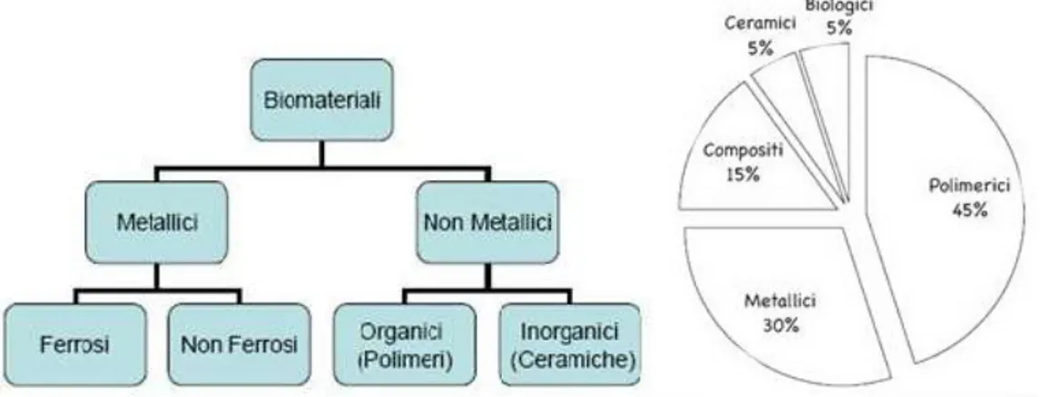
12 Biomaterials can be classified according to their chemical nature. Figure 1.7 shows the classification of biomaterials and shows the percentage of use of each family.
Fig. 1.7- Classification of biomaterials and percentage of use of the various types.
1.3.1 Metallic Biomaterials
The most used metals as biomaterials are stainless steels, cobalt-chrome alloys and titanium alloys. They are widely used mainly as materials for the construction of biomedical devices[32]. Some common applications of metal biomaterials concern the manufacture of surgical instruments, orthopedic and dental prostheses and osteosynthesis devices. In fact, the metallic materials, presenting mechanical properties that make it possible to produce prostheses able to withstand high loads with small sections, lend themselves well to solving problems related to the replacement of hard tissues such as bones and teeth. Figure 1.8 shows examples of prostheses partly or entirely constructed of biocompatible metal material.
Fig. 1.8- Examples of prostheses made from metal biomaterials: a) hip prosthesis, b) dental prosthesis.
13 The most important advantages of metal biomaterials are the following:
- high elastic modulus (about 100¸ 200 GPa);
- high yield strength (about 300¸ 1000 MPa). They can therefore withstand high loads without breaking or deforming;
- good ductility. Consequently, if the stress exceeds the yield limit, the biomaterial deforms plastically rather than breaking brittlely. This usually allows to intervene by replacing the deformed component before it breaks;
- absence of viscoelasticity;
- high resistance to mechanical fatigue. The fatigue strength also depends solely on the number of cycles and not on their frequency.
On the other hand, however, they have the following disadvantages:
· on average they have a higher specific weight than polymeric and composite biomaterials;
· they are generally more difficult to manufacture than biopolymers; · tend to corrode and wear due to contact with biological fluids.
Metal biomaterials can be processed using most of the traditional technologies and often their mechanical properties can be modified appropriately before the workpiece reaches its final shape.
To guarantee high biocompatibility it is necessary to take care of manufacturing technologies, above all with regard to surface finishing. Biocompatibility is connected, in the case of metals, to the problem of corrosion in a biological environment; in fact, in this type of environment, organic fluids have a high corrosive power towards metals. The consequences of corrosion are the loss of metallic material by the plant, with the possibility of loss of functionality of the plant itself and contamination of biological tissues.
Most of these problems, however controllable, are related to implanted devices, while for non-implanted devices, as in the case of surgical instruments, the metals have minor drawbacks and are the most used materials where high mechanical properties and reliability over time are required. From a corrosionist point of view, the human body can be assimilated to an aqueous solution with a temperature around 37 ° C, containing chloride ions and with an average pH of about 7.4 (the pH is actually variable depending on the area and can suffer strong fluctuations in
14 the presence of infections, as a result of surgical interventions and pharmacological applications). This solution is also strongly oxygenated and contains a high amount of salts and organic and inorganic compounds and therefore has a fairly high electrical conductivity. For all these reasons, the human organism is the ideal environment for the establishment of corrosion cells on metals. The corrosion resistance of metals depends on several factors, including the composition, the microstructure, the internal stresses and the surface finish.
1.3.2
Polymeric Materials
The use of polymers in medicines gave birth to the polymer science, virtually every early synthetic polymer found their way into the experimental surgical studies soon after their invention and many endured to become staples of clinical practice. The polymers remain important in clinical medicine as essential components of permanent prosthetic devices including hip implants, artificial lenses, large diameter vascular grafts, catheters, etc., and research continues to optimize the stability and performance of these materials in vivo. Whereas the original uses of polymers in surgery centered primarily on replacements for connective tissues, a host of new applications is emerging as a result of major advances in the sciences of molecular cell biology and developmental biology. An array of new protein and nucleic acid based drugs, which cannot be taken in classical pill form, is providing impetus for new implantable polymers for controlled drug delivery and gene therapy [33]. Applications in the relatively new field of tissue engineering, where polymers are used to assist the regeneration of three-dimensional tissue and organ structures, are more promising and are more assimilated with biological demands. A large number of polymers such as polyethylene (PE), polyurethane (PU), polytetrafluoroethylene (PTFE), polyacetal (PA), polymethylmethacrylate (PMMA), polyethyleneterepthalate (PET), silicone rubber (SR), polysulfone (PS), polyetheretherketone (PEEK), poly lactic acid (PLA), and poly glycolic acid (PGA) are also used in various biomedical applications. HA/PE, silica/SR, carbon fiber/ultrahigh molecular weight polyethylene (CF/UHMWPE), carbon fiber/ epoxy (CF/epoxy), and CF/PEEK are few examples of polymer composite
15 biomaterials. The application of polymeric materials for medical purposes is growing very fast. Polymers have found applications in such diverse bio-medical fields as tissue engineering, implantation of medical devices and artificial organs, prostheses, ophthalmology, dentistry, bone repair, and many other medical fields. Polymer-based delivery systems enable controlled slow release of drugs into the body. The application of synthetic polymers for gene therapy has also been investigated. They may provide a safer way of gene delivery than use of viruses as vectors. Polymeric materials have also extensively been used for biosensors, in testing devices, and for bio-regulation. Suitable polymeric material for a biomedical application must be ‘biocompatible’, at least on its surface. Strictly speaking many polymeric systems used for implantation of medical devices into the body are considered to be ‘biocompatible’, though after implantation they become isolated from the tissues of the body by collagenous encapsulation. An implanted polymeric material may be considered to be ‘biocompatible’, if its insertion into thebody does not provoke an adverse reaction. A thrombus is formed very fast when polymers contact blood cells. Materials with non-thrombogenic blood compatible surfaces must, therefore, be used in contact with the blood stream. Truly biocompatible polymers, used for medical purposes, should be able to recognize and cooperate in harmony with bio-assemblies and living cells without any non-specific interactions. Polymers have taken an important role in medical applications. In most of these applications, polymers have little or no competition from other types of materials. Their unique properties are:
1. Flexibility.
2. Resistance to biochemical attack. 3. Good biocompatibility.
4. Lightweight.
5. Availability in a wide variety of compositions with adequate physical and mechanical properties.
6. Possibility to be easily manufactured in products with the desired shape.
Biomaterials play a fundamental role in the engineering of tissue structures, working as an artificial extracellular matrix and three-dimensional support environment for cells (dragged in vitro or in vivo migration from host tissue) to
16 regenerate a wound site. Because of their mechanical versatility and resemblance to the structural characteristics of the fabric, polymers are the most popular biomaterials in tissue engineering. Polymeric biomaterials are currently dominated by thermoplastic polyesters such as polylactic acid (PLA), glycolic acid (PGA), poly caprolactone (PCL) and their mixtures or copolymers. Polyester is a category of polymers that contain the ester functional group in their main chain. Esters are chemical compounds derived from a carboxylic acid (COOH group) and a compound hydroxyl (OH), usually an alcohol. Many esters, such as fatty acids, are endogenous compared to human metabolism and therefore biocompatible. Many polyesters can degrade to natural metabolic products by simple hydrolysis. Although these biomaterials have been well characterized and fabricated to adapt to the biochemical properties of soft tissues, the mechanical compatibility between implants of thermoplastic polymers and living tissues generally lacks. Biodegradable synthetic polymers have attracted considerable attention to applications in medical devices and will play an important role in the design and function of medical devices. Drug eluting stents (DES) have been widely used as the default treatment for patients with coronary artery disease. The biodegradable polymers are always used as a biodegradable and bioabsorbable coating on stents to control drug delivery. In addition to being used as biodegradable coatings, biodegradable polymers are also candidate materials for completely biodegradable stents due to their properties for controlled drug delivery and good mechanical performance to prevent stents from deformation or fracturing. Orthopedic devices made of biodegradable materials have advantages in metallic or non-degradable materials. They can transfer stress over time to the damaged area while healing, allowing the tissues, and there is no need for a second surgery to remove the implanted devices. These biodegradable polymers have been used to prepare some single-use medical devices and will probably have an increasing commercial demand. A new dimension for the use of polymeric materials as drug delivery devices involves incorporation of biodegradability into the system. A number of degradable polymers are potentially useful for this purpose, including a variety of synthetic and natural substances. The use of intentionally degradable polymers in medicine has been brought into prominence with new innovations in drug delivery
17 systems. In some cases, erosion or dissolution of the polymer contributes to the release mechanism. Degradable polymers such as poly lactic acid and polyorthoesters, are used for drug delivery systems.
1.3.3 Ceramic Biomaterial
The field of nanotechnology is playing a pivotal role in the fields of electronics, biology and medicine. It also introduced several new concepts into medicine and thus makes these large cross disciplinary fields to join together. Nanomedicine encompasses many common technical issues like analytical tools, nanoimaging, nanomaterials and nano-devices, novel therapeutics and Drug Delivery Systems, clinical, regulatory and toxicological issues. Among the varieties of nanomaterials, nanostructured ceramics, cements and coatings are being considered for major applications in orthopaedic and dental treatments. Biocompatible Ceramics, also known as bioceramics, include both macro and nano materials mainly used for bone, teeth and other medical applications. Nanostructured ceramics, cements and coatings are being considered for major orthopaedic, dental and other medical applications. The development of novel biocompatible ceramic materials with improved biomedical functions is at the forefront of health-related applications all over the world [34]. Ceramics are also unique used biomaterials used for repairing and regenerating several parts of the human body. The widely used ceramic nanobiomaterials include (see Fig. 1.9), Calcium Phosphate (CaP), Tri-Calcium Phosphate (TCP), Hydroxy-Apatite(HAP), TCP+HAP, Si substituted HA, Calcium Sulphate and Carbonate, Bioactive Glasses, Bioactive Glass Ceramics, Titania-Based Ceramics, Zirconia Ceramics, Alumina Ceramcis and Ceramic Polymer Composites.
18
Fig. 1.9- Widely used ceramic nanobiomaterials.
Ceramics are compounds between metallic and non-metallic elements; they are most frequently oxides, phosphates, nitrides, and carbides. There are wide range of ceramic materials like clay minerals, cement, and glass used for various applications. These materials are typically insulators to electricity and heat, and are highly resistant to harsh chemical environments than metals and polymers. With regard to their mechanical behaviour, ceramics are very hard and brittle. At nanoscale also, ceramic materials exhibit higher hardness, excellent heat and corrosion resistance, and electrical insulation properties. Typical examples include china clay, firebricks, cements and glass. In addition to these properties, Fine Ceramics (also known as “advanced ceramics”) have many advanced mechanical, electrical, electronic, magnetic, optical, chemical and biochemical characteristics. One of the major field of application of bioceramics is tissue generation. The major parameters considered to optimize the biomaterials for the tissue generation include, a) Structural Components (physical, mechanical and chemical properties) and b) Biochemical Components (immobilized signals, diffusable signals, and living components).
19 The bioceramics have good biocompatibility, osteo conductivity, osteoinductivity, biodegradability, resorbability, and hydrophilicity. Yet another major field of application of bioceramic is clinical dentistry.
Bioceramics are materials used to repair and replacement of diseased and damaged parts of musculoskeletal systems. Based on their inherent properties, they are classified into three major categories as:
Bioactive ceramics (CaP, HAP, Bioactive Glass (BAG), and Glass Ceramics (GC) which form direct chemical bonds with bone or even soft tissues of living systems,
Bioresorbable ceramics (TCP) that actively participate in the metabolic process of an organism
Bioinert high strength ceramics (alumina and zirconia)
Based on their applications, ceramic biomaterials are further classified into: Cardiovascular biomaterials
Dental Biomaterials Orthopedic bomaterials
Biomaterials to promote tissue generation.
Nanomaterials of CaP, HAP, TCP, BAG, GC and Calcium sulphate form good ceramic-based bone graft substitutes. Hydroxyapatite (HAP) has been widely used as a biocompatible ceramic in many areas of medicine, but mainly for contact with bone tissue, due to its resemblance to mineral bone. HAP has exceptional biocompatibility and bioactivity properties with respect to bone cells and tissues. As a result of excellent favorable osteoconductive and bioactive properties, it is widely preferred as the biomaterial of choice in both dentistry and orthopaedics. The hydroxyapatite has a few favorable bioactive and osteoconductive properties which help in rapid bone formation, with a strong biological fixation to bony tissues. It also has very low mechanical strength and fracture toughness, which is an obstacle to its applications in load-bearing areas.
20
Fig 1.10- HA for accelerating the reconstruction of the bone tissue on the metallic implant surface creating a rapid bonding.
HAP can incorporate the drug molecules either physically or chemically so that the drug retains intact until it reaches to the target site. It could also gradually degrade and then deliver the drug in a controlled manner over time. Titanium comes under the category of Technical ceramics. The technical ceramics are divided into oxides and non-oxides like Aluminium Oxide, Ceramics, Carbide Ceramics, Nitride Ceramics, Oxide Ceramics, Silicon Carbide Ceramics, Silicon Nitride Ceramics, and Zirconium Ceramics Dioxide. The fact that titanium is strong, light, non-toxic and does not react without bodies makes it a valuable medical resource and used to make surgical implements and implants, such as hip joint replacements that can stay in place for up to 20 years. Although other photocatalytic materials are available, researchers have found that titanium dioxide provides the best performance in sunlight. Titanium nanomaterials have been clinically successful as orthopedic or dental implant material.
22
2.1. Synthesis of Nanoparticles
The importance and benefits of nanotechnology in biology and medicine are now well-recognized by scientists, technologists, as well as various governmental and private research funding agencies. The basis that enables the application of nanotechnology is the availability of nanostructured materials. Therefore, it is essential to provide an insight on the state-of-the-art methods to manufacture various nanostructures. The methods for synthesis and patterning for many of these nanostructures are founded on basic, well-known techniques and their modifications, as is described in this chapter. The nanofabrication processes can be divided into the two well-known approaches: “top-down” and “bottom-up.” The “top-down” approach uses traditional methods to guide the synthesis of nanoscale materials. The proper paradigm of its definition generally dictates that in the “top-down” approach it all begins from a bulk piece of material, which is then gradually or step-by-step removed to form objects in the nanometer-size regime. The top-down approach for nanofabrication requires a thorough understanding of the short-range forces of attraction, such as Van der Waals forces, electrostatic forces, and a variety of interatomic or intermolecular forces. Since it is not possible to have various minute things come together without some attractive force or active field of force in the region, having the fundamental forces “doing all the work” for us is the key principle underlying this approach.
23 Some examples of such a synthesis route starting from atoms and molecules are methods like self-assembly of nanoparticles or monomer/polymer molecules, chemical or electrochemical reactions for precipitation of nanostructures, sol–gel processing, laser pyrolysis, chemical vapor deposition (CVD), plasma or flame spraying synthesis, and atomic or molecular condensation [35-37].
The bio-assisted synthesis of nanomaterials also belongs to this approach. However, despite being so promising and inviting, our ability to build things from the bottom up is fairly limited in scope. While we can assemble relatively simple structures, we cannot produce complex, integrated devices using the bottom-up approach. Any kind of overall ordered arrangement aside from repeating regular patterns cannot be done without some sort of top-down influence, like lithographic patterning. Until we have fully mastered the bottom-up synthesis approach, we will not be able to fully exploit its speed and accuracy. The important factor is that they are two different approaches to creating nanostructures which can be applied according to the specific needs for each application, often in a complementary way.
24
2.2. Nanofabrication by “Top Down” Methods.
Over the past few decades, a wide variety of top-down production techniques have been developed that have been implemented using different media ranging from chemical and electrochemical media to photofabrication, laser processing, ecc. Here, we describe only the technique Laser Ablation in liquids.
2.2.1 Laser Ablation
Laser ablation is a method for fabricating various kinds of nanoparticles including semiconductor quantum dots, carbon nanotubes, nanowires, and core shell nanoparticles. In this method, nanoparticles are generated by nucleation and growth of laser-vaporized species in a background gas [38]. The extremely rapid quenching of vapor is advantageous in producing high purity nanoparticles in the quantum size range (<10 nm). The most critical characteristic of nanoparticles is that the properties electrical, optical, magnetic, and so on depend strongly on the size and size distribution of the particles. Many kinds of nanoparticles exhibit special characteristics (ferromagnetism, paramagnetism, pinned emission, fluorescence, spin quantum effect, etc.) when the size of a particle is at the nanoscale level. The special characteristics are significantly impacted by the size and size distribution of the particles. Laser ablation is a method that utilizes laser (which is an acronym for light amplification by stimulated emission of radiation) as an energy source for ablating solid target materials. In this process, extremely high energy is con centrated at a specific point on a solid surface to evaporate light-absorbing material. The term ‘ablation’ refers to the removal of surface atoms and involves not only a single photon process (breaking the chemical bonds) but also multiphoton excitation (thermal evaporation). High-purity nanoparticles can be generated by laser ablation because the purity of the particles is basically determined by the purity of the target and ambient media (gas or liquid) without contamination from the reactor. However, it is difficult to control size distribution, agglomeration, and crystal structure in the conventional laser ablation process since nanoparticles are built by random (Brownian) motion of molecules. Therefore,
25 several advanced laser ablation techniques have been developed for fabricating morphology-controlled nanoparticles.
Fig. 2.2 - Schematic of particle generation procedure in the laser ablation process.
Fig. 2.2 is a schematic of the nanoparticle formation process by laser ablation. When the laser beam is focused on the surface of a solid target material in the ambient media (gas or liquid), the temperature of the irradiated spot rapidly increases, vaporizing the target material. The collisions between the evaporated species (atom and clusters) and the surrounding molecules result in excitation of the electron state coupled with light emission and generationof electrons and ions, forming a laser-induced plasma plume. The plasma structures (size of the plume and its emission spectrum) depend on the target material, ambient media (liquid or gas), ambient pressure, and laser conditions. Laser ablation in liquid is employed to confine the plasma plume in a small region to directly disperse nanoparticles in the liquid phase. In any case, the ambient media must be carefully selected because the laser-generated particles easily react with surrounding molecules to create complexes such as oxides and other undesirable species. Coagulation is another critical phenomena that must be finely controlled in the later stages of nanoparticle formation. Since laser-generated particles have a very clean surface, agglomerated particles create chemical bonds at the contact point (neck), which significantly compromise the properties of primary particles. The low-pressure gas process is advantageous not only for reducing the size of the primary particles but also for preventing coagulation. For the fabrication of nanoparticles of the desired size and structure, the selection of a suitable laser system is one of the most critical decisions.
26 The evaporation rate of the target material is generally determined by the laser parameters (laser source, wavelength, fluence, pulse width and frequency), the light absorption efficiency of the target material, and the condition of the ambient media. Laser energy per unit area on the target material is defined as fluence of the laser F, which is given as
𝐹 = 𝐼
𝐴 (2.1) where I [J/pulse] is the laser power and A [m2] is the area of the laser spot. The
wavelength of the laser is another important parameter that determines the absorption efficiency of the target. The absorption depth and spot (focusing) area are also influenced by laser wavelength. In early studies of the nanoparticle synthesis by laser ablation, excimer lasers in the ultraviolet spectrum (193 nm for ArF, 248 nm for KrF) are often used as a light source. Recently, Q-switch pumped pulsed YAG (Yttrium Aluminum Garnet) lasers are more commonly used for laser ablation because they do not require hazardous gases. The wavelength of Nd:YAG laser (1064 nm for fundamental wave) can be changed by employing nonlinear optical crystals. The pulse width of the laser is also another important parameter that determines peak energy. Recently picosecond and femtosecond lasers have been applied to enhance the photon absorption efficiency of the target surface to break chemical bonding.
2.2.2 Our set up for Laser Ablation
A Nd:YAG laser operating at the fundamental wavelength of 1064 nm with a pulse energy of 200 mJ, 3 ns pulse duration, and a repetition rate of 10 Hz, was employed at 1010 W/cm2 intensity. The horizontal laser beam, 1 cm2 in diameter with a
Gaussian profile, was vertically deflected by a mirror and focused on the gold sample at about 1 mm2 spot size, placed in a polyethylene holder, through a lens
with a focal length of 50 cm (Fig. 2.3). The laser-generated plasma, containing the gold ions and electrons, expanded in the liquid and condensed to form spherical nanoparticles, as reported in the literature [39-40].
27 Au-NPs are immediately produced, in times comparable with the laser pulse and transforming the liquid in a solution as a consequence of the 10 Hz laser irradiation for times ranging between 10 and 30 minutes, in order to obtain different solution concentrations.
Fig.2.3- Laser Ablation in liquid set up a) scheme, b) photo of set up in our laboratory.
Generally, from 1mg/ml up to 10 mg/ml of gold nanoparticles in distilled water is obtained, but higher concentration can be easily produced. In order to reduce the effect of particle coalescence with the times, a very little quantity of tension active liquid is added (~ 10μg trisodium citrate) which covers the surface of the nanoparticles greatly reducing their coalescence [41]. The liquid and the target where particles are generated are in movement during laser ablation using the 10 Hz repetition rate. This movement is not due to motors, mechanical stirrers or continuum liquid flux, but it is due to the high used laser intensity (1010 W/cm2),
which produces high shock waves determining a continuum horizontal oscillating movement of the target (the target is clamped inside a polyethylene cavity holder so that only horizontal movements are possible) and continuum liquid oscillations and mixing (drops liquid jumping on liquid with heights up to even 10 cm, maintained inside the long used glass tube).
28
2.2.3 The Laser Ablation: Theoretical background
Comprehension of the aerosol formation as inevitable process in laser micromachining technology is established and the application of laser ablation in the production of nanoparticles is widely spread. However, characteristics of particles generated by laser ablation are not fully known. Therefore, the studies of particles concentration and their size distribution are hot topics in current laser ablation investigations recently [42]. Nanoparticle generation by laser ablation is used for a variety of applications such as biotechnology, electronic industry, etc. The laser ablation is also an intensive source of submicron particle generation and their possible leakage into ambient air, therefore investigations of the particle formation kinetics in ambient air are essential for the evaluation of potential generation of particles by laser and their impact on human health. For example, during laser processing, particles formed of metals (e. g. iron, aluminum), especially toxic (e. g. manganese, zinc) or carcinogenic substances (e. g. chromium (VI) compounds, nickel), might penetrate into a human organism and cause health concerns. The physical process of particle generation begins with the absorption of laser irradiation in the beam/material interaction zone. Depending on the wavelength of the laser beam and the material properties, kinetic energy is transformed into thermal (in metals 10–13 s), which cuts off chemical bonds in the material. The phase transition occurs if the energy threshold for that material is reached. As a result, the material can be melted, vaporized or sublimated and the particle generation process begins. Both the particle concentration and size distribution depend on laser operating parameters such as wavelength, pulse duration, energy and repetition rate, beam scanning speed. Studies show that experimental conditions during the laser ablation could be established in order to control the particles size and distribution. Besides, the medium (ambient air, argon, water, etc.) in the ablation chamber also plays an important role in the particle formation process. It was experimentally estimated that the mass of generated nanoparticles in ambient air was up to 100 times higher than in water.
29
2.2.4 Temperature Dependence
During the interaction of the laser beam and material, the light absorption is a general physical process [43-45]. The energy coupled into material causes thermal heating, melting and vaporization of material, plasma formation and particle emission. In metals, free electrons interact with the intense electromagnetic irradiation. The energy is instantly absorbed by electrons and further distributed to the lattice. The laser pulse duration τL is important for
understanding what happens with the energy in the beam / material interaction zone. τ = is the lattice heating time, τ = is the electron cooling time, where Ce and Ci are electron and lattice thermal capacities, respectively, and γ is the electron– lattice coupling parameter specific to every material. Based on the two-temperature diffusion model the laser beam/material interaction can be described by electron and lattice subsystem temperatures Te and Ti,
respectively: C ∂T ∂T = − ∂Q(z) ∂z − γ(T − T ) + S (2.2) C ∂T ∂T = γ(T − T ) (2.3) where Q(z) is the heat flux, S is the heat source (laser pulse). For picosecond pulses, the condition τe<<τL<<τi is fulfilled. The laser pulse duration is shorter than the
lattice heating time. Particles from the material are removed partly by the solid state – vapor transition and the direct breaking of chemical bonds. For picosecond pulses the electron temperature T becomes quasi-stationary:
∂ ∂z −k
∂T
∂z − γ(T − T ) + I α 𝑒𝑥𝑝(−𝛼𝑧) = 0 (2.4) where ke is the electron thermal conductivity, α is the absorption coefficient, Ia is
30 penetration into material perpendicular to the surface. In this case, lattice temperature Ti is
𝑇 = 𝑡
𝜏 𝑒𝑥𝑝 −
𝑡 − 𝜃
𝜏 𝑇 (𝜃)𝑑𝜃 + 𝑇 (2.5) where T0 is the initial electron temperature. Taking this into account Eq. (2.5) can
be simplified due to quasi-stationary condition of electron temperature:
𝑇 ≈ 𝑇 1 − 𝑒𝑥𝑝 − 𝑡
𝜏 ≈
𝑡
𝜏 𝑇 (2.6) where t is the time after the laser exposure. It means that electron cooling temperature Te stays longer than lattice heating temperature Ti after exposure to
picosecond laser pulses. Electron cooling temperature Te at the end of the pulse is
𝑇 ≈𝐼 𝛼
𝛾 𝑒𝑥𝑝(−𝛼𝑧) (2.7) Lattice heating temperature Ti at the end of the laser pulse is
𝑇 ≈𝐹 𝛼
𝐶 𝑒𝑥𝑝(−𝛼𝑧) (2.8) where Fa is the absorbed laser fluence, which is 𝐹𝑎 = 𝐼 𝜏 . By using the condition of strong evaporation it can be described as
𝐹 ≥ 𝐹 𝑒𝑥𝑝(−𝛼𝑧) (2.9)
where Fth is the threshold laser fluence for evaporation. Equations (2.7) and (2.9)
describe how laser intensity, material transmissivity and thermal capacities influence the temperature of the electron and lattice. In the second case for nanosecond pulses, the condition τL>>τi>>τe is fulfilled. Equations (2.1), (2.2) for
nanosecond pulses are
C ∂T ∂t = ∂ ∂z k ∂T ∂z + I α 𝑒𝑥𝑝(−𝛼𝑧) (2.10)
31 where k0 is the conventional equilibrium thermal conductivity of a metal and T
represents general temperature of the subsystem, because in case of ns-laser ablation Te = Ti = T.
The energy absorbed by electrons is fully transferred to the lattice during the laser pulse duration. Material is heated due to the absorbed energy of the lattice. In this case, solid material melts, the beam/interaction zone spreads and particles are evaporated from liquid state. Metals have good thermal conductivity properties, thus increasing energy loss due to lattice heating for nanosecond pulses.
2.2.5 Irradiation, Energy, Wavelength and Time Depedence
Irradiation of a solid target in a liquid with a laser beam results in the generation of metal flakes by laser fragmentation. The particles are refined by irradiation with the laser beam a second time. The laser beam reduces the size of the generated nanoparticles or produces particles with a specific size distribution. The interaction of the laser beam with the liquid also affects the particle formation. Bubbles and cavitation result from laser-induced plasma generation and affect nanoparticle fragmentation [46]. In addition, the aqueous solution influences the formation of clusters of nanoparticles. Nanoparticle size and dispersion can be controlled by varying the laser parameters. The wavelength of the laser pulse also affects the nanoparticle generation. When a laser of different wavelength with the same pulse width is used, the fragmentation effect increases with decreasing pulse wavelengths. This is because the shorter the pulse wavelength, the greater the absorption cross section. In addition, liquids other than deionized water may be used, and they can contribute to the nanoparticle formation and stability. The laser pulse width is a predominant determinant of nanoparticle size and distribution. Laser pulse widths in the femtosecond, picosecond, and nanosecond regions can be used to induce the laser fragmentation [47-49]. Several studies have evaluated the interactions of picosecond or femtosecond laser pulses with materials. Such ultrafast laser pulses cause nonlinear multiphoton absorption due to their high peak power. Because of this, energy cannot be transferred from the electrons to the ion
32 grids in femtoseconds or picoseconds, which enables the processing of materials that are little affected by heat. Irradiation with a nanosecond pulsed laser beam results in thermal diffusion at the particle surface.
It is important to compare the diffusion time and the pulse width. In general, the thermal diffusion in metal nanoparticles requires 50 to 100 ps, and the diffusion time can be used to classify laser fragmentation mechanisms into short and long pulse-width categories. At a pulse width of less than a few picoseconds, the electron and phonon temperatures in the particle are different. In this case, to determine the particle temperature distribution, a model with different electron and phonon temperatures should be applied.
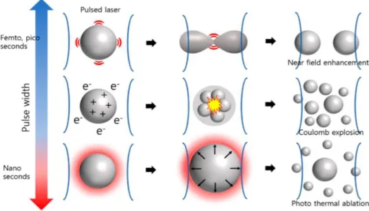
Also, since the laser beam absorption time is shorter than that of heat diffusion, it is appropriate to assume that the boundary condition of particles is adiabatic. In this model, the energy absorption by laser irradiation is a function of the fluence and absorption cross-sectional area of the laser. This correlation affects the electron temperature and phonon temperature of nanoparticles; that is, as the pulse width decreases, the peak laser power and the electron temperature increase. This results in a release of electrons from the particle; moreover, a Coulomb explosion may occur. Coulomb explosions are a major mechanism of the femtosecond laser fragmentation.
33 When the pulse width increases from femtoseconds to picoseconds, the energy transfer tends to resolve the energy imbalance in the nanoparticles. In this case, the effect of Coulomb explosions in the particles decreases. Near-field enhancement at the nanoparticle surface also occurs using ultrafast laser pulses. When a nanostructure is irradiated with a short laser pulse, the electromagnetic field is amplified at the point at which an abrupt change in shape occurs. This results in a sudden energy concentration at that point, leading to particle fragmentation. A model with constant electron and phonon temperatures can be used for lasers with nanosecond pulse widths. Also, since there is enough time for thermal diffusion within the particle, the heat at the interface interacts with the surroundings during laser irradiation. Therefore, the photothermal effect becomes dominant. Melting, evaporation, or both occur at the particle surface as a result of the temperature imbalance between the particle surface and interior, which likely also causes particle fragmentation. The three above-mentioned mechanisms of laser fragmentation are summarized in Figure 2.4. The effect on the nanoparticle formation of mechanical shock waves generated by the laser-induced generation of plasma is shown in Figure 2.5.
Figure 2.5- Effect of shockwave propagation on the laser fragmentation.
2.2.6 Photochemical effects of Laser
There are four major categories of light-tissue interactions that lead to alteration of the tissue structure/composition:
• Photochemical: Absorption of light by molecules present or added to tissue. • Photothermal: Biological effects due to deposition of thermal energy in tissue.
34 • Photoablative: in UV, photons possess sufficient energy to cause photo-dissociation of bio-polymers and subsequent desorption of fragments (a substance is released from or through a surface).
• Photomechanical: it occurs at high fluence rates where dielectric breakdown of tissue is induced which can lead to plasma formation. Rapid plasma expansion generates a shock wave which can rupture tissue [50].
Light can induce chemical effects and reactions within macromolecules or tissues. Photodynamic therapy (PDT): spectrally adapted chromophores are injected into the body.
35 Monochromatic irradiation (usually red) may then trigger photochemical reactions, resulting in certain biological transformations. A chromophore which is capable of causing light-induced reactions in other non-absorbing molecules is called photosensitizer (PS) (organic dyes). After absorption of laser photons, PS is transferred to S1. After this, a radiative decay might occur to an excited triplet state (intercrossing system) (T1) besides other alternatives such as non-radiative decays
or radiative singlet decay to the single ground state. Radiative singlet and triplet decays: fluorescence (ns) and phosphorescence (ms-s). PS are compounds whose energy difference T1 − S0 is close to the energy needed for oxygens molecules to be
excited from the triplet ground state to a excited singlet state (energy is transferred to the oxygen molecule). The excited singlet oxygen resulting from this process are very reactive and lead to lipids and protein oxygenation and other destructive processes, which could start necrosis of cancer cells (PDT: photodynamic therapy). The main disadvantage of PDT is the long time of the compound’s decay and removal from the patient body became photosensitive from several days to weeks after application of such drugs and the related effects (toxicity etc.)
2.3.
Nanofabrication by “Bottom-Up” Methods
The NPs obtained thanks to the Bottom-Up strategy, are the result of the natural manifestation of interaction between the constituent elements, that is organize to form the desired structure. The bottom up approach uses atomic or molecular raw materials as the source of the material that it must be chemically converted into larger nanoparticles. This method has the advantage of being, potentially, much more controllable than the top down approach. This has led to the development of many general strategies of bottom up.
In general, two main chemical methods can be used for the AuNP preparation: co-precipitation and chemical reduction. In both cases, the presence of a surfactant is necessary to control the process of growth. Co-precipitation reactions involve the thermal decomposition of the organometallic gold predecessors, while the chemical reduction that occurs in the colloidal solution is another approach to the formation
![Fig. 1.4- Left: illustration of absorption cross section concept. Right: picture describing transmission, absorption and scattering processes[10]](https://thumb-eu.123doks.com/thumbv2/123dokorg/4570670.38270/16.892.159.739.773.965/illustration-absorption-section-describing-transmission-absorption-scattering-processes.webp)

![Fig. 2.17- The Raman spectrum of the silver nanoparticles acquired at 785 nm excitation [59].](https://thumb-eu.123doks.com/thumbv2/123dokorg/4570670.38270/63.892.270.618.837.1098/fig-raman-spectrum-silver-nanoparticles-acquired-nm-excitation.webp)

