Nephro-Urol Mon. 2017 September; 9(5):e59535. Published online 2017 July 4.
doi:10.5812/numonthly.59535. Brief Report
Melanoma Risk in Renal Transplanted Patients
Giovanni Cavaliere,
1Elisa Zavattaro,
2Federica Veronese,
3Paolo Fava,
4and Paola Savoia
3,*1Department of Medical Science, University of Turin, Torino, Italy
2Department of Translational Medicine, University of Eastern Piedmont, Novara, Italy 3Department of Health Science, University of Eastern Piedmont, Novara, Italy 4AOU Citta Della Salute e Della Scienza, Torino, Italy
*Corresponding author: Paola Savoia, Department of Health Science, University of Eastern Piedmont, Via Solaroli 17, 28100 Novara, Italy. Tel: +39-3213733387, Fax: +39-3213733387,
E-mail: [email protected]
Received2017 April 06; Revised 2017 April 21; Accepted 2017 June 10.
Abstract
Despite the well-known increased risk to develop non-melanoma skin cancer (NMSC) due to the long-term immunosuppression, data about melanoma incidence and prognosis in solid organ transplanted patients are still debated. Literature studies report a relative risk for melanoma varying from 1.2 and 5.8 in different solid organ recipients, probably as a consequence of the difference in immunosuppressive treatments and endogenous and exogenous risk factors. Here we report data about melanoma incidence, prognosis and clinicopathological characteristics in a series of 686 kidney transplant recipients. In this series, melanoma incidence was 3.5%; most cases were represented by in situ or thin melanomas mainly related to sun exposure, and the prognosis of our patients was good except for only one case with a progressive disease. Our experience confirms the importance of a regular dermatological follow-up and of education to correct sun exposure even in transplanted patients.
Keywords:Melanoma, Skin Tumours, Solid Organ Transplantation, Immunosuppression
1. Background
The crucial role of long-term immunosuppressive treatment as a promoter for malignancy has been exten-sively established in literature (1), and the increased risk for non-melanoma skin cancer (NMSC) in solid organ trans-plant recipients is well known (2,3).
Conversely, data about melanoma incidence and be-haviour in transplanted patients are still debated, despite the fact that the impact of the immune system on the on-set and progression of this cancer has been clearly demon-strated (4,5).
A large analysis performed on an Australian cohort of kidney transplant recipients reported a melanoma inci-dence of almost 1% closely related to iatrogenic immuno-suppression and confined to the period of transplant func-tion (1). Therefore, a systematic review was recently per-formed on a 20 cohort study (6), identifying an aggregate relative risk (RR) for melanoma of 2.71 among transplanted patients; this risk was higher (5.27) for liver and heart recip-ients and lower (2.54) for kidney reciprecip-ients, probably due to the differences in immunosuppressive regimens. How-ever, several differences have been demonstrated in vari-ous studies and the RR ranged from 0.8 (7) to 5.80 (8), prob-ably due to the heterogeneity of the inclusion criteria and
the follow-up length and to the variability of the ethnic and geographic origin of patients. The well-known risk factors for melanoma such as fair skin, presence of atypical nevi, family history for melanoma, and personal history of sun-burns could also play a crucial role creating a favourable substrate on which immunosuppression acts as a trigger-ing factor.
Here we revised our series of kidney transplant recipi-ents undergoing a regular dermatological follow-up with the aim of evaluating the melanoma incidence within this group of patients, its characteristics, and the possible re-lationship between the onset of this tumour and eventual exogenous and endogenous risk factors.
Clinical outcome was also evaluated to establish the possible immunosuppression impact on the disease pro-gression.
2. Methods
2.1. Patients
A total of 686 kidney transplant recipients who under-went a regular dermatological follow-up were included in this study. Dermatological consultations were performed between April 1997 and December 2016. Our sample con-sisted in 401 males and 285 females with a median age of
Copyright © 2017, Nephro-Urology Monthly. This is an open-access article distributed under the terms of the Creative Commons Attribution-NonCommercial 4.0 International License (http://creativecommons.org/licenses/by-nc/4.0/) which permits copy and redistribute the material just in noncommercial usages, provided the original work is properly cited.
Cavaliere G et al.
50 years at transplantation and of 60 at the last follow-up visit and a median duration of immunosfollow-uppression of 8.9 years. Clinical information about the patients’ endoge-nous and exogeendoge-nous risk factors for skin tumours were recorded and a complete dermatological examination was performed at each scheduled visit. We collected, also, in-formation about immunosuppressive schedules which the patients had undergone.
3. Results
3.1. Clinical Characteristics
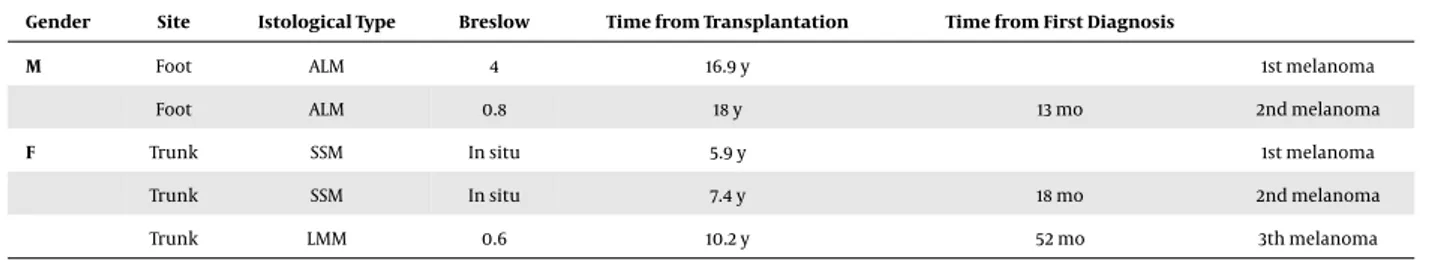
The number of the patients who developed a cuta-neous melanoma in the post-transplantation period was 24 accounting for 3.5% of our series. Seventeen patients were males and 7 females, with a median age at melanoma diagnosis of 57 years (ranging from 36 to 78). For these patients, the median age at transplantation was 49 years (ranging from 25 to 64), and the median time of immuno-suppression was 13 years (ranging from 3 to 22); the median duration of the dermatological follow-up was 8.9 years. The median time of melanoma onset from transplantation was 5.3 years (ranging from 0.7 to 14.6). Interestingly, 2 out of 24 patients developed multiple melanomas. One out of these developed a second melanoma 13 months af-ter the first diagnosis, whereas the other one developed 3 melanomas after 19 and 53 months from the first diagnosis, respectively. Clinical data of these patients are reported in
Table 1.
Moreover, 4 patients affected by melanoma developed also a NMSC. All these lesions were diagnosed as basal cell carcinoma (BCC); in two cases, the onset of BCC preceded melanoma diagnosis, while in the other two it occurred subsequently.
3.2. Histology
Eleven out of 24 of total excised melanomas were his-tologically in situ melanomas; the median Breslow’s thick-ness of other lesions was 1.12 mm (ranging from 0.5 to 4 mm). Data about histological characteristics are summa-rized inTable 2.
3.3. Risk Factors
The skin phototype predominantly represented in our series (66.6%) was III according to the characteristics of the patients pertaining to our geographical area. Two patients reported a history of sunburns at a young age and the other 6 reported frequent sun exposure without photoprotec-tion. Two patients had working outdoor activities, whereas nobody reported family history of melanoma. We found
no significant correlation between the type of immuno-suppressive schedule and melanoma onset.
From the transplanted patients affected by melanoma, of our series, only one developed metastatic melanoma with lung and distant nodal metastases.
4. Discussion
It has been demonstrated that the immune surveil-lance plays a fundamental role in the pathogenesis of melanoma (4), as confirmed by the importance of im-munotherapeutic approach in the treatment of advanced stages of this disease (9). However, while data concerning the higher risk of NMSC after transplantation are well es-tablished (2,3,10), not all authors agree about the increase in melanoma risk in transplant recipients (1,6-12).
In a previous review of our series (13), the 3.9% of kid-ney recipients who underwent a regular dermatological follow-up suffered from melanoma; the percentage of pa-tients affected by NMSC in the same series was 24.8%. Here, a series significantly increased in number and a longer follow-up enabled us to identify a percentage of patients affected by melanoma accounting for 3.5%. In literature, the pick of melanoma onset in immunosuppressed pa-tients is reported during the second year from transplan-tation (1); on the contrary, in our experience the median time of melanoma onset was 5.3 years, suggesting a po-tential role of cumulative low-dose immunosuppression. Moreover, even if cyclosporine and/or azatyoprine-based immunosuppressive regimens showed a significant corre-lation with the risk of developing skin cancer in most pub-lished studies (14-17), we did not find any significant corre-lation with the treatment schedule.
We think that endogenous and exogenous risk factors can play a major role in the pathogenesis of melanoma and, also, in transplanted patients. Differences in skin phototype and in UV radiation exposure, as well as in the ethnic and geographical origin of transplanted patients could explain differences in melanoma prevalence among the transplanted population in different countries (1,6-8,
12,18), justifying the fact that in our series, the incidence of melanoma is higher than what is described in other large studies recently published (7). In our series, most pa-tients showed a phototype III, in accordance with their ori-gin from the Mediterranean area; however, almost half of them reported previous significant sun exposure for recre-ational reasons or outdoor work, suggesting that immuno-suppression could be a simple trigger on a pre-existing condition of susceptibility. This hypothesis is supported by the onset of multiple melanomas in two of our patients and by the fact that in both, there was a concordance be-tween sites in which the lesions arise. The site concordance
Cavaliere G et al.
Table 1.Clinical Characteristics of Transplanted Patients Affected by Multiple Melanomas
Gender Site Istological Type Breslow Time from Transplantation Time from First Diagnosis
M Foot ALM 4 16.9 y 1st melanoma
Foot ALM 0.8 18 y 13 mo 2nd melanoma
F Trunk SSM In situ 5.9 y 1st melanoma
Trunk SSM In situ 7.4 y 18 mo 2nd melanoma
Trunk LMM 0.6 10.2 y 52 mo 3th melanoma
Table 2.Clinical and Histological Characteristics of Melanomas Diagnosed in Trans-planted Patients Variable Value Gender M 17 F 7 Age 57 y (range 36 - 78) Site Trunk 11 Upper limbs 1 Lower limbs 7 Foot 4 Head 3 Other 1 Histological type SSM 19 NM 1 ALM 4 LMM 2 Mucosal 1
Breslow thickness 1.12 mm (range 0.5 - 4 mm) Abbreviations: ALM, acral-lentiginous melanoma; LMM, lentigo maligna melanom; NM, nodular melanoma; SSM, superficial spreading melanoma.
for patients affected by multiple melanomas justify, in fact, the assumption that the risk is mainly related to the pre-vious UV damage (11,19). The role of the photo exposition is also confirmed by the diagnosis of BCC in other four pa-tients.
In our experience, more than 45% of our patients (11/24) had an in-situ melanoma; for other patients, the median Breslow thickness was relatively low with a high preva-lence of Superficial Spreading Melanoma. These observa-tions support, on the one hand, the relatively low aggres-siveness of melanomas diagnosed in kidney transplanted
patient; on the other hand, the major role of dermatolog-ical follow-up. In most patients, in fact, the diagnosis was made incidentally during a routinely programmed visit.
Literature data regarding the clinical course of melanoma developed in the post-transplant period are scanty. However, the thickness of the primary lesion and the known prognostic factors seems to regulate the course of the disease even in transplanted patients (20) and the outcome of patients with thicker melanoma is worse than that of patients with thinner lesions. No deaths are in fact reported for transplant recipients with in situ or thin melanomas (21). Also, in our experience, the only one patient who developed a progressive disease was affected by a 4-mm ALM that metastasised to distant lymph node and lung.
Future studies are needed to better characterize the melanoma risk and the outcome in transplant recipients. However, also in this group of patients, early diagnosis is critical and dedicated dermatologic follow-up programs are fundamental; in view of the role of sun exposure, we also underline the importance of the constant education to the photoprotection in transplanted patients.
References
1. Vajdic CM, van Leeuwen MT, Webster AC, McCredie MR, Stewart JH, Chapman JR, et al. Cutaneous melanoma is related to immune sup-pression in kidney transplant recipients. Cancer Epidemiol Biomark-ers Prev. 2009;18(8):2297–303. doi: 10.1158/1055-9965.EPI-09-0278. [PubMed:19622722].
2. Gallagher MP, Kelly PJ, Jardine M, Perkovic V, Cass A, Craig JC, et al. Long-term cancer risk of immunosuppressive regimens af-ter kidney transplantation. J Am Soc Nephrol. 2010;21(5):852–8. doi:
10.1681/ASN.2009101043. [PubMed:20431040].
3. Ingvar A, Smedby KE, Lindelof B, Fernberg P, Bellocco R, Tufveson G, et al. Immunosuppressive treatment after solid organ transplanta-tion and risk of post-transplant cutaneous squamous cell carcinoma. Nephrol Dial Transplant. 2010;25(8):2764–71. doi:10.1093/ndt/gfp425. [PubMed:19729465].
4. Dean JH, Greene MH, Reimer RR, LeSane FV, McKeen EA, Mulvihill JJ, et al. Immunologic abnormalities in melanoma-prone families. J Natl Cancer Inst.1979;63(5):1139–45. [PubMed:159376].
5. Belloni-Fortina A, Piaserico S, Tonin E, Alaibac M. Melanoma and immunosuppression. Dermatology. 2009;218(1):88. doi:
10.1159/000161125. [PubMed:18832814] author reply 89.
Cavaliere G et al.
6. Green AC, Olsen CM. Increased risk of melanoma in organ trans-plant recipients: systematic review and meta-analysis of cohort stud-ies. Acta Derm Venereol. 2015;95(8):923–7. doi:10.2340/00015555-2148. [PubMed:26012553].
7. Einollahi B, Nemati E, Lessan-Pezeshki M, Simforoosh N, Nourbala MH, Rostami Z, et al. Skin cancer after renal transplantation: Re-sults of a multicenter study in Iran. Ann Transplant. 2010;15(3):44–50. [PubMed:20877266].
8. Chatrath H, Berman K, Vuppalanchi R, Slaven J, Kwo P, Tector AJ, et al. De novo malignancy post-liver transplantation: a single center, population controlled study. Clin Transplant. 2013;27(4):582–90. doi:
10.1111/ctr.12171. [PubMed:23808800].
9. Lee CS, Thomas CM, Ng KE. An Overview of the Changing Landscape of Treatment for Advanced Melanoma. Pharmacotherapy. 2017;37(3):319– 33. doi:10.1002/phar.1895. [PubMed:28052356].
10. Jensen AO, Svaerke C, Farkas D, Pedersen L, Kragballe K, Sorensen HT. Skin cancer risk among solid organ recipients: a nationwide co-hort study in Denmark. Acta Derm Venereol. 2010;90(5):474–9. doi:
10.2340/00015555-0919. [PubMed:20814621].
11. Lindelof B, Sigurgeirsson B, Gabel H, Stern RS. Incidence of skin can-cer in 5356 patients following organ transplantation. Br J Dermatol. 2000;143(3):513–9. [PubMed:10971322].
12. Einollahi B, Rostami Z, Nourbala MH, Lessan-Pezeshki M, Simforoosh N, Nemati E, et al. Incidence of malignancy after living kidney trans-plantation: a multicenter study from iran. J Cancer. 2012;3:246–56. doi:
10.7150/jca.3042. [PubMed:22712025].
13. Savoia P, Stroppiana E, Cavaliere G, Osella-Abate S, Mezza E, Segoloni GP, et al. Skin cancers and other cutaneous diseases in renal trans-plant recipients: a single Italian center observational study. Eur J Der-matol.2011;21(2):242–7. doi:10.1684/ejd.2011.1272. [PubMed:21382788]. 14. Fekecs T, Kadar Z, Battyani Z, Kalmar-Nagy K, Szakaly P, Horvath OP, et al. Incidence of nonmelanoma skin cancer after human organ
transplantation: single-center experience in Hungary. Transplant Proc. 2010;42(6):2333–5. doi:10.1016/j.transproceed.2010.05.021. [PubMed:
20692474].
15. Keller B, Braathen LR, Marti HP, Hunger RE. Skin cancers in renal transplant recipients: a description of the renal transplant cohort in Bern. Swiss Med Wkly. 2010;140:w13036. doi:10.4414/smw.2010.13036. [PubMed:20652847].
16. Mackenzie KA, Wells JE, Lynn KL, Simcock JW, Robinson BA, Roake JA, et al. First and subsequent nonmelanoma skin cancers: in-cidence and predictors in a population of New Zealand renal transplant recipients. Nephrol Dial Transplant. 2010;25(1):300–6. doi:
10.1093/ndt/gfp482. [PubMed:19783601].
17. Bernat Garcia J, Morales Suarez-Varela M, Vilata JJ, Marquina A, Pal-lardo L, Crespo J. Risk factors for non-melanoma skin cancer in kid-ney transplant patients in a Spanish population in the Mediterranean region. Acta Derm Venereol. 2013;93(4):422–7. doi: 10.2340/00015555-1525. [PubMed:23303600].
18. Dinh QQ, Chong AH. Melanoma in organ transplant recipients: the old enemy finds a new battleground. Australas J Dermatol. 2007;48(4):199–207. doi: 10.1111/j.1440-0960.2007.00387.x. [PubMed:
17956476].
19. Kricker A, Armstrong BK, Goumas C, Litchfield M, Begg CB, Hummer AJ, et al. Ambient UV, personal sun exposure and risk of multiple primary melanomas. Cancer Causes Control. 2007;18(3):295–304. doi:
10.1007/s10552-006-0091-x. [PubMed:17206532].
20. Leveque L, Dalac S, Dompmartin A, Louvet S, Euvrard S, Catteau B, et al. [Melanoma in organ transplant patients]. Ann Dermatol Venereol. 2000;127(2):160–5. [PubMed:10739973].
21. Zwald FO, Christenson LJ, Billingsley EM, Zeitouni NC, Ratner D, Bor-deaux J, et al. Melanoma in solid organ transplant recipients. Am J Transplant. 2010;10(5):1297–304. doi:10.1111/j.1600-6143.2010.03078.x. [PubMed:20353465].