Volume 8 • Issue 5 • 10001117 J Clin Case Rep, an open access journal
ISSN: 2165-7920
Open Access Case Report
Candigliota et al., J Clin Case Rep 2018, 8:5 DOI: 10.4172/2165-7920.10001117
Journal of Clinical Case Reports
Jo ur nal o f Clinical Case R ep ort s ISSN: 2165-7920
*Corresponding author: Candigliota M, Department of Interdisciplinary Medicine, University of Bari, Piazza Giulio Cesare, Bari, Italy, Tel: 327 7427127; E-mail: [email protected]
Received May 21, 2018; Accepted May 23, 2018; Published May 28, 2018 Citation: Candigliota M, Carbonara C, Doronzo R, Spagnolo M, Suppressa P, et al. (2018) Biliary Cystadenoma in a Female Patient with HCV-Related Liver Cirrhosis: A Case Report. J Clin Case Rep 8: 1117. doi: 10.4172/2165-7920.10001117 Copyright: © 2018 Candigliota M, et al. This is an open-access article distributed under the terms of the Creative Commons Attribution License, which permits unrestricted use, distribution, and reproduction in any medium, provided the original author and source are credited.
Biliary Cystadenoma in a Female Patient with HCV-Related Liver
Cirrho-sis: A Case Report
Candigliota M*, Carbonara C, Doronzo R, Spagnolo M, Suppressa P, Sabbà C and Napoli N Department of Interdisciplinary Medicine, University of Bari, Piazza Giulio Cesare, Bari, Italy
Abstract
Biliary cystadenoma (BCA) is a rare tumor which is difficult to diagnose before surgery. We present a case of 73 years old woman with a previous histological diagnosis of biliary cystadenoma occurred in 2009, surgically removed.
In January 2018, abdomen Computerized Tomography scan (CTs) showed a 65 mm diameter cystic mass in the fifth hepatic segment. Even if the patient refused to undergo surgical resection due to her several comorbilities, imaging features and clinical history are suggestive for BCA recurrence.
Keywords: Hepatobiliary; Cystic lesion; Tomography; Diagnosis
Introduction
Biliary cystadenomas are uncommon benign cystic neoplasms of the liver. Biliary cystadenomas occur predominantly in middle-aged patients and are more common in women. Biliary cystadenomas are cystic neoplasms that may be either unilocular or multilocular. Only rarely are they found in the extrahepatic biliary tree and gallbladder. Histologically cystadenomas are composed of multiple cysts lined by cuboidal or columnar epithelium that resembles normal biliary epithelium. Diagnosis of biliary cystadenoma can be challenging and is one of exclusion because other cystic lesions, such as hepatic simple cyst, have a similar appearance. Here, we present a case of 73-years-old woman with a previous histological diagnosis of biliary cystadenoma with HCV-related liver cirrhosis.
Case Report
A 73-years-old, Caucasian woman was admitted to the Internal Medicine Unit with abdominal pain within ten days before. On general examination she had features of ascites, without evidence of palpable abdominal mass. She presented right jugular turgor related to the severe right cardiac failure. Liver function tests were normal, except for elevated serum phosphatase alcaline (114 U/L; normal range: 33-98 U/L) and gamma-glutamyl transferase (112 U/L; normal range: 5-55 U/L). Hydatid sierology was negative. The patient had a history of chronic HCV infection, probably related to a previous blood transfusion, with a high viral load (HCV-RNA was up to 2802097 IU/ml).
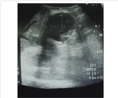
Abdomen ultrasound (US) examination revealed a circular mass (6 × 5.5 cm) in the right hypochondrium, which had no typical HCC vascular pattern (as we had expected due to her chronic HCV liver disease), nor clear anechogenic features of a cystic lesion (Figure 1). The instrumental exam also showed hepatomegaly with disomogeneous tissue and enlarged sovrahepatic veins as signs of either advanced stage of cirrhosis or severe right heart failure. Magnetic Resonance (MR) was not indicated due to the presence of cardiac mechanical valves. Abdomen computed tomography (CT) revealed a 65 mm diameter cystic mass in the fifth hepatic segment, with regular profile, small parietal calcifications and irregular content (Figures 2A and 2B). A clinical diagnosis of hydatid cyst was run out by the negative hydatid sierology and the clinical history. The patient refused to undergo surgical resection due to her high anesthesiologic risk because of several comorbilities. Although we don’t have a histological examination, according to the previous histological diagnosis of biliary cystadenoma occurred in 2009, we suspect its possible recurrence.
Discussion
BCAs are uncommon cystic benign lesions that originate from the hepatobiliary epithelium. Although the incidence of BCA is less than 5% of all hepatic biliary cystic lesions, this disease should be approached with caution due to the lack of reliable criteria for diagnosis [1].
BCAs occur commonly in middle-aged women (40-50years) with preponderance of 4:1 among males. Their clinical manifestations are variable and non-specific. Most patients are asymptomatic. The most typical symptoms are abdominal distension, abdominal pain, nausea and vomiting.
Citation: Candigliota M, Carbonara C, Doronzo R, Spagnolo M, Suppressa P, et al. (2018) Biliary Cystadenoma in a Female Patient with HCV-Related
Liver Cirrhosis: A Case Report. J Clin Case Rep 8: 1117. doi: 10.4172/2165-7920.10001117
Page 2 of 2
Volume 8 • Issue 5 • 10001117 J Clin Case Rep, an open access journal
ISSN: 2165-7920
Our patient underwent surgery for hepatic cystic lesion in 2009, when she was 64-years-old and, despite of hystotological diagnosis of BCA, in the following years she had never performed a clinical and instrumental follow-up. Because of their clinical features and slow growth, it is difficult to distinguish BCAs from simple hepatic cysts, especially if in the unilocular forms. Moreover, other hepatic cystic lesions, including hydatid cysts and a number of metastatic tumors that undergo cystic degeneration, can mimic biliary cystadenomas [2].
CT and US imaging can hardly differentiate unilocular biliary cystadenoma from other cysts. At US, biliary cystadenomas appear commonly as multiloculated cysts, whilst unilocular cysts are rare [3]. The content of the cysts may range from completely anechoic to having low-level echoes from blood products, mucin, or proteinaceous fluid. On CT scan, cystadenomas are usually seen as well demarcated lesions with contrast enhanced walls or septae. Calcifications of septae or cyst wall may be a sign of malignant degeneration [4].
The MRI and cholangio-MRI make possible to differentiate BCAs from other hepatic lesions of multilocular cystic appearance such as the hydatid cyst, hepatic abscesses, the simple or complex hepatic cyst, metastases, or other primary neoplasia.
Full surgical excision, whenever possible, followed by histopathological examination, is considered gold standard for specific diagnosis, given the high risk of malignant degeneration into cystadenocarcinoma and possible risk of recurrence of BCA if only partial excision is performed [5-7].
Conclusion
A good imaging in conjunction with a complete clinical history is always important to distinguish cystic hepatic lesions. Postoperative imaging follow-up seems to be significative to prevent recurrences and malignant degeneration of the lesion.
References
1. Jwa EK, Hwang S (2017) Clinicopathological features and post-resection outcomes of biliary cystadenoma and cystadenocarcinoma of the liver. Ann Hepatobiliary Pancreat Surg 21: 107-113.
2. Billington PD, Prescott RJ, Lapsia S (2012) Diagnosis of a biliary cystadenoma demonstrating communication with the biliary system by MRI using a hepatocyte-specific contrast agent. Br J Radiol 85: 35-36.
3. Kumar S, Gupta A, Gupta S, Noba AL, Agrawal N (2011) Giant intrahepatic biliary cystadenoma mimicking hepatic hydatid cyst. Trop Gastroenterol 32: 72-74.
4. Joel JM, Jeyasingh SD, Kalyanaraman S (2016) Biliary cystadenoma: A case report. J Clin Diagn Res 10: 19-20.
5. Arnaoutakis DJ, Kim Y, Pulitano C, Zaydfudim V, Squires MH, et al. (2015) Management of biliary cystic tumours: A multi-institutional analysis of a rare liver tumor. Ann Surg 261: 361-367.
6. Safari MT, Shahrokh S, Miri MB, Foroughi F, Sadeghi A (2016) Biliary mucinous cystic neoplasm: A case report and review of the literature. Gastroenterol Hepatol Bed Bench 9: 88-92.
7. Marrero JA, Ahn J, Rajender RK (2014) ACG clinical guideline: The diagnosis and management of focal liver lesions. Am J Gastroenterol 109: 1328-1347. Figure 2: (A and B) Cystic mass in the fifth hepatic segment, with regular profile, small parietal calcifications and irregular content.