Anno Accademico 2017-2018
Tesi di Perfezionamento
NGF steers microglia toward a
neuroprotective phenotype
Relatori
Prof. Antonino Cattaneo
Prof.ssa Simona Capsoni
Candidato
Caterina Rizzi
1 Introduction 7
1 Microglia Cell Physiology . . . 9
1.1 Morphology and Motility . . . 10
1.2 Resting (monitoring) state . . . 17
1.3 Activation state . . . 19
1.3.1 Inflammation markers . . . 25
1.3.2 Phagocytosis and pinocytosis . . . 31
1.4 Microglia and neurons interaction: Spine prun-ing and Synaptic Stripper . . . 41
2 Microglia in the Alzheimer’s Disease . . . 52
2.1 Microglia Activation: β-Amiloide and Inflam-mation . . . 58
2.1.1 Clearance of toxic peptides: Beneficial or Detrimental role of Microglia . . . . 65
3 Microglia and Neurotrophins . . . 76
2 Results 82 1 Microglia phenotype in AD11 mice . . . 82
2 Microglia express NGF receptors in vivo and in vitro . 90 3 NGF modulates the expression of genes involved in path-ways of cell motility, phagocytosis and protein degrada-tion . . . 93
4 NGF enhances microglial membrane dynamics, but not their cell speed. . . 97 5 NGF promotes microglial macropinocytosis but not
phago-cytosis . . . 98 6 NGF activates microglia currents and modulates
glu-tamatergic neurotransmission by acting on microglial cells . . . 100 7 NGF and microglia in the pathological condition: Alzheimer’s Disease . . . 103 8 NGF counteracts Aβ proinflammatory effect on microglia.104 9 NGF promotes the internalization of soluble toxic Aβ
oligomers through TrkA signaling. . . 105 10 The fate of internalized sAβ following NGF treatment . 106 11 Phagocytosis of Aβ ex vivo . . . 107 12 NGF protects against Aβ induced spine toxicity,
rescu-ing spine density and LTP . . . 107
3 Discussion 120
Microglia are the resident immune cells of the Central Nervous System (CNS). Beside classic inflammatory activities shared with macrophages, microglia actively participate in activity-dependent plasticity and learn-ing processes [1] [2], as sculptlearn-ing the neuronal circuitry durlearn-ing devel-opment [3] [4]. Microglia have been shown to be key players in the pathogenesis and progression of many neurodegenerative disorders and they are responsible for brain homeostasis and monitor the brain en-vironment with their ever-moving processes [5] [6]. However, their role, either promoting or preventing pathology, is debated. On one hand, excessive activation of microglia leads to oxidative stress, neu-roinflammation, and eventually neuronal death [7]. On the other hand, microglial activation might be harnessed to carry out protective activ-ities in the brain, such as phagocytosis of aggregates, synaptic pruning and formation, and the maintenance of healthy neuronal circuits [8]. Therefore, it is important to identify and modulate selectively the neu-roprotective activities of microglia. The idea of microglia cells as the natural scavengers of the brain becomes especially interesting when dealing with diseases with the loss of proteostasis such as Alzheimer’s disease.
In the search of neuroprotective agents against neurodegeneration, neurotrophins have been historically considered as potential therapeu-tic candidates but usually with actions targeted to natural neuronal
population. In this thesis I tested the hypothesis that microglia rep-resent a new target cell for Nerve Growth Factor (NGF) in the brain. So far sparse experiments in the literature suggest this insight.
In the literature microglia cells are known to be a source of neu-rotrophins [9] [10][11], most notably the Brain Derived Neurotrophic Factor (BDNF) which has been shown to promote synapse formation [1] and NGF [12] [13]. However, the extent of the modulation NGF might exert on physiological microglial functions and how this effect might come into play in neurodegenerative disorders has not been in-vestigated yet.
Indeed, the main cellular targets of the neurotrophin Nerve Growth Factor (NGF) [14] in the central nervous system are considered to be the cholinergic neurons of the basal forebrain (BFCNs) [15], while its sources are mainly cortical and hippocampal neurons [16].
Consistently, interference with NGF signaling (trkA-NGF signalling) in the adult brain leads to deficits of the cholinergic system that has been related to the mechanisms driving neurodegeneration, as in the AD11 transgenic mouse model [17] [18]. The expression of anti-NGF antibodies selectively neutralizing mature NGF in the adult brain de-termines a progressive comprehensive neurodegeneration with neuroin-flammation as the earliest observed change, at a presymptomatic phase [19] [20]. A similar progressive neurodegeneration is observed in trans-genic mice expressing a neutralizing anti TrkA antibody in the adult brain [21]. Changes in NGF homeostasis in the brain, with particular regard to the ratio of NGF to proNGF levels, have also been linked to Alzheimer’s disease [22].
However the overall neurodegenerative picture induced by anti-NGF or anti-TrkA antibodies in those transgenic models is much broader than what one would expect on the basis of an action of the antibodies on the BFCNs exclusively. Moreover, the loss of NGF-TrkA signaling in the CNS, obtained by conditionally deleting NGF or TrkA genes in CNS cells derived from nestin-positive cells, has proven not to
be sufficient in inducing severe cognitive impairments or neurodegen-eration in mice [23]. Altogether, this body of results has motivated our search for non neuronal targets of NGF in the adult brain. Microglia was a strong candidate, because (1) previous work had suggested that NGF could modulate some aspects of microglial cells in culture [12] and (2) transcriptomic studies in the AD11 mouse model expressing anti-NGF had shown that neuroinflammation is the earliest pheno-typic alteration, already at a presymptomatic phase (1 month of age; [19] [20]).
In this thesis I show that microglia cells are true target of NGF both in vivo and in vitro and that the activity carried out by this neurotrophin on these myeloid cells might result neuroprotective in the context of Alzheimer’s Disease.
Introduction
Microglia constitute around 10% of the total cells in the adult CNS [24]. Microglia are distributed throughout the CNS and vary in density in both rodents and humans, with subtle variations in morphology in different cytoarchitectural regions [25]. The word glia was coined in the mid 19th century and defined as the nerve glue. For decades, it was assumed to be a uniform matrix, until cell theorists raised the neuron doctrine which stipulated that nervous tissue was composed of individual cells. The term astrocytes was introduced in the late 19th century as a synonym for glial cells and later Santiago Ramon y Cajal defined a third element distinct from astrocytes and neurons.
Pio del Rio-Hortega, an alumnus of the Cajal School, introduced the modern terms and the concept of microglia that we use today. Microglia was defined cellular element of the central nervous system in a book chapter called Microglia[26] of the book Cytology and Cel-lular Pathology of the Nervous System edited by Wilder Penfield in 1932. He thoroughly described both oligodendrocytes and microglia to clearly distinguish them from astrocytes. Rio-Hortega described microglia postulating the following visionary concepts: 1) microglia enter into the brain during early development 2) these invading cells
have amoeboid morphology and are of mesodermal origin. 3) They use vessels and white matter tracts as guiding structures for migration and enter all brain regions. 4) They transform into a branched, ramified morphological phenotype in the more mature brain (known today as the resting microglia). 5) In the mature brain, they are found almost evenly dispersed throughout the central nervous system and display little variation. 6) Each cell seems to occupy a defined territory. 7) After a pathological event, these cells undergo a transformation. 8) Transformed cells acquire amoeboid morphology similar to the one observed early in development. 9) These cells have the capacity to migrate, proliferate and phagocytose [27] [28].
Microglia cells are often described as the macrophages of the cen-tral nervous system but a series of recent findings in the mouse has established that microglia are a unique cell population distinct from macrophages. Fate mapping has revealed that adult microglia are de-rived from precursors that leave the yolk sac on E8.5 E9.0, entering the neural tube via the primitive bloodstream [29] [30] as in figure 1.1. As the progenitor cells journey to the CNS, they gain lineage-specific gene expression and ultimately differentiate into mature mi-croglia. The yolk-sac-derived microglia remain throughout life, with the population being maintained by self-renewal in the healthy CNS with little contribution from bone-marrow macrophages [31] [24]. Mi-croglia are not macrophages that migrate into the brain but instead they are known to represent a distinct population of resident tissue mesenchymal cells that populate the CNS during early development [32] [33]. Remarkably, since the origins and responses of microglia and macrophages are different, the roles they play in mitigating or propa-gating pathology could be different as well. Microglial are derived from primitive erythromyeloid progenitors in the yolk sac, distinct from the definitive hematopoiesis from which the majority of macrophages is derived.
Figure 1.1: Microglia have a distinct lineage and molecular signa-ture (A) The genesis and progression of yolk-sac-derived cell lineages are illustrated. Microglial are derived from primitive erythromyeloid progenitors in the yolk sac (embryonic hematopoiesis, indicated in yel-low), distinct from the definitive hematopoiesis (shown in green) from which the majority of macrophages are derived. Figure from Salter and Beggs 2014 [24].
1
Microglia Cell Physiology
Thus, microglia cells are distinct from macrophages. In the brain, microglia, as macrophages in the other body regions, are the primary immune effector and they are key players in brain injury and disease. However, their role in the intact postnatal brain has remained elusive for a long time. Resting microglia are extremely dynamic in vivo, perpetually changing their morphology by extending and retracting highly motile processes on a time scale of minutes [34] [5]. This unex-pected finding led to a series of discoveries suggesting potential roles of microglia in postnatal development, adult neuronal plasticity, and circuit function. In the next chapters some of these physiological
mi-croglial activities and their implications for normal brain function will be described. In particular the focus will be on microglia morphology and motility, the activation state (inflammation and the engulfing abil-ity by microglia) and the interaction between microglia and neurons. Then how microglia physiology changes in pathological conditions will be discussed.
In Figure 1.2 from the paper The Role of Microglia in the Healthy Brain by Marie-Eve Tremblay et al [35], three different microglial func-tions are represented:(1) the continuous scanning of their local envi-ronment by highly motile microglial processes, (2) the structural and functional interactions with synaptic elements through direct contacts and exchanges of molecular signals, and (3) the contribution to re-structuring the neuronal circuits by phagocytosing synaptic elements and newborn cells.
Figure 1.2: Overview of microglial behavior in the healthy brain. (1) Motility, (2) Synaptic interaction (3) Phagocytosis. In green are shown dendritic branch and spines, while in blue are shown cellular inclusions.
1.1
Morphology and Motility
In 1996 Georg W. Kreutzberg published the Review Microglia: a sen-sor for pathological events in the CNS [36]. Here the microglial rapid
activation in response to minor pathological changes in CNS was de-scribed for the first time. This ability to respond quickly to a variety of signalling represents a state of vigilance to changes in their extra-cellular space. In response to any kind of brain damage or injury, microglial cells become activated and undergo morphological as well as functional transformations. In the healthy brain, microglia are characterized by cellular processes branching off from the small soma with further distal arborisation. This morphological state is typical of ramified microglia, called also resting microglia, the latter definition implying an intimate link between morphology and function [28]. The idea that microglia assume various morphological appearances corre-lated with distinct functional states arose through studies conducted in the facial nerve system [37]. It was demonstrated that depending on whether facial motoneurons regenerate after a reversible injury (facial nerve axotomy) or degenerate after irreversible injury (ricin intoxica-tion), microglia cells will respond in characteristic fashion. In figure 1.3 the original illustration from article [38] explains the new concept of functional plasticity of microglia.
Injured or diseased neurons cause resting microglia to become ac-tivated by emitting injury signals, as ATP [38] [34]. Microglia state of activation changes with the severity of neuronal damage. The mildest injuries cause hyper-ramification of microglia [39], that represents an intermediate stage between the resting and the reactive form. In the hyper-ramification state microglia start to become hypertrophic. This morphology correspond to the state of activation in vivo. Many types of neuronal damage shift resting microglia to a reactive phenotype. Nolte et al, in their paper published in 1996, investigated the regula-tion of microglia motility induced by different inflammatory mediators using time-lapse video microscopy. In this paper it was demonstrated how microglia cells exhibit a high resting motility characterized by intense ruffling of cell membranes followed by lamellipodia extension within few seconds. It was shown that this process is accompanied by
Figure 1.3: Different states of activation of microglia from Streit et al 1999.
a rapid rearrangement or the actin cytoskeleton. Later, Stence and colleagues studied microglia dynamics in rat brain tissue slices using stacks of confocal optical sections taken at intervals of 2-5 min for several hours [40]. During this time microglia became activated and shifted from a ramified phenotype to amoeboid macrophages. Ram-ified microglia progress to amoeboid macrophages through a
stereo-typical sequence of three steps, as described in figure 1.4.
Figure 1.4: Multistep model of microglial activation dynamics from Stence Glia 2001 [40].
First, in the withdrawal stage, the existing ramified branches of activating microglia do not actively extend or engulf other cells, but instead retract back (mean rate, 0.5-1.5 µm/min) and are completely readsorbed into the cell body. Second, in the motility stage, a new set of dynamic protrusions, which can exhibit cycles of rapid extension and retraction (both up to 4 µm/min), abruptly emerges. Sometimes new processes begin to emerge even before the old branches are completely withdrawn. Third, in the locomotory stage, microglia begin translo-cating within the tissue (up to 118 µm/h). The translocation starts only after the new protrusions emerge. From these experiments, it was concluded that the rapid conversion of resting ramified microglia to active amoeboid macrophages is accomplished not by converting quiescent branches to dynamic ones, but rather by replacing existing branches with an entirely new set of highly motile protrusions.
normally incapable of rapid morphological dynamics necessary for the function of activated microglial. More generally, these observations identified changes in the dynamic behavior of activating microglia and thereby helped definining distinct temporal and functional stages of activation [40].
As described above resident microglial cells in the healthy brain are thought to rest in a dormant state, whereas activation is associ-ated with structural changes, such as motile branches or migration of somata [40] [41] [38]. Most tissue preparations represent trau-matic injuries but, using time-lapse two-photon imaging of microglia, it has been demonstrated that the fine terminal of microglial processes are highly dynamic not only during the activation but also in their presumed resting state, continually surveying their microenvironment with extremely motile processes and protrusions in the intact mouse cortex [34] [5] as shown in figure 2.12.
Upon traumatic brain injury, for example when blood-brain barrier disruption occurs [5] or after ATP injection [34], microglial processes rapidly and autonomously change as described above on the site of injury without cell body movement, creating a barrier between the healthy and injured tissue. Thus microglia can be defined as busy and vigilant housekeepers in the adult brain [34]. In vivo two-photon microscopy revealed that microglial processes and arborizations are highly mobile [34] [5]. This microglia ability to monitor the envi-ronment probably depends on purinergic stimulation and involve a support from astrocytes [34] [42].
In vitro microglia cells usually do not show the ramified struc-ture characteristic of the tissue microglia. Microglia show ameboid cell morphology, no polarity, many short processes that they extend into lamellipodia to even round cells. Some cells show morphological reorganization after LPS administration [43]. Moreover, ameboid mi-croglial cells progressively ramify when co-cultured with astrocytes. Thus, microglia undergo morphological transformations from ramified
Figure 1.5: Microglial cells are highly dynamic in the resting state in vivo. (A) Maximum-intensity projections of an individual microglial cell (45 to 75 mm below the pia) at the beginning (left) and 1 hour after (center) the start of a transcranial time-lapse recording. (Right) Overlay showing extensive formation (green) and deletion (red) of mi-croglial processes. (B) Extensions (green) and retractions (red) of processes over the time course of 20 min. (C) Length changes of the processes shown in (B) as a function of time. (Right) Mean motility values in mm/min for extensions and retractions. (D) Branch motil-ity occurred at every branch order. (E) Example images of microglial protrusions (arrowheads) from a time-lapse recording. (F) Length changes over time of the two protrusions P1 and P2 indicated in (E). Vertical dashed lines mark the acquisition times of the images shown in (E). Arrows indicate protrusion lifetime. (G) Lifetime histogram of protrusions [5].
en-vironmental cues associated with astrocytes in subconfluent cultures [44].
It was through these studies on morphological changes that a croglial role in the diseased CNS was recognized. The theory of mi-croglia as sensors of pathologic changes was formulated in 1996 by Kreutzberg [36].
Given the importance of microglia morphology for its physiologi-cal role, it is important to study the changes in microglia morphology, but it is not clear from the literature how activated microglia might be accurately described. Recently, two articles, that describe an anal-ysis of microglia morphology in vivo were published. In the CNS, microglia are the only cells that express CX3CR1, also known as the fractalkine receptor [45], so Dan R. Littman and colleagues developed in 2000 a knock-in mouse model CX3CR1-GFP/+ to study the role of fractalkine [46], but later the same model was used to study mi-croglia morphology. Franck Verdonk and colleagues developed a 3D automated confocal tissue imaging system coupled with morphological modelling of many thousands of microglial cells. This study revealed a precise and quantitative assessment of major microglia cell features: cell density, cell body area, cytoplasm area and number of primary, secondary and tertiary processes. The use of two morphological cri-teria, the complexity index (CI) and the covered environment area (CEA), allowed to establish an innovative approach lying in (i) an accurate and objective study of morphological changes in healthy or pathological conditions, (ii) an in situ mapping of microglial distri-bution in different neuroanatomical regions and (iii) a study of the clustering of numerous cells, allowing us to discriminate different sub-populations. Using clustering analysis, the authors highlighted that, at resting state, microglial cells are distributed in four microglial sub-populations defined by their CI and CEA with a regional pattern and a specific behaviour. These results suggest that microglial cells are distributed in different defined sub-populations that present specific
behaviour after pathological challenge, allowing to postulate for a cel-lular and functional specialization [47]
More recently, Mar´ıa del Mar Fern´andez-Arjona and colleagues de-scribed a new method to classify microglia by their morphology, using images obtained by section scanning individual microglial cells from various regions (septofimbrial nucleus, hippocampus and hypothala-mus) at different times (2, 4 and 12 h) post-injection of a single dose of the enzyme neuraminidase (NA) within the lateral ventricle (LV). Each cell yielded a set of 15 morphological parameters. This method allowed classifying microglia population in four clusters. A linear dis-criminant analysis (LDA) suggested three specific parameters to objec-tively classify any microglia by a decision tree. In addition, a principal component
analysis (PCA) revealed two extra valuable variables that allowed to further classifying microglia in a total of eight sub-clusters or types. The spatio-temporal distribution of these different morphotypes in a rat inflammation model allowed to correlate them to microglial acti-vation status and brain location [48].
1.2
Resting (monitoring) state
It is probably reductive to apply the term resting to the non-activated microglia since they are continually involved in surveillance with their processes, which are largely responsible for this function, and therefore continually motile. The cell body of resting microglia is not motile, there is minimal expression of cell surface markers and release of cy-tokines and chemokines, and the cells are not involved in phagocytosis. However, as they clearly are not at rest, microglia under these circum-stances might be better described as monitoring microglia [49]. It is probably accurate to state that the function of resting microglia is largely unknown although key roles in homeostasis, host defense and repair have been attributed to these cells [50]. It has been
hypothe-sized that microglia are responsible for dealing with the microdamage that occurs commonly in the brain and that this ranges from forms of plasticity which are associated with resculpting (perhaps eliminat-ing) synapses to responders to capillary damages [51]. As the ma-jor resident immunocompetent cells in the brain [52] [53], microglia are responsible for sampling the microenvironment and play a role in removing cell debris [50]. This surveying role is likely to be an important factor in maintenance of homeostasis; microglia possess an array of ion channels, and expression and activation of potassium chan-nels, in particular have been suggested to be important in production of inflammatory mediators in response to stressors [54] and neuronal survival/death [55]. Similarly, at least in certain circumstances, mi-croglia, like astrocytes, express the glutamate uptake protein, GLT-1, suggesting that they may play a role in protection against glutamate toxicity [56]. The role for microglia in repair has been associated with their ability to produce neurotrophic factors, although it is also unclear whether release of neurotrophins can be achieved by resting cells [57]. What is clear is that microglia are exquisitely sensitive to stressors and rapidly change their morphology and function in response to all forms of insult. Microglia can be stimulated and may then express partic-ular cell surface markers which promote chemotaxis or infiltration of circulating cells into the CNS which permit interaction of microglia with other cells. They may be alternately non-phagocytic cells pro-ducing soluble proinflammatory molecules or they may be phagocytic and motile [36] [58]. Intermediate states may also exist: for exam-ple, it has been proposed that expression of major histocompatibility complexes (MHC) enables phagocytic function and that engulfment of pathogens or cell debris promotes release of soluble proinflammatory molecules [59] shown in figure 1.6.
Figure 1.6: At least five functional states of microglia exist but in-termediate states, and states which are multifunctional, probably also exist.
1.3
Activation state
As cited previously, microglia in healthy brain have a ramified mor-phology, with a small resting soma with fine cellular processes that scan and monitor the environment. Microglial activation is the tran-sition from resting cells to an activated state that occurs upon
dis-turbance of tissue homeostasis. This transition should be considered a change in functional phenotype. Cells shift from the surveillance mode (one type of activity) to a reactive profile, withstanding with altered homeostasis. The adjustments occur rapidly, in minutes or also seconds: cells change their chemotactic reorientations through non-transcriptional modifications. Significant transcriptional modifi-cations take place within a few hours [51].
Infection, trauma, ischemia, neurodegenerative diseases, or altered neuronal activity, ie. any disturbance or loss of brain homeostasis indicating real or potential danger to the CNS, can evoke rapid and profound changes in the microglial cell shape, gene expression and functional behavior which summarily is defined as microglial activa-tion [28] Phenotypically, the complexity of the cellular processes is reduced, and microglia revert to an amoeboid appearance. Microglia can become motile and actively move to a lesion or herd of infec-tious invaders following chemotactic gradients. Local densities of mi-croglia can also increase by proliferation, to provide more cells for the defense against invading germs and to organize for the protection and restoration of tissue homeostasis. Induction and rearrangement of surface molecules to support cell-cell and cell-matrix interactions, changes in intracellular enzymes as well as release of multiple factors and compounds with proinflammatory and immuno-regulatory effects are additional elements of the activation process. Microglia can un-fold their phagocytotic activities to clear tissue debris, damaged cells, or microbes. The release of chemoattractive factors by microglia re-cruits and guides immune cell populations to the CNS. Presentation of antigens by microglia to T cells can subsequently aid the adaptive immunity in fight against viral or bacterial invasion. The range of microglial activities also covers the production of neurotrophic factors and the physical association with endangered neurons.
Various kinds of stimulators have been predicted for microglia ac-tivation: some kinds of molecular and cellular mechanisms are shown
in figure 1.7.
Figure 1.7: Putative molecules for microglial activation and activated microglia-derived cytotoxic and neurotrophic molecules. Figure from ”Microglia: Activation and Their Significance in the Central Nervous System” Nakajima et al 2001 [60].
One category is represented by non-material stimulators, the elec-trical potential change resulting from neuronal injury and changes in the ion medium around injured neurons are candidates. The other cat-egory is that of biologically active substances, which may include low molecular weight molecules such as peptides and hormones. Growth factors or cytokines may also be able to activate microglia. Among the cytokines are macrophage colony-stimulating factors (M)-CSF and granulocyte-macrophage colony-stimulating factors (GM)-CSF [61]. As for other activators, calcitonin gene-related peptide(CGRP) and ATP [62] induce immediate early gene mRNA expression and transla-tion in microglia. Likewise, ATP can cause biological responses such
as chemotaxis [63], release of plasminogen (PGn) and interleukin IL-1β, and activation of nuclear factor (NF) KB in cultured microglia [64]. Therefore, it is likely that some molecules derived from neurons and/or astrocytes trigger the activation of microglia in vivo as shown in figure 1.7.
Microglial activation can be accompanied by proliferation. The factor(s) necessary for mitogenesis in microglia were clarified by an in vitro study. Microglia can proliferate in response to GCSF, M-CSF, and multi-CSF (IL-3) [65]. Moreover, CSFs, IL-2, IL-4, IL-5, and tumor necrosis factor (TNF)α are reported to induce proliferative activity in cultured microglia. In vivo, CSF or CSF-like factors may be responsible for the proliferation and activation of microglia.
As described above, many molecules and conditions can trigger a transformation of resting (or surveying) microglia to activated (alerted or reactive) states. These have in common that they indicate a threat to the structural and functional integrity of the CNS. Microglial cells are prepared to recognize a wide range of signs for homeostatic surveil-lance, independent of their biochemical nature (peptides, lipoproteins, glycolipids, nucleotides) or diverse (patho)physiological implications. The list is shown in figure 1.8
The activation of microglia may have serious consequences on neu-ronal and astroglial activity and survival. The effects of activated microglia on neuronal survival is debated. On one hand, activated mi-croglia in culture have been shown to produce several potentially cy-totoxic molecules, including superoxide anion, nitric oxide, and proin-flammatory cytokines [66] [67], shown in figure 1.7. Lipopolysaccha-ride (LPS), interferon γ (INFγ), and β-amyloid [68] are among the stimulators for the production of harmful factors from microglia. Re-active oxygen species (ROS) including superoxide anions, hydroxy rad-icals, and hydrogen peroxide are generally hazardous, particularly to myelin and its forming cells (oligodendrocytes) owing to their capa-bility of inducing lipid peroxidation. LPS and
phorbol-12-mynstate-Figure 1.8: Exemples of signals and modulators of microglia activation adapted from Uwe-Karsten Hanisch, Helmut Kettenmann 2007 [51].
13-acetate (PMA) are stimulators of ROS production from cultured microglia. Nitrogen oxides such as NO are highly reactive free radi-cals, of which nitrite peroxide is the strongest species. These radicals are believed to inhibit respiratory enzymes, oxidize the SH group of proteins, and enhance DNA injury, finally resulting in neuronal cell
death. LPS and β-amyloid are known stimulators of NO production from microglia. In the presence of INFγ, β-amyloid synergistically stimulates the production of NO and TNFα [69] in microglia.
Besides, recently it was demonstrated by Liddlelow and colleagues a direct role of microglia on the astrocytes activation. They demon-strated that a subtype of reactive astrocytes, named in their paper A1, is induced by classically activated neuroinflammatory microglia. They show that activated microglia induce A1 astrocytes by secreting Il-1a, TNFα and C1q, and that these cytokines together are necessary and sufficient to induce A1 astrocytes. A1 astrocytes lose the ability to promote neuronal survival, outgrowth, synaptogenesis and phago-cytosis, and induce the death of neurons and oligodendrocytes. Death of axotomized CNS neurons in vivo is prevented when the formation of A1 astrocytes is blocked. Finally, they have shown that A1 astrocytes are abundant in various human neurodegenerative diseases including Alzheimer’s, Huntington’s and Parkinson’s disease, amyotrophic lat-eral sclerosis and multiple sclerosis [70]. Effector microglia might bet-ter describe cells executing a number of adaptive responses to a given challenge.
However, on the other hand, co-cultures of microglia and neurons revealed that microglia did not kill healthy neurons in vitro, indicating that their phagocytic properties are not necessarily dangerous for neu-rons. Secondly, the effects of conditioned microglial medium (CMM) on neurons were examined. The results showed the CMM expressed no neurotoxicity. Rather, CMM significantly enhanced neuronal survival and the neurite outgrowth of neocortical and mesencephalic neurons [60].
In conclusion, activated microglia act primarily in the defence of the brain, as brain scavengers, and in tissue remodeling, an immune and/or immunoeffector role has also been proposed. Activated mi-croglia are implicated in pathogenesis and inflammation. They have also been shown to produce a variety of biological factors. Recent
bio-chemical and neurobiological studies have revealed that activated mi-croglia produce and secrete not only cytotoxic/harmful molecules but also neurotrophic molecules including neurotrophins and neurotrophic cytokines. As a whole, activated microglia appear to play a significant role in pathological and regenerative states of the brain, expressing both or either of two potentially opposing functions: cytotoxic and neurotrophic actions.
1.3.1 Inflammation markers
Neuroinflammation, and to the same degree all inflammation, is a fun-damental immune response designed to protect the body from harm, arising from both endogenous and exogenous sources. Being the sen-tinel immune cell of the brain, microglia play a role as first responders to infections or tissue injuries and initiating an inflammatory response. Using a full array of immune receptors, such as toll-like receptors (TLRs), nucleotide binding oligomerization domains (NODs), NOD-like receptors, and many scavenger receptors [71] [72], microglia (as well as other CNS cells, such as astrocytes) are able to recognize harm-ful stimuli and respond by producing inflammatory cytokines such as TNFα, IL-6, IL-1β, interferon-γ (IFNγ), and several chemokines [73]. This cytokine production is essential for the polarization of microglia into what has been termed a classically activated, M1, state [74]. Due to this ability to respond to inflammatory cytokines, as macrophages do, the microglia activation state started to be named in the same way of macrophages. In particular, the term M1 parallels the Th1 termi-nology used for T cells, and underscores the close relationship between T cells and macrophages in the periphery. Interferon-γ produced from Th1 cells was found to be instrumental in polarizing macrophages to M1 state [74]. However, the ability to produce these cytokines does not rest solely with T cells. Microglia and astrocytes have also been observed to fill this role [75], demonstrating, at least in part, that
microglia can control their own polarization through autocrine and paracrine means. For the evolution of these theories on macrophages polarization see figure 1.9.
Figure 1.9: M1 and M2: the concept of macrophage polarization [76]. In many cases, this response is protective and is downregulated once the damage or pathogen has been dealt with. However, dysreg-ulated, long-term, or chronic inflammation can lead to tissue destruc-tion [77] [7] as in the rappresentative figure 1.10.
Differently to proinflammatory M1 cells, alternatively activated macrophages (M2) express cytokines and receptors that are implicated in inhibiting inflammation and restoring homeostasis. This includes: IL-10 to downregulate inflammatory cells, extracellular matrix pro-tecting proteins like YM1, ornithine, and polyamines for wound re-pair, and higher levels of receptors associated with phagocytosis, such as scavenger receptors [78]. Just as the Th1 cytokine IFNγ has been associated with induction of proinflammatory M1 macrophages, the Th2 cytokine IL-4 has been associated with M2, or alternative, acti-vation. Interestingly, it appears that when there is a lack of M2 cell differentiation in the CNS, problems can arise. To properly understand
Figure 1.10: Reactive microgliosis drives progressive neurotoxicity: microglia can become overactivated and cause neurotoxicity through two mechanisms. First, microglia can initiate neuron damage by recog-nizing pro-inflammatory stimuli, such as lipopolysaccharide (LPS), be-coming activated and producing neurotoxic pro- inflammatory factors. Second, microglia can become overactivated in response to neuronal damage (reactive microgliosis), which is then toxic to neighbouring neurons, resulting in a perpetuating cycle of neuron death. Reactive microgliosis could be an underlying mechanism of progressive neuron damage across numerous neurodegenerative diseases, regardless of the instigating stimuli [7].
the role microglia play in neurodegeneration, understanding their phe-notypes is important. The functional effects of classical activation are geared towards antigen presentation and the killing of intracellular pathogens. Therefore, upregulation of many associated receptors and enzymes reflects that purpose. For example, MHC II, CD86, and Fcγ receptors are upregulated to allow for antigen-presenting activity of
microglia and increased crosstalk with other immune cells [79]. In addition, the ratio of particular cytokines has been used to identify inflammatory macrophages and this observation could extend to mi-croglia. For example, since M1 macrophages were found to be a key source of IL-12 [80], it was suggested that IL12 High, IL-10 Low pro-duction is a simple way to distinguish inflammatory cells [81]. Another potential distinction and an important component of M1 microglia is their ability to produce reactive oxygen species and reactive nitrogen species [82]. A key microglial enzyme associated with this process is inducible nitric oxide synthase (iNOS), which utilizes arginine to produce nitric oxide [83]. However, even though it seems straightfor-ward to identify M1 cells based on these characteristics, classifying these cells in vivo has proven to be more challenging, reflecting the plastic nature of microglia. Even for M2 cells the classification is not clear. In fact, there are many efforts to identify unique subgroups with different functions. Division of M2 cells is based on observations that stimulation with various cytokines yields different sets of receptor profiles, cytokine production, chemokine secretion, and function [81]. Even though the profiles of these cells are diverse, the one feature that places them all in the M2 classification is that they express mediators or receptors able to downregulate, repair, or protect the body from in-flammation [84]. The original alternatively activated macrophage was classified based on expression of the mannose receptor [85]. Since then a panel of different markers has been identified as M2 specific. One of the best characterized markers is the enzyme arginase 1 (Arg1) [86], which converts arginine to polyamines, proline, and ornithines that can contribute to wound healing and matrix deposition [87]. Thus, iNOS and Arg1 represent a relatively straightforward set of markers to distinguish M1 from M2 phenotypes, as shown figure 1.11.
Other markers used for identifying M2 cells include Ym1, a heparin-binding lectin [89], FIZZ1, which promotes deposition of extracellular matrix [90], and CD206, a mannose receptor [85]. Despite the benefit
Figure 1.11: Diagram of activation states of microglia based on in-flammatory profile and effector function. Based upon peripheral macrophage nomenclature, M1 and M2 polarization states of microglia have been proposed as a framework to evaluate the heterogeneity of responses with recent evaluations suggesting a framework focused on the inducing stimuli [88].
of having specific markers, using just one or two is limiting and ignores the overall diversity of M2 phenotypes. Another way to classify the function and phenotype of M2 cells is based on the cytokines that in-duce them. The prototypical cytokine used to first inin-duce alternative activation was IL-4 [85]. Both IL-4 and the closely related cytokine IL-13 signal through IL-4Rα to induce a host of downstream processes that lead to potent anti-inflammatory functions, such as Arg1 upreg-ulation, inhibition of NF-kB isoforms, and production of scavenger receptors for phagocytosis [79] [91] [92]. This type of activation has been classified as M2a. The main function of this response appears to be suppression of inflammation. A second state of alternative
acti-vation is based on macrophages exposed to IL-10, glucocorticoids, or TGF-β. This phenotype is classified as M2c [81] [93]. Originally, this state was described as being deactivated but that is not a particularly useful description. Instead of having no function, deactivated M2c macrophages appear to be involved in tissue remodeling and matrix deposition after inflammation has been downregulated [81]. A third sub-class of M2 activation has been observed following exposure to immune complexes and stimulation of TLR. This class is termed M2b or Type II [81] [94]. Of these three classes, M2b macrophages are the least understood. Interestingly, they more closely resemble M1 macrophages, owing to the lack of any M2 specific markers, such as Arg1, YM1, or FIZZ1. However, they do express the typical IL-10 High, IL-12 Low M2 cytokine profile [95]. Moreover, they have higher levels of MHCII and CD86, suggesting that they retain their ability to stimulate T cells [94].
An important consideration regarding M2 phenotypes is that these states were typically elucidated in vitro following exposure to one or two stimuli. This does not represent the complex environmental milieu seen in tissue. Therefore, some investigators have cautioned against this classification into distinct subtypes and instead propose that M2 cells should be viewed as a spectrum of phenotypes [95]. This de-tailed classification of M2 cells has been primarily carried out in the periphery. Whether or not this will extend to brain resident microglia is yet to be seen. Furthermore, microglia are not macrophages that migrate into the brain, but instead are known to represent a distinct population of resident tissue mesenchymal cells that populate the CNS during early development [32] [33]. Importantly, because the origins and responses of microglia and macrophages are different, the roles they play in mitigating or propagating pathology could be different as well.
Moreover, some other markers have been variously used as indica-tors of microglial activation but the limitations of their use are seldom
recognized. Important among these limitations is that several (if not all) of the markers are not unique to microglia and that, except in par-ticular circumstances for example where pure microglial cultures are used, their upregulation may not reflect changes in microglia alone (figure 1.12).
Figure 1.12: Proposed cell surface markers of microglial activation [49].
It is also important to recognize that, to a large extent, studies re-port changes in expression of cell surface markers under various condi-tions with minimal assessment of functional change and therefore the coupling of phenotype and function remains a challenge.
1.3.2 Phagocytosis and pinocytosis
Microglial cells are the professional phagocytes of the CNS tissue. This function is important for the normal brain, during brain development, and in pathology and regeneration [28]. As for other microglia func-tions, such as the inflammatory cytokines secretion, also for phago-cytosis there is no obvious concordance with whether it is beneficial
or detrimental for tissue health. As I described previously, microglia cells are highly sensible to different stimuli of the environment. As these cells change their motility, their surface receptors, the release of soluble chemokines and cytokines, they also change the ability of up-taking debrees. Based on the type of debris, microglia can use phagocytosis or pinocytosis to take them up [28].
Endocytosis can be classified in two types: the clathrin-dependent and clathrin independent endocytic pathways. The clathrin-dependent endocytic pathway requires the recruitment of endocytosed molecules into clathrin-coated pits that are around 100 nm in diameter [96]. Phagocytosis and pinocytosis are two clathrin-independent endocytic processes that occur in phagocytes, and both create large endocytic vacuolar compartments (≥0.2 µm) through organized membrane move-ments and actin polymerization [97]. Nevertheless, distinct models and molecular mechanisms have been suggested for the formation of phagosomes and pinosomes [97]. The well-studied Fc mediated phagocytosis is guided by a zipper-like progression of receptor-initiated membrane invagination that is shaped by the geometry of the internalized particle, whereas pinosomes, which may vary from 0.2 to 10 µm in diameter, are suggested to form spontaneously or in response to growth factor receptor activation from membrane ruffles that close at their distal margins to engulf extracellular fluid [97] [98]. Phago-cytosis is applied in many physiological and pathological processes, including development, innate immunity, and the entry of pathogens into host cells [97] [98].
The activity of microglial phagocytosis relies on ”eat me” signals expressed on the cell surface of dying cells [99] and on specific receptors expressed on microglia cell surface. Downstream signaling pathways are activated and contribute to the reorganization of actin protein and engulfment of harmful microparticles (Fig.1.13).
Microglial phagocytosis may need different types of receptors to initiate function [100], there are two distinctive types of receptors:
Figure 1.13: Signaling pathways involved in microglial phagocytosis. A Extracellular nucleotides, such as UDP and UTP, trigger microglial phagocytosis through P2Y6R/ PLC/InsP3 pathway. B Apoptotic de-bris induces phagocytosis via TREM2/DAP12/ERK/PKC pathway. C Endogenous or ectogenic detriments, such as LPS, viral nucleotides, α- synuclein, and f-Aβ, provoke phagocytosis by microglia via TLRs through activation of MyD88-dependent IRAK4/p38/ scavenger re-ceptors pathway or MyD88-independent actin- Cdc42/Rac signaling pathway.
- the first with a high affinity for foreign microbial pathogens and which simultaneously stimulates a pro-inflammatory response in the
phagocytes [101], such as Toll-like receptors (TLRs). - The second type of receptors can, recognize apoptotic cellular substances and stimulate an anti-inflammatory response in phagocytes. Triggering receptor ex-pressed on myeloid cells 2 (TREM2) is an example of this second type of receptors. The phagocytosis triggered by TREM2 takes place without inducing inflammation and it is one of the major beneficial functions of phagocytes (figure 1.13).
TREM2 is a kind of pattern receptor specific for polyanions and lo-cates mainly on the cell surface of osteoclasts in bones and in microglia of the CNS [102].
TREM2 [103] is a 40-kDa type I membrane glycoprotein with a single extracellular immunoglobulin-like domain, one trans-membrane domain, and a short cytoplasmatic tail. Its transmembrane domain interacts with the adaptor protein TYROBP/DNAX-activating pro-tein of 12 kDa (DAP12) via electrostatic interaction [103]. DAP12 is a type I transmembrane protein, which acts as a signaling adaptor protein for TREM2 and for other cell surface receptors.
TREM2 seems to be involved in two signaling pathways in mi-croglia: phagocytosis and proinflammatory cytokines secretion. In-deed, increased expression of TREM2 on microglia has been associ-ated with enhanced phagocytosis and increased alternatively activassoci-ated M2 protective microglia, while absence of TREM2 expression on mi-croglia does exactly the opposite, impairing phagocytosis and increas-ing the proinflammatory phenotype [104]. TREM2 phagocytic func-tion is not associated with inflammafunc-tion. In addifunc-tion to up-regulating of chemokine synthesis and mediating protective phagocytosis of apop-totic cell debris, activation of TREM2 receptors suppresses secretion of pro-inflammatory factors such as cytokines and ROS [102]. On mi-croglia TREM2 via binding to DNAX- activation protein 12 (DAP12), an ITAM-containing adaptor protein, triggers the reorganization of F-actin and phosphorylation of ERK/MAPK, mediating the clearance of apoptotic neurons [105] [106].
The paramount importance of these receptors has recently become clear as patients with a loss of function mutation of either TREM2 or DAP12 develop an inflammatory neurodegenerative disease leading to death at the fourth or fifth decade of life. Nasu-Hakola disease, a sys-temic bone cystic disorder with progressive presenile dementia followed by extensive sclerosis in the front-temporal lobe and the basal ganglia, occurs due to genetic mutation of TREM2 and DAP12 resulting in aberrant TREM2/DAP12 signaling pathway. Thus, even though the ligand of TREM2 is unknown, microglial TREM2/DAP12-mediated phagocytosis appears to be an essential function for CNS tissue ho-moeostasis [104].
Besides these two types of receptors, some others including Fc re-ceptors, complement receptors [107], scavenger receptors (SR), pyrim-idinergic receptor P2Y, G-protein coupled, 6 (P2RY6), macrophage antigen complex 2 (MAC-2), mannose receptor [108], and low-density lipoprotein receptor-related protein (LRP) receptor also participate in microglial clearance of misfolded, apoptotic cells and dead neurons in both acute and chronic brain injury [109].
Furthermore, TLRs are a class of proteins that play a key role in the innate immune system and the digestive system. TLRs are single, membrane-spanning, non-catalytic receptors usually expressed in pe-ripheral sentinel cells, such as macrophages and dendritic cells, that recognize structurally conserved molecules derived from microbes. An example is proposed by TLRs 1-9, which belong to interleukin (IL)-1R super-family and that are expressed exclusively on antigen presenting cells including microglia [110], macrophages, antigen presenting den-dritic cells, and on cerebral parenchyma cells including neurons, oligo-dendrocytes, and astrocytes. TLRs not only trigger the recognition of pathogen-associated molecular patterns (PAMPs), such as LPS or viral nucleotides, but also recognize so called danger-associated molec-ular patterns (DAMPs), such as deposited amyloid β (Aβ) fibril and α-synuclein [111] [112], they are host biomolecules that can initiate
and perpetuate a noninfectious inflammatory response. In contrast, PAMPs initiate and perpetuate the infectious pathogen-induced in-flammatory response. TLR4-, TLR2-, and TLR9-dependent signaling pathways are involved in mediating microglial phagocytosis of neuro-toxic Aβ deposits in AD brain and exert a protective role in nerve re-generation [113] [114]. It has been reported that TLRs regulate phago-cytosis through myeloid differentiation factor 88(MyD88)-dependent and MyD88-independent signaling pathways. The MyD88-dependent pathway is triggered by TLRs through activation of IL-1 receptor-associated kinase (IRAK)-4 and MAP p38, resulting in up-regulation of scavenger receptors [115]. On the other hand, TLRs also regu-late phagocytosis by a MyD88-independent actin-Cdc42/Rac pathway [116].
Pinocytosis can be triggered by ”drink me” signals, since in the pinosomes particles are small and a part of the extracellular fluid is also included. It can be subdivided in micro-and macro-pinocytosis.
Micropinocytotic vesicles are no larger than 0.1 µm in diameter and may be caveolin-coated. Micropinosome formation is indepen-dent of actin and occurs within cholesterol-rich lipid domains of the plasma membrane [117], while macropinocytosis results in the forma-tion of vesicles that are between 0.2 µm and 5.0 µm in diameter and are created by the enclosure of membrane ruffles. Macropinocytosis was first described morphologically by Warren Lewis in 1931 [96] and it is a clathrin-independent endocytic route, a regulated form of endo-cytosis that mediates the non-selective uptake of solute molecules, nu-trients and antigens. Macropinocytosis is a signal-dependent process that normally occurs in response to growth factor stimulation, such as macrophage colony-stimulating factor-1 (CSF-1), epidermal growth factor (EGF) and platelet-derived growth factor or tumour-promoting factor, such as phorbol myristate acetate (PMA) [118] [119]. How-ever, some specialised cell types such as antigen-presenting cells are capable of constitutive macropinocytosis [120] [121].
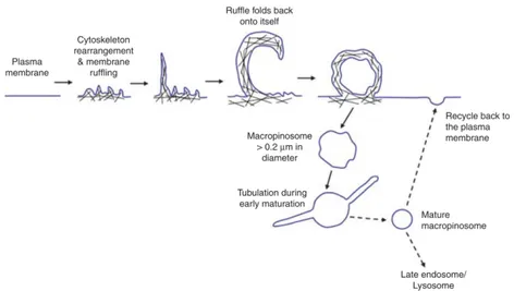
Macropinocyto-sis involves actin- mediated membrane ruffling of the plasma mem-brane leading of the formation of lamellipodia. Most of these retract back into the cell. However, a subset of lamellipodia may fold back onto themselves and fuse with the basal membrane creating large, irregular shaped vesicles named macropinosomes (Figure 1.14). How-ever, there is, no consensus on the proportion of membrane ruffles that end up forming macropinosomes. Macropinosomes are distinct from other forms of endocytic vesicles. Macropinocytic vesicles have no apparent coat structures and although heterogenous in size, they are generally considered to be ≥0.2 µm in diameter [122] [123], a size considerably larger than clathrin coated vesicles. Owing to the large size of macropinosomes, which can be up to 5 µm in diameter, macropinocytosis provides cells with a way to non-selectively inter-nalise large quantities of solute and membrane. Thus, in addition to its size, macropinosomes can also easily be identified through the use of fluid phase markers, such as Lucifer Yellow, horseradish peroxidase and dextran. Once formed, macropinosomes undergo a maturation process. However, this maturation is unlikely to occur through a path-way common to all cells (see in figure 1.14).
Macropinosomes form in response to treatment with CSF-1 [124]. They acquire and loose different endocytic protein markers as they migrate centripetally toward the lysosome. Within 1 min of colony-stimulating factor-1 treatment, macropinosomes become positive for transferrin receptors. These are considered to be macropinosomes at an early maturation stage, within 2-4 min and the macropinosomes are devoid of transferrin receptors and begin to acquire Rab7, a marker of the late endosomes. Rab7-staining coincides with the presence of lysosomal glycoprotein A (lgp-A). Finally, the macropinosomes merge with the existing tubular lysosomal compartments [125] and fuse with the plasma membrane, recycling its content to the exterior of the cell [122] [126].
Figure 1.14: Pathway of macropinocytosis. Macropinocytosis in-volves actin cytoskeleton rearrangement at the plasma membrane leading to the formation of membrane ruffles. Ruffles may fold back onto themselves and fuse at the base of plasma membrane, trapping solute and soluble substances in macropinosomes. Early maturation of macropinosomes involves extensive tubulation result-ing in mature macropinosomes that are more spherical. Content of the macropinosomes are then either degraded at the late endo-some/lysosome or recycled back to the plasma membrane. The cy-toskeleton is depicted as black lines [96].
the plasma membrane, this pathway has been implicated in cell motil-ity. Macropinocytosis is also considered important in the chemotactic response of highly mobile cells.
A key difference between clathrin-dependent endocytosis and macropinocy-tosis is that the latter requires actin cytoskeleton reorganisation.
cy-tochalasin, an actin-disrupting agent [127] has been shown to inhibit macropinocytosis but not the clathrin-dependent endocytic pathway [128] [129]. The dynamics of actin cytoskeleton during macropinocy-tosis has been visualised in cells expressing fluorescently labelled actin probes such as coronin. Coronin is an actin-associated protein, which has a role in controlling actin dynamics during processes such as cell motility, phagocytosis and cytokinesis [130]. In a separate experiment, the dynamics of actin cytoskeleton was observed using GFP-actin-binding domain probe GFP-actin-binding specifically to F-actin, a structure also recognised by phalloidin [131] [132]. The F-actin probe remained asso-ciated with the macropinosome but was completely lost 30-50 seconds after vesicle closure [131].
Araki et al. [133] investigated the distribution of F-actin in mouse macrophages using actinin-4, a non-muscle form of α-actinin [133]. α-Actinin is an actin-binding protein, which cross-links F-actin into bundles or connects F-actin to the plasma membrane. Immunofluores-cence analyses revealed that lamellipodia stained positive for actinin-4. Actinin-4 was also associated with dextran-labelled macropinosomes and this association was found to decrease as the macropinosome ma-tures [133]. These examples clearly depict the importance of actin cytoskeleton reorganisation during macropinocytosis. Therefore, com-ponents of signal transduction pathways linking receptor stimulation to actin cytoskeleton remodelling are also implicated as regulators of macropinocytosis as in table 1.15.
The remodelling of the cytoskeleton during macropinocytosis re-quires also the activity of PI3-kinase [133] [134] [135]. In the case of bone marrow-derived macrophages, PI3-kinase activity was not re-quired for cell surface ruffling but rather for the closure of macropinosomes [133]. PI3-kinase is a core signalling component in multiple signal transduction pathways such as endocytosis, membrane traffic and cell proliferation and inhibition of its activity will result in a range of other effects. Amiloride or its more potent counterpart, dimethyl amiloride,
Figure 1.15: Regulators of macropinocytosis and their respective sub-cellular localisation during macropinocytosis [96].
is an inhibitor of the Na+/H+ exchanger pump in the plasma mem-brane. It has been reported to selectively block macropinocytosis, and not the clathrin-dependent endocytic pathway, when used at the ap-propriate concentration [126] [136]. Thus, amiloride is gaining
pop-ularity as a tool to study macropinocytosis. It has recently been proposed that macropinocytosis is not directly sensitive to amiloride; rather, macropinocytosis is affected by acidification from excessive lo-calised H+ production at the site of macropinocytosis when the reg-ulatory action of Na+/H+ exchanger pump is impaired by amiloride. Acidification does not inhibit receptor activation or PI3-kinase local-isation and activation but it does affect downstream locallocal-isation and activation of Rac1 and Cdc42, GTPases required for actin remod-elling [136]. Members of the sorting nexin (SNX) family have been localised to macropinosomes and also implicated in macropinosome formation. There is a direct relationship between the level of cell surface SNX5 protein and macropinocytic activity, highlighting the role for this sorting nexin in driving this signal-activated endocytic pathway. In a recent study, 12 members of the SNX-PX-BAR fam-ily were screened in an assay to determine the effect of their over-expression on macropinosome formation [137]. SNX1, SNX5, SNX9, SNX18 and SNX33 were all found to enhance macropinosome forma-tion, an effect that appeared dependent on the increase in PI(3,4,5)P3 levels, which is regulated by PI3-kinase activity [137]. PI3-kinase, Ras, Rac1 and Cdc42, as well as sorting nexins, are some examples of macropinocytic regulators. Additional molecules, which are com-ponents of the signal transduction pathway, and which have been im-plicated in macropinocytosis are listed in table 1.15. Nonetheless it is unclear how these different components are collectively organised to regulate the process of macropinocytosis.
1.4
Microglia and neurons interaction: Spine
prun-ing and Synaptic Stripper
Recent findings challenge the concept that microglia solely function in disease states in the CNS. Rather than simply reacting to CNS injury,
infection, or pathology, emerging lines of evidence indicate that mi-croglia sculpt the structure of the CNS, refine neuronal circuitry and network connectivity, and contribute to plasticity. These physiological functions of microglia in the normal CNS begin during development and persist into maturity. Furthermore, both resting and activated mi-croglia (as defined by a morphological phenotype) have physiological functions even in the absence of pathologies. Consequently, the con-cept of resting and activated microglia is misleading because multiple phenotypic stages of these cells can influence the neuronal structure and function to maintain neural circuits.
In the absence of pathological insults, the highly dynamic motility of microglia is specifically targeted to synaptic structures as shown in figure 1.16.
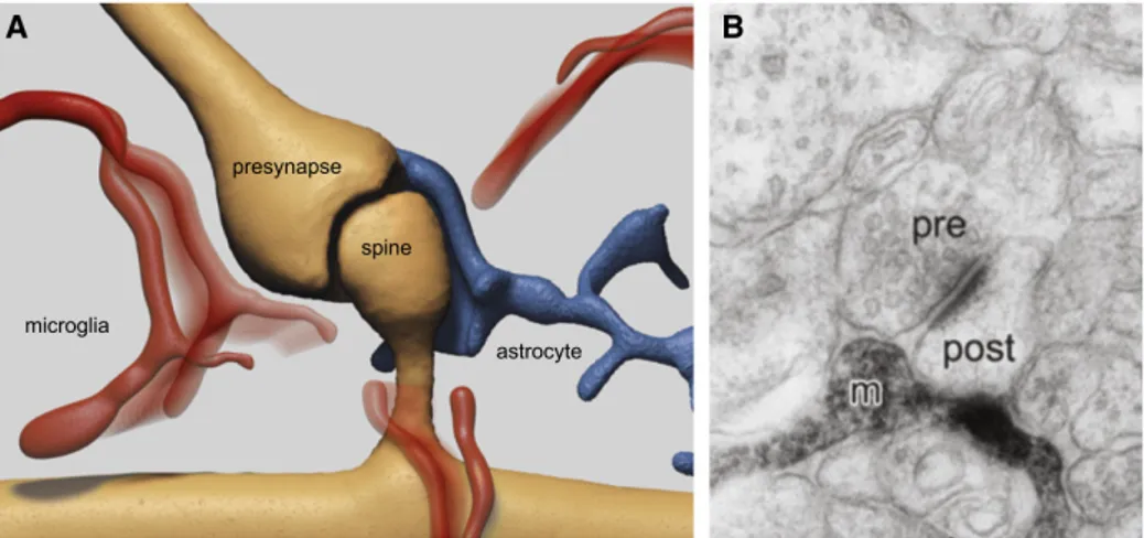
In the somatosensory and visual cortex two-photon microscopy re-vealed that microglial processes make brief, repetitive contacts with synapses at a frequency of about one per hour. These interactions were visualized by in vivo two-photon imaging in the Iba-1-EGFP/Thy-1-GFP double-transgenic mice in which microglial cells and neuronal structures can be simultaneously visualized. Wake et al. in 2009 re-ported that the microglial processes appear in a close proximity to presynaptic boutons, where they remain for about 5 min and then retract. In the visual cortex, these structural interactions are activity-dependent, as their frequency was reduced by decreased neuronal ac-tivity following either binocular eye enucleation, injection of tetrodotoxin into both retinae or a reduction of body temperature [138]. By com-bining two-photon in vivo imaging with immunohistochemistry and three-dimensional reconstructions obtained from serial section electron micrographs as shown in figure 1.16B, a rather specific apposition of microglial processes to pre as well as postsynaptic compartments was found in the visual cortex of juvenile mice [139]. Process extrusions were typically associated with small and transiently growing dendritic spines. Again, neuronal activity modulated microglial behavior: light
Figure 1.16: Dynamic Interaction of Microglial Processes with the Quadripartite Synapse: (A) Microglial processes (red) dynamically contact the cellular compartments of the tripartite synapse: pre- and postsynaptic neuronal terminals (in brown) as well as the enwrapping perisynaptic astroglial process (in blue). (B) The electron micrograph (EM) specifically shows a microglial process (m) contacting both the pre and postsynaptic compartment. The EM image is modified from Wake et al. (2009) [138].
deprivation reduced the motility of microglial processes and increased their association with larger dendritic spines. Re-exposure to light re-versed these behaviors and microglial processes enwrapped synapses more extensively [139]. In response to neuronal activity, microglia steer their processes toward active synapses (as shown in Figure1.17A), which facilitates contact with highly active neurons. The mechanism of this steering of microglia to active synapses is currently not known. Thus microglial cells respond to altered sensory experience and it remains an open question whether the interactions play any role
Figure 1.17: Microglia Control Neuronal Activity, Programmed Cell Death, and Synapse Connectivity. Taken from [24].
in experience-dependent modification or elimination of synapses, in the developing as well as in the adult CNS. The molecular cues that attract microglial processes to the synapses remain largely unknown. Considering these synapse modification or elimination that could be affected by microglia during experience-dependent plasticity, the term tripartite synapse was changed in quadripartite synapse, where pre-post synaptic neurons, astrocytes and microglia modulate the synaptic activity together.
through removal of cellular and subcellular elements (by phagocytosis) or through secreting various factors with transmitter, trophic or neu-roprotective properties. The variety of neuroactive agents that can be secreted by microglia cells has been mainly studied in cell culture, an environment that triggers transformation of microglial cells into an ac-tivated state. Thus, these factors are considered to be representative of an activated state, with possible relevance to a more pathological acti-vation. These include several types of cytokines (e.g., TNFα or ligands for receptors such as CCR1, CCR3, CCR5, and CCR7 and CXCR1 or CXCR3), trophic factors like brain derived neurotrophic factor (BDNF), the gaseous transmitter NO or neurotransmitters (ATP and glutamate) (for review, see Kettenmann et al. 2011 [28]). Some of these substances were reported to rapidly modulate neuronal function by changing excitability and synaptic strength. Moreover, several fac-tors known from pathology can mediate the interactions of microglia with synapses in the healthy brain. Recent findings indicate that some of these pathological factors mediate neuron-microglia crosstalk in the developing or uninjured adult brain.
As described in the first chapter, the primitive myeloid progenitors originating from the extra-embryonic yolk sac enter the nervous tissue very early in embryonic development, being the first existing glial cells (as both astrogliogenesis and oligodendrogliogenesis occur later in a perinatal period) [29]. This initial migration coincides with the first wave of embryonic synaptogenesis (which occurs, in rodents, around embryonic day 14-15) that proceeds in the absence of astrocytes (which assist and are indispensable for postnatal formation of synapses). In this phase microglia can assist and even promote early synaptogenesis through secretion of growth factors [140].
Miyamoto et al. in 2016 [141] demonstrated how the microglia-neurons contact induces synapse formation in the developing somatosen-sory cortex. Using in vivo multiphoton imaging of layer 2/3 pyramidal neurons in the developing somatosensory cortex, they demonstrated
that microglial contact with dendrites directly induces filopodia forma-tion. This filopodia formation occurs only around postnatal day 8-10, a period of intense synaptogenesis and when microglia have an acti-vated phenotype. Filopodia formation is preceded by contact-induced Ca2+ transients and actin accumulation, as shown in figure 1.18 [141].
Figure 1.18: Scheme of microglia functions during synaptogenesis. (a) Sequence of proposed cellular events during synapse formation: Mi-croglial contact initiates a rise in local [Ca2+]i resulting in actin
ac-cumulation and filopodia formation. Some filopodia find presynaptic partners and mature into functional synapses. (b) Schematic graph de-picting a change in microglia phenotype and function, from immature or activated microglia inducing filopodia formation and enhancing spe-cific circuit synapse formation during early synaptogenesis, followed by a putative role in synapse elimination by more mature, quiescent microglia in the latter period of circuit formation [141].
Inhibition of microglia by genetic ablation decreases subsequent spine density, functional excitatory synapses and reduces the relative connectivity from layer 4 neurons [141] demonstrating how microglia cells are crucial elements to the spine formation.
During later, pre and postnatal development, microglial processes actively engulf synaptic structures and exert a major role in control-ling the number of synapses through synaptic pruning. The chemokine fractalkine plays a role in chronic pain, inflammation, and Alzheimer’s disease and it is released by neurons and endothelial cells [142] [143]. In the brain, fractalkine receptors are expressed specifically by mi-croglia and fractalkine receptor-driven EGFP expression has become a reliable marker for identifying microglial cells [46]. In mice deficient for this receptor, there is a transient increase in the spine density dur-ing development, suggestdur-ing an underlydur-ing deficit in synaptic prun-ing. Depletion of fractalkine receptor also increased the frequency of miniature excitatory postsynaptic currents recorded in the hippocam-pus. This demonstrates that deficits in microglia function result in synaptic changes [3].
Cunningham and colleagues investigated the function of microglial cells in the developing cerebral cortex of prenatal and postnatal macaques and rats showing that microglia limit the production of cortical neu-rons by phagocytosing neural precursor cells. They show that mi-croglia selectively colonize the primate sub ventricular zone (SVZ) and phagocytose neural precursor cells as neurogenesis nears completion. These data demonstrate that microglia play a fundamental role in reg-ulating the size of the precursor cell pool in the developing cerebral cortex [144]. Paradoxically, eliminating microglia is found to increase apoptosis of lamina V cortical neurons [145].
As microglia are phagocytic, it has long been assumed that their primary role in CNS development is to engulf and clear the bodies of neurons that die as a result of programmed cell death, a mechanism that eliminates the excess of neurons generated during normal
develop-ment [146]. However, microglia are not simply reactive waste collectors in development, but rather, they are active neuronal killers, assassi-nating neurons inducing apoptosis (figure 1.17B). Several molecular mechanisms by which microglia can directly instruct neuronal apop-tosis have been identified in different CNS regions: (1) the release of superoxide ions in the cerebellum [146], (2) the secretion of nerve growth factor in the retina [147], or (3) production of TNFα in the spinal cord [148]. In the hippocampus of neonatal mice, microglia-induced neuronal apoptosis is dependent upon complement receptor 3 CR3 [149] and upon other cell surface receptors that transduce kill-me and eat-me signals [150] expressed by neurons that are committed to die. Activation of these microglial cell surface receptors initiates in-tracellular downstream signaling through KARAP/DAP12, ultimately triggering the release of neurotoxic agents. The commitment of a par-ticular neuron to die may be made cell autonomously. Alternatively, microglial cells may also participate actively in the induction of kill-me and eat-me signals in the neurons [146]. The quantity of neuronal pre-cursors, as well as of differentiated neurons, are regulated by microglia [144].
Thus, it could be speculated that a microglial dysfunction or any factor that alters the number or activation state of microglia can pro-foundly affect neural development and could be responsible for devel-opmental disorders.
Importantly the microglia physiological and pathological phago-cytosis show morphological specificity: the phagophago-cytosis of synaptic material or of apoptotic cells is performed by microglial processes or en passant branches forming ball-and-chain structures [151] without affecting the ramified microglial phenotype.
During development, CNS initially produces excessive numbers of neurons and neuronal axons typically make exuberant synaptic con-nections to more target neurons than those that are maintained in the fully developed nervous system. The projections are refined and
sculpted to the mature architecture as large numbers of synapses are eliminated. Synaptic pruning has long been known to be dependent on neuronal activity [152], but over the past decade, microglia have emerged as critical for this process (figure 1.17C). Indeed, in the cortex the peak of microglial density coincides with the peak of synaptogene-sis [153]. Developmental synaptic pruning has been extensively inves-tigated in the retinogeniculate system where the initial widespread and overlapping projections of retinal ganglion cells (RGCs) are progres-sively segregated into stereotyped eye-specific territories [150]. Prun-ing of inappropriate RGC synapses in the lateral geniculate nucleus (LGN) is achieved by microglial engulfment of the synapses, both pre-and postsynaptic elements, in a retinotopically appropriate manner. The phagocytosis of inappropriate synapses is lost in mice lacking com-plement receptor 3 (CR3), which is activated through the well-known classical complement cascade that begins with C1q and proceeds to opsonization of C3. In the CNS, CR3 is expressed only by microglia. Mice lacking C1q, CR3, or C3 have sustained defects in eye-specific segregation [154] [155], implicating complement-dependent signaling as necessary for the synaptic elimination.
In addition to synaptic pruning, microglia are required for proper maturation of excitatory synaptic transmission. In CA1 hippocampal pyramidal neurons, the frequency and amplitude of miniature excita-tory postsynaptic currents (mEPSCs) are increased in CX3CR1 null mice as compared with wild-type littermates around postnatal day 13 (P13) [3]. Mice lacking CX3CR1 show persistent alterations in excita-tory transmission, but no differences have been found with inhibiexcita-tory synaptic transmission [156].
The impairments caused by lack of CX3CR1 are suggestive of a role for microglia in maturation of synaptic functioning (Figure1.17 C).
Moreover, several studies indicate that microglial cells can influ-ence synaptic plasticity. Examples include modulation of the NMDA


![Figure 1.4: Multistep model of microglial activation dynamics from Stence Glia 2001 [40].](https://thumb-eu.123doks.com/thumbv2/123dokorg/4936033.51908/13.748.115.632.230.460/figure-multistep-model-microglial-activation-dynamics-stence-glia.webp)

![Figure 1.8: Exemples of signals and modulators of microglia activation adapted from Uwe-Karsten Hanisch, Helmut Kettenmann 2007 [51].](https://thumb-eu.123doks.com/thumbv2/123dokorg/4936033.51908/23.748.111.615.203.647/figure-exemples-modulators-microglia-activation-karsten-hanisch-kettenmann.webp)
![Figure 1.9: M1 and M2: the concept of macrophage polarization [76]. In many cases, this response is protective and is downregulated once the damage or pathogen has been dealt with](https://thumb-eu.123doks.com/thumbv2/123dokorg/4936033.51908/26.748.112.637.284.483/figure-concept-macrophage-polarization-response-protective-downregulated-pathogen.webp)
![Figure 1.12: Proposed cell surface markers of microglial activation [49].](https://thumb-eu.123doks.com/thumbv2/123dokorg/4936033.51908/31.748.111.631.320.532/figure-proposed-cell-surface-markers-microglial-activation.webp)
![Figure 1.15: Regulators of macropinocytosis and their respective sub- sub-cellular localisation during macropinocytosis [96].](https://thumb-eu.123doks.com/thumbv2/123dokorg/4936033.51908/40.748.117.483.194.664/figure-regulators-macropinocytosis-respective-sub-cellular-localisation-macropinocytosis.webp)