Università degli studi del Piemonte Orientale
“Amedeo Avogadro”
Department of Pharmaceutical Sciences
Ph.D. in Chemistry & BiologyXXXI cycle a.y. 2015-2018
Novel molecular and pharmacological
regulators of Neural Stem Cells
in physiological and disease mechanisms
Università degli studi del Piemonte Orientale
“Amedeo Avogadro”
Department of Pharmaceutical Sciences
Ph.D. in Chemistry & Biology
XXXI cycle a.y. 2015-2018
Novel molecular and pharmacological
regulators of Neural Stem Cells
in physiological and disease mechanisms
Er Xia
Supervisor: Prof. Mariagrazia Grilli
1
SUMMARY
CHAPTER 1. ... 3
INTRODUZIONE ... 4
STEM CELLS: DEFINITION AND PROPERTIES... 4
INDUCED PLURIPOTENT STEM CELLS ... 5
Induced pluripotent stem cells: an innovative technology ... 5
Properties and characterizations of IPSC... 6
In vitro maintenance ... 7
Application of IPSC in neuroscience ... 8
Generation of IPSC-derived Neural Stem Cells ... 9
ADULT STEM CELLS... 11
Adult Neural Stem Cells ... 12
ADULT NEUROGENESIS ... 12
ADULT HIPPOCAMPAL NEUROGENESIS ... 14
ROLE AND THE REGULATION OF ADULT HIPPOCAMPAL NEUROGENESIS ... 17
Regulation by endougenous factors ... 17
Regulation by environmental factors ... 18
Altered neurogenesis in disease and drug modulation ... 19
ROLE OF SEROTONINERGIC SYSTEM IN HIPPOCAMPAL NEUROGENESIS ... 20
TRAZODONE... 23
DOWN SYNDROME, A NEURODEVELOPMENTAL DISORDER ASSOCIATED WITH NEURAL STEM CELL IMPAIRMENT ... 24
General aspects ... 24
Cognitive functions ... 25
Anatomical and physiological defects ... 26
THE MURINE MODEL OF DOWN SYNDROME ... 27
NSC PROLIFERATION AND NEUROGENESIS DEFECT IN DOWN SYNDROME... 31
TIMING OF POTENTIAL DRUG THERAPIES FOR DOWN SYNDROME ... 32
REFERENCES ... 38
CHAPTER 2. ... 54
OUTLINEOFTHETHESIS ... 55
REFERENCES ... 59
CHAPTER 3. ... 62
PRONEUROGENIC EFFECTS OF TRAZODONE IN MURINE AND HUMAN NEURAL PROGENITOR CELLS .... 63
ABSTRACT ... 64
INTRODUCTION ... 65
MATERIALS AND METHODS ... 67
RESULTS AND DISCUSSION ... 73
2
REFERENCES ... 95
CHAPTER 4. ... 99
NEONATAL TREATMENT WITH CYCLOSPORINE A RESTORES NEUROGENESIS AND SPINOGENESIS IN THE TS65DN MODEL OF DOWN SYNDROME ... 100
ABSTRACT ... 101
INTRODUCTION ... 102
MATERIALS AND METHODS ... 104
DISCUSSION ... 129
CONCLUSIONS AND FUTURE PERSPERCTIVES ... 132
REFERENCES ... 135
CHAPTER 5. ... 139
DISCUSSIONANDCONCLUSIONS... 140
REFERENCES ... 146
CHAPTER 6. ... 150
LIST OF PUBLICATIONS ... 151
3
CHAPTER 1.
4
INTRODUZIONE
STEM CELLS: DEFINITION AND PROPERTIES
Stem Cells (SC) are defined as unspecialized cells characterized by an extensive self-renewal ability, while retaining their capacity to differentiate into different cell types.
The self-renewal capacity is maintained by an asymmetric cell division called "bivalent mitosis" a process in which stem cells can generate two daughter cells:
one similar to the starting cell, to maintain the pool of stem cells in the tissue;
another one with the ability to mature progressively towards phenotypically and functionally specialized cells.
SC are able to divide even in a symmetrical way generating two stem cells or two differentiated cells (Cai et al., 2004). These processes depend on the specific stimuli of the microenvironment where SC are located.
According to their differentiation potential, SC are divided in:
Totipotent Stem Cells (TSC), cells that are capable to give rise to embryonic and extra-embryonic tissue. The zygote, originating from the fusion of the male gamete and the female gamete, is a typical example of TSC (Seydoux and Braun, 2006).
Pluripotent Stem Cells reside in the internal mass of the blastocyst and are in an early embryonic differentiation stage in which the three embryonic sheets start to form: ectoderm, mesoderm and endoderm. These cells are able to differentiate in all tissues belonging to these three germinal lineages but cannot support the full development of an organism (Hadjantonakis and Papaioannou, 2001; Chou and Yabuuchi, 2011). Typical pluripotent cells are Embryonic Stem Cells (ESC).
Multipotent Stem Cells are able to generate a limited number of somatic cells, restricted to a single embryo layer. Adult stem cells belong to this category (Flickinger, 1999).
5
Unipotent Stem Cells can maturate into a single cell type of the belonging tissue and they ensure tissue repairing and maintenance. E.g. Unipotent luminar stem cells and spermatogonian stem/progenitor cells (Elias et al., 2017; Liu et al., 2016).
INDUCED PLURIPOTENT STEM CELLS
Induced pluripotent stem cells: an innovative technology
Before the birth of IPSC (Induced Pluripotent Stem Cells) technology, primary neural cell cultures isolated from animal models and immortalized cell lines have been widely used to study human diseases mechanisms and therapeutic development; however these models showed many limitations. Indeed, human patologies are not always completely reflected in animal models because of species differences and artificially manipulated cells do not represent the best choice in disease modelling (Gordon et al., 2013). Human tissues and many primary cell types isolated from donors or patients could represent a more reliable model. However, the difficulty to obtain fresh human disease tissues represents the major limitation for the use of human samples.
The pluripotency of ESC could be an excellent resource to create all cell types in vitro; nevertheless, the use of human ESC is severely limited in the clinical and preclinical fields due to ethic problems about embryo destruction. With the introduction of IPSC manipulation, different cell types were generated in vitro starting from a reprogrammed somatic cell and overcoming many ethical problems regarding ESC using. IPSC-derived cells are innovative since they can give the oppurtunity to create a patient-specific and disease-patient-specific model in vitro and accelerate the pathophysiologic understanding of different diseases.
6
Properties and characterizations of IPSC
IPSC derive from reprogramming of somatic cells with retroviral transfection of essential pluripotency genes (usually cMyc, Octamer-binding Transcription Factor 4 (OCT4), SRY-box 2 (SOX2) and Kruppel Like Factor 4 (KLF4) (Takahashi and Yamanaka, 2006)).
To generate IPSC, different cocktail of genes and different techniques were used improving the efficiency of the reprogramming methods (as shown in table 1). Furthermore, IPSC were obtained from cells belonging to different species such as monkey fibroblasts (Liu et al., 2008), pig fibroblasts (Wu et al., 2009), marmot fibroblast (Wu et al., 2010), rat liver progenitor cells (Li et al., 2009), rabbit hepatocytes and rabbit stomach cells (Honda et al., 2010). Many cells belonging to different human tissues have also been reprogrammed: endothelial cells of the umbilical cord vein, peripheral blood cells, endothelial cells, cord blood SC, adipose SC amniotic cells, neural SC, mesenchymal bone marrow SC, hepatocytes, astrocytes, keratinocytes and urine cells (Singh et al., 2015).
Table1. IPSC reprogramming: different delivery methods for transfer and different
combinations of transcription factors have variable efficiencies of reprogramming (Singh et al., 2015).
7
Human IPSC and human ESC are very similar in their morphology, proliferation rate and gene expression (including pluripotency markers and epigenetic status). Moreover, both IPSC and ESC can give rise to all the cell types belonging to ectoderm, mesoderm and endoderm. These cells are characterized by a high telomerase activity allowing unlimited replication without showing senescence phenomena (Jiang et al., 2002). The maintenance of self-renewal ability is also supported by the expression of genes encoding for transcriptional factors that guarantee pluripotency capacity (e.g. NANOG, OCT4 and SOX2) or for proteins implicated in replicative pathways such as c-MYC. Moreover, IPSC express even specific embryonic stage antigens such as SSEA-3 and SSEA-4 and surface markers such as TRA-1-60 and TRA-1-81 (Nichols et al., 1998; Carpenter et al., 2003; Chambers et al., 2003).
In vitro maintenance
IPSC are coltured in a medium contain Fibroblast Growth Factor 2 (FGF2) and they grow as colonies on feeder layer (usually mitotically inactivated fibroblasts), which provides extracellular supports (Unger et al. 2009; Llames et al., 2015).
The combination of feeder layer and growth factors allows IPSC to maintain pluripotency and self-renewal capacity (Thomson et al., 1998; Reubinoff et al., 2000; Takahashi et al., 2007). However, with modern technologies IPSC grow even on biological or synthetic substrates in factors-rich medium such as FGF2 and Transforming Growth Factor 3 (TGF3) (Ludwig et al., 2006; Hayashi et al., 2007; Higuchi et al. 2011; Kim et al., 2009; Villa-Diaz et al., 2010). The mainly used biological substrate derives from murine Engelbreth-Holm-Swarm sarcoma, such as Matrigel or Geltrex.
These substrates recreate a microenvironment that contains elements to favourite the IPSC maintenance such as soluble factors (bioactive molecules, cytokines, growth factors and nutrients), cell-cell interaction and cell-biomacromolecole (or biomaterial) interaction (Dellatore et al.,
8
2008). When removed from the feeder layer or the mentioned substrates and transferred in suspension, IPSC colonies start to differentiate by spontaneously forming Embryoid Bodies (EB). In vitro differentiation experiments demonstrated that EB are composed by a considerable variety of cell types morphologically distinguishable when transferred in adhesion, such as cardiomyocytes, showing rhythmic contraction, neuronal cells with long processes (Odorico et al., 2001) or retinal pigment epithelial cells with a dark colour (Foltz et al., 2018).
Application of IPSC in neuroscience
Many IPSC from patients affected by neurodegenerative, neuropsychiatric and neurodevelopmental diseases have been generated. These include patients with:
- neurodegenerative diseases: amyotrophic lateral sclerosis (Dimos et al., 2008), Huntington's (Liu et al., 2015), Alzheimer’s (Ooi et al., 2013) and Parkinson’s disease (Soldner et al., 2009);
- neurodevelopmental diseases: Down syndrome (Brigida and Siniscalco, 2016) or fragile-X syndrome (Vershkov and Benvenisty, 2017);
- neuropsychiatric diseases: schizophrenia (Balan et al., 2018), autism (Habela et al., 2016) and depression (Licinio and Wong, 2016);
- other diseases of nervous system: stroke (Liu, 2013), spinal cord injury (Teng et al., 2018) and brain tumors (Young et al., 2014).
Furthermore, IPSC can differentiate in functionally and regionally specialized neuronal cell types. As reported (Ghaffari et al., 2018), through addition of different exogenous stimuli, IPSC are able to differentiate directly in different neuronal subtypes such as cortical glutamatergic neurons (Shi et al., 2012), dopaminergic neurons (Sánchez-Danés et al., 2012), inhibitory GABA-ergic neurons (Yang et al., 2017) and cholinergic neurons (Dimos et al., 2008).
9
The differentiation of IPSC in glial cells could be an additional opportunity to study the role of astroglial cells in several brain diseases such as Parkinson’s disease, Alzheimer’s disease, Down syndrome, psychiatric disorders, multiple sclerosis, autism and glioblastoma. GFAP and S100B positive astrocytes could be obtained from IPSC after 180 days of differentiation (Krencik et al., 2011) while oligodendrocyte precursors could be obtained after longer differentiation time to allow maturation and generation of myelin sheaths (Hu et al., 2009). More recently, IPSC-derived microglia, called microglia-like macrophages, were generated in vitro. These cells did not show a microglial morphology, but express IBA1, CD45 and CD11b, typical microglial markers (Muffat et al., 2016).
Generation of IPSC-derived Neural Stem Cells
IPSC differentiation in Neural Stem Cells (NSC) is the main key to study in
vitro physiologic and pathologic mechanisms involved in proliferation and
differentiation processes.
IPSC are switched from the self-renewing condition to differentiation conditions to allow the three embryonic germ layers induction. Under this condition, exogenous small molecules or growth factors are added to enhance the neuroephitelial differentiation and to inhibit the extraembryionic or meso-endodermal differentiation (Chambers et al., 2009). These exogenous growth factors are dual SMAD inhibitors such as noggin, an inhibitor of BMP-SMAD1/5/8 pathway, and SB431542, an inhibitor of Activin/Nodal-SMAD2/3 pathway, as shown in figure 1.
The synergistic action of dual SMAD inhibition can promote rapid neural commitment:
- inhibition of Activin/Nodal-SMAD2/3 pathways decreases NANOG expression of IPSC and enhances ZEB2 expression, a SMAD-binding protein that limits the mesoderm-inducing effects.
- inhibition of Activin/NodalSMAD2/3 and BMP-SMAD1/5/8 induces the expression of a COUP-TFII (NR2F2), which is one of the earliest transcription
10
factors expressed during neural commitment (Tao and Zhang, 2016; Ozair et al., 2015).
Furthermore, activation of the WNT and sonic hedgehog exerts a precise dose-dependent effect to drive neuroephitelial differentiation in dorso-ventral or rostro-caudal identities (Kirkeby et al., 2012).
These cells can differentiate in neural-restricted lineages by using two different type of protocols, through EB formation or through adherent monolayer culture (Tang et al., 2017). Both methods promote the generation of neural tube-like rosettes positive for ZO1 and N-cadherin. Characterization of human IPSC-derived NSC demonstrated that these cells are positive for SOX2, SOX1, PAX6, NESTIN, Ki67 (nuclear protein which could be used as a marker for proliferative cells) and even CD133, a marker for multipotent stem cells (Meneghini et al., 2017; Palm et al., 2015). Human NSC require a serum-free, growth factor-poor medium and several weeks to differentiate and to mature into neurons, astrocytes and oligodendrocytes. These IPSC-derived cells are characterized by specific proteins expression such as PSA-NCAM as neuroblast marker, MAP2 and TUJ1 as neuronal markers, and GFAP and S100B, as glial cell markers. To identify neuronal subtypes γ-aminobutyric acid, vesicular glutamate transporter and tyrosine hydroxylase can be used to detect respectively GABAergic neurons, glutamatergic neurons and dopaminergic neurons.
Figure 1. Schematic representation of IPSC differentiation in NSC with dual SMAD
inhibitors: SB431542 inhibits the self-renewing capacity of IPSC by blocking of TGF3 and mesodermal differentiation, while Noggin inhibits trophectoderm and ectoderm differentiation (modified from Chambers el at., 2009).
11 ADULT STEM CELLS
Adult Stem Cells (aSC) are multipotent cells organized in small niches that guarantee a limited differentiation capacity. aSC have two principal functions: a functional role that consists in long-term tissue turnover (guaranteed mainly by asymmetric division), and after injury, they could switch from a functional state to a regenerative state to maintain the physiological tissue homeostasis (Wabik et al., 2015). The regeneration is due to enhanced proliferation rate in order to increase the SC population that they could differentiate into several specialized cells that participate directly in tissue regeneration (Wabik et al., 2015).
Typically, during aSC cell specialization, aSC generate an intermediate cell type, called precursor or progenitor. Progenitor cells are partially committed cells, but maintain their proliferative ability until they reach their completely differentiated state. Differentiated cells or somatic cells show mature phenotypes and are fully integrated into pre-existing circuits performing specialized functions (Clevers, 2015). Chemical modulation with small molecules and factors could improve the regeneration potency of aSC and this could be pharmacologically tagged to repair the dameged tissue (Qin et al., 2018). In such respect, study of aSC and their regulation mechanisms could be important to develop targeted therapies.
Recently, adult Neural Stem Cells (NSC) raised interest in the scientific community. NSC are multipotent stem cells that can generate neurons (neurogenesis) and glial cells (gliogenesis). During the foetal stage, NSC are involved in formation of the entire brain structure, but NSC were demonstrated to persist also in particular regions of adult mammalian brain (Altman and Das, 1965). These cells were named adult NSC or adult Neural Progenitor Cells since they were demonstrated to have limited differentiation and proliferation capacity in vivo, while in vitro, in presence of mitogens, they show long term proliferation capacity and neuronal/glial differentiation ability (Götz et al., 2015).
12
Adult Neural Stem Cells
In vitro analysis of NSC properties represents a very important approach to
investigate the cellular and molecular mechanisms regulating neurogenesis and to find stem cell-based treatments to correct neurological disorders/injuries. There are different sources to obtain NSC by using current knowledge and technology: the direct isolation of primary cultures from tissues or through differentiation of pluripotent stem cells in NSC, as described above. For the murine model, the direct isolation of primary cells is the most used method. The first successful isolation of NSC was performed in rodents from Reynolds and Weiss in 1992 (Reynolds and Weiss, 1992). The isolated NSC are cultured in presence of growth factors and they can proliferate in suspension for long periods of time forming free-floating clusters called neurospheres (a characheteristic that contributes to prove the self-renewal capacity of NSC).
Characterization of these neurospheres revealed that cells are positive for two important neural stem cell markers, SOX2 and NESTIN. NESTIN is a type VI intermediate filament, which is expressed in NSC and in neuroblasts and is implicated in the radial growth of the axon. NSC in vitro can differentiate into astrocytes, oligodendrocyte precursors and neuroblasts/neurons. ADULT NEUROGENESIS
The majority of neurons and glial cells are generated during the foetal period where they maturate and perform their specific function in the developed brain. In past years, it was firmly believed that new neurons could not be generated in adult life. This dogmatic view of an ever-immutable system has now changed as many studies demonstrated that new neuron generation occours in different brain regions from birth to adulthood (Eriksson et al., 1998). This continuous regeneration of new neurons in adult brain is called adult neurogenesis and is guaranteed by the
13
life-long persistence of adult NSC in neurogenic niches (Urbàn and Guillemot, 2015).
The neurogenic niches are a rich network of different elements that work in symphony to maintain stem cells after embryogenesis and for their progeny production in the adult brain. The other main components of neurogenic niches are different cell types, factors and extracellular matrix, which create an appropriate microenvironment with cell-cell interactions that balance NSC quiescence with proliferation or differentiation (Ma et al., 2009).
One of the main neurogenic niches, in the adult mammalian brain, is documented in the subgranular layer of the dentate gyrus (DG) of the hippocampus (Eriksson et al., 1998, Spalding et al., 2013). Adult hippocampal neurogenesis is a form of neural plasticity in the adult brain since it is important for the consolidation of new memory and for the emotional behaviour formation (Jessberger et al., 2009; Deng et al., 2010; Sahay et al., 2011; Toda et al., 2018).
Another well studied neurogenic niche is in the Subventricular Zone (SVZ) of the lateral ventricles. NSC in SVZ contribute to form new neurons, granule neurons or periglomerular inhibitory interneurons, which migrate to the olfactory bulb (Lois and Alvarez-Buylla, 1993; Doetsch et al., 1999). The continuous replacement of new neurons in the olfactory bulb may allow for adjustment of olfactory circuitry in response to odour experience and environmental changes (Alvarez-Buylla and Garcı ́a-Verdugo, 2002). More recent reports demonstrated that adult neurogenesis also occurs in other brain regions in many animal models:
- the striatum of adult mice, monkeys and rabbits (Willaime-Morawek et al., 2006, Bédard et al., 2006; Luzzati et al., 2006);
- the amygdala of adult mice and monkeys (Bernier et al., 2002; Jhaveri et al., 2018);
- the spinal cord of adult mice (Sabelström et al., 2014); - the cerebellum of adult mice (Lee et al., 2005);
14
Many of these NSC niches were found also in human nervous system (Kempermann et al., 2018). Post-mortem tissue analysis using Ki-67, a marker of proliferative cells, and BrdU, a synthetic nucleoside that was incorporated in the new synthetized DNA (Eriksson et al., 1998), identified neurogenesis in the human hippocampal region. Furthermore, metabolic biomarkers were used in humans to localize hippocampal NSC (Manganas et al., 2007). Striatal neurogenesis was discovered by using histological and a carbon-14 dating approach in human post-mortem tissue (Ernst et al., 2014).
ADULT HIPPOCAMPAL NEUROGENESIS
Adult ippocampal neurogenesis (aHpNG) is characterized by complex processes that begin with NSC proliferation (Bonaguidi et al., 2011).
Subsequently, neural progenitors differentiate in excitatory dentate granule neurons. These neurons receive information from the entorhinal cortex (from perforant pathway) and converge their axons, called mossy fibers, in the CA3 area giving excitatory input to the pyramidal cells. From radial glia-like precursor cells, adult neurogenesis progresses over four phases to generate new granule cells and this process was estimated to take several weeks, as shown in figure 2 (Kempermann et al., 2015)
Precursor cell phase: radial glial-like precursor cells expand with high proliferative activity and maturate in three main stages to create a pool of stem cells that is an important source from which progenies differentiate. These three progenitor stages are:
Radial glia-like cells type 1 that are positive for GFAP, NESTIN and SOX2.
These cells are quiescent and, under particular stimuli, are able to asymmetrically divide and generate a cell identical to the starting one and another cell with restricted neuronal lineage, called type 2 cell.
Transient amplyfing progenitor cells type 2 express glial markers, such as
GFAP, but they lost the typical morphology of radial cells. These cells express also Eomes (Tbr2), a transcription factor which appears to inhibit SRY-box 2 expression. This is a crucial point for the transition from stem
15
cells to more commitment cells called radial type 3 cells (Hodge et al., 2012).
Transient amplyfing progenitor cells type 3 start to express Doublecortin
(DCX) and down-regulate the expression of NESTIN. They are characterized by reduced proliferative activity compared to that of type 1 and type 2 cells (Kempermann et al., 2004).
In the early survival phase, type 3 cells enter in a quiescent state and start to send their axon to target the pyramidal cells in CA3 area forming appropriate synapses. At this stage, only a very small proportion of the newborn neurons survives and will be integrated into the dentate gyrus network (Sun et al., 2013; Kempermann et al., 2003).
The establishment of functional connections of newborn neurons characterizes the post-mitotic maturation phase where axons, dendrites, spines and synapsis start to develop (Kempermann et al., 2015b).
A late survival phase: after complete structural integration into the existing network, the newborn neurons become electrophysiological indistinguishable from their older neighbours and contribute to increase synaptic plasticity (van Praag et al., 2002; Ambrogini et al., 2004).
16
Figure 2. Schematic representation of adult hippocampal neurogenesis: developmental
stages of adult hippocampal neuronal differentiation and the expression of stage specific markers (Kempermann et al., 2015b).
17
ROLE AND THE REGULATION OF ADULT HIPPOCAMPAL NEUROGENESIS Hippocampus is a crucial structure implicated in the formation of emotional behaviour, cognitive functions, learning and spatial associative memory (Scoville and Milner, 1957; Oomen et al., 2014). Adult hippocampal neurogenesis confers an extra degree of neural plasticity in the DG and is important for the consolidation of new cognitive functions (Toda et al., 2018). Imparment in hippocampal neurogenesis is associated to alterated cognitive functions (Kempermann, 2002; Shohayeb et al., 2018).
Shors at al. demonstrate that the administration of low dose of the DNA methylating agent in rats, such as methylazoxymethanol (MAM), inhibits hippocampal NSC to complete the cell cycle with consequent impairment in hippocampus-mediated learning and memory functions (Shors et al., 2001). Moreover, recent studies demonstrated that mice, with significant numbers of new generated neurons, are able to learn faster in the Morris water maze (Merritt et al., 2015).
Regulation by endougenous factors
Adult hippocampal neurogenesis was demonstrated to be modulated by several extrinsic and intrinsic factors present in neural niche: ephrins, synapsins, growth factors, cell cycle regulators, neutrophines, paracrine signalling molecules, free radicals, transcriptional factors, neuropeptides, endogenous psychotropic systems and sex hormones. Furthermore, neurotransmitters solve an important role in the neurogenesis regulation. DG receives imputs from several brain regions where afferent terminals release different type of neurotransmitters that could modulate aHpNG process (Balu and Lucki, 2009; Shohayeb et al., 2018). The neurotransmitters involved are:
Catecholamines in the DG are released by noradrenergic neurons that are localized massively in the locus coeruleus. A study in rats demonstrated that depletion of norepinephrine reduces the proliferation rate of NSC and
18
the derived neurons (Kulkarni et al., 2002) while a study in mice demonstrated that the reduced dopamine level is linked merely to an aHpNG alterations (Schlachetzki et al., 2016).
Glutamatergic fibers originate from the entorhinal cortex and innervate the hippocampal DG via the perforant path. Depletion of glutamatergic-mediated neurons by inhibition of NMDA receptos was shown to increase neurogenesis in the adult hippocampus rats (Gould, 1994; Cameron et al., 1995).
Cholinergic neurons reside in the septum and the nucleus basalis of Meynert and innervate different brain regions, mainly hippocampus. In
vivo studies demonstrated that depletion of cholinergic pathway in rats
do not affect the hippocampal proliferative cells rate (BrdU+ cells), but on the contrary reduces neurogenesis (Cooper-Kuhn et al., 2004).
GABA levels is maintained by the activities of seven types of interneurons in the DG and these cells are involved in the modulation of newborn granule cell (Freund and Buzsaki, 1996) as well as in neurogenesis regulation (Sibbe and Kulik, 2017).
Serotonergic neurons are localized in median and dorsal raphe nuclei and converge to multiple forebrain structures such as hippocampus (Oleskevich et al., 1991). The serotoninergic neurotransmission is suggested to be one of the central modulator of hippocampal neurogenesis, neuronal maturation and synaptogenesis (Djavadian et al., 2004; Daubert and Condron, 2010). This aspect will be deeply analysed below.
Regulation by environmental factors
Neurogenesis in the adult brain is influenced also by other extrinsic factors such as environmental variables. Adult hippocampal neurogenesis was found to correlate positively with voluntary physical activity that contributes to increase the survival and the number of newborn neurons (Van Praag et al, 1999). Environmental enrichment and running are shown
19
to positively modulate neurogenesis. Mice and rats living in enriched cages have a more efficient learning capacity compared to that of control rodents (Kempermann et al., 1997). These animals have also increased synaptogenesis (Hosseiny et al., 2015) and enhanced 5-HT1a receptor expression (Rasmuson et al., 1998). Moreover, learning and healthy diet consumption have a good effect on rodent neurogenesis (Lee et al., 2002). The same mechanisms are potentially translatable in humans, diet and exercise intervention could promote aHpNG and increasing cognitive function (Shors et al., 2014; Hueston et al., 2017).
Stress is another strong inhibitor of aHpNG in different mammalian species, including mice and humans (Mirescu and Gould, 2006). Stress reduces the survival of newborn neurons in the DG with consequences on behavioural functions such as reduction of learning and memory (Czéh et al., 2002; Westenbroek et al., 2004). Control mice show more neurogenesis compared to stressed mice: they use better spatially complex arrangements of food storage sites and have incresed learning and memory tasks compared to stressed mice (Garthe et al., 2016).
Moreover, aHpNG decreases drastically with age in rodents and also in human (Kempermann, 2015a; Manganas et al., 2007; Kempermann, 2015). Different studies carried out in rats suggested that with aging there is a reduction of proliferative and migrating cells into the granule cell layer and originating from the SGZ (Heine et al., 2004; McDonald and Wojtowicz, 2005).
Altered neurogenesis in disease and drug modulation
Interesting, aHpNG impairment is involved in different neurodegenerative diseases, including Alzheimer's, Parkinson's and Huntington's disease. Moreover, an alteration of aHpNG occurs in several neuropsychiatric and neurodevelopmental diseases, such as major depression, bipolar disorder, schizophrenia, Down syndrome, Fragile X syndrome and Rett syndrome (Winner and Winkler, 2015).
20
Patients suffering from depression show impairment in cognitive flexibility, learning and memory (Austin et al., 2001; Fossati et al., 2002). These defects correlate with reduced hippocampal volume (Bremner et al., 2000) and neurogenesis (Sahay et al., 2007). Rats, mice and humans treated with antidepressants show enhanced aHpNG (Schmidt and Duman, 2007; Boldrini et al., 2009, Lee et al., 2013).
Lithium, a typical drug to treat bipolar disorder, is able to induce neuronal differentiation of NSC from the adult murine hippocampus (Bianchi et al., 2010; Guidi et al., 2017; Trazzi et al., 2014).
Furthermore, impaired hippocampal neurogenesis correlates with cognitive defects in mouse models of Alzheimer's disease. Some studies demonstrated that donepezil, a typical AD drugs, enhances the survival of newborn neurons in the DG (Kotani S et al., 2008) and improves spatial learning and memory deficits in adult rats.
Conversely, adult hippocampal neurogenesis is inhibited by addictive drugs (Xu et al., 2016) such as opiates (Bortolotto and Grilli, 2017), cocaine (Castilla-Ortega E et al., 2017) and alcohol (Morris et al., 2010). If these negative effects on hippocampal neurogenesis correlate with impairment of cognitive functions in addicted patients remains to be fully established. ROLE OF SEROTONINERGIC SYSTEM IN HIPPOCAMPAL NEUROGENESIS
Serotonin, 5-hydroxytryptamine (5-HT), is a monoaminergic
neurotransmitter. In the developing foetal brain, neurotransmitters like amino acids and monoamines, and in particular 5-HT, are expressed relatively early before they exert their functional role in the CNS maturation. In the human developing brain, the serotoninergic system is evident from 5th week of gestation and increase rapidly through the 10th
week of gestation.
In adult brain, serotonergic terminals originate from the median and dorsal raphe nuclei of the brainstem and innervate multiple forebrain structures such as striatum, spinal cord, substantia nigra, cerebellum, nucleus accumbens and in the hippocampal formation, hilus, molecular and
21
granular layers of the dentate gyrus. In these regions, serotoninergic neurons synapses connect preferentially with interneurons of the stratum lacunosum-molecolare of the CA1 and CA3 regions.
5-HT plays a variety of roles in physiologic mechanisms including developmental, cardiovascular, gastrointestinal and endocrine functions. In the CNS, 5-HT is a regulatory neurotransmitter that modulates different brain functions like sleep, aggression, feeding, sex behaviour, thermoregulation and mood, depending on different target regions. In hippocampus, serotonin plays an important role in cognitive functions such as spatial learning and memory, and in stress regulation, including emotional behaviour and anxiety (Gould, 1999; Djavadian, 2004; Kraus et al., 2017).
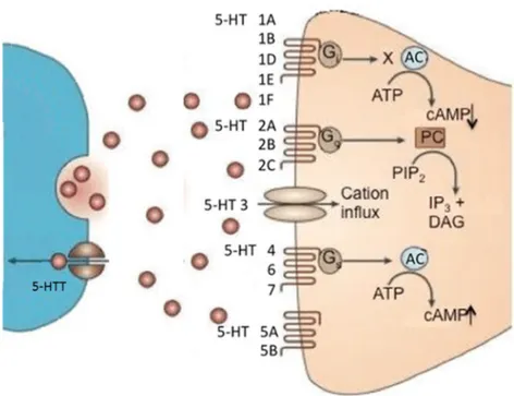
Serotonin binds 15 specific receptors, grouped into 7 families, with distinct characteristics and expression patterns. These receptors have both excitatory and inhibitory action and many of them can regulate neurotransmitter release such as dopamine, acetylcholine, GABA, glutamate and noradrenaline.
Almost all these receptors are expressed in the dentate gyrus, on both stimulatory glutamatergic cells and inhibitory interneurons (Berumen et al., 2012). This evidence supports the possibility that 5-HT signalling may influence adult hippocampal neurogenesis and functions.
Many studies, in fact, demonstrated that administration of serotoninergic agonists results in increased cell proliferation and neurogenesis both in SVZ and in SGZ of the hippocampus. Following chronic or acute serotonin depletion there is a drastic decrease in the number of new generated neurons in the subventricular zone and dentate gyrus of rodents (Benninghoff et al., 2010; Ueda et al., 2005).
The 5-HT3 receptors are the only serotonin-gated ion channels and their activation in hippocampus is potentially involved in proneurogenic effects associated with exercise-induced hippocampal neurogenesis (Kondo et al., 2015).
The other six serotonin receptor families are G protein-coupled receptors (GPCRs):
22
The HT1 receptors are divided in subtypes HT1a, HT1b, HT1d, 5-HT1e and 5-HT1f and are coupled to the Gi/o protein. Agonists for these receptor family decrease adenylyl cyclase activation and cAMP levels. Activation of 5-HT1 receptors increases cell membrane conductance for the potassium ions.
The 5-HT4, 5-HT6 and 5-HT7 receptors are associated with Gs proteins and their activation supports the opposite action: increased cAMP level and decreased potassium conductance with consequent increase in neuronal excitability.
The 5-HT2 receptors are classified in the subtypes 5-HT2a, 5-HT2b and 5-HT2c and are associated to the Gq proteins. Activation of these receptors induces phospholipase C (PLC) to catalyse the hydrolysis of PIP2 with consequent increase of inositol triphosphate (IP3), diacylglycerol (DAG) and intracellular calcium levels.
The HT5 receptors are divided in subtype HT5a and ht5b: as the 5-HT1 receptors, they are coupled to Gi/o proteins and their activation decreases cAMP levels.
The role of each serotonin receptor subtype is not yet fully elucidated in the aHpNG process. In the DG, neurons usually co-express different types of 5-HT receptors that could have similar or opposite effects on a specific function. Moreover, 5-HT receptors can combine each other forming homodimers or heterodimers so to contribute to further complexity in the HT signalling. E.g., heterodimers of HT1a with 5HT7 receptors and 5-HT2A with mGlu2 receptors (metabotropic glutamate 2 receptors) have been shown to have characteristics that differ from their individual counterparts (Renner et al., 2012; Delille et al., 2013). Finally, the resulting effect of 5-HT on hippocampal neurogenesis depends also on local serotonin receptor concentration, on the ratio of different receptor subtypes in loco and on the density of 5-HT receptors in a specific cell population (Sahay et al., 2007; Alenina and Klempin, 2015; Dale et al., 2016).
Another important protein that binds 5-HT is the serotonin transporter (SERT or 5HTT). This transporter, localized on the presynaptic terminal
23
membrane, is able to reuptake 5-HT from the synaptic gap and to interrupt the action of the neurotransmitter, as shown in figure 3.
Figure 3. Schematic representation of 5-HT receptors and transpoters: 5-HT, released from
pre-synaptic neurons in the synaptic cleft, are able to bind different post-synaptic 5-HT receptors that activates various signal cascades. 5-HTT is able to reuptake serotonin in the pre-synaptic neurons interrupting neurotransmitter effect. AC= adenylate cyclase, ATP= adenosine triphosphate, cAMP= cyclic adenosine monophosphate, PIP2 = phosphatidylinositol 4,5biphosphate, IP 3 = inositol triphosphate, DAG= diacylglycerol, PC= phospholipase C (adapted from Waider et al., 2012)
TRAZODONE
Trazodone (TZD), among antidepressants, belongs to SARIs drug class, since it is a serotonin receptor antagonist and reuptake inhibitor. It is used to treat psychiatric conditions including anxiety, insomnia, obsessive and compulsive disorder, post-traumatic stress disorder, substance use disorders, feeding and eating disorders, sexual dysfunction, behavioral disturbances associated with cognitive dysfunction, certain pain conditions
24
and rehabilitation after acute ischemic stroke (Lance et al., 1995; Roth et al., 2011; Khouzam, 2017). Recently, there has been an increasing interest in reconsidering trazodone as an effective antidepressant and as a drug with additional indications (Fagiolini et al., 2013).
Trazodone is a phenylpiperazine and a triazolopyridine derivative that is structurally uncorrelated to other major classes of antidepressants. Trazodone is an antagonist of 5-HT2a and 5-HT2c receptors and has a partial agonism of serotonin 5-HT1a receptors. It inhibits serotonin reuptake by action on serotonin transporter. Furthermore, trazodone is an antagonist of α1- adrenergic receptors and, with lower affinity, of α2-adrenergic receptors. Lastly, TZD shows minimal anticholinergic effects. For its characteristics, TZD can be recognized as the first ever multimodal antidepressant.
TZD was demonstrated to have neuroprotective effects via the expression of mTOR, CREB and BDNF in neurons derived from human neural stem cells (Daniele et al., 2012). Electrofisiological studies in rat brain demonstrated that TZD is able to increase the serotoninergic neurotransmission (Ghanbari et al., 2010). Moreover, TZD was demonstrated to significantly improve cognitive performance in rat treated with the toxin 3-nitropropionic acid, which induces cognitive impairment, oxidative stress (glutathione) and mitochondrial dysfunction (Kumar et al., 2010). These studies suggest that TZD is involved in diffent mechanisms that are potentially correlated to the neurogenesis process.
DOWN SYNDROME, A NEURODEVELOPMENTAL DISORDER ASSOCIATED WITH NEURAL STEM CELL IMPAIRMENT
General aspects
Down syndrome (DS) is a neurodevelopmental disorder affecting 1 in every 787 live born babies each year (De Graaf et al., 2017a; De Graaf et al., 2017b; De Graaf et al., 2015). Among individuals affected by DS, in most cases (around 88% of DS patients), the disorder is caused by meiotic non-disjunction of the more likely maternal chromosome 21 resulting in a triplication of the entire chromosome 21. In some cases (around 4%), the
25
disorder is caused by robertsonian translocation of part or entire chromosome 21 to the long arm of the acrocentric chromosomes 14 or 22. Rarely (around 1%) DS presents as a mosaic conditions where only some cells have an extra copy of chromosome 21. These last cases are likely due to a non-disjunctional event occuring during the first stages of embryo formation. The phenotypic effects are very similar in all three DS forms, except for the severity of the disease: the mosaicism cases have a clinical phenotype milder than the typical full trisomy. Patients have reduced muscle tone that results in floppiness (hypotonia) and show typical craniofacial features that are geometrically well descripted (Cornejo et al., 2017). These facial abnormalities are microgenia (abnormally small chin), round face, slanting eye fissures with prominent ephicanthic folds, Brushfield spots in the iris, a flat facial profile, a flat nasal bridge, a protruding tongue, a shorter neck, a smaller nose and smaller ears than euploid individuals. Patients with DS are predisposed to a wide range of medical conditions. About 40-50% of newborns with DS present cardiac malformations, gastrointestinal defects, immune system anomalies, thyroid disorders, metabolic problems and increased frequency of leukaemia (Whooten et al., 2018; Hasle et al., 2016). However, with advances of medical technologies and increased access to medical care most of these medical issues have become treatable and life expectancy of DS patients has dramatically increased from 9 years old in 1929 to an average age of 60 years in the 2002 (Carfì et al., 2014). At present the most debilitating and unsolved problem of DS individuals is cognitive impairment.
Cognitive functions
Intellectual disabilities and mental retardation are invariably present in DS population, in a degree ranging from mild to severe. DS is the commonest identifiable cause of mental retardation (around 15-20% of the intellectually disabled population).Down syndrome children show memory profiles that are completely different from other genetic syndromes with
26
intellectual disabilities. DS childrens are characterized, in fact, by normal immediate visual-spatial short memory, but poor verbal working memory skills that worsen in the adolescence period (Conners et al., 2011; Edgin et al., 2010; Vicari and Carlesimo, 2006). Furthermore, cognitive functions degenerate with age due to several co-morbid factors such as sleep disruption, depression, sensory impairments, seizures, autism and other medical and psychiatric conditions. In the middle-late age, DS patients frequently develop dementia of the Alzheimer type. The increased risk of dementia results from the extra copy of the gene that codes for amyloid precursor protein (APP) which is strongly associated with the Alzheimer’s disease development.
Anatomical and physiological defects
Mental disorders in DS patients derive from a combination of reduced neural development and functional alterations that appear in 4-5-months foetuses and drastically worsen in the last three months of gestation (Guihard-Costa et al., 2006). In the two to six months of gestation period, cortical neurons born in the proliferative ventricular zone migrate into the cortical plate where they assume a committed phenotype to form the specific layers of the cortex (Weitzdoerfer et al., 2001). Alteration in this period cold impact the final brain function. The late prenatal DS brain, in fact, shows delayed and disorganized cortical lamination with smaller and hypocellular hippocampal DG and hypomorphic cerebellum. Consequently, MRI and post-mortem studies of brain size, in children with DS between 10 and 20 years, show an approximate 17% decrease in volume, with selective loss in the hippocampus, cerebellum and frontal, temporal and occipital lobes. In adult age, brain size and weight is about 20% smaller than euploid brain and there are further reductions in the proportion between frontal lobe and temporal lobe volumes (Beacher et al., 2010; Anderson et al., 2013, Pinter et al., 2001). At the histological level, the cerebral cortex of adult brain shows a reduced number of pyramidal neurons and altered
27
granule cells distribution in the cortical layers II and IV. Ultra-structural studies show that trisomic neurons possess reduced dendritic arborisations, reduced synaptic density and length, and profound alterations in dendritic spine, which appear smaller and immature. Furthermore, there is an unbalance between the excitatory and inhibitory tone due to complex impairment of neurotransmitter receptor functions, but also of both excitatory and inhibitory neurons numbers. These defects have several consequences such aberrant maturation of neurons and defective synaptic transmission (Stagni et al., 2018). The cyto-architectonic alterations and their effect on the cognitive development in DS is not very well elucidated; however, some alterations may be associated with specific intellectual disabilities in DS. (Couzens et al., 2011; Tsao and Kindelberger, 2009). Deficits in memory consolidation may be consequent to temporal lobe and hippocampal dysfunction as well as to cerebellum and prefrontal cortex alterations (Pennington et al., 2003; Lott and Dierssen, 2010). It seems plausible, in fact, that cognitive dysfunctions in domains such as attention, executive control, language learning, spatial memory and emotional behaviours could be correlated with cerebellar–cortical–limbic circuitry impairment (Lott and Dierssen, 2010; Vicari, 2006).
THE MURINE MODEL OF DOWN SYNDROME
Over the last several years, very promising results have been obtained with a mouse model of DS, the Ts65Dn model. The murine chromosome 10, 16 and 17 show conserved synteny with human chromosome 21. The highest proportion of orthologues genes are on murine chromosome 16 (about 80%) and the remaining syntenic genes are on murine chromosome 10 (around 14% of syntenic gene) and murine chromosome 17 (around 6% of syntenic gene). In 1990 Davisson et al. created the first viable trisomy murine model, the Ts65Dn mouse, that is still the best-studied model and contains partial and segmental trisomy 16. The Ts65Dn mice have an extra chromosome composed by a region of murine chromosome 16
28
translocated onto a short segment of murine chromosome 17 and this correspond to trisomy of 104 genes orthologous to human chromosome 21 genes. In particular, Ts65Dn mice carry a triplication part between Mrpl39 and Znf295 genes that contain several studied genes like App, Sod1, Sim2 and Mx1, important in many human pathologic conditions. However, Ts65Dn mice have also three copies of 19 genes of murine chromosome 17 that are not syntenic and some their phenotypes might not be correlated to human DS (Gupta et al., 2016).
Figure 4. Schematic representation of the long-arm of human chromosome 21 comprised
in the mouse chromosome 16, 17 and 10: Ts65Dn mice show triplication of approximately 104 of the 170 orthologous genes, but they include the most studied genes such as APP, SOD1, DSCR1 and DYRK1A (modified from Lockrow et al., 2012).
29
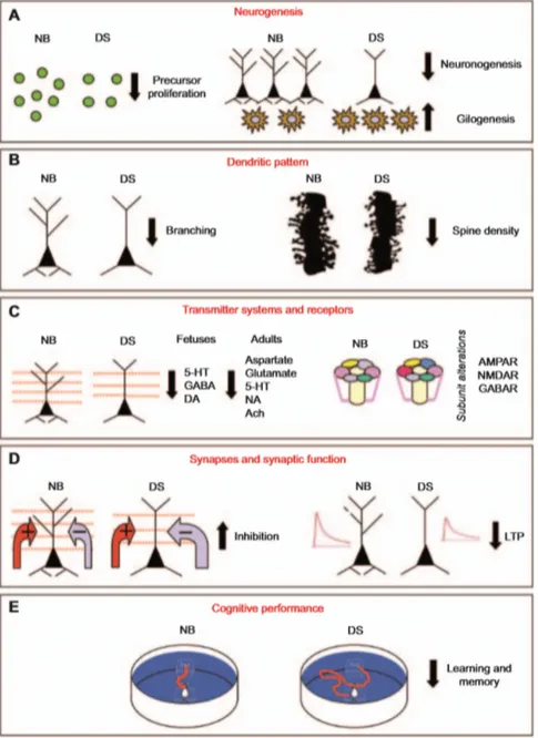
The Ts65Dn mice show several phenotypes and behavioural alterations that recapitulate the human counterpart, as shown in figure 5. As reported in a recent review (Stagni et al., 2018), they have several anatomical, biochemical and molecular alterations and, very importantly, they display cognitive alterations as in the human disease. Ts65Dn mice have altered cerebral architecture and reduced brain volume. Like DS patients, the size of the cerebellum and hippocampal granule cells layer are reduced very early and further decrease with age in Ts65Dn mice. Ts65Dn hippocampus shows reduced granule cell number in the DG associated with reduced dendritic spine density/shape and decreased synaptic structures across all postnatal ages (Benavides-Piccione et al., 2004).
Furthermore, the ratio between excitatory and inhibitory neurotrasmitters is altered in Ts65Dn mice due to increased inhibitory synapses and increased activity of GABAergic neurons, while the excitatory components are decreased (Créau, 2012). Many studies suggested that increased inhibitory interneurons in Ts65Dn forebrain is due to triplication of the genes Oligodendrocyte Transcription Factor 1 and 2 that are strongly involved in oligodendrogenesis and neurogenesis (Chakrabarti et al., 2010). The over-expression of inhibitory neruons and neurotrasmission was demonstrated to alter LTP and LTD in DG of trisomic mice, and these defects strongly correlate with learning and memory impairment (Kleschevnikov et al., 2004). Similarily to the human disease, in Ts65Dn mice there is also an unbalance between the astroglial and neuronal components, with a reduced number of neurons and increased number of astrocytes in multiple regions (Guidi et al., 2008; Stagni et al., 2018). Another similarity between the mouse model and the human disease is at the onset of Alzheimer's disease (AD). Starting from 12 months of age Ts65Dn mice show, in fact, an increased expression of APP protein in hippocampus and cortex and degeneration of basal forebrain cholinergic neurons. For this reason, Ts65Dn mice can be used also as a model of AD-like neurodegeneration.
30
Figure 5. Schematic representation of brain abnormalities in Ts65Dn mice: impairment of
NSC proliferation, reduced neuronogenesis and increased gliogenesis (A), dendritic atrophy and reduced density of dendritic spines (B), reduced levels of various neurotransmitter systems and receptor alterations (C), increased inhibition and impairment of long-term potentiation (D). The overall outcome is a severe impairment of cognitive functions, including hippocampus-dependent learning and memory (E) (from Bartesaghi et al., 2011).
31
NSC PROLIFERATION AND NEUROGENESIS DEFECT IN DOWN SYNDROME Key deficiencies were demonstrated to occur during the NSC expansion and neurogenesis in the early foetal stage (Guidi et al., 2008). Stagni et al. show that the reduced proliferation rate and proneurogenic capacity in Ts65Dn mice and DS brain are likely due to common mechanisms (Stagni et al., 2018). Through immunohistological analysis of human foetus tissues for the cell cycle-associated marker such as Ki-67 (expressed in S+G2+M phases) and phospo-histone 3 (H3, mainly expressed in M phase), the NSC proliferative status was investigated in DS brain. In foetus during the first three months of gestation, cells expressing Ki-67 and H3 were drastically reduced in dentate gyrus, cortex and cerebellum in comparison with euploid NSC (Contestabile et al., 2007; Guidi et al., 2011). Studies in Ts65Dn mice confirmed the observations found in human brain. Histological analysis of Ts65Dn mice show that the numbers of BrdU+ cells is reduced in
the same areas of the human brain since prenatal stage (Contestabile et al., 2007).
It seems that reduced NSC proliferation rate is due to elongation of cell cycle and precocious exit from the cell cycle. In trisomic mice, cyclins and cyclin dependent kinases (CDK), that regulate cell cycle, are altered: 1. cyclin D1 is overexpressed causing prolongation of the G1 phase; 2. cyclin-dependent kinase inhibitor p27KIP1 is increased inducing the cells to enter in
G0 and premature neuronal differentiation; 3. p53 and p21CIP1
(cyclin-dependent kinase inhibitor 1) are overexpressed impairing the transition from G1/G0 to S phase. It has been reported that triplication of the most studied genes such as Dirk1A, App, Rcan1 and Olig 1/2 has an active role in increasing activation and overexpression of cyclin D1, P27KIP1, p53 and
p21CIP1 (Stagni et al, 2018).
In the same studies, defects in NSC proliferation were shown to result in a reduction of neurogenesis in the developing DS brain. Neurogenesis is highly compromised in favour of gliogenesis. Studies in vivo and in vitro,
32
with murine and human foetal NSC, demostrated that the newborn neurons significantly decrease in number, with paraller increased number of the astrocyte counterpart, in the hippocampus, parahippocampus gyrus and cerebellum (Guidi et al., 2008; Trazzi et al., 2014). This umbalance is due, at least in part, to triplication of genes that encode for several interferon receptors (such as IFNAR1, IFNAR2, IFNGR2) and IL10RB. The activation of interferon-receptors activates the JAK/STAT pathway, and in particular STAT3, that in turn activates transcription of GFAP and S100, whose products are typical markers of astroglial cells (Ferrando-Miguel et al., 2003). Moreover, increased expression of APP and its intracellular domain portion (AICD) can repress Shh pathway through activation of PTCH1 expression. Consequently, the transcription factors GLI1 and GLI2, mediated by Shh pathway activation, are considerably reduced. GLI2 can induce neurogenesis through positive regulation of Mash1 and its downregulation affects neuronal differentiation (Trazzi et al., 2011). TIMING OF POTENTIAL DRUG THERAPIES FOR DOWN SYNDROME
In the last 20 years, since the discovery that drugs are able to modulate neurogenesis and potentially restore cognitive functions, a growing interest had turned towards novel pharmacological approaches in DS. Experimental evidence further confirmed that there is the possibility to correct the DS NSC deficits. The target mechanisms of these studies involve different signalling pathways and molecules. The most important pharmacological approaches could be divided in 5 types, depending on targeted mechanisms:
A. Restoring of the physiological neurotransmission since in the DS brain there is an unbalance between reduced excitatory activity and increased inhibitory activity (Créau, 2012), both playing an important role in cognitive and neurodevelopment processes.
B. Prevention of neurodegeneration through antioxidant, neurotrophic molecules and free radical scavengers. Enhanced production of ROS, due to triplication of SOD1 gene (that plays a role in ROS scavenging) was
33
correlated with neurodegeneration and intellectual disabilities in DS (Perluigi and Butterfield, 2012). Treatments such as melatonin, vitamin E and oestrogens have shown some positive effects on learning and memory in Ts65Dn mice (Corrales et al., 2013; Lockrow et al., 2009; Granholm et al., 2002).
C. Recovery of disrupted downstream signalling pathways such as GSK3 signalling, whose dysregulation was suggested to be implicated in the neural impairment in DS (Trazzi et al., 2014). Inhibition of GSK3 by lithium reverted DS-associated defects not only at the cellular and anatomical levels, but ameliorated neurogenesis and cognitive functions in Ts65Dn mice (Stagni et al., 2013).
D. Correction of the protein dosage encoded by triplicated genes such as App and Dirk1A, which were demonstrated to be implicated in DS brain deficit. Several studies that blocked APP and DYRK1A downstream pathways using inhibitors such as DAPT (a -secretase inhibitor) and Epigallocatechin Gallate (EGCG, a Dirk1A kinase inhibitor) showed amelioration of behavioural functions in Ts65Dn mice (Netzer et al., 2010; Stagni et al., 2014).
E. Increasing neurogenesis by proneurogenic molecules such as P7C3 that was a well-know molecule to induce hippocampal neurogenesis in Ts65Dn mice (Latchney et al., 2015). Moreover, certain fatty acids demonstrated to have proneurogenic effects in Ts65Dn mice (Stagni et al., 2017).
More than half of these studies aimed to correct neurotransmission: I. Inactivation of GABAergic neurotransmission by antagonizing GABAergic receptors. GABA neurotransmission is increased and is strongly correlated to cognitive and learning deficits in Ts65Dn mice. GABA antagonists or GABAA 5 negative allosteric modulators were demonstrated to ameliorate neurogenesis, LTP and to improve cognition in trisomic mice (Kleschevnikov et al., 2012; Martínez-Cué et al., 2013).
II. Restoring the cholinergic system that is degenerated in TS65dn mice (starting from 4-6 months and becoming evident at 10-12 months of age) and in DS individuals (Godrige et al, 1987; Chang and Gold, 2008). Reduction of the number of basal forebrain cholinergic neurons is correlated to
34
working memory/attention impairment. Drugs such as donezepil have been shown to partially restore learning and memory in Ts65Dn mice (Rueda et al., 2008).
III. Restoring noradrenergic neurotransmission that is largely affected in DS patients and in Ts65Dn mice. This impairment strongly correlates with cholinergic system degeneration, inflammation and cognitive decline in Ts65Dn. Restoration of the noradrenergic tone, using a norepinephrine precursor or adrenergic receptor agonists, caused an improvement of cognitive functions in Ts65Dn mice (Salehi et al., 2009).
IV. Restoring the glutamatergic system: in DS patients and in Ts65Dn mice it has been demonstrated that glutamatergic neurotransmission and N-methyl-D-aspartate (NMDA) receptor signalling are altered and this might potentially contribute to behavioural disabilities. Mice trated with memantine, a moderate-affinity antagonist of NMDA receptors, show increased spatial learning capacity and restored electrophysiological abnormalities in trisomic mice (Lockrow et al., 2011).
V. Restoring the serotonergic system whose impairment in DS brain correlates to neurogenesis impairment and to declined cognitive functions (Whittle et al., 2007). Studies in vivo demonstrated that disruption of the serotoninergic tone in neonatal mice results in brain development dysfunctions, including generation of precursor cells, their migratory abilities and their differentiation in mature neurons and glial cells (Durig and Hornung, 2000). At the molecular level, the reason why the 5-HT pathway appears to be implicated in the DS may have also to do with S100β gene triplication since this gene product negatively regulates the outgrowth of serotonin terminals and reduces serotonin levels (Shapiro et al., 2010). In this respect, treatments that increase 5-HT availability may counteract the defects and even the mental retardation in DS individuals.
VI. Restoring other pathways such as histamine neurotransmission that was demonstrated, when deregulated, to contribute to cognitive impairment in DS (Kim et al., 2001).
Most of the studies listed above were performed in adult Ts65Dn mice and this is very relevant because it gives the demonstration that something
35
could be restore even in adult DS individuals. Among the studies done in adult Ts65Dn mice: 58% showed recovery of cognitive defects such as memory and learning, 19% only a partial improvement and 28% no effects. Unfortunately, very few studies concerned the neurogenesis process. Moreover, few studies took into consideration the duration of the treatment effects after treatments ending. Based on these considerations, some of the preclinical studies have built the rationale for studing in clinical trials the effects of drugs such as RG1662 (a GABAA5 negative allosteric modulator), Memantine, donepezil and rivastagmine (acetylcholinesterase inhibitors), EGCG and vitamin E in adult and children affected by DS (Stagni et al., 2015).
An important critical aspect of the summarized preclinical studies performed in adult mice has to do with the fact that the overall brain development and function depends principally on events occurring during the early foetal stages. Moreover, after the birth neurogenesis is maintained in hippocampus until adulthood, while neurogenesis in the cerebellum occurs and ends in the first two post-natal weeks. For this reason, therapies tested in adult mice could modulate hippocampal neurogenesis with limited changes in the neural plasticity and with a partial recovery of the intellectual disabilities, while it may be too late to recover cerebellum-associated functions, such as attention and language capacity. Consequently, therapies to improve neurogenesis defects should be started as soon as possible to allow the formation of appropriate neuronal connections. The two potential best time windows are likely neonatal and prenatal/perinatal treatments.
The most relevant studies in neonatal mice showed increased in hippocampal and cerebellum neurogenesis. E.g. in vitro, newborn Ts65Dn mice, treated with single injection of Shh agonist at birth (such as SAG-1), recovered cerebellar development and hippocampal LTP when they reached adult age (4 month-old), but with no effect on cerebellar-mediated cognitive functions (Das et al., 2013; Gutierrez-Castellanos et al., 2013). In another study, chronic treatment of fluoxetine in trisomic mice from
36
postnatal day 3 to day 15 (P3-P15) restored hippocampal neurogenesis and the number of granule cells in the neonatal mice. 1 and 4 months after treatment cessation, Ts65Dn mice maintained restored dendritic structure, spine density and cellular connectivity (Stagni et al., 2015). In addition, 7, 8-DHF, a natural small molecule, promoted neurogenesis and maturation of newborn neurons in neonatal Ts65Dn mice treated from P3 to P15 (Stagni et al., 2017). Furthermore, neonatal mice exposed to EGCG from P3-P15 fully restored hippocampal cellularity and neurogenesis (Stagni et al., 2016). However, with the aim of fully correcting DS pathophysiology, prenatal stage seems to be the best treatment period and many studies demonstrated that drugs given to mothers had a larger impact to correct the whole brain functions. E.g. High concentration of choline in the diet of pregnant Ts65Dn mothers (beginning from embryonic day 1, or E1, to when pups reach P21) increased organization of cholinergic neurons in their progeny. When they reached 6 months-old age, mice showed increased cognitive functions as far as attention behaviour. The same diet, in combination with environmental enrichment, increased also hippocampal neurogenesis, spatial cognition and cholinergic neuron numbers in prenatally treated mice when they reached adulthood (Moon et al., 2010). The peptides NAPVSIPQ (that mimics the activity of the neuroprotective protein ADNP) and SALLRSIPA (that mimics the activity of neurotrofic factor ADNF) prevented neurodevelopmental delay, restored the altered subunit of GABA and improved learning in Ts65Dn mice treated in the prenatal period from E8-E12 (Vink et al., 2009). Prenatal treatment with tocoferol, the active form of vitamin E, increased cell density in DG and ameliorated learning and retention memory in Ts65Dn mice treated from E0 to adulthood P84 (Shichiri et al., 2011). Chronic administration of ECGC was able also to correct brain weight, thalamo-hypothalamic volume proportion and to completely restore the long-term memory (Guedj et al., 2009) in DYRK1A transgenic mice treated from gestation to adulthood (3 month-old).
37
Fluoxetine treatment in Ts65Dn mice from E10 to P2 (10 mg/kg for pregnant mother and 150 ug/g body weigth for pups) was able to restore proliferation and cellularity in all main brain regions of prenatally treated mice. When these mice reached adulthood (P43), they showed increased numbers of NSC in the SVZ and SGZ, corrected neuronal/astroglial cells balance, normalized number of granule cells, restored maturation of dendritic spines and improved cognitive function (Guidi et al., 2014). Altogether, drug treatments in the prenatal phase showed a clear improvement in brain structures and functions to indicate that sooner treatment had more beneficial effects. However, in order to translate this protocol on human counterpart, it must be remembered that the human ontology is different from mice one. Mice development occurs mainly after birth, while in human nervous system maturation occurs during the prenatal stage and the main brain regions, except for SVZ, hippocampus and cerebellum, enter in a state of replicative quiescence after birth. This is an important critical issue to plan in the future any safe treatment in a human brain development phase. Since hippocampal and cerebellum neurogenesis in human begin from the 12th gestation week (Seress et al., 2011; ten Donkelaar et al., 2003) and in the same week it is possible to perform non-invasive prenatal testing to confirm DS, the remaining 28 weeks could potentially represent the optimal time window to treat foetus. Many studies are currently active with the final goal to develop new therapeutic drugs that have minimal side effects in mothers and unborn children, that are not teratogenic and that could pass through the placenta barrier and the blood brain barrier.
38
REFERENCES
Alenina N, Klempin F. The role of serotonin in adult hippocampal neurogenesis. Behav Brain Res. 2015 Jan 15;277:49-57.
Allen DD, Cárdenas AM, Arriagada C, Bennett LB, García CJ, Caviedes R, Rapoport SI, Caviedes P. A dorsal root ganglia cell line derived from trisomy 16 fetal mice, a model for Down syndrome. Neuroreport. 2002 Mar 25;13(4):491-6.
Altman J, Das GD. Autoradiographic and histological evidence of postnatal hippocampal neurogenesis in rats. J Comp Neurol. 1965 Jun;124(3):319-35.
Alvarez-Buylla A, Garcia-Verdug o JM. Neurogenesis in adult subventricularzone. J Neurosci. 2002 Feb 1;22(3):629-34.
Ambrogini P, Lattanzi D, Ciuffoli S, Agostini D, Bertini L, Stocchi V, Santi S, Cuppini R. Morpho-functional characterization of neuronal cells at different stages of maturation in granule cell layer of adult rat dentate gyrus. Brain Res. 2004 Aug 13;1017(1-2):21-31.
Anderson JS, Nielsen JA, Ferguson MA, Burback MC, Cox ET, Dai L, Gerig G, Edgin JO, Korenberg JR. Abnormal brain synchrony in Down Syndrome. Neuroimage Clin. 2013 May 24;2:703-15.
Austin MP, Mitchell P, Goodwin GM. Cognitive deficits in depression: possible implications for functional neuropathology. Br J Psychiatry. 2001 Mar;178:200-6.
Balan S, Toyoshima M, Yoshikawa T. Contribution of induced pluripotent stem cell technologies to the understanding of cellular phenotypes in schizophrenia. Neurobiol Dis. 2018 May 3.
Balu DT, Lucki I. Adult hippocampal neurogenesis: regulation, functional implications, and contribution to disease pathology. Neurosci Biobehav Rev. 2009 Mar;33(3):232-52.
Bartesaghi R, Guidi S, Ciani E. Is it possible to improve neurodevelopmental abnormalities in Down syndrome? Rev Neurosci. 2011;22(4):419-55.
Beacher F, Daly E, Simmons A, Prasher V, Morris R, Robinson C, Lovestone S, Murphy K, Murphy DG. Brain anatomy and ageing in non-demented adults with Down's syndrome: an in vivo MRI study. Psychol Med. 2010 Apr;40(4):611-9.
Bédard A, Gravel C, Parent A. Chemical characterization of newly generated neurons in the striatum of adult primates. Exp Brain Res. 2006 Apr;170(4):501-12.
Benavides-Piccione R, Ballesteros-Yáñez I, de Lagrán MM, Elston G, Estivill X, Fillat C, Defelipe J, Dierssen M. On dendrites in Down syndrome and DS murine models: a spiny way to learn. Prog Neurobiol. 2004 Oct;74(2):111-26.
Benninghoff J, Gritti A, Rizzi M, Lamorte G, Schloesser RJ, Schmitt A, Robel S, Genius J, Moessner R, Riederer P, Manji HK, Grunze H, Rujescu D, Moeller HJ, Lesch KP, Vescovi AL. Serotonin depletion hampers survival and proliferation in neurospheres derived from adult neural stem cells. Neuropsychopharmacology. 2010 Mar;35(4):893-903.