Long-term effects of inhaled
corticosteroids on sputum bacterial
and viral loads in COPD
Marco Contoli
1, Alessia Pauletti
1, Maria Rita Rossi
2, Antonio Spanevello
3,
Paolo Casolari
1, Andrea Marcellini
1, Giacomo Forini
1, Giulia Gnesini
1,
Brunilda Marku
1, Neil Barnes
4,5, Andrea Rizzi
6, Giacomo Curradi
6,
Gaetano Caramori
7, Paolo Morelli
8and Alberto Papi
1Affiliations:1Research Centre on Asthma and COPD, Department of Medical Sciences, University of Ferrara, Ferrara, Italy.2Laboratory of Clinical Microbiology, Arcispedale S. Anna, Ferrara, Italy.3University of Insubria and Fondazione Maugeri, Varese, Italy.4GlaxoSmithKline, Brentford, UK.5William Harvey Institute, Barts and the London School of Medicine and Dentistry, London, UK. 6GlaxoSmithKline, Medical and Scientific Department, Verona, Italy.7Unità Operativa Complessa di Pneumologia, Dipartimento di Scienze Biomediche, Odontoiatriche e delle Immagini Morfologiche e Funzionali (BIOMORF), Università degli Studi di Messina, Messina, Italy.8Cros NT, Verona, Italy.
Correspondence: Alberto Papi, Research Centre on Asthma and COPD, Department of Medical Sciences, University of Ferrara, Italy, Via Rampari di S. Rocco, 27 - 44121 Ferrara, Italy. E-mail: [email protected]
@ERSpublications
Long term inhaled corticosteroids increase airway bacterial load in COPD patients with low eosinophil countshttp://ow.ly/8nO530eMSza
Cite this article as:Contoli M, Pauletti A, Rossi MR, et al. Long-term effects of inhaled corticosteroids on sputum bacterial and viral loads in COPD. Eur Respir J 2017; 50: 1700451 [https://doi.org/10.1183/ 13993003.00451-2017].
ABSTRACT Inhaled corticosteroid-containing medications reduce the frequency of COPD exacerbations (mainly infectious in origin) while paradoxically increasing the risk of other respiratory infections. The aim was to determine the effects of inhaled corticosteroids on airway microbial load in COPD patients and evaluate the influence of the underlying inflammatory profile on airway colonisation and microbiome.
This is a proof-of-concept prospective, randomised, open-label, blinded endpoint study. Sixty patients with stable moderate COPD were randomised to receive one inhalation twice daily of either a combination of salmeterol 50μg plus fluticasone propionate 500 μg or salmeterol 50 μg for 12 months. The primary outcome was the change of sputum bacterial loads over the course of treatment.
Compared with salmeterol, 1-year treatment with salmeterol plus fluticasone was associated with a significant increase in sputum bacterial load ( p=0.005), modification of sputum microbial composition and increased airway load of potentially pathogenic bacteria. The increased bacterial load was observed only in inhaled corticosteroid-treated patients with lower baseline sputum or blood eosinophil (⩽2%) levels but not in patients with higher baseline eosinophils.
Long-term inhaled corticosteroid treatment affects bacterial load in stable COPD. Lower eosinophil counts are associated with increased airway bacterial load.
This article has supplementary material available from erj.ersjournals.com Received: March 03 2017 | Accepted after revision: July 10 2017
First published online October 5, 2017; republished October 12, 2017 with amendments to the author’s affiliation details. This study is registered at ClinicalTrials.gov with number NCT01213693.
Support statement: The study was supported by an unrestricted grant for research from GlaxoSmithKline. Funding information for this article has been deposited with the Crossref Funder Registry.
Conflict of interest: Disclosures can be found alongside this article at erj.ersjournals.com Copyright ©ERS 2017
Introduction
Exacerbations of chronic obstructive pulmonary disease (COPD) have a major impact on the quality of life, prognosis and progression of the disease [1].
Inhaled corticosteroids (ICSs) in association with long-acting β2 agonists (LABAs) are recommended treatments for COPD patients at high risk of exacerbation [1]. Interestingly, recent analyses suggest that blood eosinophilia predicts a greater efficacy of ICS/LABA combination in preventing exacerbations over LABA alone, thus providing a possible biomarker to identify patients who are more likely to benefit from this treatment [2–4].
There is an apparent paradox related to ICS treatment in COPD: on the one hand, ICS/LABAs effectively prevent COPD exacerbations that are commonly related to an infective aetiology [5] and, on the other hand, the same combinations increase the risk of pneumonia [6, 7].
Many COPD patients have chronic bacterial colonisation of the airways, and airway bacterial load in stable COPD is related with both the frequency of exacerbations [8] and decline in lung function [9]. An increase of the airway bacterial load over 1 year has been previously observed in a population largely treated with ICS-containing medications [9]. A greater sputum bacterial load has been also reported in COPD patients receiving high-dose ICSs [10]. No control group was included in these observational reports.
With this background, we conducted a proof-of-concept prospective, randomised, open-label, blinded endpoint (PROBE) study specifically powered to determine the effects on sputum bacterial load of 12-month treatment with either LABA or LABA/ICS combination in COPD patients. In addition, exploratory analyses stratified by sputum/blood eosinophil counts were performed to evaluate the influence of the underlying inflammatory profile on chronic airway colonisation and microbiome.
Methods
Study design
Consecutive COPD patients, according to Global Initiative for Chronic Obstructive Lung Disease definition [1], were screened for eligibility from May 4, 2009 to May 7, 2012 among the cohort of patients visiting the outpatient clinic of the Research Centre on Asthma and COPD, University of Ferrara, Italy. 60 steroid-naïve COPD patients with stable disease ( post-bronchodilator FEV1⩾50 and <80% predicted) on treatment with the LABA salmeterol (SALM) were recruited to participate in this PROBE study [11].
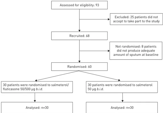
Assessed for eligibility: 93
Recruited: 68
Randomised: 60
Not randomised: 8 patients did not produce adequate amount of sputum at baseline
Excluded: 25 patients did not accept to take part to the study
30 patients were randomised to salmeterol/ fluticasone 50/500 µg b.i.d.
30 patients were randomised to salmeterol 50 µg b.i.d.
Analysed: n=30 Analysed: n=30
This is an exploratory proof-of-concept study designed to investigate the effect of long-term treatment with an inhaled corticosteroid (fluticasone propionate) added to a LABA on sputum bacterial load in stable COPD patients. For ethical reasons, we were not allowed to enrol patients with more severe disease, for which treatment with LABA alone, as the comparator arm, is considered inappropriate and patients would have been undertreated.
Patients were randomised to receive either SALM alone (50μg one inhalation twice daily, n=30) or the ICS/LABA inhaled fixed combination (salmeterol 50μg plus fluticasone propionate 500 μg (SALM/FP one inhalation twice daily, n=30)) for 12 months. After baseline evaluation and randomisation (visit 0), patients were seen every 3 months in an outpatient setting. Patients were instructed to contact the centre for an unscheduled visit in case of worsening symptoms. Exacerbation episodes were treated with antibiotic and systemic corticosteroids [1]. The study conformed to the Declaration of Helsinki, the work was approved by the institutional ethics committee, and informed written consent was obtained from each subject. A detailed description of the study design, randomisation and masking procedures, exacerbation definition, lung function measurements and quality of life assessment is reported in the supplementary material.
This study is registered with ClinicalTrials.gov, number NCT01213693.
Randomisation and masking
Patients were randomised to receive either SALM/FP (50/500μg twice daily; study group, n=30) or SALM (50μg twice daily only; control group, n=30) for 12 months (figure 1). Patients were randomly assigned to a treatment group according to a list prepared with the use of a random number generator.
The physicians who performed the medical evaluation and collection of clinical and functional data of the patients did not have access to the microbiological data of the scheduled visits. The microbiological loads for both bacteria and viruses were assessed in a blind manner. Therefore, the data used for the evaluation of both primary and secondary outcomes were acquired in a blinded fashion (PROBE study) [11].
Microbiological assays
Sputum collection and analyses, quantitative bacteriology, respiratory virus and atypical bacteria detection and microbial identification and profiling assays are detailed in the supplementary material and in the supplementary tables S1 and S2.
Eosinophil counts and bacterial load
Because a differential count of ⩾2% in blood has been previously reported to be predictive of an eosinophil count ⩾3% in induced sputum [12], we performed post hoc analyses to evaluate changes in bacterial load according to the level of baseline blood (⩾2% or <2%) and sputum eosinophils. Recurrent sputum eosinophilia was defined as high eosinophil counts (above cut-off values) in ⩾50% of available samples obtained in at least four visits (at baseline, at the end of treatment and at least in two scheduled visits during the study).
Analyses
The primary outcome was the assessment of bacterial load in the sputum of COPD patients after 12 months of treatment with SALM/FP compared with that in COPD patients treated with SALM. Secondary outcomes included assessment of viral detection; the clinical and inflammatory outcomes are detailed in the supplementary material.
In a previous study [9], mean increase in bacterial load of 0.46 log10colony-forming unit (CFU) mL−1was
observed over 1 year in sputum samples of 30 stable COPD patients. The vast majority of these subjects (93%) were treated with an ICS containing medication. Based on these premises: 1) we postulated no change in sputum bacterial load over 1 year in COPD patients not receiving an ICS-containing medication and 2) we calculated that the bacterial load would increase up to a mean value of about 0.5 log10CFU mL−1in a COPD population that was 100% treated with ICS-containing medications. As we
recruited only steroid-naïve subjects, we considered that a greater increase in sputum bacterial load was to be expected than the reference considered [9], and a 20% increased value of 0.6 log10CFU mL−1 was
assumed for our primary analysis. The common standard deviation for the change is assumed to be 0.8. The type I error is set to 0.05 and the power should be at least 80%. Under these assumptions a sample size of 30 patients per treatment group have to be available for the analysis. A similar number of subjects were enrolled in the study where increased sputum bacterial load was observed over 1 year [9].
The tests used for statistical comparisons are detailed in the Methods section of the supplementary material. p-values of 0.05 or less were considered to indicate statistical significance.
Results
Study design and patient characteristics
In total, 93 patients met the criteria for inclusion; however, 25 patients refused to take part in the study. 68 patients were recruited, but eight patients were not randomised because they did not produce adequate amount of sputum at baseline (figure 1). Thirty patients were randomised to receive one inhalation twice daily of a combination of SALM/FP 50/500 µg, and 30 were randomly selected to receive SALM 50 µg twice daily. No differences were found between the two groups of patients for the demographic characteristics (table 1) and comorbid conditions (supplementary material). Further patients’ clinical and functional characteristics are detailed in the supplementary material.
Sputum sampling
As per the inclusion criteria, sputum samples were collected for all patients at baseline (n=30 in each group) and at the end of the 1-year study treatment (for the primary analysis of the study). Sputum was obtained in 90%, 80%, and 83% of patients treated with ICS/LABA and 86%, 73%, and 83% of patients treated with LABA alone at the 3-, 6-, and 9-month study visits, respectively. Samples for all visits were available in 24 (80%) and 22 (73%) patients treated with ICS/LABA and LABA alone, respectively. No differences in demographic characteristics and/or lung functions were found between patients with samples for all study visits and the entire study population.
Bacterial and viral load at stable state
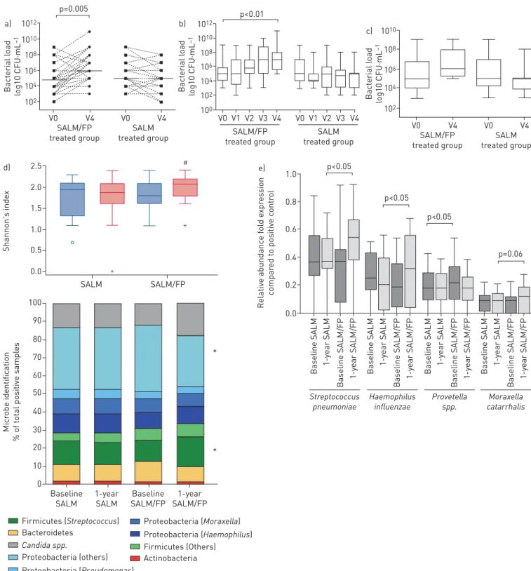
Baseline bacterial loads were similar between the two groups (figure 2a and 2c).
The total bacterial load, which includes any type of bacteria grown in cultures from the sample, was significantly increased with respect to baseline ( p=0.005) at the end of the study in the SALM/FP group, whereas the end-study total bacterial load had not changed from baseline in the SALM group (figure 2a). A progressive increase in sputum bacterial load was confirmed in the SALM/FP group when the analysis was confined to patients for whom sputum samples were obtained and assessed at every visit (figure 2b). Supplementary table S3 lists the potentially pathogenic airway bacteria (PPB) detected by conventional culture assays in the study for each visit. A high variability in the detection of specific potential pathogens was observed for patients from visit 1 (baseline) to visit 4 (12 months) in both treatment groups. No correlation was found between the ICS treatment and detection of specific individual pathogens. In stable conditions, the rate of detection of PPB in the sputum sample did not significantly change throughout the study (PPB were identified in approximately one-third of sputum samples at each study visit) (supplementary table S3). Conversely, an increase of PPB load was observed at the end of the study in the SALM/FP group (figure 2c), although the change, in the relatively low number of samples considered, did not reach statistical significance. These data were confirmed by the PCR assay performed in sputum samples for selected bacteria pathogens (supplementary figure S1) [10].
TABLE 1Demographic and clinical characteristics of the study population
Study population SALM/FP SALM p-value
Patients n 60 30 30
Age years 70.6±0.9 70.5±1.2 70.6±1.3 >0.05
Male/female n 48/12 23/7 25/5 >0.05
Smoking habit pack-years 25±4 28±6 23±5 >0.05
Chronic bronchitis 27% 23% 30% >0.05
FEV1pre-bronchodilator L 1.42±0.03 1.44±0.03 1.41±0.04 >0.05 FEV1pre-bronchodilator % pred 58.6±0.7 58.8±1.0 58.3±1.1 >0.05 FEV1post-bronchodilator L 1.56±0.04 1.55±0.05 1.57±0.06 >0.05 FEV1post-bronchodilator % pred 63.9±0.9 63.1±1.3 64.6 ± 1.2 >0.05 Number of moderate-to-severe
exacerbations in the previous year
0.91±0.09 0.86±0.13 0.96±0.13 >0.05 Patients with⩾1 moderate-to-severe
exacerbation in the previous year
72% 73% 70% >0.05
Data are presented as mean±SEM, unless otherwise stated. SALM/FP: salmeterol/fluticasone propionate-treated group; SALM: salmeterol-treated group; FEV1: forced expiratory volume in 1 s.
No differences in respiratory virus detection were observed at baseline and over the study period within and between groups. By real-time PCR analysis, low rates of viral detection were found throughout the study as detailed in the supplementary material. The detected viruses are detailed in supplementary table S3. 1012 a) 1010 108 106 104 102 Bact erial l oad log10 CFU·mL –1 SALM/FP treated group SALM treated group V0 V4 V0 V4 p=0.005 1010 c) 108 106 104 102 Bact erial l oad log10 CFU·mL –1 SALM/FP treated group SALM treated group V0 V4 V0 V4 1012 b) 1010 108 106 104 102 100 Bact erial l oad log10 CFU·mL –1 SALM/FP treated group SALM treated group V0 V1 V2 V3 V4 V0 V1 V2 V3 V4 p<0.01 d) 2.0 1.5 1.0 0.5 0.0 2.5 Shannon's inde x SALM SALM/FP 10 20 30 40 50 60 70 80 90 100 0 Micr obe identific ation % of t o tal positiv e sampl es Baseline SALM 1-year SALM Baseline SALM/FP 1-year SALM/FP * Actinobacteria Firmicutes (Streptococcus) Firmicutes (Others) Candida spp. Proteobacteria (others) Proteobacteria (Pseudomonas) Proteobacteria (Moraxella) Proteobacteria (Haemophilus) Bacteroidetes * 1.0 e) 0.6 0.8 0.4 0.2 0.0 Relativ e abundanc e f old e xpr es sion c ompar ed t o positiv e c ontr ol Baseline SALM Baseline SALM/FP 1-y ear SALM 1-y ear SALM/FP Streptococcus pneumoniae Haemophilus influenzae Provetella spp. Moraxella catarrhalis p=0.06 Baseline SALM Baseline SALM/FP 1-y ear SALM 1-y ear SALM/FP Baseline SALM Baseline SALM/FP 1-y ear SALM 1-y ear SALM/FP Baseline SALM Baseline SALM/FP 1-y ear SALM 1-y ear SALM/FP p<0.05 p<0.05 p<0.05 #
FIGURE 2 Airway bacterial load and microbiome analysis. a) Total bacterial load is shown as colony-forming units (CFU) per mL and was assessed at baseline (V0) and after 12 months of therapy (V4) in sputum samples from patients in both the salmeterol/fluticasone (SALM/FP) and SALM alone groups.b)Total bacterial load of patients for whom sputum samples were obtained and assessed at every visit.c)Airway bacterial load of pathogenic airway bacteria (PPB) at baseline (V0) and after 12 months of therapy (V4).d)Analysis of sputum microbial composition (alpha diversity evaluated by Shannon’s index) restricted to the 41 bacterial or fungal pathogens detected by multiplex 16s RNA qPCR assay (*: p<0.05 compared to baseline form Proteobacteria and Firmicutes phyla in the salmeterol/fluticasone treated group; #: p<0.05 versus baseline). e)Microbial quantification by multiplex 16s RNA qPCR assay in the sputum samples.
Sputum microbial identification
The sputum microbial analysis was performed in 120 paired sputum samples (60 samples at baseline versus 60 samples at the end of 1-year treatment) of the patients treated with SALM alone (n=30) or SALM/FP combination (n=30). The analysis was performed by multiplex quantitative PCR assay designed to detect bacterial 16S rRNA and fungal ribosomal rRNA gene sequences for 41 bacterial or fungal pathogens (supplementary material). Overall, we found a significant increase in the number of microbes identified in the ICS/LABA group (+12%; p<0.05) but not in the LABA treated group (−3%; p=0.23). No composition dissimilarity was found in the microbial profiling of the two groups at baseline and at 1-year (figure 2d, supplementary table S6). However, in patients treated with SALM/FP (but not in SALM-alone treated patients) we found a significant increased meanα diversity (figure 2d, supplementary table S7 and table S8) after 1 year of treatment. The microbial composition also shifts toward an increased detection of the Firmicutes phylum (+6%; p<0.05) and Candida spp. (+5%; p=0.10) which was paralleled by a significant reduction in Proteobacteria phylum detection (−9%; p<0.05) (figure 2d).
Increased relative abundance in Streptococcus pneumoniae ( p<0.05) and Haemophilus influenzae ( p<0.05) was found after 1 year of treatment with SALM/FP but not with SALM alone. In patients treated with LABA only, we found a significant increase in the relative abundance of Prevotella spp. ( p<0.05). No other significant changes in sputum microbial composition were found (figure 2e).
Clinical outcomes
Although the study was not designed to assess the effects of the treatment arms on COPD clinical outcomes, we found a small improvement in patients receiving ICS/LABA over LABA alone in relevant clinical outcomes (e.g. SGRQ) as detailed in the supplementary material (supplementary figure S2). A nonsignificant trend was found in the SALM/FP group in the reduction of exacerbation rate, when compared with the previous year. No change in the exacerbation frequency was observed in the SALM group (supplementary material).
Airway inflammation at stable state
No difference in terms of total and differential inflammatory cell counts was documented at baseline and over the study period within and between study groups in stable conditions (supplementary table S4).
Airway inflammation and microbiology during COPD exacerbations
Twenty-two and 28 exacerbations occurred in patients treated with SALM/FP and SALM alone, respectively, none of which required hospitalisation.
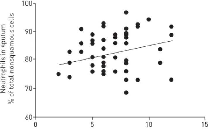
A significant correlation was found between neutrophil cell counts and total bacterial load at exacerbations ( p=0.041; r=0.29 figure 3). A detailed description of the changes of airway inflammation and bacterial (including PPB) and viral loads at exacerbation compared to stable state is available in the supplementary material (supplementary figure S3).
Predictors for changes in bacterial load in patients treated with ICS/LABA combination
The changes in bacterial load in the SALM/FP group did not correlate with any of the baseline demographic characteristics, functional parameters, and/or SGRQ values, nor with the changes of clinical outcomes throughout the study (supplementary material).
FIGURE 3 Correlation between sputum neutrophil cell counts and total airway bacterial load at exacerbation. 100 90 80 70 60 Neutr ophils in sputum % of t o tal nonsquamous c ells
Bacterial load log10 CFU·mL–1
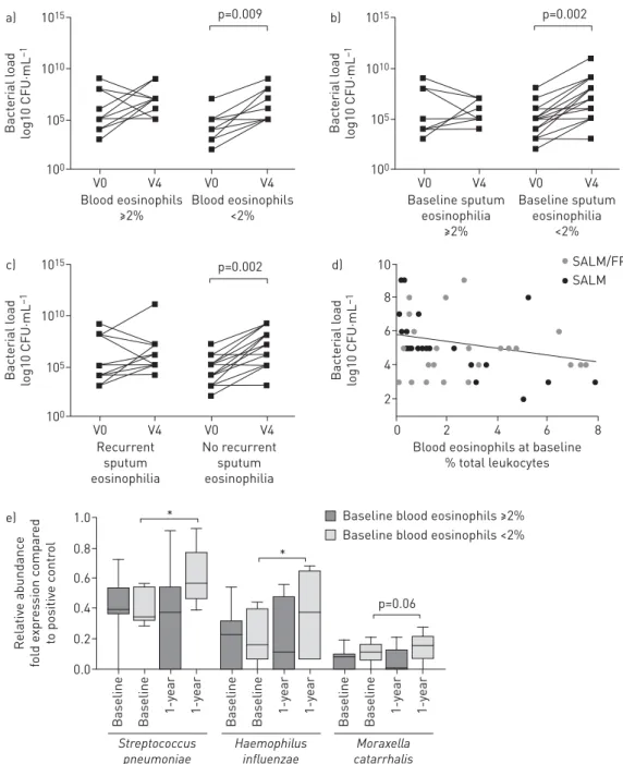
Blood eosinophil cell count was available at recruitment in 87% and 83% of patients in the SALM/FP and SALM groups, respectively. A significant correlation was found between blood eosinophil cell count and sputum eosinophil count at baseline ( p=0.004; r=0.37; supplementary figure S4). When patients were stratified based on blood eosinophils, the 1-year ICS/LABA treatment resulted in a significant increase (+2.47±0.44 log10CFU·mL−1) in the total bacterial load in patients with <2% blood eosinophil counts at
baseline but not in patients with ⩾2% baseline blood eosinophil levels (+0.91±0.37 log10CFU·mL−1)
(figure 4a). The latter group consisted of 53% of the ICS/LABA-treated patients assessed in the study. Because few patients (10%) reported sputum eosinophil levels⩾3% at baseline, we performed stratification
1015 a) 1010 105 100 Bact erial l oad lo g10 CFU·mL –1 Blood eosinophils ≥2% Blood eosinophils <2% V0 V4 V0 V4 p=0.009 1015 c) 1010 105 100 Bact erial l oad lo g10 CFU·mL –1 Recurrent sputum eosinophilia No recurrent sputum eosinophilia V0 V4 V0 V4 p=0.002 1015 b) 1010 105 100 Bact erial l oad lo g10 CFU·mL –1 Baseline sputum eosinophilia ≥2% Baseline sputum eosinophilia <2% V0 V4 V0 V4 p=0.002 1.0 e) 0.6 0.8 0.4 0.2 0.0 Relativ e abundanc e fold e xpr es sion c ompar ed to positiv e c ontr o l Baseline Baseline 1-y ear 1-y ear 10 d) 6 8 4 2 Bact erial l oad lo g10 CFU·mL –1
Blood eosinophils at baseline % total leukocytes 2 0 4 6 8 SALM/FP SALM Streptococcus pneumoniae Baseline Baseline 1-y ear 1-y ear Haemophilus influenzae Baseline Baseline 1-y ear 1-y ear Moraxella catarrhalis p=0.06 *
* Baseline blood eosinophils ≥2% Baseline blood eosinophils <2%
FIGURE 4 a)Total bacterial load at baseline (V0) and after 12 months of therapy (V4) in sputum samples of patients treated with salmeterol/fluticasone propionate (SALM/FP) with baseline blood eosinophil counts⩾2% or <2% of total leukocytes. b) Total bacterial load at baseline (V0) and after 12 months of therapy (V4) in sputum samples of patients treated with SALM/FP with baseline sputum eosinophil counts⩾2% or <2% of total inflammatory cells.c)Total bacterial load at baseline (V0) and after 12 months of therapy (V4) in sputum samples of patients treated with SALM/FP with or without recurrent sputum eosinophilia⩾2% in at least 50% of available samples obtained at scheduled visits (at least 2 visits) during the 1-year study.d) Correlation between blood eosinophils and bacterial load at baseline.e)Sputum microbial quantification of the SALM/FP treated patients with baseline blood eosinophil levels <2% or⩾2%. *: p<0.05.
analysis with a cut-off value of 2% sputum eosinophil. One-year treatment with ICSs resulted in a significant increase in total bacterial load that was limited to patients with <2% baseline sputum eosinophils (+2.14±0.31 log10CFU·mL−1) with no change in the bacterial load (+0.34±0.42 log10CFU·mL−1)
of patients with ⩾2% baseline sputum eosinophils (the latter group comprising 37% of ICS/LABA-treated population) (figure 4b). Multiple regression analysis confirmed the interaction between treatment regimen and sputum eosinophil level at baseline as a predictor of increased bacterial load (supplementary material and table S9).
Similarly, the microbial analysis showed that among COPD patients treated with SALM/FP, the increased relative abundance in S. pneumoniae and H. influenzae was limited to those with blood eosinophils <2%. In these patients, we also found a numerical increase in relative abundance in Moraxella catarrhalis after 1 year of SALM/FP treatment compared with baseline (figure 4e).
Furthermore, the 1-year ICS treatment did not modify total bacterial load in patients with recurrent sputum eosinophilia (+0.75±0.39 log10CFU·mL−1), whereas a significant bacterial load increase was
observed compared with baseline (+2.15±0.32 log10CFU·mL−1) in patients with no recurrent sputum
eosinophilia (the latter group comprising 60% of ICS/LABA-treated patients with at least four sputum samples tested during the study) (figure 4c).
Of note, a significant negative correlation was found at baseline between blood eosinophil counts and total sputum bacterial load ( p=0.003; r=−0.28; figure 4d).
PPB tended to be frequently detected during exacerbation in patients with baseline blood eosinophil cell count <2%, compared with patients with baseline blood eosinophil cell counts ⩾2% (85% versus 61%, respectively; p=0.082).
Discussion
This study was designed in a controlled fashion to evaluate the effect of 1-year treatment with the inhaled corticosteroid FP added to SALM (a LABA) as the primary outcome, on sputum microbiological loads. Corticosteroid-naïve COPD patients who received SALM alone were recruited to the study.
By conventional sputum cultures, and confirmed by molecular techniques, we found that FP added to SALM increased bacterial load in sputum samples from patients with moderate COPD with moderate airflow limitation. Conversely, no change was found in the frequency of detection of respiratory viruses. The reasons for the observed differential effects of ICS treatment on bacterial and viral loads are unclear. The differences between the mechanisms involved in combatting bacterial and viral infections of the airways may explain the different susceptibility to steroid treatment. In addition, viral detection is rather low in stable-state disease [13].
Our results show that treatment with high doses of FP in addition to SALM increases airways bacterial load in the airways of stable-state COPD patients. At variance with previous occasional reports [9, 10], this is the first prospective study, to our best knowledge, powered to test this hypothesis as the primary outcome. Several studies investigated the effects of ICS on pulmonary host defence against bacterial pathogens. FP can impair bacterial clearance by interfering with key elements of innate antimicrobial activity, such as 1) inhibition of macrophage antimicrobial activity [14], 2) inhibition of the macrophage release of cytokines such as TNF and IP-10 [15], 3) downregulation of the expression of MHC class II molecules in macrophages [16] and reduction of adaptive immune responses [17]. However, there are contrasting data on the modulatory effects of ICSs on bacterial immune responses, with some studies showing that FP potentiates PPB clearance in human airway epithelial cells [18] and enhances the epithelial expression of molecules that are involved in innate immune responses in airway mucosa [19]. Further studies are needed to explore the effects of inhaled corticosteroids on host defence mechanisms against bacterial infection, particularly in COPD patients with airway bacterial colonisation and documented impaired immune response to infections [20]. Our study proves that in this clinical condition, the ultimate result of long-term treatment with ICS is a net increase of the airways’ bacterial load.
On examination of the correlations between inflammatory markers and changes in bacterial load, we found that ICS-containing treatment led to a significant increase in bacterial load only in patients with low eosinophil counts. Eosinophils are known to contribute to the immune response to pathogens. They can act as antigen-presenting cells to CD4+T-cells, thereby promoting T-cell proliferation and polarisation [21]. Moreover, eosinophils exhibit a potent bactericidal activity through the release of eosinophil cationic protein (ECP) and major basic protein (MBP) [22]. Thus, they can play an important role in both the innate and adaptive immune responses. Indeed, eosinopenia is regarded as an independent predictor of poor clinical outcomes of severe infection, such as bacteraemia [23] and COPD exacerbations [24, 25]. In line with these
concepts, and with recent publication [26], we found a negative correlation between eosinophils and airway bacterial load in COPD patients at baseline.
ICS/LABA prevents exacerbations more effectively than LABA alone in eosinophilic patients [2, 3], in which eosinophilic exacerbations most frequently occur [12], and in whom bacterial aetiology is less likely [12, 27]. In this eosinophilic environment, our study suggests that bacterial load is not modified by chronic ICS treatment. Conversely, the low eosinophil group that is less prone to benefit from a chronic ICS treatment for exacerbation prevention in addition to LABAs, develops increased airway bacterial load and infectivity and thus increased risk of infective events [7]. Consistent with these concepts is also our finding that patients with low blood eosinophil levels have higher detection rates of PPB at exacerbation. Although small, the sample size was adequately powered to test the primary hypothesis of the study. We acknowledge the obvious limitation that the study was not powered to capture clinical outcomes and investigate the stratification of the sub-analyses performed; in particular, whether the increased bacterial load leads to increased infectious events. Despite the lack of power, a significant result emerged from this assessment, which is in line with the results of a very recent meta-analysis reporting an increased risk of pneumonia in ICS-treated COPD patients with low blood eosinophil counts [28]. We are aware that the studied population (∼1 exacerbation in the previous year) (table 1) was not entitled to regular ICS/LABA treatment [1]. For ethical reasons, this is the only population where this proof-of-concept study could be conducted. Notably, the population of our study is broadly similar to the population that has been previously tested in some randomised controlled trials evaluating the effects of SALM/FP fixed combination and mono-components [6, 29] and on average complies with the approved indications for SALM/FP in COPD (50/500μg one inhalation twice daily) in Europe.
Previous studies have showed that the composition of the lung microbiome changes between healthy, smoking and COPD patients (mainly characterised by Proteobacteria and Firmicutes phyla) [30–32]. Moreover, it has been documented that the changes in the lung microbiome are associated with COPD exacerbation events and are potentially implicated in mediating host inflammatory responses, with the Proteobacteria phylum mainly expressed in exacerbations of bacterial aetiology and Firmicutes spp. in eosinophilic exacerbations [33]. To our best knowledge, our study is the first to longitudinally evaluate the effect of adding an inhaled corticosteroid to LABA in COPD on sputum microbial composition. We recognise that our microbial analysis is limited in that it does not cover the entire airway ecology. However, since it includes the most representative pathogens belonging to the main phyla of airway microbiology [30–33], it can be considered representative of airway microbial composition. We confirmed that the composition of sputum microbiome in COPD is relatively stable over time [33]. SALM/FP (but not SALM alone) treatment leads to specific perturbations in microbiome diversity with an increase in the relative expression of firmicutes and reduction in proteobacteria. Moreover, in line with the data from sputum cell cultures, we found that the addition of ICS to LABA resulted in increased abundance of potential pathogen microbes (Pneumococcus, Haemophilus and Moraxhella) only in COPD patients with baseline eosinophil levels <2%. The mechanisms by which these communities of different micro-organisms interact with the epithelium and/or influence the immune system in COPD are virtually unknown. In conclusion, the results of our randomised controlled trial indicate that during stable disease, ICS increases the total bacterial load, as well as the potentially pathogenic bacterial load, but not the viral load, and modifies microbiome composition. Whether the increased bacterial load [8] or the change in airway microbial composition [34] reported in COPD patients treated with ICS/LABA results in infectious clinical consequences must be evaluated with properly designed studies. The increase in bacterial load appears limited to patients with low blood and/or sputum eosinophil levels. Larger randomised controlled trials are required to evaluate whether such a biomarker-driven pharmacological approach would result in significant improvement of clinical outcomes in COPD.
Acknowledgements
We thank Elisa Veratelli (University of Ferrara, Ferrara, Italy) for assistance with manuscript preparation. We thank Giulia Zardi (Cros NT, Verona, Italy) for the support in the statistical analysis elaboration and revision.
References
1 Global initiative for chronic obstructive lung disease (GOLD). Global Strategy for the diagnosis, management and prevention of chronic obstructive pulmonary disease (updated 2017). www.goldcopd.com
2 Siddiqui SH, Guasconi A, Vestbo J, et al. Blood eosinophils: a biomarker of response to extrafine beclomethasone/ formoterol in chronic obstructive pulmonary disease. Am J Respir Crit Care Med 2015; 192: 523–525.
3 Pascoe S, Locantore N, Dransfield MT, et al. Blood eosinophil counts, exacerbations, and response to the addition of inhaled fluticasone furoate to vilanterol in patients with chronic obstructive pulmonary disease: a secondary analysis of data from two parallel randomised controlled trials. Lancet Respir Med 2015; 3: 435–442.
4 Pavord ID, Lettis S, Locantore N, et al. Blood eosinophils and inhaled corticosteroid/long-acting β-2 agonist efficacy in COPD. Thorax 2016; 71: 118–125.
5 Wedzicha JA, Seemungal TAR. COPD exacerbations: defining their cause and prevention. Lancet 2007; 370: 786–796.
6 Calverley PMA, Anderson JA, Celli B, et al. Salmeterol and fluticasone propionate and survival in chronic obstructive pulmonary disease. N Engl J Med 2007; 356: 775–789.
7 Suissa S, Patenaude V, Lapi F, et al. Inhaled corticosteroids in COPD and the risk of serious pneumonia. Thorax 2013; 68: 1029–1036.
8 Patel IS, Seemungal TAR, Wilks M, et al. Relationship between bacterial colonisation and the frequency, character, and severity of COPD exacerbations. Thorax 2002; 57: 759–764.
9 Wilkinson TMA, Patel IS, Wilks M, et al. Airway bacterial load and FEV1 decline in patients with chronic obstructive pulmonary disease. Am J Respir Crit Care Med 2003; 167: 1090–1095.
10 Garcha DS, Thurston SJ, Patel ARC, et al. Changes in prevalence and load of airway bacteria using quantitative PCR in stable and exacerbated COPD. Thorax 2012; 67: 1075–1080.
11 Hansson L, Hedner T, Dahlöf B. Prospective randomized open blinded end-point (PROBE) study. A novel design for intervention trials. Prospective randomized open blinded end-point. Blood Press 1992; 1: 113–119.
12 Bafadhel M, McKenna S, Terry S, et al. Acute exacerbations of chronic obstructive pulmonary disease: identification of biologic clusters and their biomarkers. Am J Respir Crit Care Med 2011; 184: 662–671.
13 Mallia P, Contoli M, Caramori G, et al. Exacerbations of asthma and chronic obstructive pulmonary disease (COPD): focus on virus induced exacerbations. Curr Pharm Des 2007; 13: 73–97.
14 Stolberg VR, McCubbrey AL, Freeman CM, et al. Glucocorticoid-augmented efferocytosis inhibits pulmonary pneumococcal clearance in mice by reducing alveolar macrophage bactericidal function. J Immunol 2015; 195: 174–184.
15 Patterson CM, Morrison RL, D’Souza A, et al. Inhaled fluticasone propionate impairs pulmonary clearance of Klebsiella pneumoniae in mice. Respir Res 2012; 13: 1–9.
16 van de Garde MDB, Martinez FO, Melgert BN, et al. Chronic exposure to glucocorticoids shapes gene expression and modulates innate and adaptive activation pathways in macrophages with distinct changes in leukocyte attraction. J Immunol 2014; 192: 1196–1208.
17 Lee J, Machin M, Russell KE, et al. Corticosteroid modulation of immunoglobulin expression and B-cell function in COPD. FASEB J 2016; 30: 2014–2026.
18 Barbier M, Agusti A, Alberti S. Fluticasone propionate reduces bacterial airway epithelial invasion. Eur Respir J 2008; 32: 1283–1288.
19 Zhang N, Truong-Tran QA, Tancowny B, et al. Glucocorticoids enhance or spare innate immunity: effects in airway epithelium are mediated by CCAAT/enhancer binding proteins. J Immunol 2007; 179: 578–589.
20 Mallia P, Message SD, Gielen V, et al. Experimental rhinovirus infection as a human model of chronic obstructive pulmonary disease exacerbation. Am J Respir Crit Care Med 2011; 183: 734–742.
21 Shi H-Z. Eosinophils function as antigen-presenting cells. J Leukoc Biol 2004; 76: 520–527.
22 Malik A, Batra JK. Antimicrobial activity of human eosinophil granule proteins: involvement in host defence against pathogens. Crit Rev Microbiol 2012; 38: 168–181.
23 Terradas R, Grau S, Blanch J, et al. Eosinophil count and neutrophil-lymphocyte count ratio as prognostic markers in patients with bacteremia: a retrospective cohort study. PLoS ONE 2012; 7: e42860.
24 Holland M, Alkhalil M, Chandromouli S, et al. Eosinopenia as a marker of mortality and length of stay in patients admitted with exacerbations of chronic obstructive pulmonary disease. Respirology 2010; 15: 165–167.
25 Steer J, Gibson J, Bourke SC. The DECAF Score: predicting hospital mortality in exacerbations of chronic obstructive pulmonary disease. Thorax 2012; 67: 970–976.
26 Kolsum U, Donaldson GC, Singh R, et al. Blood and sputum eosinophils in COPD; relationship with bacterial load. Respir Res 2017; 1–11.
27 Papi A, Bellettato C, Braccioni F, et al. Infections and airway inflammation in chronic obstructive pulmonary disease severe exacerbations. Am J Respir Crit Care Med 2006; 173: 1114–1121.
28 Pavord ID, Lettis S, Anzueto A, et al. Blood eosinophil count and pneumonia risk in patients with chronic obstructive pulmonary disease: a patient-level meta-analysis. Lancet Respir Med 2016; 4: 731–741.
29 Jenkins CR, Jones PW, Calverley PM, et al. Efficacy of salmeterol/fluticasone propionate by GOLD stage of chronic obstructive pulmonary disease: analysis from the randomised, placebo-controlled TORCH study. Respir Res 2009; 10: 59.
30 Hilty M, Burke C, Pedro H, et al. Disordered microbial communities in asthmatic airways. PLoS ONE 2010; 5: e8578.
31 Sin D, Sze M, Hogg J. Bacterial microbiome of lungs in COPD. Int J Chron Obstruct Pulmon Dis 2014; 229. 32 Pragman AA, Kim HB, Reilly CS, et al. The lung microbiome in moderate and severe chronic obstructive
pulmonary disease. PLoS ONE 2012; 7: e47305.
33 Wang Z, Bafadhel M, Haldar K, et al. Lung microbiome dynamics in COPD exacerbations. Eur Respir J 2016; 47: 1082–1092.
34 Sethi S, Evans N, Grant BJB, et al. New strains of bacteria and exacerbations of chronic obstructive pulmonary disease. N Engl J Med 2002; 347: 465–471.