Ryanodine receptors are targeted by
anti-apoptotic Bcl-X
L
involving its BH4
domain and Lys87 from its BH3 domain
Tim Vervliet1, Irma Lemmens2, Elien Vandermarliere3, Elke Decrock4, Hristina Ivanova1, Giovanni Monaco1, Vincenzo Sorrentino5, Nael Nadif Kasri6, Ludwig Missiaen1, Lennart Martens3, Humbert De Smedt1, Luc Leybaert4, Jan B. Parys1, Jan Tavernier2& Geert Bultynck11KU Leuven, Laboratory of Molecular and Cellular Signaling, Department of Cellular and Molecular Medicine, B-3000 Leuven,
Belgium,2University of Gent, Cytokine Receptor Lab, VIB Department of Medical Protein Research, B-9000 Gent, Belgium, 3University of Gent, Computational Omics and Systems Biology Group, VIB Department of Medical Protein Research, B-9000 Gent,
Belgium,4University of Gent, Physiology Group, Department of Basic Medical Sciences, B-9000 Gent, Belgium,5University of Siena,
Molecular Medicine Section, Department of Molecular and Developmental Medicine, and Interuniversitary Institute of Myology, 53100 Siena, Italy,6Radboud University Medical Center, Donders Institute for Brain, Cognition and Behaviour, Department of
Cognitive Neuroscience, Department of Human Genetics, 6500HB Nijmegen, The Netherlands.
Anti-apoptotic B-cell lymphoma 2 (Bcl-2) family members target several intracellular Ca21-transport systems. Bcl-2, via its N-terminal Bcl-2 homology (BH) 4 domain, inhibits both inositol 1,4,5-trisphosphate receptors (IP3Rs) and ryanodine receptors (RyRs), while Bcl-XL, likely independently of its BH4 domain,
sensitizes IP3Rs. It remains elusive whether Bcl-XLcan also target and modulate RyRs. Here, Bcl-XL
co-immunoprecipitated with RyR3 expressed in HEK293 cells. Mammalian protein-protein interaction trap (MAPPIT) and surface plasmon resonance (SPR) showed that Bcl-XLbound to the central domain of RyR3
via its BH4 domain, although to a lesser extent compared to the BH4 domain of Bcl-2. Consistent with the ability of the BH4 domain of Bcl-XLto bind to RyRs, loading the BH4-Bcl-XLpeptide into
RyR3-overexpressing HEK293 cells or in rat hippocampal neurons suppressed RyR-mediated Ca21release. In silico superposition of the 3D-structures of Bcl-2 and Bcl-XLindicated that Lys87 of the BH3 domain of
Bcl-XLcould be important for interacting with RyRs. In contrast to Bcl-XL, the Bcl-XLK87Dmutant displayed
lower binding affinity for RyR3 and a reduced inhibition of RyR-mediated Ca21release. These data suggest that Bcl-XLbinds to RyR channels via its BH4 domain, but also its BH3 domain, more specific Lys87,
contributes to the interaction.
T
he B-cell lymphoma 2 (Bcl-2) protein family has long been studied with respect to its prominent role in the regulation of apoptosis1,2. Beyond this, it is becoming increasingly clear that both the pro- and anti-apoptotic Bcl-2 family proteins are crucial regulators of intracellular Ca21signaling. In this way, Bcl-2 proteins affect various targets related to intracellular Ca21 homeostasis3–5. More specific, this protein family was found to regulate the mitochondrial voltage-dependent anion channels6–8, plasma-membrane Ca21-ATPases9, sarco/ endoplasmic-reticulum Ca21-ATPases (SERCA)10, Bax inhibitor 111,12, inositol 1,4,5-trisphosphate (IP3)
recep-tors (IP3R)13–15and ryanodine receptors (RyRs)16.
Anti-apoptotic Bcl-2 proteins are characterized by the presence of four Bcl-2 homology (BH) domains import-ant for their biological function17. Although their structural organization is very similar, Bcl-2 and Bcl-X
Lmay act
in very different ways on their targets. As such, the BH4 domain of Bcl-2 is critical for binding to a site in the regulatory domain of the IP3R (a.a. 1389–1408 for mouse IP3R1) thereby inhibiting IP3-induced Ca21release14,18.
In contrast, the BH4 domain of Bcl-XLfails to bind to this IP3R domain and to inhibit IP3Rs19. Moreover, we
showed that this difference between the BH4 domains of Bcl-2 and Bcl-XLcan largely be attributed to a single
amino acid change (Lys17 in BH4-Bcl-2 corresponding to Asp11 in BH4-Bcl-XL) in the center of their respective
BH4 domains. Indeed, the mutated BH4K17D domain of Bcl-2 and mutated full-length Bcl-2K17D are greatly impaired in targeting and regulating the IP3R.
We recently showed that, similar to its interaction with the IP3R, Bcl-2 via its BH4 domain targets a RyR region
(a.a. 2263–2688 for mink RyR3) containing a highly conserved regulatory site (a.a. 2309–2330 for mink RyR3), which shows striking resemblance to the known Bcl-2 binding site on the IP3R16. The interaction of Bcl-2 and the
SUBJECT AREAS: CALCIUM SIGNALLING CALCIUM CHANNELS Received 28 September 2014 Accepted 13 March 2015 Published 31 March 2015 Correspondence and requests for materials should be addressed to G.B. (geert.bultynck@ med.kuleuven.be)
identified Lys87, located in the BH3 domain of Bcl-XL, as an
import-ant contributor of Bcl-XLbinding to the RyR.
Results
Bcl-XLbinds to RyR3.Bcl-2K17Dis a Bcl-2 mutant based on a critical
difference between the BH4 domains of Bcl-2 and Bcl-XLand is
impaired in binding to and regulating IP3Rs19. However, this
mutant still binds to and regulates RyRs with similar efficiencies as wild-type Bcl-216, suggesting that Bcl-X
Lmay also bind to and regulate
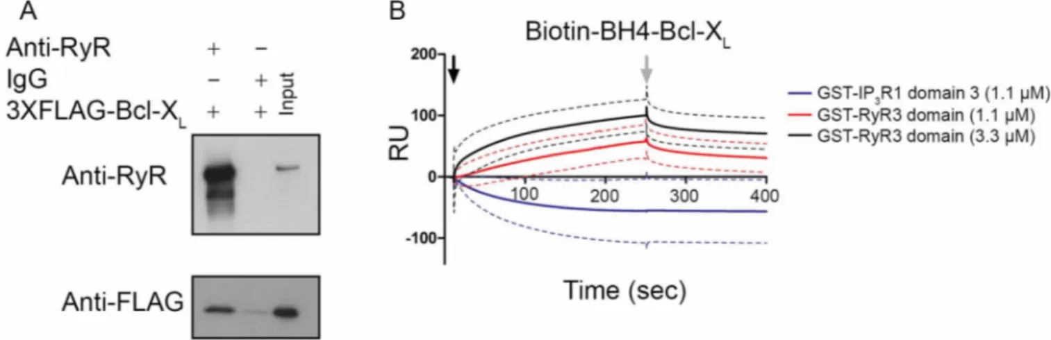
RyRs. Hence, we performed co-immunoprecipitation studies using lysates from HEK293 cells stably overexpressing RyR3 (HEK RyR3). In these cells, transiently overexpressed 3XFLAG-tagged Bcl-XL
co-immunoprecipitated with RyR3 indicating the formation of RyR3/Bcl-XLcomplexes (Fig. 1A and Supplementary Fig. 1A for uncropped
Western-blot images).
In our previous work we reported that the interaction between Bcl-2 and the RyR occurred via the BH4 domain of Bcl-Bcl-2 and a central regulatory domain of the RyR (a.a. 22632263–2688 for mink 2688 for mink RyR3)16. To examine whether a direct interaction between RyRs and the BH4 domain of Bcl-XLexists and whether this
inter-action occurs via the same or similar domains, surface plasmon resonance (SPR) experiments were performed (Fig. 1B). A
concen-XLwith the RyR3 is direct and that Bcl-XLvia its BH4 domain targets
the same domain as Bcl-2 on the RyR. However, the BH4 domain of Bcl-XLseems to have a lower affinity for the GST-RyR3 domain
compared to the BH4 domain of Bcl-2. This could indicate that biotinylation of the BH4 domain of Bcl-XLinfluences its binding
capabilities more than is the case for the BH4 domain of Bcl-2. Alternatively, other domains besides Bcl-XL’s BH4 domain may be
involved in the interaction of full-length Bcl-XL with the RyR.
Therefore, we wanted to identify if other domains besides the BH4 domain of Bcl-XLare important for interacting with the RyR.
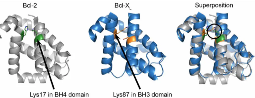
Superposition of the 3D-structures of Bcl-2 and Bcl-XLreveals a
spatial resemblance of Lys17 in the BH4 domain of Bcl-2 with Lys87 in the BH3 domain of Bcl-XL.To identify the contribution
and involvement of other Bcl-XL domains for targeting RyR
channels, an in silico superposition of the Bcl-2 (PDB-entry 4AQ320) and Bcl-X
L(PDB-entry 1R2D21) structures was performed
with the aid of PyMOL (The PyMOL Molecular Graphics System, Version 1.5.0.4 Schro¨dinger, LLC.). This superposition allowed the comparison of corresponding residues in the 3D-structures of Bcl-2 and Bcl-XL(Fig. 2). This analysis revealed that the positively charged
e-amino terminus of the side chain of Lys87 in Bcl-XL, located in the
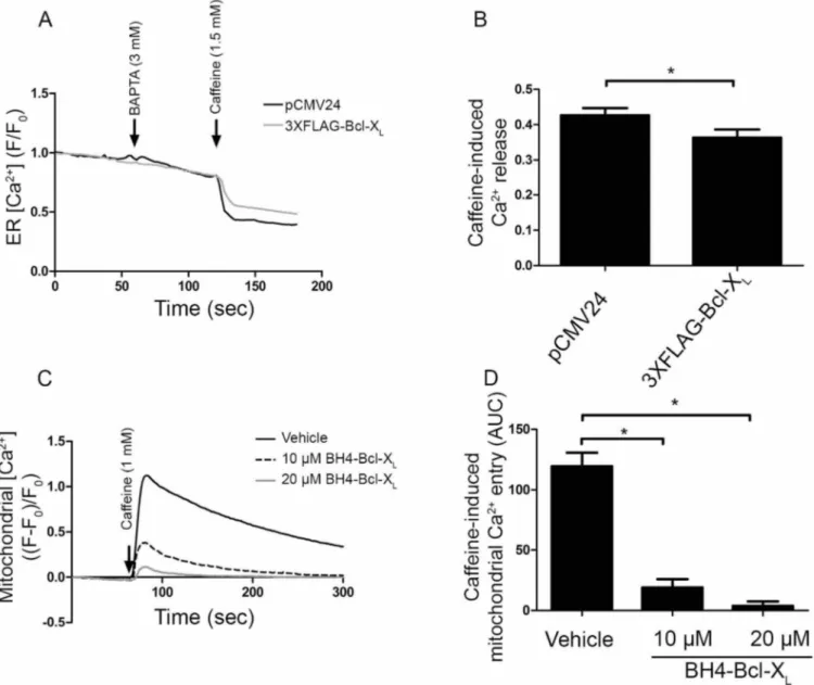
Figure 1|Bcl-XLbinds to a central regulatory region of RyR3. (A) Co-immunoprecipitation experiments were performed utilizing cell lysates from HEK RyR3 cells transiently overexpressing 3XFLAG-Bcl-XL. RyR3 was immunoprecipitated from these lysates utilizing a pan-RyR antibody. An anti-FLAG-HRP conjugated antibody was used for detecting co-immunoprecipitated 3XFLAG-Bcl-XL. Immunoblot showing the immunoprecipitated RyR3 (top) and co-immunoprecipitated 3XFLAG-tagged Bcl-XL(bottom). Immunoprecipitations using non-specific IgG antibodies were applied as negative controls. All experiments were performed at least three times utilizing each time independently transfected cells and freshly prepared HEK RyR3 lysates. All samples were run using the same experimental conditions on the same gel/blot. The uncropped image is shown in Supplementary Fig. 1A. (B) Sensorgrams of the surface plasmon resonance experiments expressed in RU as a function of time. The biotin-BH4-Bcl-XLpeptide and the scrambled peptide were immobilized on different channels of a streptavidin-coated sensor chip. The channels on the chip were exposed to the indicated concentrations of purified GST-fusion proteins (GST-IP3R1 domain 3 and GST-RyR3 domain). Binding of the GST-tagged proteins to the scrambled peptides was subtracted from each sensorgram. GST-IP3R1 domain 3 bound stronger to the scrambled peptide than to the biotin-BH4-Bcl-XLresulting in apparent negative values after this correction. The black arrow indicates the start of the association phase (addition of the GST-tagged proteins) and the grey arrow indicates the start of the dissociation phase (running buffer alone). Each sensorgram depicts the average of three experiments (full line) 6 S.D. (dashed lines).
BH3 domain, is in the same spatial constraints as the positively charged e-amino terminus of the side chain of Lys17 located in the BH4 domain of Bcl-2. Furthermore, Lys87 did not seem to be part of the hydrophobic cleft of Bcl-XL, as it was directed towards the space
facing the BH4 domain.
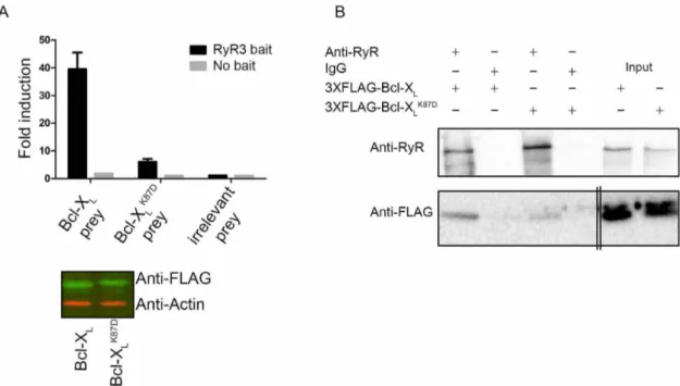
The Bcl-XLK87Dmutant is impaired in RyR3 binding.The relevance
of Lys87 in Bcl-XLfor RyR binding was addressed via mammalian
protein-protein interaction trap (MAPPIT)22, an in cellulo protein-protein interaction assay. MAPPIT is based on the functional complementation of cytokine receptor signaling. To study the possible existence of RyR/Bcl-XLcomplexes, the RyR3 domain was
cloned downstream of a chimeric cytokine receptor (RyR3 bait), consisting of the extracellular domain of the erythropoietin (Epo) receptor fused to the transmembrane and cytosolic part of the leptin receptor. In the latter, three tyrosines were mutated to phenylalanine to down regulate receptor signaling. Bcl-XLor the Bcl-XLK87Dmutant
were cloned downstream of a part of the glycoprotein 130 receptor (Bcl-XL or Bcl-XLK87D prey). If the Bcl-XL and Bcl-XLK87D prey
constructs interact with the RyR3 bait construct, functional complementation of the chimeric cytokine receptor occurs, leading to ligand-dependent downstream STAT signaling. The latter is monitored via a luciferase reporter assay driven by a STAT-sensitive promoter. We also used the SV40 large antigen T (irrelevant prey) as a prey to monitor the signal representing the non-specific binding to RyR3. As a negative control, binding of the chimeric cytokine receptor without the RyR3 fragment (no bait) to the two Bcl-XL preys was also assessed. These MAPPIT results
confirmed the data obtained via SPR and co-immunoprecipitation experiments, showing that Bcl-XL could interact with the RyR3
domain in a cellular context (Fig. 3A, top). Moreover, the Bcl-XLK87Dmutant was severely impaired in interacting with the RyR3
domain without affecting its expression (Fig. 3A, bottom panel and Supplementary Fig. 1B for uncropped Western-blot images). No binding was detected when the RyR3 domain was not present in the bait vector (Fig. 3A, top panel), indicating that the interaction was specific.
The impact of mutating Lys87 into Asp was also examined in the context of the full-length RyR3 protein using co-immunoprecipita-tion experiments. Consistent with the MAPPIT data, 3XFLAG-tagged Bcl-XLK87Ddisplayed a reduced affinity for full-length RyR3
channels (Fig. 3B and Supplementary Fig. 1C for uncropped Western-blot images).
Taken together, these data indicate that Bcl-XL, similarly to Bcl-2,
binds via its BH4 domain to the same regulatory domain on RyR3. However, whereas for Bcl-2 the BH4 domain appears to be the main determinant for complex formation with RyR channels, it seems that
for Bcl-XLboth the BH4 domain and the BH3 domain, likely via
Lys87, contribute to the interaction with RyR channels.
Bcl-XL, but not Bcl-XLK87D, inhibits RyR3-mediated Ca21release.
Driven by the fact that Bcl-XL can bind to RyR3, we examined
whether Bcl-XL could modulate RyR-mediated Ca21 release
(Fig. 4). Single-cell cytosolic [Ca21] measurements in HEK RyR3 cells loaded with Fura-2-AM were performed (Fig. 4A). An empty vector (pCMV24) control, 3XFLAG-tagged Bcl-XLor the
3XFLAG-tagged Bcl-XLK87Dmutant were transiently transfected into the HEK
RyR3 cells. An mCherry coding plasmid was co-transfected (at a 1:3 ratio) to identify transfected cells. After chelating extracellular Ca21 with BAPTA (3 mM), caffeine (1.5 mM) was applied to induce RyR-mediated Ca21 release. Overexpression of 3XFLAG-tagged Bcl-X
L
inhibited caffeine-induced Ca21 release compared to the empty vector control. The Bcl-XLK87D mutant failed to inhibit
caffeine-induced Ca21 release (Fig. 4B), correlating with its poor RyR3-binding properties. To exclude that the observed reduction in caffeine-induced Ca21 release upon Bcl-X
Loverexpression would
have been due to an indirect effect via lowering of the Ca21-filling state of the endoplasmic reticulum (ER), we determined the amount of thapsigargin (1 mM)-releasable Ca21. This irreversible SERCA inhibitor causes a depletion of the ER Ca21stores and provides a good measure for the ER Ca21-store content. The ER Ca21-store content was not affected by overexpression of 3XFLAG-tagged Bcl-XL(Fig. 4C). This supports the view that Bcl-XL, similarly to Bcl-2,
suppresses RyR-mediated Ca21release.
The BH4 domain of Bcl-XLby itself seems sufficient to inhibit
RyR-mediated Ca21release. In order to assess whether the BH4 domain of Bcl-XLis sufficient for inhibiting RyR-mediated Ca21
release, Fluo-3-AM loaded HEK RyR3 cells were loaded acutely with the BH4 domain of Bcl-XL, a control peptide or the vehicle
via electroporation (Fig. 5A). The BH4 domain of Bcl-XL, but not a
control peptide, suppressed caffeine (1 mM)-induced Ca21release. The BH4 domain of Bcl-XLinhibited caffeine-induced Ca21release
in a concentration-dependent manner (Fig. 5B). This indicates that the BH4 domain of Bcl-XL was sufficient for inhibiting
RyR-mediated Ca21release.
We also assessed whether the BH4 domain of Bcl-XLcould inhibit
endogenous RyR channels by using 14- to 18-day-old rat hippocam-pal cultures known to express different RyR isoforms23. The experi-mental set-up was identical to the one previously used for characterization of the effect of the BH4 domain of Bcl-2 on native RyRs16. Cytosolic [Ca21] was monitored in GCaMP3-expressing hip-pocampal neurons. The BH4 domain of Bcl-XL, a control peptide or
the vehicle were introduced into the neurons via a patch pipette. After loading the neuron for five minutes with the peptides or
Figure 2|Spatial resemblance of Lys17 in the BH4 domain of Bcl-2 and Lys87 in the BH3 domain of Bcl-XL. Image showing the 3D-structures for Bcl-2 (left), Bcl-XL(middle) and their in silico superposition (right). Lys17 in the BH4 domain of Bcl-2 and Lys87 in the BH3 domain of Bcl-XL
are indicated with arrows. The a.a. in green represent Lys17 and His94 in the BH4 and BH3 domain of Bcl-2 respectively. The a.a. in orange represent Asp14 and Lys87 in the BH4 and BH3 domains of Bcl-XLrespectively. The images were obtained by using PyMOL.
vehicle, cytosolic [Ca21] measurements were started. RyR-mediated Ca21release was triggered via a local puff of caffeine (10 mM) deliv-ered via a second patch pipette positioned next to the neuron. A time lapse (Fig. 5C) and a [Ca21] trace (Fig. 5D) of a typical experiment are shown for each condition. Loading of the neurons with the BH4 domain of Bcl-XL(20 mM) caused a significant reduction of the
caffeine-induced Ca21 release compared to the control peptide (Fig. 5 D, E). These results indicate that the BH4 domain of Bcl-XL
can regulate endogenously expressed RyR channels.
Bcl-XLand its BH4 domain directly inhibit RyRs at the level of the
ER.Bcl-XLand its isolated BH4 domain as a synthetic peptide inhibit
the caffeine-induced [Ca21] rise in the cytosol. Bcl-X
Lhas also been
implicated in the control of mitochondrial Ca21transport at the level of VDAC1. Bcl-XL was shown to inhibit Ca21 uptake into the
mitochondria6,24. However, it was also reported that Bcl-X
Lcould
stimulate mitochondrial Ca21 uptake25. The latter effect could result in a decrease in caffeine-induced [Ca21] rise in the cytosol. Therefore, we set out to document whether the decrease in caffeine-induced Ca21release in the cytosol by Bcl-X
Lis due to a decreased
Ca21release from the ER or to an increased Ca21accumulation into the mitochondria. Direct ER-Ca21measurements were performed in HEK RyR3 cells utilizing a recently described green fluorescent CEPIA1 protein, that is targeted to the lumen of the ER (G-CEPIA1er)26. HEK RyR3 cells were transiently transfected with the empty vector (pCMV24) as control or with 3XFLAG-tagged Bcl-XL
in combination with the G-CEPIA1er-encoding vector (at a 351 ratio). G-CEPIA1er-positive cells were selected and measurements
were performed as in Fig. 4A. A typical average trace of one experiment and the quantification of all performed experiments are shown in Fig. 6A and B, respectively. These results indicate that overexpression of 3XFLAG-Bcl-XL suppressed the
caffeine-induced Ca21 release from the ER, supporting a model in which the inhibitory effect of Bcl-XLon RyR-mediated [Ca21] rise in the
cytosol occurs at least in part due to inhibition of the Ca21release from the ER. Finally, we set out to directly measure the effect of the BH4 domain of Bcl-XL on caffeine-induced mitochondrial Ca21
entry. Rhod-FF-loaded HEK RyR3 cells were electroporated with either the vehicle (DMSO) or the BH4 domain of Bcl-XL(10 and
20 mM) and then stimulated with caffeine. Caffeine stimulation resulted in an increase in mitochondrial [Ca21] (Fig. 6C). Compared to the vehicle control however, the BH4 domain of Bcl-XLpotently inhibited the mitochondrial Ca21entry (Fig. 6 C, D).
Furthermore, the effectiveness of BH4-Bcl-XLto inhibit
caffeine-induced [Ca21] rise in the mitochondria seemed higher than for inhibiting the caffeine-induced [Ca21] rise in the cytosol, because 10 mM BH4-Bcl-XLinhibited caffeine-induced Ca21release in the
cytosol by about 50% but inhibited caffeine-induced Ca21uptake in the mitochondria by about 90%. Taken together these data suggest that BH4-Bcl-XL likely inhibits, rather than stimulates,
mitochondrial Ca21 accumulation. This is consistent with our recent findings showing that BH4-Bcl-XL directly interacts with
VDAC1 and suppressed VDAC1-mediated Ca21transfer into the mitochondria27. These experiments indicate that Bcl-X
L can
directly inhibit the caffeine-induced Ca21release at the level of the ER and potently inhibit mitochondrial Ca21 uptake under these
Figure 3|The Bcl-XLK87Dmutant is impaired in RyR3 binding. (A) Top: Representative example of a MAPPIT experiment. The binding is shown as fold induction value, calculated by dividing the average luciferase value of erythropoietin-stimulated cells by the average of non-stimulated cells. Binding of Bcl-XL(Bcl-XLprey), the Bcl-XLK87Dmutant (Bcl-XLK87Dprey) or irrelevant prey control (SV40 large T antigen) to the RyR3 domain (RyR3 bait) and as negative control the bait vector without RyR3 (No bait) are shown. Fold induction values at least 4 times higher than the irrelevant prey control are considered as bona fide protein-protein interactions. Values represent the average of three repeats within the same experiment 6 S.D. All experiments were independently performed at least three times. Bottom: Odyssey Western blot analyses staining for the FLAG tag of the prey vector containing Bcl-XL or the Bcl-XLK87Dmutant fusion proteins (green) or for actin (red) as a loading control. All samples were run using the same experimental conditions on the same gel/blot. The uncropped image is shown in Supplementary Fig. 1B. (B) Co-immunoprecipitations were performed in HEK RyR3 cells transiently overexpressing 3XFLAG-Bcl-XLor 3XFLAG-Bcl-XLK87Dsimilarly as in Fig. 1A. Non-specific IgG antibodies were applied as negative controls. These experiments were performed at least three times utilizing each time independently transfected and freshly prepared HEK RyR3 cell lysates. All samples were run using the same experimental conditions and were derived from the same gel/blot, i.e. 3-8% tris-acetate gels for RyRs and 4-12% bis-tris gels for 3xFLAG-Bcl-XL. The double lines indicate that an additional empty lane separating the immunoprecipitated samples and the input samples was removed for the 3XFLAG-Bcl-XLblot. The uncropped image is shown in Supplementary Fig. 1C.
experimental settings. We therefore conclude that the observed decrease in caffeine-induced Ca21release in the cytosol (Fig. 4 and 5) is mainly due to a direct inhibition of RyR3.
Discussion
The main conclusion of this paper is that Bcl-XLbinds to and
reg-ulates RyR3 channels. Similarly to Bcl-2, Bcl-XLtargets the central
modulatory domain of the RyR protein, thereby suppressing RyR-mediated Ca21release. Moreover, the BH4 domain of Bcl-X
Lwas
sufficient to inhibit both over- and endogenously expressed RyR channels in HEK293 cells or primary rat hippocampal neurons
respectively. Consistent with this, the BH4 domain of Bcl-XLcould
bind to the purified RyR3 domain. However, the RyR3-binding effi-ciency of the BH4 domain of Bcl-XLseemed much lower than that of
the BH4 domain of Bcl-2. Via an in silico superposition of the Bcl-2 and Bcl-XLcrystal structures, a spatial overlap was observed between
Lys17 in the BH4 domain of Bcl-2 and Lys87 in the BH3 domain of Bcl-XL: the positively charged e-amino groups of their side chains
coincide in space. Consistent with the moderate RyR3-binding prop-erties of the isolated BH4 domain of Bcl-XL, we found that Lys87
from Bcl-XLplayed a prominent role in binding to and regulating
RyR3.
Figure 4|Bcl-XLbut not Bcl-XLK87Dinhibits RyR-mediated Ca21release. Single-cell cytosolic [Ca21] measurements were performed in HEK RyR3 cells utilizing Fura-2-AM. (A) Average calibrated [Ca21] trace of 15 to 20 HEK RyR3 cells transfected (mCherry positive) with an empty vector
as control (pCMV24), 3XFLAG-Bcl-XLor 3XFLAG-Bcl-XLK87D. Addition of BAPTA and caffeine is indicated by the arrows. (B) Quantitative analysis of the single-cell cytosolic [Ca21] measurements. Values indicate averages of all peak values 6 S.E.M. These experiments were independently performed at
least four times (.120 cells/condition) (p50.008). (C) Quantitative analysis of the ER Ca21-store content. ER-store content was determined by
performing similar experiments as in A except that 1 mM thapsigargin was used as the stimulus. The values indicate the average area under the curve (AUC) 6 S.E.M. of at least three independent experiments (.80 cells/condition).
The association of Bcl-XLwith RyR channels and its functional
implications appear to be very similar as the ones observed for Bcl-2, since i) RyR3/Bcl-XLbinding is direct; ii) the binding of Bcl-XLto
RyR3 occurs, at least in part, via the BH4 domain; iii) Bcl-XL
over-expression inhibits RyR-mediated Ca21 release; and iv) the BH4 domain of Bcl-XLis also sufficient to suppress RyR activity. These Figure 5|The BH4 domain of Bcl-XLby itself was sufficient to inhibit RyR-mediated Ca21release. (A) Representative trace of the performed Fluo-3-AM single-cell cytosolic [Ca21] measurements in HEK RyR3 cells loaded by electroporation with either the vehicle (DMSO), a control peptide or the BH4
domain of Bcl-XL. The addition of caffeine is indicated by the arrow. Traces were normalized to the baseline fluorescence ((F-F0)/F0). (B) Quantitative analysis of the single-cell cytosolic [Ca21] measurements with indicated concentrations of the BH4 domain of Bcl-X
L. Values indicate caffeine-induced Ca21release after electroporation loading with different concentrations of the BH4 domain of Bcl-X
Lrelative to the response after electroporation loading with the same concentration of the control peptide. Values depict average 6 S.E.M. of at least four independent experiments (p-values were 0.0037, 0.001 and 0.0039 for 10 mM, 20 mM and 40 mM of the BH4 domain of Bcl-XLrespectively). (C-E) Single-cell [Ca21] measurements performed in 14- to 18-day-old hippocampal cultures. GCaMP3, introduced into these neurons via adeno-associated infection, was used as cytosolic Ca21indicator. Utilizing
whole-cell voltage clamp the membrane potential of the neurons was clamped at 260 mV. 20 mM of the BH4 domain of Bcl-XL, a control peptide or the vehicle (DMSO) was introduced into each measured neuron via the patch pipette. All experiments were performed in the presence of 1 mM tetrodotoxin. A 10 mM caffeine puff was locally administered via a second patch pipette positioned 15-25 mm from the soma of the neuron. (C) Time lapse of a typical experiment for each of the tested conditions. Caffeine was administered after 60 sec. The scale bar depicts 5 mm. (D) Typical responses to caffeine after loading the neurons with 20 mM of either the control peptide the BH4 domain of Bcl-XLor the vehicle. Traces were normalized to the baseline fluorescence ((F-F0)/F0). The arrow indicates when caffeine was administered. (E) Scatter plot showing peak responses of all performed measurements and the median (horizontal line). All values were normalized to the caffeine response after vehicle control treatment (p50.0037, N512 and N515 for the control peptide and BH4-Bcl-XLrespectively).
findings correlate with the fact that the Bcl-2K17Dmutant and BH4-Bcl-2K17Dremain capable of binding to and regulating RyR channels, although this mutation changes the lysine critical for binding to the IP3R into the Asp11 residue in the BH4 domain of Bcl-XL16. This lack
of selectivity between Bcl-2 and Bcl-XLmay illustrate an important
difference between IP3R- and RyR-mediated Ca21release. However,
the binding of Bcl-XLversus Bcl-2 to RyRs in native tissues
expres-sing RyRs ought to be further explored. In particular, it will be important to carefully analyze the Bcl-2- and Bcl-XL-expression
levels in the relevant tissues and to determine whether a preferential binding of Bcl-2 or Bcl-XLto RyR channels exists in cells expressing
both Bcl-2 and Bcl-XL. Despite these similarities, the molecular
determinants underlying RyR/Bcl-XL-complex formation do not
seem identical to those of 2, because the BH4 domain of
Bcl-XLby itself displays rather moderate RyR3-binding properties. As a
consequence, additional domains seem to be involved in RyR/Bcl-XL-complex formation. Here, we identified Lys87, located in the BH3
domain of Bcl-XL, as a critical determinant contributing to binding to
and regulating RyR channels. Despite the importance of Lys87, the BH4 domain of Bcl-XLalone was able to suppress RyR activity.
The BH4 domain of Bcl-XLhas been implicated in numerous
studies to display strong anti-apoptotic and protective effects against a wide variety of insults and triggers, including in the heart28-30, endothelial cells31,32, blood cells33-35, pancreatic islets36and neurons37. Many of the cell types and tissues reported to benefit from the BH4 domain of Bcl-XLfor their survival endogenously express RyR
chan-nels (cardiomyocytes, lymphocytes, pancreatic islets and neurons). Furthermore, in many apoptotic paradigms, reactive oxygen species
Figure 6|Bcl-XLand its BH4 domain directly inhibit RyR-mediated Ca21release from the ER. (A) Typical average normalized (F/F0) traces of single-cell ER [Ca21] measurement performed in HEK RyR3 cells transfected with G-CEPIA1er plasmid. G-CEPIA1er-positive cells transfected with the empty
control vector (pCMV24) or 3XFLAG-Bcl-XLwere selected for these measurements. After chelating extracellular Ca21with BAPTA, caffeine was added to stimulate RyR-mediated Ca21release (arrows). (B) Quantitative analysis of the performed experiments. For each trace the caffeine-induced Ca21release
was determined by subtracting the fluorescence after caffeine addition (during plateau phase) from the fluorescence just before caffeine addition after normalization. Values depict average 6 S.E.M. These experiments were independently repeated at least four times (.100 cells/condition) (p50.0018). (C) Normalized ((F-F0)/F0) representative traces of mitochondrial [Ca21] measurements. The vehicle (DMSO) or the BH4 domain of Bcl-XL(10 mM and 20 mM) were introduced into Rhod-FF-loaded HEK RyR3 cells via electroporation loading. Mitochondrial Ca21was measured after caffeine (arrow)
stimulation. (D) Quantification of the performed experiments. Values show the average caffeine-induced mitochondrial Ca21entry as area under the
death.
RyRs have important physiological functions in a variety of excit-able cells and tissues, including skeletal muscle, cardiac muscle, neu-rons and pancreatic cells43–46. Furthermore, dysregulation of RyRs, either by somatic mutations or by altered expression levels, has been implicated in a variety of pathophysiological conditions, including malignant hyperthermia and central core disease47,48, cardiac dis-eases49–51 and neurodegenerative diseases like Alzheimer’s dis-ease52–54 and Huntington’s disease55. At this point, the existence and physiological relevance of RyR/Bcl-2- and RyR/Bcl-XL-complex
formation in these tissues and their potential disturbance in RyR-associated pathophysiologies will require further research.
In conclusion, our data further expand the number of Bcl-2-family members that are able to form protein complexes with RyR channels, thereby underpinning their critical role in regulating intracellular Ca21dynamics at the level of intracellular Ca21-release channels. Methods
Chemicals, antibodies and peptides.Unless otherwise specified, all chemicals were purchased from Sigma-Aldrich (St. Louis, MO, USA). The following antibodies were used: mouse monoclonal anti-actin antibody, anti-FLAG M2 antibody and HRP-conjugated anti-FLAG M2 antibody (Sigma-Aldrich), mouse monoclonal anti-RyR antibody 34C (Thermo Scientific, Rockford, IL, USA, or Developmental Studies Hybridoma Bank, University of Iowa, Iowa, USA) and mouse monoclonal anti-Bcl-XLantibody YTH-2H12 (Trevigen, Gaithersburg, WV, USA). The sequences of the
peptides used in this study were:
Biotin-BH4-Bcl-XL: Biotin-MSQSNRELVVDFLSYKLSQKGYSW (also used
without the biotin tag)
Biotin-scrambled BH4-Bcl-XL: Biotin-WYSKQRSLSGLVMYVLEDKNSQFS
Control peptide: WYEKQRSLHGIMYYVIEDRNTKGYR
These peptides were synthesized by Life Tein (Hillsborough, NJ, USA) with a purity of at least 85%.
Plasmids, constructs and protein purifications.3XFLAG-Bcl-XLwas obtained as
previously described19. The 3XFLAG-Bcl-X
LK87Dmutant was obtained by PCR
site-directed mutagenesis utilizing the following primers: forward:
59ATCCCCATGGCAGCAGTAGATCAAGCGCTGAGGGAGGCA39, and reverse: 59TGCCTCCCTCAGCGCTTGATCTACTGCTGCCATGGGGAT39. The pCMV G-CEPIA1er containing plasmid was a gift from Dr. Masamitsu Iino (Addgene plasmid # 58215)26. The GST-IP
3R1 domain 3 construct and the GST-RyR3 construct
were obtained and purified as described16.
Cell culture, transfections and dissociated hippocampal cultures.All media and supplements added to the medium used in this paper were purchased from Life Technologies (Ghent, Belgium). HEK293 cells stably overexpressing RyR3 were cultured at 37uC in a 5% CO2incubator in a-Minimum Essential Medium
supplemented with 10% fetal calf serum, 100 IU/mL penicillin, 100 mg/mL streptomycin, 2 mM glutamax and 800 mg/mL G41856. HEK293 cells were grown in
Dulbecco’s Modified Eagle Medium containing 4500 mg/L glucose, 10% fetal bovine serum and 50 mg/mL gentamicin57.
24 hours after seeding, the 3XFLAG-Bcl-XLor the 3XFLAG-Bcl-XLK87Dmutant
construct were introduced into the HEK RyR3 cells utilizing JETPrime transfection reagent (Polyplus Transfections, Illkirch, France) according to the manufacturer’s protocol. 48 hours later the cells were harvested and lysed utilizing a CHAPS-based lysis buffer (pH 7.5, 50 mM Tris-HCl, 100 mM NaCl, 2 mM EDTA, 50 mM NaF, 1 mM Na3VO4, 1% CHAPS and protease inhibitor tablets (Roche, Basel,
Switzerland)). For single-cell cytosolic [Ca21] measurements the same constructs or
the empty pCMV24 vector were introduced 48 hours after seeding in the HEK RyR3 cells utilizing X-tremeGENE HP DNA transfection reagent (Roche) according to the manufacturer’s protocol. A pcDNA 3.1(-) mCherry expressing vector was co-trans-fected at a 153 ratio as a selection marker. For direct ER [Ca21] measurements, the
G-(green) or anti-rabbit-IRDye700 (red) as secondary antibodies (Thermo Scientific).
Co-immunoprecipitation experiments.Co-immunoprecipitation experiments were performed utilizing a co-immunoprecipitation kit (Thermo Scientific). RyR antibody or mouse IgG control antibody (Santa Cruz Biotechnology, Heidelberg, Germany) was immobilized according to the manufacturer’s protocol. Gelatine was removed from the IgG control antibody utilizing a Pierce Antibody Clean-up Kit (Thermo scientific). Precleared HEK RyR3 lysates containing the 3XFLAG-Bcl-XL
constructs (150 mg) were added to the resin to which the antibodies were immobilized and allowed to incubate overnight at 4uC. The next day, the resin was washed at least five times utilizing the CHAPS-based lysis buffer. The immune complexes were eluted by boiling (95uC) in 50 mL 23 LDS (Life Technologies) supplemented with 1/200 b-mercaptoethanol for 5 min.
MAPPIT.The RyR3 domain was amplified by PCR using the following primers, forward: 59TAGTTGTCGACGAAGAGAGAAGTCATGGAGGA39, and reverse: 59TAGTTGCGGCCGCCTATTTGGTCCTCTCCACA39, and cloned in the pSEL12L bait vector59, using the restriction enzymes SalI and NotI. Bcl-X
Lwas
cloned in the pMG1-GW plasmid (prey vector)22using the Gateway recombination
technology as described by the manufacturer (Life Technologies). Utilizing the same primers as described before, the Bcl-XLK87Dmutation was also introduced in this
construct via site directed mutagenesis. The MAPPIT analyses were done as previously described22with minor changes. Briefly, HEK293 cells were seeded in
96-well plates. Six 96-wells per condition were transfected with the different combinations of bait, prey and reporter plasmid (rPAP1-luci) using the calcium phosphate method. The next day, half of the wells were stimulated with 5 ng/mL Epo while the other half were left untreated. 24 hours later the cells were lysed and after the addition of substrate the luciferase activity was determined using a luminometer. The fold induction was obtained by dividing the average value of the stimulated cells by the average value of the non-stimulated cells.
Electroporation loading.Electroporation loading of HEK RyR3 cells was performed as previously described16,60.
Single-cell cytosolic Ca21imaging.Fura-2-AM and Fluo-3-AM [Ca21]
measurements in HEK RyR3 cells and GCaMP3 single-cell [Ca21] measurements in
dissociated hippocampal neurons were performed as previously described16.
Single-cell ER Ca21imaging.The G-CEPIA1er construct was introduced into HEK
RyR3 cells as described above. A Zeiss Axio Observer Z1 Inverted Microscope equipped with a 203 air objective and a high-speed digital camera (Axiocam Hsm, Zeiss, Jena, Germany) were used for these measurements. Changes in fluorescence were monitored in the GFP channel (480/520 excitation/emission). To chelate extracellular Ca21, 3 mM BAPTA (Alfa Aesar, Ward Hill, MA, USA) was added. One
minute later 1.5 mM caffeine was added to trigger RyR-mediated Ca21release. All
traces were normalized (F/F0) where F0is the starting fluorescence of each trace.
Single-cell mitochondrial Ca21imaging.HEK RyR3 cells were loaded for 30 min
with 5 mM Rhod-FF-AM. Subsequently, cells were subjected to de-esterification over 15 min. During this time the BH4 domain peptides were introduced into the cells using the in situ electroporation technique60. Fluorescence-intensity changes in
mitochondria were analyzed with custom-developed FluoFrames software. For each individual trace, the relative change of fluorescence (DF/F) was calculated. DF/F equals [Ft-F0/F0], with F0denoting the fluorescence before stimulation with caffeine
and Ftthe fluorescence at different time points after caffeine stimulation.
Subsequently, relative mitochondrial [Ca21] changes were quantified as the area
under the curve of the various Ca21traces.
Statistical analysis.Two-tailed student’s t-tests were performed when two conditions were compared. When comparing three conditions a one-way ANOVA with Bonferroni’s multiple comparison test was performed. * indicates significantly different results (p,0.05). Exact p-values are indicated in the figure legends, where available.
1. Brunelle, J. K. & Letai, A. Control of mitochondrial apoptosis by the Bcl-2 family. J Cell Sci 122, 437–441 (2009).
2. Chipuk, J. E. & Green, D. R. How do BCL-2 proteins induce mitochondrial outer membrane permeabilization? Trends Cell Biol 18, 157–164 (2008).
3. Chami, M. et al. Bcl-2 and Bax exert opposing effects on Ca21 signaling, which do not depend on their putative pore-forming region. J Biol Chem 279, 54581–54589 (2004).
4. Ferrari, D. et al. Endoplasmic reticulum, Bcl-2 and Ca21 handling in apoptosis. Cell Calcium 32, 413–420 (2002).
5. Monaco, G., Vervliet, T., Akl, H. & Bultynck, G. The selective BH4-domain biology of Bcl-2-family members: IP3Rs and beyond. Cell Mol Life Sci 70,
1171–1183 (2013).
6. Arbel, N., Ben-Hail, D. & Shoshan-Barmatz, V. Mediation of the antiapoptotic activity of Bcl-xL protein upon interaction with VDAC1 protein. J Biol Chem 287, 23152–23161 (2012).
7. Arbel, N. & Shoshan-Barmatz, V. Voltage-dependent anion channel 1-based peptides interact with Bcl-2 to prevent antiapoptotic activity. J Biol Chem 285, 6053–6062 (2010).
8. Plotz, M., Gillissen, B., Hossini, A. M., Daniel, P. T. & Eberle, J. Disruption of the VDAC2-Bak interaction by Bcl-xSmediates efficient induction of apoptosis in
melanoma cells. Cell Death Differ 19, 1928–1938 (2012).
9. Ferdek, P. E. et al. A novel role for Bcl-2 in regulation of cellular calcium extrusion. Curr Biol 22, 1241–1246 (2012).
10. Kuo, T. H. et al. Modulation of endoplasmic reticulum calcium pump by Bcl-2. Oncogene 17, 1903–1910 (1998).
11. Ahn, T., Yun, C. H., Kim, H. R. & Chae, H. J. Cardiolipin, phosphatidylserine, and BH4 domain of Bcl-2 family regulate Ca21/H1antiporter activity of human Bax
inhibitor-1. Cell Calcium 47, 387–396 (2010).
12. Xu, Q. & Reed, J. C. Bax inhibitor-1, a mammalian apoptosis suppressor identified by functional screening in yeast. Mol Cell 1, 337–346 (1998).
13. Oakes, S. A. et al. Proapoptotic BAX and BAK regulate the type 1 inositol trisphosphate receptor and calcium leak from the endoplasmic reticulum. Proc Natl Acad Sci U S A 102, 105–110 (2005).
14. Rong, Y. P. et al. Targeting Bcl-2-IP3receptor interaction to reverse Bcl-2’s
inhibition of apoptotic calcium signals. Mol Cell 31, 255–265 (2008). 15. White, C. et al. The endoplasmic reticulum gateway to apoptosis by Bcl-XL
modulation of the InsP3R. Nat Cell Biol 7, 1021–1028 (2005).
16. Vervliet, T. et al. Bcl-2 binds to and inhibits ryanodine receptors. J Cell Sci 127, 2782–2792 (2014).
17. Letai, A. G. Diagnosing and exploiting cancer’s addiction to blocks in apoptosis. Nat Rev Cancer 8, 121–132 (2008).
18. Rong, Y. P., Barr, P., Yee, V. C. & Distelhorst, C. W. Targeting Bcl-2 based on the interaction of its BH4 domain with the inositol 1,4,5-trisphosphate receptor. Biochim Biophys Acta 1793, 971–978 (2009).
19. Monaco, G. et al. Selective regulation of IP3-receptor-mediated Ca21signaling and
apoptosis by the BH4 domain of Bcl-2 versus Bcl-Xl. Cell Death Differ 19, 295–309 (2012).
20. Perez, H. L. et al. Identification of a phenylacylsulfonamide series of dual Bcl-2/ Bcl-xL antagonists. Bioorg Med Chem Lett 22, 3946–3950 (2012).
21. Manion, M. K. et al. Bcl-XL mutations suppress cellular sensitivity to antimycin A. J Biol Chem 279, 2159–2165 (2004).
22. Eyckerman, S. et al. Design and application of a cytokine-receptor-based interaction trap. Nat Cell Biol 3, 1114–1119 (2001).
23. Martin, C., Chapman, K. E., Seckl, J. R. & Ashley, R. H. Partial cloning and differential expression of ryanodine receptor calcium-release channel genes in human tissues including the hippocampus and cerebellum. Neuroscience 85, 205–216 (1998).
24. Shimizu, S., Konishi, A., Kodama, T. & Tsujimoto, Y. BH4 domain of antiapoptotic Bcl-2 family members closes voltage-dependent anion channel and inhibits apoptotic mitochondrial changes and cell death. Proc Natl Acad Sci U S A 97, 3100–3105 (2000).
25. Huang, H. et al. An interaction between Bcl-xL and the voltage-dependent anion channel (VDAC) promotes mitochondrial Ca21 uptake. J Biol Chem 288, 19870–19881 (2013).
26. Suzuki, J. et al. Imaging intraorganellar Ca21at subcellular resolution using
CEPIA. Nat Commun 5, 4153 (2014).
27. Monaco, G. et al. The BH4 domain of anti-apoptotic Bcl-XL, but not that of the related Bcl-2, limits the voltage-dependent anion channel 1 (VDAC1)-mediated transfer of pro-apoptotic Ca21signals to mitochondria. J Biol Chem Epub ahead
of print(2015).
28. Boisguerin, P. et al. Systemic delivery of BH4 anti-apoptotic peptide using CPPs prevents cardiac ischemia-reperfusion injuries in vivo. J Control Release 156, 146–153 (2011).
29. Ono, M. et al. BH4 peptide derivative from Bcl-xL attenuates ischemia/ reperfusion injury thorough anti-apoptotic mechanism in rat hearts. Eur J Cardiothorac Surg 27, 117–121 (2005).
30. Sugioka, R. et al. BH4-domain peptide from Bcl-xL exerts anti-apoptotic activity in vivo. Oncogene 22, 8432–8440 (2003).
31. Cantara, S., Donnini, S., Giachetti, A., Thorpe, P. E. & Ziche, M. Exogenous BH4/ Bcl-2 peptide reverts coronary endothelial cell apoptosis induced by oxidative stress. J Vasc Res 41, 202–207 (2004).
32. Cantara, S., Thorpe, P. E., Ziche, M. & Donnini, S. TAT-BH4 counteracts Abeta toxicity on capillary endothelium. FEBS Lett 581, 702–706 (2007).
33. Hotchkiss, R. S. et al. TAT-BH4 and TAT-Bcl-xL peptides protect against sepsis-induced lymphocyte apoptosis in vivo. J Immunol 176, 5471–5477 (2006). 34. McConnell, K. W. et al. Anti-apoptotic peptides protect against radiation-induced
cell death. Biochem Biophys Res Commun 355, 501–507 (2007). 35. Santamaria, B. et al. Bcl-xL prevents peritoneal dialysis solution-induced
leukocyte apoptosis. Perit Dial Int 28 Suppl 5, S48–52 (2008).
36. Klein, D. et al. Delivery of Bcl-XL or its BH4 domain by protein transduction inhibits apoptosis in human islets. Biochem Biophys Res Commun 323, 473–478 (2004).
37. Martorana, F. et al. The BH4 domain of Bcl-XLrescues astrocyte degeneration in
amyotrophic lateral sclerosis by modulating intracellular calcium signals. Hum Mol Genet 21, 826–840 (2012).
38. Niggli, E. et al. Posttranslational modifications of cardiac ryanodine receptors: Ca21signaling and EC-coupling. Biochim Biophys Acta 1833, 866–875 (2013).
39. Marengo, J. J., Hidalgo, C. & Bull, R. Sulfhydryl oxidation modifies the calcium dependence of ryanodine-sensitive calcium channels of excitable cells. Biophys J 74, 1263–1277 (1998).
40. Xu, L., Eu, J. P., Meissner, G. & Stamler, J. S. Activation of the cardiac calcium release channel (ryanodine receptor) by poly-S-nitrosylation. Science 279, 234–237 (1998).
41. Mochizuki, M. et al. Scavenging free radicals by low-dose carvedilol prevents redox-dependent Ca21 leak via stabilization of ryanodine receptor in heart failure. J Am Coll Cardiol 49, 1722–1732 (2007).
42. Oda, T. et al. Defective regulation of interdomain interactions within the ryanodine receptor plays a key role in the pathogenesis of heart failure. Circulation 111, 3400–3410 (2005).
43. Islam, M. S. Calcium signaling in the islets. Adv Exp Med Biol 654, 235–259 (2010). 44. Lanner, J. T., Georgiou, D. K., Joshi, A. D. & Hamilton, S. L. Ryanodine receptors:
structure, expression, molecular details, and function in calcium release. Cold Spring Harb Perspect Biol 2, a003996 (2010).
45. Stutzmann, G. E. & Mattson, M. P. Endoplasmic reticulum Ca21handling in
excitable cells in health and disease. Pharmacol Rev 63, 700–727 (2011). 46. Van Petegem, F. Ryanodine receptors: structure and function. J Biol Chem 287,
31624–31632 (2012).
47. Lyfenko, A. D., Goonasekera, S. A. & Dirksen, R. T. Dynamic alterations in myoplasmic Ca21in malignant hyperthermia and central core disease. Biochem
Biophys Res Commun 322, 1256–1266 (2004).
48. Robinson, R., Carpenter, D., Shaw, M. A., Halsall, J. & Hopkins, P. Mutations in RYR1 in malignant hyperthermia and central core disease. Hum Mutat 27, 977–989 (2006).
49. Blayney, L. M. & Lai, F. A. Ryanodine receptor-mediated arrhythmias and sudden cardiac death. Pharmacol Ther 123, 151–177 (2009).
50. Marx, S. O. & Marks, A. R. Dysfunctional ryanodine receptors in the heart: new insights into complex cardiovascular diseases. J Mol Cell Cardiol 58, 225–231 (2013).
51. Priori, S. G. & Chen, S. R. Inherited dysfunction of sarcoplasmic reticulum Ca21
handling and arrhythmogenesis. Circ Res 108, 871–883 (2011).
52. Bruno, A. M. et al. Altered ryanodine receptor expression in mild cognitive impairment and Alzheimer’s disease. Neurobiol Aging 33, 1001 e1001–1006 (2012).
53. Del Prete, D., Checler, F. & Chami, M. Ryanodine receptors: physiological function and deregulation in Alzheimer disease. Mol Neurodegener 9, 21 (2014). 54. Liu, J. et al. The role of ryanodine receptor type 3 in a mouse model of Alzheimer
disease. Channels (Austin) 8, 230–242 (2014).
55. Chen, X. et al. Dantrolene is neuroprotective in Huntington’s disease transgenic mouse model. Mol Neurodegener 6, 81 (2011).
56. Rossi, D. et al. RyR1 and RyR3 isoforms provide distinct intracellular Ca21signals
in HEK 293 cells. J Cell Sci 115, 2497–2504 (2002).
57. Eyckerman, S., Broekaert, D., Verhee, A., Vandekerckhove, J. & Tavernier, J. Identification of the Y985 and Y1077 motifs as SOCS3 recruitment sites in the murine leptin receptor. FEBS Lett 486, 33–37 (2000).
58. Nadif Kasri, N., Nakano-Kobayashi, A. & Van Aelst, L. Rapid synthesis of the X-linked mental retardation protein OPHN1 mediates mGluR-dependent LTD through interaction with the endocytic machinery. Neuron 72, 300–315 (2011). 59. Lemmens, I. et al. Heteromeric MAPPIT: a novel strategy to study modification-dependent protein-protein interactions in mammalian cells. Nucleic Acids Res 31, e75 (2003).
60. Decrock, E. et al. Electroporation loading and flash photolysis to investigate intra-and intercellular Ca21signaling in Calcium Techniques: A Laboratory Manual (ed.
Parys, J. B., Bootman, M., Yule, D. I. & Bultynck, G.) 93–112 (Cold Spring Harbor, 2014).
Acknowledgments
We would like to thank Marco Benevento, Martijn Selten, Wei Ba, Kirsten Welkenhuyzen, Marina Crabbe´, Steffi de Rouck, and Anja Florizoone for their excellent technical assistance. We thank Dr. Masamitsu Iino (The University of Tokyo, Japan) for providing the G-CEPIA1er plasmid. This work was supported by the Research Foundation-Flanders (FWO) grants 6.057.12 to G.B., H.D.S., J.B.P. and L.L. and G.0134.09N to L.L., by the Research Council of the KU Leuven via an OT START grant (STRT1/10/044 and OT/14/ 101) to G.B., by the Interuniversity Attraction Poles Program (Belgian Science Policy; P7/13
![Figure 4 | Bcl-X L but not Bcl-X L K87D inhibits RyR-mediated Ca 21 release. Single-cell cytosolic [Ca 21 ] measurements were performed in HEK RyR3 cells utilizing Fura-2-AM](https://thumb-eu.123doks.com/thumbv2/123dokorg/4673783.43302/5.892.68.820.66.812/figure-inhibits-mediated-single-cytosolic-measurements-performed-utilizing.webp)