https://doi.org/10.1177/1756283X16673981 https://doi.org/10.1177/1756283X16673981 Ther Adv Gastroenterol
2017, Vol. 10(4) 353 –360 DOI: 10.1177/ 1756283X16673981 © The Author(s), 2017. Reprints and permissions: http://www.sagepub.co.uk/ journalsPermissions.nav
Therapeutic Advances in Gastroenterology
journals.sagepub.com/home/tag 353
Introduction
Mast cells (MCs) play a role in tumour
angiogen-esis and their involvement has been demonstrated
in several animal and human malignancies
[Gulubova and Vlaykova, 2009; Marech et al.
2014a]. MCs can secrete classical
proangio-genic factors, including vascular endothelial
growth factor (VEGF), fibroblast growth factor-2,
thymidine phosphorylase and interestingly, a
non-classical proangiogenetic factor, namely tryptase,
stored in their secretory granules [Marech et al.
2014b; Ammendola et al. 2013a, 2014]. With
spe-cial reference to tryptase, it induces in vitro
micro-vascular endothelial cells’ (ECs) proliferation in
Tryptase mast cell density,
protease-activated receptor-2 microvascular density,
and classical microvascular density
evaluation in gastric cancer patients
undergoing surgery: possible translational
relevance
Michele Ammendola, Rosario Sacco, Giuseppina Vescio, Valeria Zuccalà,
Maria Luposella, Rosa Patruno, Nicola Zizzo, Claudia Gadaleta, Ilaria Marech,
Roberta Ruggieri, Ibrahim Furkan Kocak, Taner Ozgurtas, Cosmo Damiano Gadaleta,
Giuseppe Sammarco and Girolamo Ranieri
Abstract
Background: Mast cells (MCs) can stimulate angiogenesis, releasing several proangiogenic
cytokines stored in their cytoplasm. In particular, MCs can release tryptase, a potent in vivo
and in vitro proangiogenic factor via protease-activated receptor-2 (PAR-2) activation and
mitogen-activated protein kinase (MAPK) phosphorylation. Nevertheless, no data are available
concerning the relationship among tryptase MC density (TMCD), endothelial cells (ECs)
positive to PAR-2 microvascular density (PAR-2-MVD) and classical MVD (C-MVD) in gastric
cancer (GC) angiogenesis.
Methods: In this study, we analyzed the correlation of TMCD, PAR-2-MVD, C-MVD with each
other and with the main clinicopathological features in GC patients who underwent surgery. A
series of 77 GC patients with stage T
2-3N
2-3M
0(classified by the American Joint Committee on
Cancer for Gastric Cancer, 7th edition) were selected and then underwent surgery.
Results: Tumour tissue samples were evaluated by mean of immunohistochemistry and
image analysis methods in terms of numbers of TMCD, PAR-2-MVD and C-MVD. A significant
correlation between the TMCD, PAR-2-MVD and C-MVD groups with each other was found by
Pearson t-test analysis (r ranged from 0.64 to 0.76; p value ranged from 0.02 to 0.03). There
was no other significant correlation between the above parameters and clinicopathological
features.
Conclusions: Our in vivo preliminary data suggest that TMCD and PAR-2-MVD may play a role
in GC angiogenesis and they could be further evaluated as a target of antiangiogenic therapy.
Keywords: angiogenesis, gastric cancer, mast cells, PAR-2, tryptase
Correspondence to:
Michele Ammendola, MD
Department of Medical and Surgical Sciences, Clinical Surgery Unit, University of Catanzaro ‘Magna Graecia’ Medical School, Viale Europa – Germaneto, 88100, Catanzaro, Italy michele.ammendola@ libero.it Rosario Sacco, MD, Professor Giuseppe Sammarco, MD, Professor Giuseppina Vescio, MD
Department of Medical and Surgical Sciences, Clinical Surgery Unit, University of Catanzaro ‘Magna Graecia’ Medical School, Catanzaro, Italy
Valeria Zuccalà, MD
Health Science Department, Pathology Unit, University of Catanzaro ‘Magna Graecia’ Medical School, Catanzaro, Italy
Maria Luposella, MD
Cardiovascular Disease Unit, ‘San Giovanni di Dio’ Hospital, Crotone, Italy
Claudia Gadaleta, MD Rosa Patruno, MD Nicola Zizzo, MD, Professor
Chair of Pathology, University ‘Aldo Moro’ Veterinary Medical School, Bari, Italy
Ibrahim Furkan Kocak, MD Taner Ozgurtas, MD, Professor
Department of Biochemistry, Gulhane Military Medical Academy Etlik, Ankara, Turkey
Roberta Ruggieri, MD Ilaria Marech, MD Cosmo Damiano Gadaleta, MD
Girolamo Ranieri, MD
Diagnostic and Interventional Radiology Unit with Integrated Section of Translational Medical Oncology, National Cancer Research Centre, ‘Giovanni Paolo II’, Bari, Italy
the matrigel assay and displayed in vivo the
capil-lary growth on the chick embryo chorioallantoic
membrane, conversely, suppressed by tryptase
inhibitors [Ammendola et al. 2013b; Marech et al.
2016b; Ribatti et al. 2011]. This proangiogenic
stimulus induced by tryptase is mainly mediated
via protease-activated receptor-2 (PAR-2) that
belongs to the G protein-coupled receptor family
[Blair et al. 1997; Stack and Johnson, 1994;
Fajardo and Pejler, 2003; Itoh et al. 2005]. Four
forms of PARs have been reported (PAR-1
through PAR-4). In particular, PAR-2 can be
acti-vated by proteases such as trypsin and tryptase.
These proteases cleave the N terminus to generate
a tethered ligand, which interacts and activates the
receptor [Rickard et al. 2005; Matej et al. 2007;
Morris et al. 2006; Ammendola et al. 2013c, 2014;
Donato et al. 2014; Ranieri et al. 2009]. Signaling
via PAR-2, expressed on ECs, elicits activation of
the major members of the mitogen-activated
pro-tein kinase (MAPK) phosphorylation family and
induces EC proliferation. PAR-2 activation also
leads to the production of other proangiogenic
factors, such as VEGF, interleukin-8 (IL-8), IL-6,
granulocyte-macrophage colony-stimulating factor
(GM-CSF) and macrophage colony-stimulating
factor (M-CSF) [Ammendola et al. 2015; Ribatti
and Ranieri, 2015; Malfettone et al. 2013;
Soreide et al. 2006; Darmoul et al. 2001;
Uusitalo-Jarvinen et al. 2007; Liu and Mueller, 2006].
In literature, no data have been published on
the relationship among tryptase MC density
(TMCD), ECs positive to PAR-2 forming
micro-vascular density (PAR-2-MVD) and classical
MVD (C-MVD) in gastric cancer (GC)
angio-genesis [Yano et al. 1999; Sedda et al. 2014;
Ribatti et al. 2010; Wang et al. 2013a; Zhang
et al. 2012].
In this preliminary study, we analyzed the
num-bers of TMCD, PAR-2-MVD and C-MVD to
analyse whether they correlated with each other
in primary tumour tissue from GC patients
undergoing surgery. The correlation among the
above analysed parameters and the main
clinico-pathological features has been also performed.
Materials and methods
Study population
A series of 77 GC patients with stage T
2-3N
2-3M
0(classified by the American Joint Committee on
Cancer for Gastric Cancer, 7th edition)
diag-nosed with preoperative gastric endoscopy were
selected and underwent to curative resection.
Surgical approaches used were open total and
subtotal gastrectomy, with D2 lymph node
dis-section. Patients were staged according to the
American Joint Committee on Cancer 7th edition
(AJCC-TNM) classification [Washington, 2010;
Tamura et al. 2011; Verlato et al. 2014; Mrena
et al. 2015]. All patients did not have distant
metastases on computed tomography of the
tho-rax, abdomen and pelvis. All samples evaluated in
this study were of adenocarcinomas’ histological
type. The clinicopathological features of the
patients are summarized in Table 1. Full ethical
approval and signed consent from individual
patients were obtained. The study was conducted
in accordance with the Declaration of Helsinki,
and the protocol was approved by the Ethics
Committee of ‘Mater Domini’ Hospital, ‘Magna
Graecia’ University, Catanzaro.
Immunohistochemistry
For the evaluation of TMCD, PAR-2-MVD and
C-MVD, a three-layer biotin–avidin–peroxidase
system was utilized [Ranieri et al. 2007]. Briefly,
6 μm-thick serial sections of formalin-fixed and
paraffin-embedded tumour sample were cut.
Sections were then microwaved at 500W for 10
min-utes, after which, endogenous peroxidise activity
was blocked with 3% hydrogen peroxide solution.
Tumour sections were incubated with the
follow-ing primary antibodies: antitryptase (clone AA1;
Dako, Glostrup, Denmark) diluted 1:100 for
1 hour at room temperature specific for MC
iden-tification; anti-PAR-2 (C-17; sc-8205, Santa Cruz
Biotechnology, Texas, USA), diluted 1:50 for
1 hour at room temperature and CD34
anti-body (QB-END 10; Bio-Optica Milan, Italy)
diluted 1:50 for 1 hour at room temperature as a
pan-endothelial marker, respectively. The bound
antibody was visualized using a biotinylated
sec-ondary antibody, avidin–biotin–peroxidase
com-plex and liquid permanent red (LPS, K0640,
Dako, Glostrup, Denmark). Nuclear
counterstain-ing was performed with Gill’s haematoxylin no.2
(Polysciences, Warrington, PA, USA). The
pri-mary antibody was omitted in negative controls.
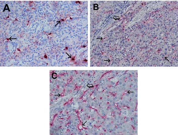
Morphometrical assay
Light microscopy integrated with an image
analysis system (Quantimet-500 Leica, Wetzlar,
Germany) was utilized [Ranieri et al. 2007]. In
tumour sections, immunostained areas (hot
spots) were selected at ×100 magnification and
TMCD (Figure 1A), PAR-2-MVD (Figure 1B)
and C-MVD (Figure 1C) were assessed at ×400
magnification (0.19 mm
2area).
Statistical analysis
Mean values ± 1 standard deviation (SD) of all
the tissue-evaluated parameters are reported in
Table 2. Correlations between TMCD,
PAR-2-MVD, and C-MVD were calculated using
Pearson’s (r) analysis. Correlations among the all
analyzed parameters and the main
clinicopatho-logical features listed in Table 1 were performed
by Chi-square test (χ
2). A p
< 0.05 was
consid-ered significant. All statistical analyses were
per-formed with the SPSS statistical software package
(SPSS, Inc., Chicago, IL).
Results
Immunohistochemical staining by using the
anti-bodies antitryptase, anti-PAR-2 and anti-CD34,
demonstrates that tryptase-positive MCs are well
recognizable as red-stained ovoid cells with thin
prolongations and generally, they are located in
perivascular position (Figure 1A). A close
topo-graphic association between TMCD and
PAR-2-MVD and between TMCD and C-PAR-2-MVD was
often observed. The mean value ± SD of TMCD,
PAR-2-MVD and C-MVD was 10.87 ± 4.21,
24.32 ± 8.67 and 27.22 ± 9.12 respectively, and
these results are summarized in Table 2.
There was a significant correlation between
TMCD and C-MVD (r = 0.71, p = 0.03), between
PAR-2-MVD and C-MVD (r = 0.76, p = 0.02),
and between TMCD and PAR-2-MVD (r = 0.64
p
= 0.02) (Figure 2). There was no correlation
between TMCD, PAR-2-MVD and C-MVD and
the main clinicopathological features found.
Discussion
Currently, a lot of data supported the central role
of angiogenesis in GC development and
progres-sion but few data regarding the role of MCs in
GC angiogenesis have been published [Geng
et al. 2014; Wang et al. 2013a; Zhao et al. 2012].
In particular, in a study performed by Mukherjee
and colleagues, the authors studied MC density
in tissue from patients with gastric ulcers and in
tissue from GC patients [Mukherjee et al. 2009].
In the above study, the histochemical method of
toluidine blue was employed to identify and count
MC density. Data from this study indicated that
MC density in benign gastric ulcers and in
can-cers was much higher than control and correlated
with angiogenesis.
Ribatti and colleagues studied tumour samples
from GC patients by mean of
immunohistochem-istry employing antitryptase and antichymase
antibodies to stain MCs. In this study, a
correla-tion between MVD and tryptase and
chymase-positive MCs with histopathological type was
found [Ribatti et al. 2010].
Interestingly, MCs have been shown as important
players in tumour angiogenesis because of the
release of proangiogenic factors stored in their
secretory granules [Bhattacharyya et al. 1998;
Marech et al. 2016a].
In the tumour microenvironment, MCs can be
activated in different ways such as: c-Kit receptor
activation and phosphorylation by stem cell factor,
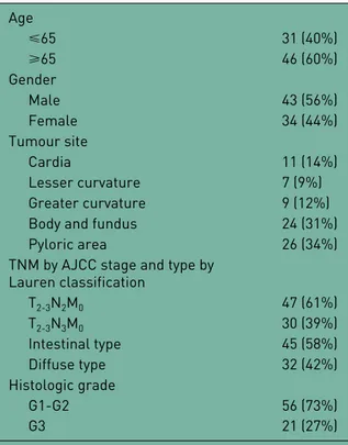
Table 1. Clinicopathological features of patients (n = 77). Age ⩽65 31 (40%) ⩾65 46 (60%) Gender Male 43 (56%) Female 34 (44%) Tumour site Cardia 11 (14%) Lesser curvature 7 (9%) Greater curvature 9 (12%)
Body and fundus 24 (31%)
Pyloric area 26 (34%)
TNM by AJCC stage and type by Lauren classification T2-3N2M0 47 (61%) T2-3N3M0 30 (39%) Intestinal type 45 (58%) Diffuse type 32 (42%) Histologic grade G1-G2 56 (73%) G3 21 (27%)
TNM, classification of cancer staging; AJCC, American Joint Committee on Cancer.
IgE mechanism mediated by T-lymphocytes–MC
interaction and other microenvironmental stimuli
[Ammendola et al. 2016a; Patruno et al. 2014].
After intensive or piecemeal activation,
degranula-tion of secretory granules occurs, depending on
the MC-activation mechanism, and MC-derived
Figure 2. Correlation between tryptase mast cells’ density (TMCD) and classical microvascular density (C-MVD) (r = 0.71, p = 0.03), protease-activated receptor-2 microvascular density (PAR-2-MVD) and C-MVD (r = 0.76, p = 0.02), TMCD and PAR-2-MVD (r = 0.64, p = 0.02).
TMCD, tryptase mast cell density; PAR-2-MVD, protease-activated receptor-2 microvascular density; C-MVD, classical microvascular density.
Figure 1. (A) Primary gastric cancer (GC) tissue section. Many scattered red immunostained mast cells positive to the antitryptase antibody. Each arrow indicates single mast cells, ×400 magnification (0.19 mm2). (B) Primary GC tissue section. Each small arrow indicates single red immunostained microvessel positive to the antiprotease-activated receptor-2 antibody. Big arrows indicate stromal collagen and lymphocytes infiltrate, ×400 magnification (0.19 mm2). (C) Primary GC tissue section. Each small arrow indicate single red immunostained microvessel positive to the anti-CD34 antibody. Big arrow indicates stromal collagen with scattered lymphocytes, ×400 magnification (0.19 mm2).
proangiogenic factors are released in the tumour
microenvironment, stimulating angiogenesis
[Ribatti and Ranieri, 2015; Wasiuk et al. 2009].
Among them, tryptase has been characterized as a
powerful nonclassical angiogenic factor [Marech
et al. 2016b; Norrby, 2002; Visciano et al. 2015;
Marone et al. 2015].
Preclinical data showed that tryptase is an agonist
of PAR-2 in vascular ECs that stimulates their
proliferation. Signaling via PAR-2 on ECs elicits
activation of the major members of the MAPK
phosphorylation family and contributes to
prolif-eration of ECs and angiogenesis. Experimental
data also suggest that PAR-2 activation leads to
the production of other proangiogenic factors,
such as VEGF, IL-8, IL-6, GM-CSF and M-CSF
[Malfettone et al. 2013; Soreide et al. 2006;
Darmoul et al. 2001; Uusitalo-Jarvinen et al.
2007; Liu and Mueller, 2006; Yano et al. 1999;
Sedda et al. 2014; Ribatti et al. 2010; Wang et al.
2013b; Zhang et al. 2012; Washington, 2010;
Tamura et al. 2011; Verlato et al. 2014; Mrena
et al. 2015; Loffredo et al. 2014; Zhang et al.
2013; Rasmussen et al. 2012; Chang et al. 2013;
Ammendola et al. 2016b]. With special regard to
GC, it is important to underscore that the role of
TMCD in angiogenesis has been little
investi-gated and no data have been published regarding
MVD in terms of PAR-2 endothelial expressing
cells. Our results demonstrated an association
between TMCD, PAR-2-MVD and C-MVD,
supporting the central role of tryptase as a main
proangiogenic factor in primary GC tissue. On
the other hand, these first data need to be
inter-preted with caution, due to following limitations:
the medium size of analyzed samples series; the
lack of a previous standardized method to
evalu-ate PAR-2-MVD and the possible inter-observer
variability in the evaluation of PAR-2-MVD.
Although our results are preliminary and they
need to be confirmed to further awaited studies, it
is intriguing to speculate that the inhibition of
MC degranulation by means of c-Kit receptor
tyrosine kinase inhibitors (e.g. masitinib) or the
inhibition of tryptase by means of gabexate
mesi-late or nafamostat mesimesi-late, which could be novel
antiangiogenic strategies worthy of clinical
inves-tigation [Erba et al. 2001; Mori et al. 2003;
Humbert et al. 2010; Marech et al. 2014c;
Deplanque et al. 2015; Ammendola et al. 2016c].
Author contributions
Ammendola M and Ranieri G conceived the
research. Vescio G, Kocak IF and Ozgurtas T
performed the critical review of the literature.
Ammendola M Sammarco G Sacco R performed
surgical procedures. Zuccalà V, Patruno R, C
Gadaleta, Zizzo N contributed to
immunohisto-chemistry and tissue’s study. Marech I, Ruggieri
R, Luposella M and Gadaleta CD elaborated
data analysis. All authors wrote the manuscript.
Ranieri G reviewed the manuscript.
Funding
This research received no specific grant from any
funding agency in the public, commercial, or
not-for-profit sectors.
Conflict of interest statement
The authors declare that there is no conflict of
interest.
References
Ammendola, M., Leporini, C., Marech, I., Gadaleta, C., Scognamillo, G., Sacco, R. et al. (2014) Targeting mast cells tryptase in tumor microenvironment: a potential antiangiogenetic strategy. Biomed Int Res 2014: 154702.
Ammendola, M., Marech, I., Sammarco, G., Zuccalà, V., Luposella, M., Zizzo, N. et al. (2015) Infiltrating mast cells correlate with angiogenesis in bone metastases from gastric cancer patients. Int J Mol Sci 16: 3237–3250.
Ammendola, M., Patruno, R., Sacco, R., Marech, I., Sammarco, G., Zuccalà V. et al. (2016a) Mast Table 2. Tryptase mast cell density, protease-activated receptor-2 microvascular density and classical microvascular density means ± standard deviation as a function of gastric cancer tumour tissue, respectively.
Tissue TMCD
400× (0.19 mm2) PAR-2-MVD400× (0.19 mm2) C-MVD400× (0.19 mm2)
Primary tumour 10.87 ± 4.21 24.32 ± 8.67 27.22 ± 9.12
TMCD, tryptase mast cell density; PAR-2-MVD, protease-activated receptor-2 microvascular density; C-MVD, classical microvascular density.
cells positive to tryptase and tumour-associated macrophages correlate with angiogenesis in locally advanced colorectal cancer patients undergone to surgery. Expert Opin Ther Targets 20: 533–540.
Ammendola, M., Sacco, R., Donato, G., Zuccalà, V., Russo, E., Luposella, M. et al. (2013a) Mast cell positivity to tryptase correlates with metastatic lymph nodes in gastrointestinal cancer patients treated surgically. Oncology 85: 111–116.
Ammendola, M., Sacco, R., Sammarco, G., Donato, G., Montemurro, S., Ruggieri, E. et al. (2014) Correlation between serum tryptase, mast cells positive to tryptase and microvascular density in colo-rectal cancer patients: possible biological-clinical significance. PLoS One 9: e99512.
Ammendola, M., Sacco, R., Sammarco, G., Donato, G., Zuccalà, V., Luposella, M. et al. (2014) Mast cells density positive to tryptase correlates with angiogenesis in pancreatic ductal adenocarcinoma patients having undergone surgery. Gastroenterol Res
Pract 2014: 951957.
Ammendola, M., Sacco, R., Sammarco, G., Donato, G., Zuccalà, V., Romano, R. et al. (2013b) Mast cells positive to tryptase and c-Kit receptor expressing cells correlates with angiogenesis in gastric cancer patients surgically treated. Gastroenterol Res Pract 2013: 703163.
Ammendola, M., Sacco, R., Sammarco, G.,
Luposella, M., Patruno, R., Gadaleta C. et al. (2016b) Mast cell-targeted strategies in cancer therapy.
Transfus Med Hemother 43: 109–113.
Ammendola, M., Sacco, R., Sammarco, G., Piardi, T., Zuccalà, V., Patruno, R. et al. (2016c) Mast cells positive to tryptase, endothelial cells positive to protease-activated receptor-2, and microvascular density correlate among themselves in hepatocellular carcinoma patients who have undergone surgery. Onco
Targets Ther 9: 4465–4471.
Ammendola, M., Zuccalà, V., Patruno, R., Russo, E., Luposella, M., Amorosi, A. et al. (2013c) Tryptase-positive mast cells and angiogenesis in keloids: a new possible post-surgical target for prevention. Updates
Surg 65: 53–57.
Bhattacharyya, S., Drucker, I., Reshef, T.,
Kirshenbaum, A., Metcalfe, D. and Mekori, Y. (1998) Activated T lymphocytes induce degranulation and cytokine production by human mast cells following cell-to-cell contact. J Leukoc Biol 63: 337–341. Blair, R., Meng, H., Marchese, M., Ren, S., Schwartz, L., Tonnesen, M. et al. (1997) Human mast cells stimulate vascular tube formation. Tryptase is a novel, potent angiogenic factor. J Clin Invest 99: 2691–2700.
Chang, L., Pan, S., Lai, C., Tsai, A. and Teng, C. (2013) Activated PAR-2 regulates pancreatic cancer progression through ILK/HIF-α-induced TGF-α expression and MEK/VEGF-A-mediated angiogenesis. Am J Pathol 183: 566–575.
Darmoul, D., Marie, J., Devaud, H., Gratio, V. and Laburthe, M. (2001) Initiation of human colon cancer cell proliferation by trypsin acting at protease-activated receptor-2. Br J Cancer 85: 772–779. Deplanque, G., Demarchi, M., Hebbar, M., Flynn, P., Melichar, B., Atkins, J. et al. (2015) A randomized, placebo-controlled phase III trial of masitinib plus gemcitabine in the treatment of advanced pancreatic cancer. Ann Oncol 26: 1194–1200.
Donato, G., Conforti, F., Camastra, C., Ammendola, M., Donato, A., Renzulli, A. et al. (2014) The role of mast cell tryptases in cardiac myxoma: histogenesis and development of a challenging tumor. Oncol Lett 8: 379–383.
Erba, F., Fiorucci, L., Pascarella, S., Menegatti, E., Ascenzi, P. and Ascoli, F. (2001) Selective inhibition of human mast cell tryptase by gabexate mesylate, an antiproteinase drug. Biochem Pharmacol 61: 271–276. Fajardo, I. and Pejler, G. (2003) Human mast cell beta-tryptase is a gelatinase. J Immunol 171: 1493– 1499.
Geng, Y., Chen, X., Qiu, J., Zhou, Y., Wang, J. and Liu, L. (2014) Human epidermal growth factor receptor-2 expression in primary and metastatic gastric cancer. Int J Clin Oncol 19: 303–311. Gulubova, M. and Vlaykova, T. (2009) Prognostic significance of mast cell number and microvascular density for the survival of patients with primary colorectal cancer. J Gastroenterol Hepatol 24: 265–275. Humbert, M., Castéran, N., Letard, S., Hanssens, K., Iovanna, J., Finetti, P. et al. (2010) Masitinib combined with standard gemcitabine chemotherapy:
in vitro and in vivo studies in human pancreatic
tumour cell lines and ectopic mouse model. PLoS One 5: e9430.
Itoh, Y., Sendo, T. and Oishi, R. (2005) Physiology and pathophysiology of proteinase-activated receptors (PARs): role of tryptase/PAR-2 in vascular endothelial barrier function. J Pharmacol Sci 97: 14–19.
Liu, Y. and Mueller, B. (2006) Protease-activated receptor-2 regulates vascular endothelial growth factor expression in MDA-MB-231 cells via MAPK pathways. Biochem Biophys Res Commun 344: 1263–1270.
Loffredo, S., Staiano, R., Granata, F., Genovese, A. and Marone, G. (2014) Immune cells as a source and target of angiogenic and lymphangiogenic factors.
Malfettone, A., Silvestris, N., Saponaro, C., Ranieri, G., Russo, A., Caruso, S. et al. (2013) High density of tryptase-positive mast cells in human colorectal cancer: a poor prognostic factor related to protease-activated receptor 2 expression. J Cell Mol Med 17: 1025–1037.
Marech, I., Ammendola, M., Gadaleta, C., Zizzo, N., Oakley, C., Gadaleta, C. et al. (2014a) Possible biological and translational significance of mast cells density in colorectal cancer. World J Gastroenterol 20: 8910–8920.
Marech, I., Ammendola, M., Sacco, R., Capriuolo, G., Patruno, R., Rubini, R. et al. (2014b) Serum tryptase, mast cells positive to tryptase and
microvascular density evaluation in early breast cancer patients: possible translational significance. BMC
Cancer 14: 534.
Marech, I., Ammendola, M., Sacco, R., Sammarco, G., Zuccalà, V., Zizzo, N. et al. (2016a) Tumour-associated macrophages correlate with microvascular bed extension in colorectal cancer patients. J Cell Mol
Med 20: 1373–1380.
Marech, I., Leporini, C., Ammendola, M., Porcelli, M., Gadaleta, C., Russo, E. et al. (2016b) Classical and non-classical proangiogenic factors as a target of antiangiogenic therapy in tumor microenvironment.
Cancer Lett 28; 380: 216–226.
Marech, I., Patruno, R., Zizzo, N., Gadaleta, C., Introna, M., Zito, A. et al. (2014c) Masitinib (AB1010), from canine tumour model to human clinical development: where we are? Crit Rev Oncol
Hematol 91: 98–111.
Marone, G., Varricchi, G., Loffredo, S. and Granata, F. (2015) Mast cells and basophils in inflammatory and tumor angiogenesis and lymphangiogenesis. Eur J
Pharmacol 778: 146–151.
Matej, R., Mandàkovà, P., Netikovà, I., Poucková, P. and Olejár, T. (2007) Proteinase-activated receptor-2 expression in breast cancer and the role of trypsin on growth and metabolism of breast cancer cell line MDA MB-231. Physiol Res 56: 475–484.
Mori, S., Itoh, Y., Shinohata, R., Sendo, T., Oishi, R. and Nishibori, M. (2003) Nafamostat mesilate is an extremely potent inhibitor of human tryptase. J
Pharmacol Sci 92: 420–423.
Morris, D., Ding, Y., Ricks, T., Gullapalli, A., Wolfe, B. and Trejo, J. (2006) Protease-activated receptor-2 is essential for factor VIIa and Xa-induced signaling, migration, and invasion of breast cancer cells. Cancer
Res 66: 307–314.
Mrena, J., Mattila, A., Böhm, J., Jantunen, I. and Kellokumpu, I. (2015) Surgical care quality and
oncologic outcome after D2 gastrectomy for gastric cancer. World J Gastroenterol 21: 13294–13301. Mukherjee, S., Bandyopadhyay, G., Dutta, C., Bhattacharya, A., Karmakar, R., Barui, G. et al. (2009) Evaluation of endoscopic biopsy in gastric lesions with a special reference to the significance of mast cell density. Indian J Pathol Microbiol 52: 20–24.
Norrby, K. (2002) Mast cells and angiogenesis.
APMIS 110: 355–371.
Patruno, R., Marech, I., Zizzo, N., Ammendola, M., Nardulli, P., Gadaleta, C. et al. (2014) C-Kit expression, angiogenesis, and grading in canine mast cell tumour: a unique model to study c-Kit driven human malignancies. Biomed Res Int 2014: 730246. Ranieri, G., Ammendola, M., Patruno, R., Celano, G., Zito, F., Montemurro, S. et al. (2009) Tryptase-positive mast cells correlate with angiogenesis in early breast cancer patients. Int J Oncol 35: 115–120.
Ranieri, G., Grammatica, L., Patruno, R., Zito, A., Valerio, P., Iacobellis, S. et al. (2007) A possible role of thymidine phosphorylase expression and 5-fluorouracil increased sensitivity in oropharyngeal cancer patients. J Cell Mol Med 11: 362–368. Rasmussen, J., Riis, S., Frobert, O., Yang, S., Kastrup, J., Zachar, V. et al. (2012) Activation of protease-activated receptor 2 induces VEGF independently of HIF-1. PLoS One 7: e46087. Ribatti, D., Guidolin, D., Marzullo, A., Nico, B., Annese, T., Benagiano, V. et al. (2010) Mast cells and angiogenesis in gastric carcinoma. Int J Exp Pathol 91: 350–356.
Ribatti, D. and Ranieri, G. (2015) Tryptase, a novel angiogenic factor stored in mast cell granules. Exp Cell
Res 332: 157–162.
Ribatti, D., Ranieri, G., Nico, B., Benagiano, V. and Crivellato, E. (2011) Tryptase and chymase are angiogenic in vivo in the chorioallantoic membrane assay. Int J Dev Biol 55: 99–102.
Rickard, A., Portell, C., Kell, P., Vinson, S. and McHowat, J. (2005) Protease-activated receptor stimulation activates a Ca2+-independent phospholipase A2 in bladder microvascular endothelial cells. Am J Physiol Renal Physiol 288: F714–F721.
Sedda, S., Marafini, I., Caruso, R., Pallone, F. and Monteleone, G. (2014) Proteinase activated-receptors-associated signaling in the control of gastric cancer. World J Gastroenterol 20: 11977–11984. Soreide, K., Janssen, E., Körner, H. and Baak, J. (2006) Trypsin in colorectal cancer: molecular
biological mechanisms of proliferation, invasion, and metastasis. J Pathol 209: 147–156.
Stack, M. and Johnson, D. (1994) Human mast cell tryptase activates single-chain urinary-type plasminogen activator (pro-urokinase). J Biol Chem 269: 9416–9419.
Tamura, S., Takeno, A. and Miki, H. (2011) Lymph node dissection in curative gastrectomy for advanced gastric cancer. Int J Surg Oncol 2011: 748745. Uusitalo-Jarvinen, H., Kurokawa, T., Mueller, B., Andrade-Gordon, P., Friedlander, M. and Ruf, W. (2007) Role of protease activated receptor 1 and 2 signaling in hypoxia-induced angiogenesis. Arterioscler
Thromb Vasc Biol 27: 1456–1462.
Verlato, G., Giacopuzzi, S., Bencivenga, M., Morgagni, P. and De Manzoni, G. (2014) Problems faced by evidence-based medicine in evaluating lymphadenectomy for gastric cancer. World J
Gastroenterol 20: 12883–12891.
Visciano, C., Prevete, N., Liotti, F. and Marone, G. (2015) Tumor-associated mast cells in thyroid cancer.
Int J Endocrinol 2015: 705169.
Wang, G., Wang, Y., Li, D. and Deng, B. (2013a) Expression of protease-activated receptor-2 in human gastric stromal tumor and its clinicopathological significance. Hepatogastroenterology 60:
2125–2128.
Wang, X., Chen, X., Fang, J. and Yang, C. (2013b) Overexpression of both VEGF-A and VEGF-C in gastric cancer correlates with prognosis, and silencing
of both is effective to inhibit cancer growth. Int J Clin
Exp Pathol 6: 586–597.
Washington, K. (2010) 7th edition of the AJCC cancer staging manual: stomach. Ann Surg Oncol 17: 3077–3079.
Wasiuk, A., de Vries, V., Hartmann, K., Roers, A. and Noelle, R. (2009) Mast cells as regulators of adaptive immunity to tumours. Clin Exp Immunol 155: 140–146.
Yano, H., Kinuta, M., Tateishi, H., Nakano, Y., Matsui, S., Monden, T. et al. (1999) Mast cell infiltration around gastric cancer cells correlates with tumour angiogenesis and metastasis. Gastric Cancer 2: 26–32.
Zhang, C., Gao, G., Lv, C., Zhang, B., Zhang, Z. and Zhang, X. (2012) Protease-activated receptor-2 induces expression of vascular endothelial growth factor and cyclooxygenase-2 via the mitogen-activated protein kinase pathway in gastric cancer cells. Oncol
Rep 28: 1917–1923.
Zhang, X., Wang, W., Mize, G., Takayama, T., True, L. and Vessella, R. (2013) Protease-activated receptor 2 signaling up regulates angiogenic growth factors in renal cell carcinoma. Exp Mol Pathol 94: 91–97.
Zhao, Y., Wu, K., Cai, K., Zhai, R., Tao, K., Wang, G. et al. (2012) Increased numbers of gastric-infiltrating mast cells and regulatory T cells are associated with tumor stage in gastric adenocarcinoma patients. Oncol Lett 4: 755–758.
Visit SAGE journals online journals.sagepub.com/ home/tag