Clin Case Rep. 2019;7:2215–2219. wileyonlinelibrary.com/journal/ccr3
|
22151
|
INTRODUCTION
Injectable hyaluronic acid derivatives are the most used re‐ absorbable dermal fillers for soft tissue augmentation and volume expansion as they provide a nonsurgical alternative procedure for the temporary correction of age‐related skin defects (facial rejuvenation), postsurgical, and post‐traumatic skin facial alterations, increasing of the lip volume. Their utilization is considered overall safe and well tolerable be-cause of biocompatibility and biodegradability of hyaluronic acid, with minimal adverse events secondary to the intrader-mic injection. Nevertheless, late or early adverse reactions may occur although with an incidence ranging from 0.02% to 0.4%. We report on a female patient showing a delayed onset (10 years later) of a sclerosing granulomatous reaction due to the intradermal filler (poly‐hydroxyethyl‐methacrylate suspended in hyaluronic acid) injection for lip augmentation, with histochemical and confocal laser scanning microscopi-cal analysis of the lesion.
On the basis of manufacturers' and some authors' claims, all commonly used injection materials for esthetic correction and different formulations of hyaluronic acid (HA), with or without adjunctive substances, result in no immunogenic re-actions or other complications1-3; nevertheless, unexpected,
late, or early adverse reactions have been reported.4-6 Overall,
poly‐hydroxyethyl‐methacrylate (HEMA) suspended in HA can promote the formation of late foreign body granulomas (FBGs).7,8 Authors report on the histologic (conventional and
confocal laser scanning microscopy) features of a case oc-curred 10 years after the injection of HA + HEMA in the lower lip of a female patient. HA is a constituent of several normal tissues and, as such, does not lead to adverse reac-tions; therefore, when FBGs develop, one should argue that additional components were bound to HA.9,10 HEMA has
been used as a stabilizer of HA‐based fillers but it is known to induce transient macrophagic reaction, fibroblast prolif-eration with scarce collagen deposition, and multinucleated giant cells.8,11 The morphological features of the present case C A S E R E P O R T
Delayed sclerosing granulomatous reaction to dermal filler
injection of poly‐hydroxyethyl‐methacrylate suspended in
hyaluronic acid: Histochemical and confocal laser scanning
microscopical analysis
Saverio Capodiferro
1|
Pasquale Sportelli
1|
Luisa Limongelli
1|
Fabio Dell’Olio
1|
Angela Tempesta
1|
Gianfranco Favia
1|
Eugenio Maiorano
2This is an open access article under the terms of the Creative Commons Attribution‐NonCommercial‐NoDerivs License, which permits use and distribution in any medium, provided the original work is properly cited, the use is non‐commercial and no modifications or adaptations are made.
© 2019 The Authors. Clinical Case Reports published by John Wiley & Sons Ltd. 1Department of Interdisciplinary
Medicine, University of Bari Aldo Moro, Bari, Italy
2Department of Emergency and Organ
Transplantation, University of Bari Aldo Moro, Bari, Italy
Correspondence
Saverio Capodiferro, Department of Interdisciplinary Medicine, University of Bari, Piazza G. Cesare, 11, 70124 Bari, Italy.
Email: [email protected]
Abstract
Re‐absorbable dermal fillers of poly‐hydroxyethyl‐methacrylate suspended in hyalu-ronic acid are considered overall safe and well tolerable because of biocompatibility; nevertheless, rarely, late, or early adverse reactions may occur.
K E Y W O R D S
are consistent with previous injection of HA + HEMA, and the prolonged time interval from injection to clinical manifes-tations indicates the adverse reaction was slowly progressive. Also, it was postulated that macrophages would incorporate foreign particles, thus keeping the foreign particles in a latent stage. Subsequently, additional priming events (eg, superven-ing infections) would be needed to re‐activate macrophages, lead to multinucleated giant cell accumulation, and, finally, to fully developed granulomatous reaction.6,8,10,12-14 Such
pathogenetic mechanism may explain the prolonged course of the disease, with only late development of clinically de-tectable nodular lesions.
2
|
CASE REPORT
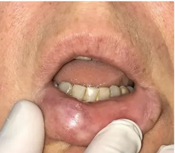
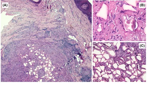
The patient, a 50‐year‐old woman, had undergone filler in-jection for lip augmentation 10 years earlier. She presented a slowly growing nodular lesion (Figure 1) of the right lower hemi‐lip of hard consistency, not ulcerated, painless, and without signs of infection. With the provisional clinical di-agnosis of benign nodular lesion, surgical excision was per-formed followed by histologic examination. The nodular lesion (Figure 2A) was mainly composed of many clear po-lygonal spaces, surrounded by fibrous collagen, small lym-phocytes, macrophages with occasional cytoplasmic clearing, and sparse multinucleated giant cells (Figure 2B), pointing at long‐standing FBG. The polygonal spaces were 20‐120 µm in size and partly filled with translucent particles, with a broken‐glass appearance (Figure 2C). Histochemical stains (Alcian, Giemsa, May‐Grunwald‐Giemsa, Grocott) and con-focal laser scanning microscopical analysis were performed
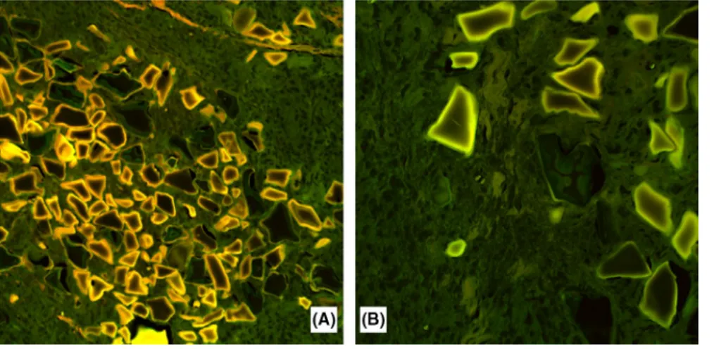
in order to better characterize the morphological features of the foreign material. With Alcian Blue stain (Figure 3A), the material filling the polygonal spaces was stained in light blue and appeared irregularly condensed toward the center, leaving an empty‐looking space at the periphery. Giemsa and May–Grunwald–Giemsa (Figure 3B,C) stains highlighted a homogeneous, centrally placed component, which was deeply stained in blue, which appeared encircled, in most in-stances, by unstained or light brown, granular to crystalline material. The Grocott stain only showed very light purplish discoloration of the acellular material, centrally placed in the lacunae (Figure 3D). By confocal laser scanning microscopy, we could detect (Figure 4A) more intense autofluorescence of the peripheral content of the lacunae, which progressively vanished toward the center, with a sort of onion‐skin pattern. The most intensely autofluorescent material more or less cor-responded to crystalline‐like material demonstrated by the Giemsa stain (Figure 4B).
3
|
DISCUSSION
Hyaluronic acid (HA) fillers in cosmetic medicine are widely used and considered relatively safe, though not all the fill-ers used in European countries and throughout the world have been approved by the Regulatory Agencies.2,5,6,15,16
Nevertheless, a wide range of medical compounds has been authorized as fillers for HA injections.8,9,16,17 Despite the
ob-vious cosmetic benefits allowed by such fillers, a wide range of possible complications, such as immediate, late, delayed, temporary, or irreversible inflammatory reactions, has been reported in the literature.1,5,11
Filler adverse effects can be classified according to the technical procedure, onset (early or late), filler type, and host factors.6,11 Technical errors may be due to too much or too
lit-tle volume injection, incorrect depth or wrong localization of filler placement, and inappropriate product choice, the latter possibly being the most relevant topic. In fact, fillers are usu-ally divided into reversible and irreversible; in the first group are included early or temporary fillers (collagens and hyal-uronic acid), late or long‐term (HA with dextranomer beads, poly‐L‐lactic acid [PLL] and calcium hydroxylapatite), and delayed or permanent fillers (paraffin, silicon preparations, polymethyl methacrylate microspheres, hydroxyethyl‐meth-acrylate fragments, polyalkylimide gel, polyacrylamide hy-drogel, polyvinyl hydroxide microspheres in polyacrylamide gel).10,13,15 Although it is generally accepted that temporary
fillers are better tolerated than permanent ones, the frequency of short‐term adverse reactions is similar because filler into tissues is perceived as a foreign substance, with an initial challenge to the host's immune system and with possible early complications (erythema, edema, and allergy) in less than 2 weeks.10,15
FIGURE 1 Slowly growing nodular lesion of the right lower hemi‐lip of hard consistency, not ulcerated, painless, showing no signs of previous infection
It is generally accepted that HA (crosslinked or not) in-duced erythema and swelling few days after injection, as resulting from the little foreign body reaction observable on the histologic examination; the blue‐stained hyaluronic acid is slowly degraded by macrophages and invading cap-illaries at the circumference of the HA implant in the fol-lowing 30‐60 days; this process goes along with a slower resorption for the cross‐linked HA fillers, but generally with a gradual and scarce inflammatory infiltrate, absence or lit-tle deposition of structures resembling new collagen fibers at 3‐4 months, while clusters of macrophages and giant cells are detectable also after 4‐6 months. Overall, no residue of HA is usually identifiable at 6‐9 months.8-11
Bumps and lumps following superficial filler placement usually are visible immediately after the injection or shortly
thereafter, while necrosis due to intra‐arterial injection be-comes evident within a day.13
Late complications, instead, may develop over weeks, months, or even years after the injection and may include diffuse chronic inflammation, nodular outgrowth (usually granulomas), late allergic reactions, hypertrophic scars, and telangiectasia.4,5,12 In such instances, patients usually are
unable to properly recall which kind of filler was injected. HA preparations with a longevity of approximately 6 months (with highly variable molecular weight and cross‐linking properties) are the most commonly used fillers and consid-ered the safest in view of HA being universally present in all animal species, its non‐species specificity, free from foreign proteins, with very low propensity to adverse effects in good filler preparations by well‐reputed manufacturers.
FIGURE 2 A‐C, Histologic examination showing several clear polygonal spaces surrounded by fibrous collagen, small lymphocytes, macrophages with occasional cytoplasmic clearing, and sparse multinucleated giant cells (A, H&E, original magnification ×2) (B, H&E, original magnification ×10); the polygonal spaces were 20‐120 µm in size and partly filled with translucent particles, with a broken‐glass appearance (C, H&E, original magnification ×20)
FIGURE 3 A‐D, Alcian Blue stain (A, original magnification ×20) showing a light blue coloration of the filling material, irregularly condensed toward the center, leaving an empty‐looking space at the periphery. Giemsa (B, original magnification ×20) and May–Grunwald– Giemsa (C, original magnification ×20) stains highlighting a homogeneous, centrally placed component deeply stained in blue, appearing encircled in most instances, by unstained or light brown, granular to crystalline material. Grocott stain (D) showing a very light purplish discoloration of the acellular material centrally in the lacunae
Indeed, many complications can occur, like in the cur-rent case, when using long‐term filler reinforced with hy-droxyethyl‐methacrylate fragments, and they usually are represented by late granuloma formations.10,11 The results
of previous histologic studies on tissues examined over the first few months after injection of these reinforced fillers appear promising, having shown the hydrophilic polygonal, translucent, not birefringent particles were surrounded by normal tissues, in the absence of inflammatory reactions, thus appearing fully biocompatible products.1,10,13,18,19 The
HA carriers were included in empty lacunae within the first days after the injection.8,10 In cases in which granulomatous
reactions developed, inflammatory lymphocytic infiltrates, foreign body giant cells, sometimes containing asteroid bod-ies, and increased fibrillary collagen accumulation usually appeared suddenly.8,10,17-19
Many possible causes may lead to early and late gran-uloma formation, such as the volume of injected dermal fillers, impurities within the filler substance with bio-film formation, local infection unrelated to the filler site, and severe systemic infections with subsequent immuno-logic alterations.6,8,12 Surely, the presence of impurities
may facilitate late granuloma formation, especially when including particles <20 µm in size, as phagocytosis and immunologic memory are more efficiently stimulated by smaller particles than by larger ones.9,10,13,14 Furthermore,
irregularities of the particle surface, with pointed edges and corners, seem to further stimulate late granuloma formation.7,8,10,11,17,18
In conclusion, clinicians should be aware that adverse re-actions to fillers should be expected, especially when stabi-lizers or carriers are suspended in HA; therefore, prolonged follow‐up of the patients should be carried out, to possibly detect such events at early stages.
CONFLICT OF INTEREST
Authors declare no conflict of interest.
AUTHORS CONTRIBUTION
SC: involved in surgical procedure and confocal laser scan-ning examination. PS: involved in surgical procedure. LL: prepared the manuscript. FD: prepared the manuscript and reviewed the literature. AT: corrected the manuscript. GF: involved in surgical procedure and review of the manuscript. EM: involved in histologic examination and review of the manuscript.
ORCID
Saverio Capodiferro https://orcid.
org/0000-0002-9819-6229 REFERENCES
1. Woodward J, Khan T, Martin J. Facial filler complications. Facial
Plast Surg Clin North Am. 2015;23(4):447‐458.
2. Tamiolakis P, Piperi E, Christopoulos P, Sklavounou‐ Andrikopoulou A. Oral foreign body granuloma to soft tissue fill-ers. Report of two cases and review of the literature. J Clin Exp
Dent. 2018;10(2):e177‐e184.
3. Abtahi‐Naeini B, Faghihi G, Shahmoradi Z, Saffaei A. Filler mi-gration and extensive lesions after lip augmentation: Adverse effects of polydimethylsiloxane filler. J Cosmet Dermatol. 2018;17(6):996‐999.
4. Mundada P, Kohler R, Boudabbous S, Toutous Trellu L, Platon A, Becker M. Injectable facial fillers: imaging features, complica-tions, and diagnostic pitfalls at MRI and PET CT. Insights Imaging. 2017;8(6):557‐572.
5. Alcântara C, Noronha MS, Cunha JF, Flores IL, Mesquita RA. Granulomatous reaction to hyaluronic acid filler material in oral and perioral region: A case report and review of literature. J
Cosmet Dermatol. 2018;17(4):578‐583.
6. Wagner RD, Fakhro A, Cox JA, Izaddoost SA. Etiology, preven-tion, and management of infectious complications of dermal fillers.
Semin Plast Surg. 2016;30(2):83‐86.
7. Wolkow N, Jakobiec FA, Yoon MK. Dermalive facial filler granu-lomas masquerading as neurofibromas. Ophthalmic Plast Reconstr
Surg. 2018;34:e99‐e103.
FIGURE 4 A, B, Confocal laser scanning microscopy showing (A, original magnification ×10) an intense autofluorescence of the peripheral material within the lacunae progressively vanished toward the center with an onion‐ skin appearance. The most intensely autofluorescent material corresponded to the crystalline‐like material demonstrated by the Giemsa stain (B, original magnification ×20)
8. Lemperle G, Gauthier‐Hazan N, Wolters M, Eisemann‐Klein M, Zimmermann U, Duffy DM. Foreign body granulomas after all in-jectable dermal fillers: part 1. Possible causes. Plast Reconstr Surg. 2009;123(6):1842‐1863.
9. Lemperle G, Morhenn V, Charrier U. Human histology and per-sistence of various injectable filler substances for soft tissue aug-mentation. Aesthetic Plast Surg. 2003;27(5):354–366.
10. Ledon JA, Savas JA, Yang S, Franca K, Camacho I, Nouri K. Inflammatory nodules following soft tissue filler use: a review of causative agents, pathology and treatment options. Am J Clin
Dermatol. 2013;14(5):401‐411.
11. Rongioletti F, Atzori L, Ferreli C, et al. Granulomatous reactions after injections of multiple aesthetic micro‐implants in tempo-ral combinations: a complication of filler addiction. J Eur Acad
Dermatol Venereol. 2015;29(6):1188‐1192.
12. Ibrahim O, Overman J, Arndt KA, Dover JS. Filler nodules: in-flammatory or infectious? A review of biofilms and their implica-tions on clinical practice. Dermatol Surg. 2018;44(1):53‐60. 13. Lee JM, Kim YJ. Foreign body granulomas after the use of dermal
fillers: pathophysiology, clinical appearance, histologic features, and treatment. Arch Plast Surg. 2015;42(2):232‐239.
14. Seok J, Jang YJ, Li K, Mun SK, Kim BJ. Streptococcus sanguinis isolated from filler granuloma: Successful treatment with incision and drainage. Dermatol Ther. 2016;29(6):463‐465.
15. Wang LL, Thomas WW, Friedman O. Granuloma formation sec-ondary to silicone injection for soft‐tissue augmentation in facial cosmetics: mechanisms and literature review. Ear Nose Throat J. 2018;97(1–2):E46‐E51.
16. Hu HC, Fang HW, Chiu YH. Delayed‐onset edematous foreign body granulomas 40 years after augmentation rhinoplasty by sil-icone implant combined with liquid silsil-icone injection. Aesthetic
Plast Surg. 2017;41(3):637‐640.
17. El‐Khalawany M, Fawzy S, Saied A, Al Said M, Amer A, Eassa B. Dermal filler complications: a clinicopathologic study with a spectrum of histologic reaction patterns. Ann Diagn Pathol. 2015;19(1):10‐15.
18. Eversole R, Tran K, Hansen D, Campbell J. Lip augmentation der-mal filler reactions, histopathologic features. Head Neck Pathol. 2013;7(3):241‐249.
19. Park KG, Dhong ES, Namgoong S, Han JK, Han SK, Kim WK. Atypical Facial filler granuloma: comparative histologic analysis with paraffinoma. Arch Craniofac Surg. 2016;17(3):169‐172.
How to cite this article: Capodiferro S, Sportelli P,
Limongelli L, et al. Delayed sclerosing granulomatous reaction to dermal filler injection of poly‐
hydroxyethyl‐methacrylate suspended in hyaluronic acid: Histochemical and confocal laser scanning microscopical analysis. Clin Case Rep. 2019;7:2215– 2219. https ://doi.org/10.1002/ccr3.2478