Summary. Although the use of probiotics in human and animal medicine is growing, their mode of action remains poorly understood. This study examined the effects of a multi-strain probiotic (SLAB51™) on the morphology and carbohydrate composition of mucins secreted by goblet cells of intestinal crypts in growing-finishing pigs. Sections of duodenum, caecum and colon from pigs fed for 12 weeks with an orally administered control basal diet (No-Pro) or one with a probiotic blend (Pro) were processed for microscopic analysis and stained with (1) haematoxylin-eosin for structural and morphometrical investigation; (2) conventional histochemistry (periodic acid-Schiff, Alcian Blue pH 2.5, high iron diamine staining) for neutral, acidic non-sulphated, and sulphated mucin analysis; and (3) FITC-labelled MAA-II and SNA lectins for α2,3- and α2,6-sialomucin identification. Compared with No-Pro samples, Pro samples displayed (1) increased goblet cell numbers in all investigated tract crypts; (2) an increase in acidic non-sulphomucins but a decrease in neutral, sulphated and α2,6-sialomucin-secreting goblet cells in the duodenum; (3) decreased crypt depth, an increase in α2,6-sialomucin secretory goblet cells, and a loss of goblet cell-secreting α2,3-sialomucins, which appeared on the apical surface of crypt fundus epithelial cells in
the caecum; and (4) an increase in α2,6-sialomucin-producing goblet cells in the colon. Results suggest that treatment with SLAB51™ induces region-specific changes in the morphology and carbohydrate composition of mucins secreted along intestinal tracts of growing-finishing pigs. These changes could ameliorate the health status of the animals, which displayed higher growth performance and meat quality than controls (Tufarelli et al., 2017).
Key words: Mucins, Intestine, Glycohistochemistry, Probiotic, Diet, Swine
Introduction
Mucins are high molecular weight glycoproteins that constitute the major component of the gel-like mucus layer covering the epithelium of the gastrointestinal tract. Their functions include protecting the epithelium against physical and chemical injuries caused by ingested food, microbes and microbial products; promoting the elimination of gut content (Kim and Ho, 2010); and lubricating and modulating water and electrolyte absorption (Forstner and Forstner, 1994).
The mucins are secreted by the goblet cells whose positive or negative regulation of mucin transcription depends on several bioactive factors, including microbes and their products (Kim and Ho, 2010). The intestinal commensal microbiota depends on mucus and undigested dietary carbohydrates for binding sites and
Effects of a probiotic on the morphology
and mucin composition of pig intestine
Salvatore Desantis1, Maria Mastrodonato2, Gianluca Accogli1, Giacomo Rossi3and Alberto Maria Crovace1 1Section of Veterinary Clinics and Animal Productions, Department of Emergency and Organ Transplantation (DETO), University of Bari “Aldo Moro”, Valenzano, (Ba), 2Department of Biology, University of Bari “Aldo Moro”, Bari and 3School of Biosciences and Veterinary Medicine, University of Camerino, Matelica, (MC), Italy
Offprint requests to: Salvatore Desantis, Section of Veterinary Clinics
and Animal Productions, Department of Emergency and Organ Transplantation (DETO), University of Bari Aldo Moro, S.P. Casamassima Km 3, 70010 Valenzano (Ba), Italy. e-mail: [email protected]
energy sources; moreover, it affects the functions of the intestinal epithelium, including those of goblet cells and mucus layers, by a “cross-talk” feedback mechanism (Lievin-Le and Servin, 2006; Martens et al., 2009). The microbiota and microbial products can also modulate mucin synthesis and secretion, either by direct activation of diverse signalling cascades or through bioactive factors generated by epithelial and lamina propria cells. Intestinal deregulation of mucin expression provides a microenvironment where bacteria initiate an inflammatory response (Johansson et al., 2008). The importance of the intestinal microbiota for gastrointestinal function and health has been shown in many studies (Heinritz et al., 2013; Büsing and Zeyner, 2015; Liu et al., 2017).
Probiotics are preparations of viable microorganisms that induce beneficial effects in both animals and humans by influencing gut flora or modifying immune status, as well as by stimulating digestive processes (Fuller, 2006; Meng et al., 2010). Thus, the use of probiotics is suggested as an alternative for antibiotics or anti-inflammatory drugs. The mode of action of probiotics is poorly understood, however, and the reported mechanisms of action are often the result of in
vitro experiments. Therefore, these results should be
confirmed by in vivo studies (Oelschlaeger, 2010). Several studies have investigated the effects of dietary administration of probiotics on intestinal physiology and immunology (Lallés et al., 2007), as well as on gut morphology and mucus composition in pig intestine (Baum et al., 2002; Bontempo et al., 2006; Di Giancamillo et al., 2007, 2008). The results of these investigations indicate that the effects on gut morphology and mucins secreted by goblet cells depend on probiotic composition.
Pigs are not only important farm animals, but, because of their similarities to humans in gastrointestinal tract functions, structure, metabolism, nutritional requirements, major bacteria phyla and genetics, they have a superior position over other non-primates, such as rodents, in their frequency of use as animal models (Heinritz et al., 2013). Thus, this species is considered a promising animal model in translational biomedical research, including studies of human nutrition and health problems (Heinritz et al., 2013; Prather et al., 2013; Gonzalez et al., 2015). Recently, it has been shown for the first time that a commercial multi-strain probiotic (SIVOY51™) intended for humans, apart from enhancing the growth performance and meat quality of growing-finishing pigs (Tufarelli et al., 2017), also affects the glycan composition of glycoproteins secreted by Brunner’s glands of the pig duodenum (Accogli et al., 2018). With such evidence in mind, the aim of this study was to examine the effect of the above-cited probiotic complex on the glycan composition of mucins produced in the intestinal goblet cells of growing-finishing pigs. This study could therefore establish a valuable baseline for further studies with possible translational value in humans.
Materials and methods
Probiotic sources
The probiotic blend SLAB51™ (Mendes SA, Lugano, Switzerland) contained 2×10¹¹ lactic acid bacteria composed of the following strains:
Streptococcus thermophilus DSM 32245; a mixture of
two strains of Bifidobacterium animalis spp., Lactis DSM 32246 and DSM 32247; Lactobacillus acidophilus DSM 32241; Lactobacillus helveticus DSM 32242;
Lactobacillus paracasei DSM 32243; Lactobacillus plantarum DSM 32244; and Lactobacillus brevis DSM
27961. Animals
The trial received ethical approval from the Italian Ministry of Health (n.597/2015-PR del 23/06/2015) and was conducted in strict accordance with the recommendations of the Guide for the Care and Use of Laboratory Animals of the National Institutes of Health (Art. 18 D.L. 4 March 2014, no. 26).
Twenty female pigs [(Landrace × Yorkshire) × Talent] with an average initial body weight (BW) of 22.80±0.95 kg (SE) were used in a 12-week experiment. Pigs were assigned to two dietary treatments: a control basal diet without the probiotic blend (No-Pro) and an experimental diet that included the probiotic blend (Pro). The probiotic mixture was used as a dietary supplement for the pigs during the entire feeding period at a dose of 100 mg/kg of BW. The basal diet was formulated to meet or exceed the nutrient requirements of pigs according to the NRC (1998). Pigs were housed in an environmentally controlled room with a concrete floor and were fed ad libitum.
Sampling and histology processing
At the end of the trial, pigs aged 112±3 days were slaughtered and the gastrointestinal tract was removed. Tissue samples were collected from the duodenum (5 cm aborally from the pylorus), caecum (middle of caecum) and proximal colon (20 cm aborally from the ileocaecal entrance) and fixed in 4% (v/v) phosphate-buffered-saline-buffered paraformaldehyde for 24 h at 4°C. The samples were then dehydrated through a graded series of ethanol and embedded in paraffin wax. Serial sections (4-μm thick) were cut and, after being de-waxed with xylene and hydrated in an ethanol series of descending concentrations, stained with haematoxylin-eosin for morphological and morphometric studies and by conventional histochemical procedures or lectin histochemistry for sialylated mucin characterization. Conventional histochemistry
Sections were treated with (1) the periodic acid-Schiff (PAS) reaction for neutral glycans (Mc Manus,
1948); (2) Alcian Blue pH 2.5 (AB 2.5) for sulphate esters and carboxyl groups in mucins (Pearse, 1968); (3) combined high iron diamine/Alcian Blue pH 2.5 (HID/AB 2.5) for simultaneous staining of sulphated and non-sulphated acidic glycans (Spicer, 1965); and (4) an AB 2.5/PAS sequence to reveal combinations of acidic and neutral mucins.
Lectin histochemistry
To differentiate the sialylated mucins, we stained tissue sections with FITC-labelled Maackia amurensis agglutinin (MAA)-II (specific for NeuNAcα2,3Galβ1,4 GlcNAc) (EY Laboratories, San Mateo, CA, USA) and
Sambucus nigra agglutinin (SNA) (specific for
Neu5Acα2,6Gal/GalNAc) (Vector Laboratories, Burlingame, CA, USA). Tissue sections were rinsed in 0.05 M Tris-HCl-buffered saline (TBS), pH 7.4, and incubated in 20 µg/mL of each lectin diluted in TBS for 1 h at 25°C in the dark. After three rinses in TBS, slides were mounted in Vectashield mounting medium (Vector Laboratories Inc., Burlingame, CA, USA). Each experiment was repeated twice for each sample. Controls for lectin staining included (1) substitution of the substrate medium with buffer without lectin and (2) incubation with each lectin in the presence of its hapten sugar (Neu5Ac 0.5 M in TBS). All control experiments gave negative results.
Slides were observed with the light photomicroscope Eclipse Ni-U (Nikon, Japan) at 20× magnification and
photographed with a digital camera (DS-U3, Nikon, Japan). The images were analysed by the image-analysing program NIS Elements BR (Version 4.20) (Nikon, Japan).
Morphometry and statistical analysis
Haemoxylin-eosin-stained sections were used to measure the depth of 15 well-oriented duodenum, caecum and colon crypts from each No-Pro and Pro pig. Microscopic fields were photographed with a 10x lens. Images were analysed with the image-analysing program NIS Elements BR (Version 4.30) (Nikon, JP). The total number of goblet cells, the number of goblet cells with different types of mucins as distinguished by AB 2.5/PAS staining, the HID/AB 2.5 reaction, and MAL II and SNA affinity were determined by counting both sides of 15 well-oriented crypts of the duodenum, caecum and colon tracts with a 10x lens.
The fluorescence signal intensity of FITC-labelled MAA-II and SNA was measured as described by Mastrodonato et al. (2017) and the fluorescence intensity (FI) for each goblet cell computed as described by McCloy et al. (2014). The integrated density (ID) and the area (A) of each cell were measured, after which10 regions in the field around the cells without fluorescence were selected as background and their mean density (BD) value was computed. FI was then calculated as FI = ID – (A× BD).
The cell count of all staining methods was manually
Fig. 1. Histological profile of the duodenum (A), caecum (B) and colon (C) of probiotic-fed pigs. Goblet cells were negatively stained. Bg, Brunner's gland; dc, duodenal crypt; dv, duodenal villum; s, submucosa; arrow, muscularis mucosae. Haematoxylin-eosin staining. Scale bars: 130 µm.
performed in a double-blind test by at least two of the authors. Values were expressed as cell percentage per crypt and as cell means ± standard deviation (SD) per crypt. The results were evaluated for statistical significance by Student’s t test.
Results
Morphometry
The examined organs from No-Pro and Pro samples did not show either macroscopic or histological lesions (Fig. 1). The morphometric evaluations revealed that the probiotic blend significantly reduced (P<0.01) the depth of caecal crypts, whereas it did not affect the length of crypts in other intestinal tracts (Fig. 2).
Histochemistry
The differences between the untreated and treated animals in the number of goblet cells secreting the different types of mucins in duodenal, caecal and colonic crypts are summarized in Figs. 3, 5, and 7. The number of goblet cells per crypt, as revealed by AB 2.5/PAS staining, which detects all types of acidic mucins and neutral mucins at the same time, was significantly higher (P<0.05 in the duodenum, P<0.001 in the caecum and colon) in the probiotic-fed pigs than in the untreated pigs (Fig. 3).
AB 2.5/PAS sequential staining showed that goblet cells of the intestinal crypts from both No-Pro and Pro-fed animals produced mostly a mixture of neutral and acidic mucins (violet staining). However, there was a very low incidence of crypt goblet cells producing only PAS-positive (magenta) mucins. These cells were observed in the duodenal and colonic crypts but not in the caecal crypts of both samples (Fig. 4A-F). Caecal
goblet cells secreting both neutral and acidic mucins were distributed along the entire crypts in Pro-samples, whereas they were missing at the opening of the crypts in the No-Pro pigs (Fig. 4C,D). The number of goblet cells with only neutral mucins was higher in the duodenum than in the colon and was significantly (P<0.001) decreased in Pro pigs (Fig. 3). This trend was distinctly evident when the cell percentage was compared (Fig. 5).
Fig. 2. Crypt length in the intestine of no-probiotic (No-Pro)-fed and probiotic (Pro)-fed pigs. Data show the mean with error bars representing ± SD and Student’s t-test results (**P<0.01).
Fig. 3. Box-and-whisker plots of goblet cell number in duodenal, caecal and colonic crypts of no-probiotic (No-Pro)-fed and probiotic (Pro)-fed pigs stained by conventional and lectin histochemical techniques. In all plots, the upper and lower bounds of the box denote the upper (Q3) and lower (Q1) quartiles, respectively; the symbol in the box interior represents the group mean; the horizontal line in the box interior represents the group median; and the vertical lines (whiskers) issuing from the box extend to the group minimum and maximum values. Student’s t-test results: *P<0.05; **P<0.001.
Fig. 4. Alcian Blue pH 2.5 and PAS staining of pig duodenum (A,B), caecum (C,D) and colon (E,F). A,C,E: No-fed samples; B,D,E: probiotic-fed samples. Most of the goblet cells exhibit both PAS and AB2.5 positivity (violet). Inset shows some PAS-positive (magenta) goblet cells. Bg, Brunner's gland; s, submucosa; arrow, muscularis mucosae. Scale bars: A-F, 90 µm. Insets: A, B, 50 µm; E, F, 40 µm.
The combination HID/AB 2.5 method showed that HID-positive (brown) goblet cells predominated over AB 2.5-positive (blue) goblet cells in the intestinal crypts from both No-Pro and Pro pigs (Figs. 3,6). The presence of goblet cells showing only AB 2.5 positivity or a mixture of both HID and AB 2.5 staining was detected in duodenal (Fig. 6A-C) and caecal (Fig. 6D-F) crypts of both No-Pro and Pro samples. In the duodenum, AB 2.5-positive goblet cells were widespread in the basaland intermediate zones of the crypts (Fig. 6A,C), whereas HID/AB 2.5-positive goblet cells were found in the intermediate zone of the crypts (Fig. 6B). In the caecum, AB 2.5-positive goblet cells were detected at the bases of the crypts (Fig. 6D), and the very few HID/AB 2.5 goblet cells (Fig. 7) were localized at the bottom and intermediate zone of the crypts. AB 2.5-positive and HID/AB2.5-positive goblet cells were not detectable in the colonic crypts of either No-Pro (Fig. 6G,H) or Pro (Fig. 6I) pigs. Probiotic supplementation significantly (P<0.001) increased the number of AB 2.5-positive and HID/AB2.5-positive goblet cells in the duodenum (Figs. 3,7), which displayed AB 2.5-positive goblet cells mainly located in the basal zone of the crypts (Fig. 6C). The probiotic blend did not significantly affect the staining pattern of the goblet cells in the caecal and colonic crypts (Figs. 3, 6E,F,I, 7), although a significant (P<0.001) increase in HID-positive goblet cells in colonic crypts was detected. Lectin histochemistry
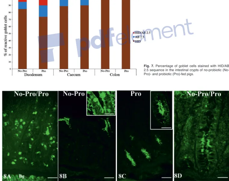
In No-Pro samples, MAA-II stained scattered goblet cells along the entire crypts in the duodenum (Fig. 8A) and a few goblet cells at the opening of the caecal crypts (Fig. 8B), whereas it reacted with the apical region of the absorptive enterocytes in the basal half of the colonic crypts (Fig. 8D). The probiotic blend affected the MAA-II binding pattern only in the caecum, which lost goblet cell reactivity (Fig. 3), whereas an appearance of affinity was exhibited in the apical region of epithelial cells at
the bottom of the crypts (Fig. 8C).
SNA stained the goblet cells along the crypts of all investigated intestinal tracts in both the No-Pro and Pro samples (Fig. 9A-F). Supplementation with the probiotic blend induced a significant decrease in signal intensity of the duodenal crypt goblet cells (Figs. 9B, 10), whereas it significantly (P<0.001) increased the number and the staining signal in the goblet cells along the caecal and colonic crypts (Figs. 3, 9D-F, 10).
Discussion
This study demonstrated that a dietary probiotic complex containing Streptococcus, Bifidobacterium and
Lactobacillus strains is able to induce regional effects on
intestinal crypt morphology as well as on the mucins secreted by goblet cells in growing-finishing pigs.
Intestinal crypts consist of several cell types, including pluripotent stem cells residing at the bottom of the crypts, and cells that differentiate into mature cell lineages during migration along the crypts, such as the mucin secretory goblet cells. Histomorphometric analysis in this study showed that probiotic supplementation did not affect the depth of the duodenal crypts, whereas it induced a general reduction in crypt depth in the large intestine, which was significant in the caecum but not in the colon. Although a decrease in crypt depth was unexpected, it is possible for commensal microbiota to decrease intestinal crypt depth. For example, Lactobacillus reuteri, an important member of the intestinal commensal microbiota, is able to reduce the crypt depth of pig small intestine (Le et al., 2016; Yi et al., 2018). It has also been reported that a lower crypt depth could be related to a reduced intestinal cell turnover rate and therefore may indicate an energy-saving mechanism (Hedeman et al., 2014; Metzler-Zebeli et al., 2017). Crypt lengthening may be mediated by a spectrum of local, immune and neurohumoral factors (Pearson and Brownlee, 2010). Gut microbiota can affect intestinal motor functions either directly or
Fig. 5. Percentage of goblet cells stained with both Alcian Blue pH 2.5 (AB) and PAS staining or with PAS only in the intestinal crypts of no-probiotic (No-Pro) and probiotic (Pro)-fed pigs.
indirectly via mediators released by the gut immune response, intestinal neuroendocrine factors or the end products of bacterial fermentation (Wu et al., 2013). Recently, an increase in the interstitial cells of Cajal
(ICC), the pacemaker cells of spontaneous motility in the gut (Takaki, 2003), has been detected in cats after probiotic (SLAB51) therapy (Rossi et al., 2018). If the decrease in crypt depth observed in this study is related
Fig. 6. HID/AB 2.5 staining sequence of duodenal (A, B, C), caecal (D, E, F) and colonic (G, H, I) crypts. A, B, D, G, H. Instead of no-probiotic-fed samples. C, E, F, I. Instead of probiotic-fed samples. Most of the goblet cells exhibit HID positivity (brown). Picture B shows goblet cells producing both HID-positive (brown) and AB positive (blue) mucins. Pictures H, I clearly show the absence of goblet cells that synthesize both HID- and AB 2.5-positive mucins in the colonic crypts. Bg, Brunner's gland; s, submucosa; arrow, muscularis mucosae. Scale bars: A, C-E, G, 90 µm; F, I, 50 µm; B, H, 25 µm.
to the contraction of muscular wall via ICC, this could be the subject of further specific investigations.
Histochemical analysis revealed that probiotic supplementation induced a significant increase in the number of goblet cells per crypt in the investigated intestinal tracts. Previous studies demonstrated that the application of micro-organisms as probiotics could change the number of these cells in the crypts of pig intestine. A significant increase in the number of goblet cells per crypt as also been detected in the caecum but not in the small intestine or the proximal colon of pigs fed with Saccharomyces boulardii (Baum et al., 2002). This increased secretion of mucins was related to the change in concentrations of the end products of microbial fermentation of non-digestible carbohydrates
such as short-chain fatty acids (Cummings and MacFarlane, 1991; Sakata and Setoyama, 1995; Shimotoyodome et al., 2000; Willemsen et al., 2003). Interestingly, it has been demonstrated that SLAB51 administration affects the concentration of some short-chain fatty acids in pigs (Tufarelli et al., 2017). The comparison of our results with those obtained by using other probiotics shows that the probiotic effects on gut morphology arise from their variation in bacterial composition. For example, no effects on duodenal and colonic crypt depth have been found in pigs fed with probiotics containing either Bacillus cereus var. toyoi or
Saccharomyces boulardii (Baum et al., 2002) or in those
fed fermented wheat grain with Lactobacillus reuteri (Le et al., 2016). Moreover, dietary supplementation with
Fig. 7. Percentage of goblet cells stained with HID/AB 2.5 sequence in the intestinal crypts of no-probiotic (No-Pro)- and probiotic ((No-Pro)-fed pigs.
Fig. 8. Reactivity pattern of MAA-II, specific for Neu5Acα2,3Galβ1,4GlcNAc, with the duodenal (A), caecal (B, C) and colonic (D) mucosa of pigs. No-Pro, No-probiotic fed pigs; No-Pro, probiotic-fed pigs. Bg, Brunner's gland. Scale bars: A-C, 80 µm; D, 50 µm; Insets in B, C, 40 µm.
Fig. 9. Reactivity pattern of the SNA, specific for Neu5Acα2,6Gal/GalNAc, with the duodenal (A, B), caecal (C, D) and colonic (E, F) mucosa of no-probiotic (A, C, E) and no-probiotic (B, D, F)-treated pigs. Bg, Brunner's gland. Scale bars: 80 µm.
Pediococcus acidilactici was able to increase the crypt
depth of pig caecum (Di Giancamillo et al., 2008). These results are congruent with the view that probiotic effects depend on a complex interaction between the type of probiotic supplementation, the gut flora and the administrator (Fuller, 2006; Metzler-Zebeli et al., 2017).
Mucins are glycoproteins that can be classified into neutral and acidic types. When the latter terminate with sialic acids or sulphate groups, they constitute the sialomucins and the sulphomucins, respectively (Deplancke and Gaskins, 2001). In the duodenum of no-probiotic fed animals, the percentage of crypt goblet cells producing a mixture of both neutral and acidic mucins (PAS and AB 2.5 positivity) predominated over those synthesizing only neutral glycans (85% vs 15%). The secretion of neutral mucins, together with the secretion of bile and pancreatic juice, has been related to the neutralization of the acidic pH of gastric juices entering the duodenum (Georgiades et al., 2014). Concerning acidic mucins, most of the duodenal goblet cells (85%) produced sulphoglycans (HID positivity), whereas goblet cells that produced only non-sulphoglycans (AB 2.5 positivity) or mixed sialosulphomucins were detected to a lesser extent (10% and 5%, respectively). Goblet cells with non-sulphomucins were mainly located in the fundus of the crypts. This location could be related to goblet cell differentiation, which starts from precursor cells in the crypt fundus and continues during migration towards the upper portion (Kim and Ho, 2010). Moreover, if the sulphation of mucins is considered a modification of acidic mucins (Freeze et al., 2017), the presence of only acidic non-sulphated glycans in the bases of the crypts could represent an immature phase of mucin biosynthesis. In turn, this could justify the presence of goblet cells that produce mixed sialosulphomucins in the intermediate zone of the crypts. It has been reported that sulphomucins are added to the secretory product of goblet cells as they migrate towards the intestinal
mucosa surface in mice (Liquori et al., 2012). The use of lectin histochemistry demonstrated that the acidic non-sulphomucins contain α2,6- and α2,3-sialylated glycoproteins (SNA and MAA-II affinity, respectively). Administration of probiotics induced a significant decrease in the percentage of goblet cells secreting either neutral mucins (15% No-Pro vs 3% Pro) or sulphoglycans (85% No-Pro vs 74% Pro) in duodenal crypts and, in contrast, a significant increase in goblet cells producing acidic non-sulphated mucins (10% No-Pro vs 16% No-Pro). Notably, we observed increased synthesis of acidic non-sulphated glycans in duodenal Brunner's glands from the same pigs used in this study (Accogli et al., 2018). A higher secretion of sialomucins than sulphomucins in the small intestine has been related to improved defence in pigs (Liu et al., 2014). Lectin reactivity demonstrated that the probiotic blend induced a decrease in α2,6-sialylated mucin secretory goblets. At first glance, this result seems inconsistent with the results concerning acidic non-sulphoglycans obtained with conventional histochemistry. The discrepancy can be explained if we consider that, unlike other monosaccharides, sialic acid exhibits pronounced chemical diversity in structure and linkage so that the total diversity of sialoglycans constitute the “sialome” (Varki et al., 2017). Moreover, in contrast with conventional histochemistry, which differentiates main categories of complex carbohydrates, lectin histochemistry distinguishes sugar isomers, as well as branching, linkage and terminal modifications of complex glycans (Spicer and Schulte, 1992; Sharon and Lis, 2004). However, further studies on the structure of sialic acids that use other lectins in association with chemical and enzymatic treatments, or other techniques used in glycoanalysis, could provide a deeper knowledge of the sialomucins produced by pig intestinal goblet cells.
In both experimental groups, caecal goblet cells secreted mixed neutral and acidic mucins. The latter
Fig. 10. Quantification of SNA-FITC signals in crypt goblet cells of no-probiotic (No-Pro)- and probiotic (Pro)-fed pigs. Data show the mean with error bars representing ± SD and Student’s t-test results (*P<0.05; **P<0.01).
mainly constituted sulphoglycans and very few acidic non-sulphomucins, which were secreted by cells detected at the bases of the crypts. In pig caecum, the percentage of sulphoglycan secretory goblet cells was higher than in the duodenum. This finding agrees with the results of previous studies of humans (Croix et al., 2011; Holmén Larsson et al., 2013). The probiotic complex that was used did not affect the percentage of goblet cells secreting neutral and acidic (non-sulphated and sulphated) glycans in the caecal crypts. However, in Pro samples, caecal goblet cells that secreted both neutral and acidic mucins were distributed along the entire crypts, whereas they were absent at the opening of the crypts in the No-Pro pigs. This suggests that the administration of the probiotics increased the secretion of mucins in the caecum. In addition, the probiotic blend induced an increase in α2,6-sialylated mucins and abolished the synthesis of α2,3-sialoglycoproteins in the goblet cells. This latter modification is crucial for swine resistance to pathogen colonization, principally to prevent infection of gastrointestinal cells by avian strains of influenza viruses (Cone, 2009). These strains attach to the host cell by binding to NeuNAca2,3Gal (Cohen et al., 2013), a sialic acid modification to glycans that is also present in the epithelial cells of the upper respiratory tract of swine and humans (Skehel and Wiley, 2000); in these two species, the α2,3-linked sialoglycoproteins are essential for successful infection by influenza A (Shynia et al., 2006; Nicholls et al., 2007).
Probiotic modification of the α2,6-/α2,3-sialoglycoproteins in the goblet cells is also critical from an immunopathological point of view. It is indeed known that recognition by CD22, a molecule belonging to the SIGLEC family of lectins (Crocker et al., 1998) described on the surface of mature B cells, is specific for α2,6 linkage, with no binding to α2,3-linked sialic acid (Powell et al., 1993). Additional studies defined the highly conserved preference of CD22 for this linkage and further characterized the interactions (Powell and Varki, 1994). CD22 is a regulatory molecule that prevents over activation of the immune system and the development of severe inflammation and/or autoimmune diseases (Hatta et al., 1999).
Our results showed that glycans terminating with α2,3-linked sialic acid appeared in the apical surface of epithelial cells at the crypt fundus. This result demonstrates for the first time that administration of probiotics could modify the glycan pattern and consequently the function of non-secretory cells located at the base of crypts. Membrane-bound sialoglycans are known to provide a cell surface charge that acts as a barrier and regulates cell surface functions (Varki et al. 2017). The marked presence of Neu5Acα2,3 Galβ1,4GlcNAc has been demonstrated on the luminal cell surface of the fundus of the distal colon crypts in rats (Accili et al., 2008). We cannot compare our results with those of previous studies concerning probiotic effects on the composition of sialomucins secreted by
goblet cells of pig caecum, because to our knowledge, similar studies are lacking. However, in contrast to our results, Di Giancamillo et al. (2008) did not detect a change in the mucin types secreted by caecum goblet cells of pigs fed with the probiotic Pediococcus
acidilactici (Di Giancamillo et al., 2008).
Regarding the colon, the goblet cells involved in the secretion of a mixture of both neutral and acidic glycans predominated over the goblet cells that produced only neutral mucins (93% vs 7%). The acidic mucins were almost exclusively sulphomucins. These results are consistent with the view that because of the vulnerability of sialomucins to bacterial degradation, sulphomucin-secreting cells are expected to dominate in the large intestine where the bacterial load is high (Deplancke and Gaskins, 2001). Previous studies reported a predominance of sulphomucins in the colonic crypts from the small intestine to the distal colon in rodents (Karlsson et al., 1997; Liquori et al., 2012; Holmén Larsson et al., 2013) and in humans (Matsuo et al., 1997). In addition, the expression of N-acetylglucosamine 6-O-sulfotransferase, one of the major sulfotransferases involved in mucin sulphation, is regulated by butyrate, a metabolite from colonic anaerobic bacteria (Tobisawa et al., 2010; Kawashima, 2012). Sulphated groups increase the negative charge of mucins and thus the already-mentioned effects of sialic acid. Sulphomucins are a crucial source of sulphate for sulphate-reducing bacteria as well because they play a prominent protective role in the increase of mucus viscosity (Croix et al., 2011). It has been reported that pathogenic bacteria are trapped in this dense mucus state, and so sulphomucins ensure a local defensive response, acting as a barrier against mucosal inflammation (Deplancke and Gaskins, 2001; Montagne et al., 2004; Kawashima, 2006, 2012; Tobisawa et al., 2010). In addition, a high negative charge contributes to the stiffness of mucin peptides and permits mucins to adsorb large amounts of water and ions (Forstner and Forstner, 1994). The probiotic complex used in this study also contained Bifidobacterium animalis subsp.
lactis, which recognizes and adheres to sialo- and/or
sulphomucins of porcine colon (Nishiyama et al., 2014). In rats, it has been reported that the administration of this bifidobacterium increased the production and secretion of sulphomucin in the proximal colon, consequently increasing the thickness of the mucus layer, which in turn prevented loperamide-induced constipation (Aoki et al., 2016). Interestingly, the probiotic used in our study has more recently been shown to induce significant clinical improvement in cats with chronic constipation and idiopathic megacolon (Rossi et al., 2018).
SNA and MAA reactivity showed a diffuse presence of α2,6-sialomucins that produced goblet cells along the colonic crypts and sialomucins that terminated with Neu5Acα2,3Galβ1,4GlcNAc on the apical surface of epithelial cells at the crypt bottom, respectively. Probiotic dietary supplementation induced a significant
increase in Neu5Acα2,6Gal/GalNAc sialomucins that produced goblet cells. Interestingly, the Neu5Ac residue that linked α2,6 to the GalNAc constitutes an epitope commonly found in the human colon MUC2, the major gel-forming mucin in the gastrointestinal tract (Thomsson et al., 2012). This effect, in our opinion, is crucial because, as demonstrated in mice that are deficient in MUC2 (Johansson et al., 2008), the loss or absence of this mucin fails to restrict bacterial attachment to the mucosal tissues, predisposing mice to severe spontaneous colitis and colorectal cancer (Velcich et al., 2002; Van der Sluis et al., 2006).
One bacterial constituent of the probiotic preparation used in the present study was the Lactobacillus
plantarum strain. Notably, this probiotic increased the
expression of MUC2 in colon cell cultures (Mack et al., 1999). This finding has been related to the ability of
Lactobacillus strains to protect the intestinal epithelium
against invasion of enteropathogenics by inhibiting intestinal adherence (Mack et al., 1999).
In conclusion, this study demonstrates that dietary supplementation with a commercial multi-strain probiotic (SLAB51™) containing a mixture of different species of lactic acid bacteria and bifidobacteria induces region-specific changes in crypt depth and in the mucins secreted along the intestinal tract of growing-finishing pigs. These findings add further data about the positive effects of probiotics on mammalian intestine, which enhanced the growth performance of growing-finishing pigs (Tufarelli et al., 2017) and produced significant clinical amelioration in cats with chronic constipation and idiopathic megacolon (Rossi et al., 2018). Bearing in mind that pigs are a promising animal model in translational biomedical research and that the commercial probiotic blend used herein is designed for human use, these results may allow progress in the understanding of its action on the human intestine.
References
Accili D., Menghi G. and Gabrielli M.G. (2008). Lectin histochemistry for in situ profiling of rat colon sialoglycoconjugates. Histol. Histopathol. 23, 863-875.
Accogli G., Crovace A., Mastrodonato M., Rossi G., Francioso E.G. and Desantis S. (2018). Probiotic supplementation affects the glycan composition of mucins secreted by Brunner’s glands of the pig duodenum. Ann. Anat. 218, 236-242.
Aoki R., Tsuchida S., Arai Y., Ohno K., Nishijima T., Mawatari T., Mikami Y. and Ushida K. (2016). Effect of Bifidobacterium animalis subsp. Lactis GCL250 on the physiological function of intestine in a rat model. Food. Sci. Nutr. 4, 782-790.
Baum B., Liebler-Tenorio E.M., Enss M.L., Pohlenz J.F and Breves G (2002). Saccharomyces boulardii and Bacillus cereus var. Toyoi influence the morphology and the mucins of the intestine of pigs. Z. Gastroenterol. 40, 277-284.
Bontempo V., Di Giancamillo A., Savoini G., Dell’Orto V. and Domeneghini C. (2006). Live yeast dietary supplementation acts upon intestinal morpho-functional aspects and growth in weanling piglets. Animal Feed. Sci. Technol. 129, 224-236.
Büsing K. and Zeyner A. (2015). Effects of oral Enterococcus faecium strain DSM 10663 NCIMB 10415 on diarrhoea patterns and performance of sucking piglets. Benef. Microbes. 6, 41-44. Cohen M., Zhang X.Q., Senaati H.P., Chen H.W., Varki N.M., Schooley
R.T. and Gagneux P. (2013). Influenza A penetrates host mucus by cleaving sialic acids with neuraminidase. Virol. J. 10, 321.
Cone R.A (2009). Barrier properties of mucus. Adv. Drug. Deliv. Rev. 61, 75-85.
Crocker P.R., Clark E.A., Filbin M, Gordon S., Jones Y, Kehrl J.H., Kelm S., Le Douarin N., Powell L., Roder J., Schnaar R.L., Sgroi D.C., Stamenkovic K., Schauer R., Schachner M., van den Berg T.K., van der Merwe P.A., Watt S.M. and Varki A. (1998). Siglecs: a family of sialic-acid binding lectins. Glycobiology 8, v-vi.
Croix J.A., Carbonero F, Nava G.M., Russell M., Greenberg E. and Gaskins H.R. (2011). On the relationship between sialomucin and sulfomucin expression and hydrogenotrophic microbes in the human colonic mucosa. PLoS One 6, e24447.
Cummings J.H. and Macfarlane G.T. (1991). The control and consequences of bacterial fermentation in the human colon. J. Appl. Bacteriol. 70, 443-459.
Deplancke B. and Gaskins H.R. (2001). Microbial modulation of innate defense: goblet cells and the intestinal mucus layer. Am. J. Clin. Nutr. 73, 1131S-1141S.
Di Giancamillo A., Bontempo V., Savoini G., Dell’Orto V., Vitari F. and Domeneghini C. (2007). Effects of live yeast dietary supplementation to lactating sows and weaning piglets. Int. J. Probiot. Prebiot. 2, 55-66.
Di Giancamillo A., Vitari F., Savoini G., Bontempo V., Bersani C, Dell’Orto V. and Domeneghini C. (2008). Effects of orally administered probiotic Pediococcus acidilactici on the small and large intestine of weaning piglets. A qualitative and quantitative micro-anatomical study. Histol. Histopathol. 23, 651-664.
Forstner J,F and Forstner G.G. (1994). Gastrointestinal mucus. In: Physiology of the gastrointestinal tract. Johnson L.R. (ed). Raven Press. New York. pp 1245-1283.
Freeze H.H., Hart G.W. and Schnaar R.L. (2017). Glycosylation precursors. In: Essentials of glycobiology. 3rd ed. Varki A (ed), Cold Spring Harbor Laboratory Press. New York. pp 51-63.
Fuller R. (2006). Reasons for the apparent variation in the probiotic response. Biologia, Bratislava, 61, 751-754.
Georgiades P., Pudney P.D., Thornton D.J. and Waigh T.A. (2014). Particle tracking microrheology of purified gastrointestinal mucins. Biopolymers 101, 366-377.
Gonzalez L.M., Moeser A.J. and Blikslager A.T. (2015). Porcine models of digestive disease: the future of large animal translational research. Transl. Res 166, 12-27.
Hatta Y., Tsuchiya N., Matsushita M., Shiota M., Hagiwara K. and Tokunaga K. (1999). Identification of the gene variations in human CD22. Immunogenetics 49, 280-286.
Hedemann M.S., Jensen B.B and Poulsen H.D. (2014). Influence of dietary zinc and copper on digestive enzyme activity and intestinal morphology in weaned pigs. J. Anim. Sci. 84, 3310-3320.
Heinritz S.N., Mosenthin R. and Weiss E. (2013). Use of pigs as a potential model for research into dietary modulation of the human gut microbiota. Nutr. Res. Rev. 26, 191-209.
Holmén Larsson J.M., Thomsson K.A., Rodriguez-Pineiro A.M., Karlsson H. and Hansson G.C. (2013). Studies of mucus in mouse stomach, small intestine, and colon. III. Gastrointestinal Muc5ac and Muc2 mucin O-glycan patterns reveal a regiospecific distribution.
Am. J. Physiol. Gastrointest. Liver. Physiol. 305, G357-G363. Johansson M.E., Phillipson M., Petersson J., Velcich A., Holm L. and
Hansson G.C. (2008). The inner of the two Muc2 mucin-dependent mucus layers in colon is devoid of bacteria. Proc. Natl. Acad. Sci. USA 105, 15064-15069.
Karlsson N.G., Herrmann A., Karlsson H., Johansson M.E., Carlstedt I. and Hansson G.C. (1997). The glycosylation of rat intestinal Muc2 mucin varies between rat strains and the small and large intestine. A study of O-linked oligosaccharides by a mass spectrometric approach. J. Biol. Chem. 272, 27025-27034.
Kawashima H. (2006). Roles of sulfated glycans in lymphocyte homing. Biol. Pharm. Bull. 29, 2343-2349.
Kawashima H. (2012). Roles of the gel-forming MUC2 mucin and its O-glycosylation in the protection against colitis and colorectal cancer. Biol. Pharm. Bull. 35, 1637-1641.
Kim Y.S. and Ho S.B. (2010). Intestinal goblet cells and mucins in health and disease: recent insights and progress. Curr. Gastroenterol. Rep. 12, 319-330.
Lallés J.P., Bosi P., Smidt H. and Stokes C.R. (2007) Nutritional management of gut health in pigs around weaning. Proc. Nutr. Soc. 66, 260-268.
Le M.H., Galle S., Yang Y., Landero J.L., Beltranena E., Gänzle M.G. and Zijlstra R.T. (2016). Effects of feeding fermented wheat with
Lactobacillus reuteri on gut morphology, intestinal fermentation,
nutrient digestibility, and growth performance in weaned pigs. J. Anim. Sci. 94, 4677-4687.
Lievin-Le Moal V. and Servin A.L. (2006). The front line of enteric host defense against unwelcome intrusion of harmful microorganisms: mucins, antimicrobial peptides, and microbiota. Clin. Microbiol. Rev. 19, 315-337.
Liquori G.E., Mastrodonato M, Mentino D., Scillitani G., Desantis S., Portincasa P. and Ferri D. (2012). In situ characterization of O-linked glycans of Muc2 in mouse colon. Acta Histochem. 114, 723-732. Liu P., Pieper R., Tedin L., Martin L., Meyer W., Rieger J., Plendl J.,
Vahjen W. and Zentek J. (2014). Effect of dietary zinc oxide on jejunal morphological and immunological characteristics in weaned piglets. J. Anim. Sci. 92, 5009-5018.
Liu C., Zhu Q., Chang J., Yin Q., Song A., Li Z., Wang E. and Lu F. (2017). Effects of Lactobacillus casei and Enterococcus faecalis on growth performance, immune function and gut microbiota of suckling piglets. Arch. Anim. Nutr. 71, 120-133.
Mack D.R., Michail S., Wei S., McDougall L. and Hollingsworth M.A. (1999). Probiotics inhibit enteropathogenic E. coli adherence in vitro by inducing intestinal mucin gene expression. Am. J. Physiol. 276, G941-G950.
Martens E.C., Roth R., Heuser J.E. and Gordon J.I. (2009). Coordinate regulation of glycan degradation and polysaccharide capsule biosynthesis by a prominent human gut symbiont. J. Biol. Chem. 284, 18445-18457.
Mastrodonato M., Mentino D., Lopedota A., Cutrignelli A. and Scillitani G. (2017). A histochemical approach to glycan diversity in the urothelium of pig urinary bladder. Microsc. Res. Tech. 80, 239-249. Matsuo K., Ota H., Akamatsu T., Sugiyama A. and Katsuyama T.
(1997). Histochemistry of the surface mucous gel layer of the human colon. Gut 40, 782-789.
McCloy R.A., Rogers S., Caldon C.E., Lorca T., Castro A. and Burgess, A. (2014). Partial inhibition of Cdk1 in G 2 phase overrides the SAC and decouples mitotic events. Cell Cycle 13, 1400-1412.
Mc Manus J.F.A. (1948). Histological and histochemical uses of periodic
acid. Stain. Technol. 23, 99-108.
Meng Q.W., Yan L., Ao X., Zhou T.X., Wang J.P., Lee J.H. and Kim I.H. (2010). Influence of probiotics in different energy and nutrient density diets on growth performance, nutrient digestibility, meat quality, and blood characteristics in growing-finishing pigs. J. Anim. Sci. 88, 3320-3326.
Metzler-Zebeli B.U., Lawlor P.G., Magowan E., McCormack U.M., Curião T., Hollmann M., Ertl R., Aschenbach J.R. and Zebeli Q. (2017). Finishing pigs that are divergent in feed efficiency show small differences in intestinal functionality and structure. PLoS One 12, e0174917.
Montagne L., Piel D. and Lallès J.P. (2004). Effect of diet on mucin kinetics and composition: nutrition and health implications. Nutr. Rev. 62, 105-114.
Nicholls J.M., Bourne A.J., Chen H., Guan Y. and Peiris J.S.M. (2007). Sialic acid receptor detection in the human respiratory tract: evidence for widespread distribution of potential binding sites for human and avian influenza viruses. Respir. Res. 8, 73.
Nishiyama K., Kawanabe A., Miyauchi H., Abe F., Tsubokawa D., Ishihara K., Yamamoto Y. and Mukai T. (2014). Evaluation of bifidobacterial adhesion to acidic sugar chains of porcine colonic mucins. Biosci. Biotechnol. Biochem. 78, 1444-1451.
NRC (1998) Nutrient requirements of swine. 10th rev. ed. Natl Acad Press. Washington, DC. pp 110-142.
Oelschlaeger T.A. (2010). Mechanisms of probiotic actions – A review. Int. J. Med. Microbiol. 300, 57-62.
Pearse A.G.E. (1968). Histochemistry theoretical and applied. 3rd ed. Vol. I. Churchill. London.
Pearson J.P. and Brownlee I.A. (2010).The interaction of large bowel microflora with the colonic mucus barrier. Int. J. Inflam. 2010, 321426.
Powell L.D. and Varki A. (1994). The oligosaccharide binding specificities of CD22 beta, a sialic acid-specific lectin of B cells. J. Biol. Chem. 269, 10628-10636.
Powell L.D., Sgroi D., Sjoberg E.R., Stamenkovic I. and Varki A. (1993). Natural ligands of the B cell adhesion molecule CD22 beta carry N-linked oligosaccharides with alpha-2,6-N-linked sialic acids that are required for recognition. J. Biol. Chem. 268, 7019-7027.
Prather R.S., Lorson M., Ross J.W., Whyte J.J. and Walters E. (2013). Genetically engineered pig models for human diseases. Annu. Rev. Anim. Biosci. 1, 203-219.
Rossi G., Jergens A., Cerquetella M., Berardi S., Di Cicco E., Bassotti G., Pengo G. and Suchodolski J.S. (2018). Effects of a probiotic (SLAB51™) on clinical and histologic variables and microbiota of cats with chronic constipation/megacolon: a pilot study. Benef. Microbes 9, 101-110.
Sakata T. and Setoyama H. (1995). Local stimulatory effect of short-chain fatty acids on the mucus release from the hindgut mucosa of rats (Rattus norvegicus). Comp. Biochem.Physiol. A Physiol.111, 429-432.
Sharon N. and Lis H. (2004). History of lectins: from hemagglutinins to biological recognition molecules. Glycobiology 14, 53R-62R. Shimotoyodome A., Meguro S., Hase T., Tokimitsu I. and Sakata T.
(2000). Short chain fatty acids but not lactate or succinate stimulate mucus release in the rat colon. Comp. Biochem. Physiol. A Mol. Integr. Physiol. 125, 525-531.
Shynia K., Ebina M., Yamada S., Ono M, Kasai N. and Kawaoka Y. (2006). Avian flu: influenza virus receptors in the human airway. Nature 440, 435-436.
Skehel J. and Wiley D.C. (2000). Receptor binding and membrane fusion in virus entry: the influenza hemagglutinin. Annu. Rev. Biochem. 69, 531-569.
Spicer S.S. (1965). Diamine methods for differentialing mucosubstances histochemically. J. Histochem. Cytochem. 13, 211-234.
Spicer S.S. and Schulte B.A. (1992). Diversity of cell glycoconjugates shown histochemically: a perspective. J. Histochem. Cytochem. 40, 1-38.
Takaki M. (2003). Gut pacemaker cells: the interstitial cells of Cajal (ICC). J. Smooth Muscle Res. 39, 137-161.
Thomsson K.A., Holmen-Larsson J., Angstrom J., Johansson M.E.V., Xia L. and Hansson G.C. (2012). Detailed O-glycomics of the Muc2 mucin from colon of wild-type, core 1- and core 3-transferase-deficient mice highlights differences compared with human MUC2. Glycobiology 22, 1128-1139.
Tobisawa Y., Imai Y., Fukuda M. and Kawashima H. (2010). Sulfation of colonic mucins by N-cetylglucosamine 6-O-sulfotransferase-2 and its protective function in experimental colitis in mice. J. Biol. Chem. 285, 6750-6760.
Tufarelli V., Crovace A.M., Rossi G. and Laudadio V. (2017). Effect of a dietary probiotic blend on performance, blood characteristics, meat quality and faecal microbial shedding in growing-finishing pigs. S. Afr. J. Anim. Sci. 47, 875-882.
Van der Sluis M., De Koning B.A., De Bruijn A.C., Velcich A., Meijerink J.P., Van Goudoever J.B., Buller H.A., Dekker J., Van Seuningen I.,
Renes I.B. and Einerhand A.W. (2006). Muc2-deficient mice spontaneously develop colitis, indicating that MUC2 is critical for colonic protection. Gastroenterology 131, 117-129.
Varki A., Schnaar R.L. and Schauer R. (2017). Sialic acids and other nonulosonic acids. In: Essentials of glycobiology, 3rd ed. Varki A (ed.) Cold Spring Harbor Laboratory Press. New York. pp. 179-195. Velcich A., Yang W., Heyer J., Fragale A, Nicholas C., Viani S.,
Kucherlapati R., Lipkin M, Yang K. and Augenlicht L. (2002). Colorectal cancer in mice genetically deficient in the mucin Muc2. Science 295, 1726-1729.
Willemsen L.E, Koetsier M.A., van Deventer S.J. and van Tol EA. (2003). Short chain fatty acids stimulate epithelial mucin 2 expression through differential effects on prostaglandin E(1) and E(2) production by intestinal myofibroblasts. Gut 52, 1442-1447. Wu R.Y., Pasyk M., Wang B., Forsythe P., Bienenstock J., Mao Y.K.,
Sharma P., Stanisz A.M. and Kunze W.A. (2013). Spatiotemporal maps reveal regional differences in the effects on gut motility for Lactobacillus reuteri and rhamnosus strains. Neurogastroenterol. Motil. 25, e205-214
Yi H., Wang L., Xiong Y., Wen X., Wang Z., Yang X., Gao K. and Jiang Z. (2018). Effects of Lactobacillus reuteri LR1 on the growth performance, intestinal morphology, and intestinal barrier function in weaned pigs. J. Anim. Sci. 96, 2342-2351.