Contents lists available atScienceDirect
European Journal of Pharmacology
journal homepage:www.elsevier.com/locate/ejpharFull length article
PLGA nanoparticles loaded with beta-lactoglobulin-derived peptides
modulate mucosal immunity and may facilitate cow's milk allergy
prevention
Atanaska I. Kostadinova
a,b,⁎, Jim Middelburg
a,c, Michele Ciulla
c, Johan Garssen
a,b,
Wim E. Hennink
c, Leon M.J. Knippels
a,b, Cornelus F. van Nostrum
c, Linette E.M. Willemsen
aaDivision of Pharmacology, Utrecht Institute for Pharmaceutical Sciences, Utrecht University, Universiteitsweg 99, 3584CG Utrecht, The Netherlands bDepartment of Immunology, Nutricia Research, Uppsalalaan 12, 3584CT Utrecht, The Netherlands
cDivision of Pharmaceutics, Utrecht Institute for Pharmaceutical Sciences, Utrecht University, Universiteitsweg 99, 3584CG Utrecht, The Netherlands
A R T I C L E I N F O
Keywords: Beta-lactoglobulin Cow's milk allergy Peptide PLGA nanoparticle Oral tolerance Prevention
A B S T R A C T
Beta-lactoglobulin (BLG)-derived peptides may facilitate oral tolerance to whey and prevent cow's milk allergy (CMA). Loading of BLG-peptides in poly(lactic-co-glycolic acid) (PLGA) nanoparticles (Pep-NP) may improve this. Here we studied the uptake of NP and the capacity of NP and Pep-NP to activate bone marrow dendritic cells (BMDC). Furthermore, CMA prevention was evaluated by orally exposing three-week-old female C3H/ HeOuJ mice to Pep-NP, NP or free peptides (PepMix) for 6 days before oral sensitization with whole whey protein and effects on the spleen and small intestine lamina propria (SI-LP) were studied. In BMDC, NP and Pep-NP enhanced CD40 expression and IL-6 and TNF-α secretion, while tended to decrease CD80 expression and prevented PepMix-induced IL-12 secretion. In vivo, oral exposure to Pep-NP, but not NP or PepMix, prior to whey sensitization tended to partially prevent the acute allergic skin response to whole whey protein. Splenocytes of NP-pre-exposed mice secreted increased levels of whey-specific IL-6, but this was silenced in Pep-NP-pre-exposed mice which also showed reduced TNF-α and IFN-γ secretion. In the SI-LP, Pep-NP pre-exposure reduced the CD4+T cell frequency in CMA mice compared to PBS pre-exposure. In addition, while NP increased whey-specific IL-6 secretion in the SI-LP, Pep-NP did not and maintained regulatory TGF-β secretion. This study presents a proof-of-concept that PLGA nanoparticles facilitate the capacity of BLG peptides to suppress the allergic response to whole whey protein. Hence, PLGA nanoparticles may be further developed as an adjunct strategy for BLG-peptide-based oral tolerance induction and CMA prevention.
1. Introduction
Cow's milk allergy (CMA) is the most prevalent food allergy in children younger than 5 years (Koletzko et al., 2012; Venter and Arshad, 2011). Even though most children outgrow CMA by developing tolerance, they have an increased risk of developing other atopic dis-orders later in life, such as asthma, rhinoconjunctivitis or functional gastrointestinal disorders (Nissen et al., 2013; Saps et al., 2011). The many approaches being investigated for the prevention or treatment of CMA aim to induce or restore oral tolerance– the state of local and systemic immune unresponsiveness established upon oral exposure to harmless antigens, such as food proteins (Pabst and Mowat, 2012). Oral tolerance is an antigen-dependent process and therefore the use of the allergen is unavoidable (Prescott et al., 2008). Recent clinical studies reported that early introduction of whole peanut protein to high-risk
infants has some efficacy in reducing the prevalence of peanut allergy (Du Toit et al., 2015; Perkin et al., 2016), but studies with egg protein were less successful and reported safety concerns in a relevant number of patients (Bellach et al., 2017; Palmer et al., 2013). Furthermore, whole allergen is introduced only after 4 months of age (Du Toit et al., 2015), which encourages the search for other, safer, strategies that would allow active intervention in the first months of life when breastfeeding is not possible.
To avoid potential sensitization or allergic symptoms in infants at high risk of developing CMA, specific allergen-derived peptide se-quences containing T cell epitopes may be utilized instead. When taken up and presented by antigen-presenting cells in the intestine, those epitopes will possibly initiate the development of a tolerogenic re-sponse. For instance, beta-lactoglobulin (BLG)-derived peptides have been shown to decrease the sensitizing capacity of the BLG protein
http://dx.doi.org/10.1016/j.ejphar.2017.10.051
Received 20 July 2017; Received in revised form 22 October 2017; Accepted 23 October 2017
⁎Corresponding author at: Division of Pharmacology, Utrecht Institute for Pharmaceutical Sciences, Utrecht University, Universiteitsweg 99, 3584CG Utrecht, The Netherlands.
E-mail address:[email protected](A.I. Kostadinova).
Available online 25 October 2017
0014-2999/ © 2017 The Authors. Published by Elsevier B.V. This is an open access article under the CC BY license (http://creativecommons.org/licenses/BY/4.0/).
(Bogh et al., 2013) and to facilitate oral tolerance to BLG (Pecquet et al., 2000a) or to whole whey protein in mice (Kostadinova et al., 2016; Meulenbroek et al., 2013). However, proteolytic enzymes present in the stomach can lead to peptide degradation and consequently loss of bioavailability, emphasizing the interest in a delivery vehicle.
Poly(lactic-co-glycolic acid) (PLGA) is a FDA-approved biodegrad-able polymer which is under investigation for the oral and sub-cutaneous delivery of vaccines (Bolhassani et al., 2014; McHugh et al., 2015; Pavot et al., 2014; Rahimian et al., 2015). In oral antigen de-livery, PLGA nanoparticles offer protection from enzymatic degradation and facilitate uptake via M-cells (Brotons-Canto et al., 2016). Further-more, PLGA particles are taken up by macrophages and dendritic cells (DC) (Lutsiak et al., 2002; Nicolete et al., 2011) and might also directly activate humoral or cellular responses (Nicolete et al., 2011; Silva et al., 2015). Previous reports describe that delivering BLG in PLGA micro-particles increased the efficacy of BLG to induce oral tolerance (Pecquet et al., 2000b).
In the current study, we encapsulated a low dose of BLG-derived peptides in PLGA nanoparticles to investigate whether their capacity to prevent the allergic response to the cow's milk protein whey in a mouse model of CMA will be improved.
2. Materials and methods 2.1. Peptides
Four 18-AA-long peptides spanning the B variant of the BLG se-quence were purchased from JPT Peptide Technologies (Berlin, Germany). The peptide sequences are sequential and each peptide has a 12-AA-long overlap with the preceding peptide (Table 1). The peptides, as indicated here by 1, 2, 3 and 4, were previously tested in human T cell lines (Meulenbroek et al., 2013) as well as in an in vivo mouse model of CMA (Kostadinova et al., 2016; Meulenbroek et al., 2013). Compared to a previous study where PepMix alone was found effective in preventing the allergic response in a CMA model (Meulenbroek et al., 2013), in the current study a 100-fold lower dose was administered to allow for the better detection of a potential improving effect due to the encapsulation as well as for cost reduction. The four peptides were ei-ther dissolved in sterile phosphate buffered saline (PBS; Lonza, Walk-erville, MD, USA) and combined in a mixture until each peptide was at a concentration of 0.08 mg/ml or were used for encapsulation in the PLGA nanoparticles as described in theSupplementary information. 2.2. PGLA nanoparticles preparation and characterization
Methods on PLGA NP preparation, characterization and peptide release are available in theSupplementary information.
2.3. In vitro studies
2.3.1. Culture of bone marrow-derived dendritic cells (BMDC)
Bone marrow cells were isolated from sham-sensitized C3H/HeOuJ mice as previously described (Lutz et al., 1999). Bone marrow cells were resuspended in RPMI1640 medium (Lonza) and transferred through a 70 µm strainer. Red blood cells were removed by 1 min
incubation in 5 ml lysis buffer (8.3 g/L NH4Cl, 1 g/L KHCO3, and 37.2 mg/L EDTA) and the bone marrow cells were resuspended in complete medium (RPMI 1640 supplemented with 10% fetal bovine serum (FBS; Bodinco, Alkmaar, The Netherlands), penicillin (100 U/ ml)/streptomycin (100μg/ml) (Sigma-Aldrich), 1 mM sodium pyruvate (Gibco, Carlsbad, USA), 1 mM MEM NEAA (Gibco) and 50μM 2-mer-captoethanol (Gibco)). For the BMDC culture, 10 ml bone marrow cell suspension (4 × 10⁵cells/ml) was plated in a petri dish (Corning Inc., Corning, NY, USA) in the presence of 20 ng/ml recombinant mouse GM-CSF (R & D Systems, Minneapolis, USA) and incubated at 37 °C and 5% CO2. After three days of culture, an equal amount of medium containing 20 ng/ml GM-CSF was added. At day 6, 10 ml medium from the culture was replaced by 10 ml fresh medium containing 20 ng/ml GM-CSF and 7–8-day old BMDC were used for further analysis.
2.3.2. Analysis of nanoparticle uptake by BMDC
To analyze the uptake of nanoparticles by BMDC, 1 ml of cell sus-pension (1 × 10⁶ cells) per well was plated in a 24-well plate. Fluorescent particles (concentrations from 0.01 to 0.5 mg/ml) were added and the cells were incubated at 37 °C and 5% CO2.Cells were then harvested after 2, 4, 6 or 24 h of incubation, passed through 70 µm cell strainer to remove potential clumps, washed and analyzed byflow cytometry as described below.
2.3.3. BMDC stimulation and cytokine production
At day 7 or 8, BMDC were collected and plated at 1 × 10⁶cells/well (1 ml) in a 24-well culture plate. For investigating the capacity of the empty (NP) and the mixture of the peptide-loaded nanoparticles (NP) to stimulate DC, BMDC were incubated with 0.2 mg/ml NP or Pep-NP. The Pep-NP mixture contained 0.067 mg for each of the peptide-loaded nanoparticles (peptide 1, 3, 4). As a control, the amount of the free peptides corresponding to the peptides present in the Pep-NP (0.36μg/ml, 0.96 μg/ml and 1.36 μg/ml for peptides 1, 3 and 4, re-spectively) was directly added to the BMDC (referred to as PepMix). As positive controls for BMDC activation, 100 ng/ml LPS (Invivogen, San Diego, CA, USA) or 10 ng/ml IFN-γ (R & D Systems) and 1 μg/ml prostaglandin E2 (Pfizer, New York, NY, USA)) (referred to as OX40L-inducing mixture) were used. After 24 h of incubation, concentrations of the cytokines IL-6, IL-10, IL-12p70 and TNF-α were measured in the supernatants by means of an ELISA (all from eBioscience) according to manufacturer's instructions. Cells were harvested forflow cytometric analysis.
2.4. In vivo studies 2.4.1. Animals
Three-week-old pathogen-free female C3H/HeOuJ mice were pur-chased from Charles River Laboratories (Sulzfeld, Germany) and housed under normal conditions at the animal facility of Utrecht University. The study protocol was approved by the Animal Ethics committee of Utrecht University and the Central Commission for Animal use (CCD) (approval number AVD108002015262). Animal care and use were according to the guidelines of the Animal Ethics com-mittee of Utrecht University.
2.4.2. Tolerance induction, sensitization and challenge
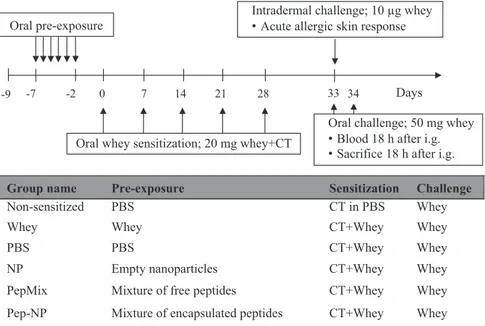
To study oral tolerance induction prior to whey sensitization, mice received daily, for a period of 6 days, an oral gavage with 1) 0.5 ml PBS, 2) 50 mg whey protein (DMV International, Veghel, The Netherlands), 3) a mixture of free peptides (0.04 mg of each BLG-derived peptide; referred to as PepMix), 4) empty nanoparticles (21.6 mg; referred to as NP), 5) a mixture of the encapsulated peptides (peptide 1, 3 and 4) combined with free peptide 2 because this peptide could not be loaded (referred to as Pep-NP; Pep-NP contained 0.04 mg of each peptide and 21.6 mg PLGA matrix) (all amounts were dissolved or dispersed in 0.5 ml PBS) (Fig. 1). The NP and Pep-NP suspensions were prepared
Table 1
Sequences and chemical characteristics of the BLG-derived peptides.
Sequence pI logP
Peptide 1 QKVAGTWYSLAMAASDIS 6.03 −11 Peptide 2 WYSLAMAASDISLLDAQS 3.48 −10.51 Peptide 3 AASDISLLDAQSAPLRVY 4.20 −11.62 Peptide 4 LLDAQSAPLRVYVEELKP 4.40 −8.64 pI, isoelectric point.
fresh and administered within 30 min after preparation. Next, mice were sensitized by means of weekly oral gavage with 20 mg whey protein (in 0.5 ml PBS) supplemented with 10μg cholera toxin (CT; List Biological Laboratories, Inc. California, USA) as an adjuvant. Sham-sensitized control mice received 10μg CT in 0.5 ml PBS. Five days after the last sensitization, an intradermal (i.d.) and an oral challenge were conducted as previously described (van Esch et al., 2011) with 10μg and 50 mg whey protein respectively. 18 h After the oral challenge with whey, mice were bled and euthanized.
2.4.3. Evaluation of the allergic response
To evaluate the possible allergic response against whey, mice were i.d. challenged in the ear pinnae with 10μg whey protein/20 μl PBS per ear. Before and 1 h after the i.d. injection, the ear thickness was eval-uated using a digital micrometer (Mitutoyo, Veenendaal, The Netherlands). Isoflurane was used for inhalational anesthesia during the ear measurements and the i.d. injection. The acute allergen-specific skin response, measured as the ear swelling, is calculated as the difference between the average ear thickness at 1 h time point and the average basal ear thickness (Δ = ear thickness at 1 h – basal ear thickness) and is expressed as delta micrometer.
2.4.4. Antibody detection in serum
Whey-specific antibody titers were detected in sera 18 h after the oral challenge with whey as previously described (van Esch et al., 2011).
2.4.5. Cell isolation from tissues
Spleen and mesenteric lymph nodes (MLN) cells were obtained by crushing the tissue through 70 µm cell strainers. Red blood cells in splenocyte suspensions were removed by 4-min incubation on ice with lysis buffer (composition described in Section 2.3.1.). For the small intestine lamina propria (SI-LP) cell isolation, the small intestine was cleared of Peyer's patches, washed in cold PBS, opened longitudinally, and minced in 0.5 cm segments. After an initial wash in Hank's Ba-lanced Salt Solution (HBSS; Invitrogen, Life Technologies, Carlsbad, CA, USA) supplemented with 15 mM HEPES (Gibco, Life Technologies, Carlsbad, CA, USA) pH 7.2, fragments were incubated 4 × 15 min in HBSS supplemented with 15 mM HEPES, 5 mM Na2-EDTA, 10% FBS and penicillin (100 U/ml)/streptomycin (100μg/ml), pH 7.2. Samples were washed in RPMI 1640 supplemented with 5% FBS and penicillin (100 U/ml)/streptomycin (100μg/ml) and incubated 2 × 45 min in RPMI 1640 containing 5% FBS, penicillin (100 U/ml)/streptomycin
(100μg/ml) and 0.5 mg/ml collagenase type VIII (Sigma-Aldrich). Tissue fragments were vortexed for 10 s after each incubation and poured over a 70 µm cell strainer to collect cells. Digestion was stopped by adding FBS up to 10% and washing cells in HBSS/15 mM HEPES solution. SI-LP cells were then purified by means of Percoll gradient centrifugation.
2.4.6. Allergen-specific re-stimulation and cytokine measurements Spleen and SI-LP cells were resuspended in RPMI 1640 supple-mented with 10% FBS and penicillin (100 U/ml)/streptomycin (100μg/ml). Next, 6 × 10⁵splenocytes or 4 × 10⁵SI-LP cells (200μl) per well were cultured in a round-bottom culture plate and either in-cubated with medium or with 500μg/ml whey protein at 37 °C, 5% CO2. Supernatants were collected 5 days later and stored at −20 °C until further analysis. The concentrations of the cytokines IL-5, IL-13, IL-10, IL-17A and IFN-γ in the supernatants were measured using a Cytometric Bead Array (CBA) Flex Set assay (BD Biosciences) following manufacturer's instructions. Cytokine measurements in serum samples were performed using the CBA Mouse Th1/Th2/Th17 Cytokine kit (BD Biosciences) and IL-13 CBA Flex Set assay according to BD Biosciences protocol. Results were obtained with BD FACSCanto IIflow cytometer (Becton Dickinson, Franklin Lakes, NJ, USA) and analyzed with FCAP v.3.0 software (Becton Dickinson). IL-6, TNF-α and TGF-β concentra-tions were measured using ELISA according to manufacturer's protocol (eBioscience). Absorbance was measured at 450 nm on a Benchmark plate reader (Bio-Rad, Veenendaal, The Netherlands). To determine the whey-induced cytokine response, cytokine concentrations measured in the medium-stimulated wells were subtracted from the cytokine con-centrations measured in the whey-stimulated wells. Any negative va-lues resulting from the subtraction were replaced by zero in order to reflect levels specifically induced by the re-exposure to the whole whey allergen.
2.5. Flow cytometry
Spleen, MLN, SI-LP or BMDC cells were resuspended in PBS-1% BSA. Non-specific binding sites were blocked by incubating the cells for 15 min with anti-mouse CD16/CD32 (Mouse BD Fc Block; BD Pharmingen, San Jose, CA, USA) in PBS- 1% BSA-5% FBS buffer. DC and BMDC were extracellularly stained for 30 min on ice with CD11c-PerCp-Cy5.5, CD11b-PE, CD11b-PE-Cy7, MHC class PE, MHC class II-PE-Cy7, CD40-FITC, CD80-APC, CD86-FITC, OX40L-APC and F4/80-APC-Cy7 (all from eBiosciences, San Diego, CA, USA). For analyzing T
Group name Pre-exposure Sensitization Challenge
Non-sensitized PBS CT in PBS Whey
Whey Whey CT+Whey Whey
PBS PBS CT+Whey Whey
NP Empty nanoparticles CT+Whey Whey
PepMix Mixture of free peptides CT+Whey Whey
Pep-NP Mixture of encapsulated peptides CT+Whey Whey -7 -2
Oral pre-exposure
0 7 14 21 28
Oral whey sensitization; 20 mg whey+CT 33
Intradermal challenge; 10 µg whey Acute allergic skin response
Oral challenge; 50 mg whey Blood 18 h after i.g. Sacrifice 18 h after i.g.
-9 34 Days
Fig. 1. Treatment groups and a schematic overview of the animal model for CMA prevention; CT, cholera toxin.
cell subsets in the in vivo study, cells werefirst extracellularly stained with CD4-PerCp-Cy5.5, and CD25-AlexaFluor488 (all from eBiosciences). To exclude dead cells, fixable viability dye eFluor780 (eBioscience) was used. For detecting Foxp3 transcription factor, cells were first fixated and permeabilized with Foxp3 Staining Buffer Set (eBioscience) according to manufacturer's protocol and then incubated with Foxp3-PE-Cy7 antibody (eBioscience). BMDC from the uptake study were only extracellularly stained with CD11c-FITC andfixable viability dye eFluor450 (both from eBioscience). Results were collected with BD FACSCanto II flow cytometer and analyzed with FlowLogic software (Inivai Technologies, Mentone, VIC, Australia).
2.6. Statistical analysis
All data were analyzed with GraphPad Prism 6.0 software for Macintosh (GraphPad Software, San Diego, CA, USA). Multiple com-parison test for selected pairs was applied with the following compar-isons: i) all treatment groups were compared to PBS, ii) NP was com-pared to Pep-NP and iii) PepMix was comcom-pared to Pep-NP. For data analysis, one-way ANOVA followed by Bonferroni's multiple compar-ison post hoc test was used. When data were not normally distributed or variance between the groups significantly differed, an appropriate data transformation was applied before the one-way ANOVA analysis. When ANOVA's assumptions were not met after transformation, the non-parametric Kruskal-Wallis test followed by a Dunn's post hoc test was used. Data are presented as mean ± S.E.M. of 5–8 animals per group and immunoglobulin levels are presented in Tukey box-and-whisker plots. P < 0.05 is considered statistically significant.
3. Results
3.1. Preparation and characteristics of the PLGA nanoparticles loaded with synthetic BLG-derived peptides
Both the empty and the peptide-loaded PLGA formulations were prepared by a double emulsion solvent evaporation technique and
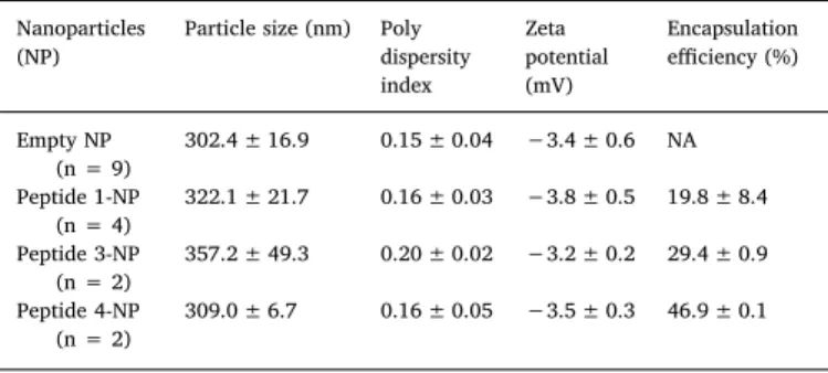
showed similar characteristics with sizes of 302 ± 17 nm (n = 9 in-dependent batches) and 328 ± 30 nm (n = 8 inin-dependent batches) respectively (Table 2). Peptide 2 could not be successfully encapsulated (encapsulation efficiency 0–3%), resulting in the addition of peptide 2 in free form to the Pep-NP mixture.
The in vitro release kinetics of the peptide-loaded NP revealed that two of the encapsulated peptides presented a biphasic release profile with an initial burst release of 55% for peptide 3 and 10% for peptide 4 (Fig. S1). After a week of low or no release, a phase of sustained release was observed for those peptides. Peptide 1, on the other hand, was rapidly released from the PLGA nanoparticles (complete burst release within 2 days; data not shown).
3.2. BMDC efficiently internalize empty PLGA nanoparticles
BMDC uptake studies showed that already after 2 h of incubation, the lowest amount of NP (0.01 mg/ml) resulted in about 70% of cells being positive for thefluorescent label. Increasing the incubation time as well as increasing the amount of NP resulted in higher nanoparticle uptake (Fig. 2A). Cell viability was not compromised when BMDC were incubated with NP for an increasing amount of time (Fig. 2B). However, the highest dose of NP (0.5 mg/ml) resulted in lower percentages of live cells. After 24 h incubation, already the lowest NP concentration (0.01 mg/ml) led to approximately 90% of the cells being positive for the fluorescent label demonstrating that the cells efficiently inter-nalized the PLGA NP (Fig. 2C). Importantly, a dose-dependent increase was observed in the amount of label per cell as indicated by the increase in medianfluorescence intensity (MFI) (Fig. 2C). Based on these data and literature (Elamanchili et al., 2004), in follow-up BMDC experi-ments the 0.2 mg/ml dose of NPs was used and cells were incubated for 24 h.
3.3. Activation of BMDC by peptide-loaded PLGA nanoparticles
After 24 h of incubation, surface expression of activation and co-stimulation markers was studied. Representativeflow cytometry plots and histograms show the gating strategy used (Fig. 3A). Both NP and Pep-NP increased the expression of CD40 compared to medium whereas Pep-NP led to more CD40 expression compared to PepMix (Fig. 3B). CD80 expression, on the other hand, tended to be lower in the Pep-NP group compared to medium (P = 0.798) (Fig. 3B). Incubation of BMDC with NP, PepMix and Pep-NP did not affect the expression of CD86 and OX40L (Fig. 3B). In contrast, LPS reference control induced the ex-pression of all four co-stimulatory molecules, while the OX40L mix stimulated the expression of OX40L, CD80 and CD86.
Next, either NP or Pep-NP resulted in BMDC secreting more IL-6 and TNF-α compared to medium (Fig. 3C). Further, comparing PepMix with Pep-NP, it is observed that encapsulating the BLG-peptides stimulates production of IL-6 and TNF-α, while preventing IL-12 secretion (Fig. 3C). IL-10 remained below detection limit for all groups.
Fig. 2. Cellular uptake of the empty PLGA delivery vehicle. Fluorescent-labeled empty PLGA NP were incubated with BMDC and the uptake (A) and the viability of the BMDC (B) were evaluated byflow cytometry. Percentage of cells positive for the fluorescent dye and the amount of dye taken up per cell (presented as MFI) for the 24 h time point are presented in (C). MFI, medianfluorescent intensity.
Table 2
Characteristics of nanoparticle preparations (mean ± S.D.).
Nanoparticles (NP)
Particle size (nm) Poly dispersity index Zeta potential (mV) Encapsulation efficiency (%) Empty NP (n = 9) 302.4 ± 16.9 0.15 ± 0.04 −3.4 ± 0.6 NA Peptide 1-NP (n = 4) 322.1 ± 21.7 0.16 ± 0.03 −3.8 ± 0.5 19.8 ± 8.4 Peptide 3-NP (n = 2) 357.2 ± 49.3 0.20 ± 0.02 −3.2 ± 0.2 29.4 ± 0.9 Peptide 4-NP (n = 2) 309.0 ± 6.7 0.16 ± 0.05 −3.5 ± 0.3 46.9 ± 0.1
3.4. Oral delivery of BLG-derived peptides loaded in PLGA nanoparticles tends to reduce the acute allergic skin response to whole whey protein
After an i.d. challenge with whey, it was observed that prior ex-posure to the empty NP showed a similar result as the PBS control, while pre-exposure to the Pep-NP mixture containing encapsulated peptides partially prevented the acute allergic skin response to whole protein when compared to the empty NP (Fig. 4A). The same tendency was observed when Pep-NP was compared to PBS-pre-exposed allergic mice. Administration of PepMix alone was not effective possibly due to the low dose used. The effect of Pep-NP on the acute allergic skin re-sponse was associated with the encapsulation as the simultaneous ad-ministration of the PepMix and the PLGA matrix was not effective (data not shown).
When examining the effect of the pretreatments on the sensitization to whey protein, we observed that PBS-pre-exposed whey-sensitized mice had significantly elevated and BLG-specific IgE and whey-specific IgG1 levels, while IgG2a was not affected (Fig. 4B and C). Pre-exposure to whole whey protein, but no other pretreatments, prevented the increase in these immunoglobulins.
3.5. No effect on co-stimulatory molecules expression on DC and regulatory T cells in vivo
To investigate the effect of peptide-loaded PLGA nanoparticles on DC activation in vivo, we studied the expression of co-stimulatory mo-lecules on CD11c+MHC-II+F4/80-DC in the MLN and SI-LP. Exposure to the various treatments prior to sensitization did not affect the
Fig. 3. Effect of empty and peptide-loaded PLGA NP on BMDC activation in vitro. 7–8 days-old BMDC were incubated for 24 h with empty NP, PepMix or Pep-NP, followed by BMDC phenotyping and cytokine measurement in the supernatants. Stimulation with LPS or OX40L-inducing mix was included as a positive control. Representative plots of the gating strategy including MFI are presented in (A). The expression of CD40, CD80, CD86 and OX40L was determined using the MFI of the CD11+MHCII+CD11b+BMDC population (B). Concentrations
of TNF-α, IL-6 and IL-12 measured in the BMDC supernatants are presented in (C). Data are presented as mean ± S.E.M. of n = 5–6, except for CD86 and OX40L where n = 2–3. * P < 0.05, ** P < 0.01, *** P < 0.001, **** P < 0.0001 as analyzed with one-way ANOVA followed by Bonferroni's post hoc test for selected pairs (TNF-α, IL-6 and IL-12 after LOG transformation). Comparisons between medium and LPS or OX40L were analyzed with one-way ANOVA (TNF-α, 6 and 12 after LOG transformation), except for CD40, 6 and IL-12 where the non-parametric Kruskal-Wallis test, followed by Dunn's post hoc test was used; FMO,fluorescence minus one control.
frequency of the DC (data not shown) or the expression of CD40, CD80 and CD86 molecules (Fig. S2) in both the MLN and SI-LP. Further, in all groups hardly any OX40L expression was detected on in vivo DC (data not shown).
Investigating the involvement of regulatory T (Treg) cells in this CMA mouse model, it was observed that the frequency of CD25+Foxp3+Tregsin the spleen, MLN and SI-LP remained unaffected in all groups (data not shown).
3.6. Low cytokine secretion by spleen cells from Pep-NP-pretreated mice after re-exposure to the allergen
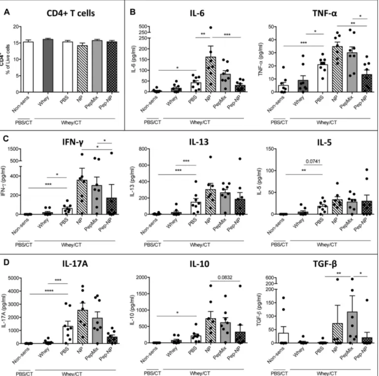
To determine the functional response of immune cells upon direct exposure to the whole whey protein, splenocytes and SI-LP cells were ex vivo re-stimulated with whey. Spleen cells did not differ in their fre-quency of CD4+T cells at the start of the re-stimulation (Fig. 5A). After a 5-day incubation with whole whey protein, whey-specific IL-6 se-cretion was increased in the NP group but this was prevented when the BLG-derived peptides were loaded in the NP (Fig. 5B). Pep-NP pre-ex-posure also resulted in significantly lower production of the in-flammatory TNF-α (Fig. 5B), the T helper 1 (Th1)-polarizing IFN-γ (Fig. 5C) and the regulatory TGF-β (Fig. 5D) cytokines compared to the NP and/or PepMix pre-exposures, maintaining these levels similar to those in the PBS pre-exposed mice. Furthermore, whey-induced IL-17A and IL-10 secretion remained low in the Pep-NP group (Fig. 5D), while no effect of the different pretreatments was detected on the T helper 2 (Th2)-associated IL-5 and IL-13 production compared to PBS-pretreat-ment (Fig. 5C). In addition, cytokine levels in serum were measured 18 h after the oral challenge with whole whey and they were found to follow the same pattern as in the splenocyte supernatants (Fig. S3). 3.7. Exposure to Pep-NP prior to sensitization lowers the CD4+T-cell frequency in the SI-LP and maintains whey-specific TGF-β secretion
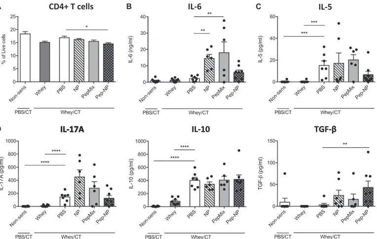
SI-LP cells were isolated and re-stimulated with whole whey protein to study the effect of the NP, PepMix and Pep-NP pretreatments on the mucosal immune response in the gut. Prior to re-stimulation,flow cy-tometric analysis revealed that Pep-NP pretreatment reduced the CD4+
T cell frequency in the SI-LP compared to PBS-pretreatment (Fig. 6A). NP or PepMix pre-exposures increased IL-6 secretion (Fig. 6B) com-pared to the PBS-pre-exposed group, but on the contrary, IL-6 remained low in the Pep-NP group and resembled levels in the PBS-pretreatment group. Also IL-5 (Fig. 6C), IL-17A and IL-10 (Fig. 6D) concentrations remained low in the Pep-NP group and resembled those in the PBS-pretreated mice. On the other hand, whey-specific TGF-β concentration in the SI-LP supernatant of the Pep-NP group was increased compared to this of the PBS-pretreated CMA controls (Fig. 6D). IL-13, IFN-γ, and TNF-α levels remained below detection limit in all SI-LP supernatants. 4. Discussion
In the current study, the applicability of encapsulating a low dose of BLG-derived peptides in PLGA nanoparticles for CMA prevention was investigated.
For the current study, three of the four BLG-derived peptides were successfully loaded in PLGA nanoparticles, of which peptide 3 and 4 were released in a biphasic manner as expected for PLGA particles (Makadia and Siegel, 2011), indicating successful encapsulation with a sustained release profile. Indeed oral administration of the mixture containing the encapsulated peptides tended to partially prevent the acute allergic skin reaction to the whole whey allergen while the free PepMix did not. This shows a proof-of-concept for the PLGA delivery system to facilitate the tolerogenic properties of the PepMix.
PLGA NP were successfully taken up in vitro by BMDC. However, incubation of BMDC with NP and Pep-NP did not enhance the expres-sion of conventional co-stimulatory molecules, except for CD40. High CD40 expression during antigen presentation to T cells is suggested to result in effector T cell activation (Chen and Flies, 2013), however, in vivo cross-linking of CD40 is shown not to abrogate oral tolerance in-duction (Chung et al., 2004). Importantly, Pep-NP had no effect on the expression of the Th2-skewing OX40L, while, as expected, the pros-taglandin E(2)-containing cytokine cocktail which served as a positive control, did induce the expression of this surface molecule (Blazquez and Berin, 2008; Krause et al., 2009). This suggests that uptake of the BLG peptides-loaded PLGA nanoparticles does not induce Th2-skewing DC.
Fig. 4. Acute allergic skin response and allergen-specific serum immunoglobulin levels. Five days after the last sensitization, mice were i.d. challenged with whey and the acute allergic skin response was measured (A). Whey-specific (B) and BLG-specific (C) IgE, IgG1 and IgG2a levels were measured in serum 18 h after oral challenge with whey. Data are presented as Tukey box-and-whisker plots for n = 7–8 per group. * P < 0.05, ** P < 0.01, *** P < 0.001. **** P < 0.0001; analysis with one-way ANOVA followed by Bonferroni's post hoc test for selected pairs without (A), or after LOG transformation (B); (C) is analyzed with Kruskal-Wallis non-parametric test, followed by Dunn's post hoc test for selected pairs. BLG, beta-lactoglobulin; CT, Cholera toxin.
BMDC exposed to NP or Pep-NP produced more IL-6 and TNF-α, pointing at a more mature and pro-inflammatory state of the BMDC (Lutz and Schuler, 2002), although these levels were very low com-pared to the LPS control. Alike Braun et al., the current study demon-strates that the expression of CD80 and CD86 co-stimulatory molecules does not necessarily associate with IL-6 and TNF-α production (Braun et al., 2010). Interestingly, PepMix enhanced IL-12 secretion by BMDC which may contribute to Th1 polarization (Berin and Shreffler, 2008), however loading the peptides in PLGA NP prevented this IL-12 secre-tion. This suggests that Pep-NP may induce incomplete maturation and activation of the BMDC, which might link to the underlying
immunomodulatory mechanism (Lutz and Schuler, 2002). In addition, recent data revealed that GM-CSF-induced BMDC cultures comprise a heterogeneous population of CD11c+MHC-II+ DC and macrophages (Helft et al., 2015). Even though in our study we observed clear LPS-driven expression of maturation markers, whichHelft et al. (2015) re-port to be more pronounced in CD11c+MHC-II+DC rather than mac-rophages, the measured cytokines might not be exclusively produced by DC, but also macrophages may have contributed.
Even though the in vitro results revealed some cell activation by NP and Pep-NP, the in vivo pretreatment with Pep-NP significantly pre-vented the acute allergic skin response to the whole whey protein
Fig. 5. Cytokine production by splenocytes. Spleen cells were isolated and the percentage of CD4+cells was evaluated byflow cytometry (A). Spleen cells were re-stimulated with
medium or whole whey for 5 days and inflammatory (B), Th1- and Th2-associated (C) or Th17- and Treg-associated (D) cytokines were measured in the supernatants. Data are presented as
mean ± S.E.M. for n = 7–8 per group. * P < 0.05, ** P < 0.01, *** P < 0.001. **** P < 0.0001; as analyzed with one-way ANOVA followed by Bonferroni's post hoc test for selected pairs (IL-10, IL-6 and TNF-α after square root transformation, the rest after LOG transformation); CT, Cholera toxin.
Fig. 7. Overview of the proof-of-concept. Encapsulating the BLG-derived peptides in PLGA NP results in maintaining low levels of whey-induced cytokines, while enhancing whey-induced reg-ulatory TGF-β. This suggests that Pep-NP stimulates a more tol-erogenic cytokine microenvironment in the small intestine which might contribute to the tendency of Pep-NP to partially suppress the acute allergic response.
Fig. 6. Cytokine production by cells in the gut. SI-LP cells were isolated and used forflow cytometry or re-stimulated with medium or whole whey protein for 5 days. Percentage CD4+T
cells within the live cells (A) was determined byflow cytometry. Concentrations of inflammatory (B), Th2-associated (C) or Th17- and Treg-associated (D) cytokines was measured in the
re-stimulation supernatants. Data are presented as mean ± S.E.M. of n = 6–8 per group. ** P < 0.01, *** P < 0.001. **** P < 0.0001; as analyzed with one-way ANOVA followed by Bonferroni's post hoc test for selected pairs (IL-6, IL-10 after square root transformation, the rest after LOG transformation); CT, Cholera toxin.
compared to the NP. However, this effect was not accompanied by re-duction of the allergen-specific immunoglobulin levels. Also in previous reports, suppression of allergic symptoms by pretreatments using pro-tein fragments, such as hydrolyates (van Esch et al., 2011) or peptides (Hoyne et al., 1997; Kostadinova et al., 2016; Meulenbroek et al., 2013; Pecquet et al., 2000a) was shown despite unaltered levels of allergen-specific immunoglobulins. In addition, the PepMix covers only a limited part of the BLG sequence which may be insufficient to prevent the generation of a broad panel of immunoglobulins directed against all allergenic epitopes present in whole whey protein. Therefore, in our future studies we will aim to expand the number of BLG-derived pep-tides and add peppep-tides of other major allergens in whey, such as alpha-lactalbumin, in order to cover a broader spectrum of epitopes. Inter-estingly, however, NP stimulated high whey-specific IgG2a levels, which, by contrast to IgE in mice, reflect a Th1 or regulatory T cell driven humoral response driving away from the allergic phenotype. This indicates some immune priming by NP which hereby may con-tribute to allergy prevention induced by the encapsulated BLG-derived peptides.
In contrast with the in vitro data, the CD40 surface expression on DC in the mouse model was not affected. This discrepancy might be due to different times between exposure to Pep-NP and the DC phenotyping (24 h for in vitro and 6 weeks for in vivo). It further implies that in vivo mechanistic investigation at earlier stages is necessary to reveal the location of nanoparticles uptake as well as any effects due to the po-tential uptake of NP, PepMix or Pep-NP by DC. Interestingly, IL-6 and TNF-α concentrations in supernatants of re-stimulated splenocytes of NP-pretreated mice resembled the results from the in vitro studies, while in vivo Pep-NP exposure resulted in significantly less IL-6 and TNF-α compared to the NP-pretreated mice. This suggests a different me-chanism of action of the Pep-NP when administered in vivo and un-derlies the partial silencing of the adaptive immune response for the whole whey protein when NP are loaded with the BLG-peptides.
Pretreatment with Pep-NP showed a tendency to reduce the acute skin response upon intradermal challenge with whole whey compared to the PBS control group. Neither NP, nor PepMix alone showed such tendency. This suggests that the Pep-NP approach might have a po-tential to improve the preventive capacity of PepMix. Even though Pep-NP did not ameliorate the levels of several cytokines (IL-6, TNF-α, IFN-γ, IL-5, IL-17A, and IL-10) as compared to the PBS group, we observed that in mice pretreated with Pep-NP the frequency of CD4+
T cells in the SI-LP was decreased while TGF-β was increased upon re-stimulation with the whole whey allergen when compared to the PBS group. Hence, these results found locally in the SI-LP suggest modulation in the mu-cosal immunity which is associated with the effect observed on the acute skin response. It further suggests that the Pep-NP might have an immune suppressive effect compared to the PBS control group. This, in combination with maintaining low levels of whey-induced pro-in-flammatory cytokines, suggests that Pep-NP might stimulate a more tolerogenic cytokine microenvironment in the small intestine (Fig. 7) upon whole whey exposure (Yu et al., 2016). Further improvements of the Pep-NP approach, such as improving the encapsulation efficiency and covering more epitopes from BLG, might enhance the preventive effect of Pep-NP.
Typically, NP alone enhanced IL-6 secretion by splenocytes and SI-LP cells, which indicates NP to prime the mucosal immune response. However, this priming might facilitate the tolerogenic capacity of the BLG-derived peptides since in the Pep-NP group this whey-induced immune activation was silenced. This was further associated with low levels of most of the whey-induced cytokines in the systemic com-partment and in the gut, and most importantly, associated with a partial suppression of the allergic symptoms. This suggests again that the NP create a milieu that facilitates the tolerogenic properties of the BLG-derived peptides.
In conclusion, this study provides a proof-of-concept that PLGA nanoparticles are a promising strategy for BLG peptides delivery which
also facilitates the capacity of those peptides to induce tolerance to the whole whey protein. Here we show that BLG-derived peptides en-capsulated in PLGA tend to reduce the allergic skin response in CMA mice. PLGA NP induce local intestinal and systemic immune priming resulting in increased production of whey-induced cytokines. However, this was silenced when the NP were loaded with BLG-peptides, while regulatory TGF-β production in the intestine was preserved. This ad-juvant action of PLGA may possibly underlie the mechanism by which PLGA NP facilitate tolerance induction by the BLG-derived peptides. Optimizing the efficacy of the Pep-NP approach by broadening the specter of T cell epitope-containing peptides could contribute to im-proving the effect on the humoral and effector cell responses and hence to contribute to the future development of effective strategies for CMA prevention in early life.
Conflict of interest
None of the authors have a competingfinancial interest in relation to the presented work; L.M.J. Knippels is employed by Nutricia Research. J. Garssen is partly employed by Nutricia Research. A. Kostadinova received funding from Nutricia Research.
Acknowledgements
This work wasfinancially supported by Nutricia Research as part of the strategic alliance between Nutricia Research and Utrecht Institute for Pharmaceutical Sciences. The funder had no involvement in the study design, the collection, analysis and interpretation of the results or the writing and submission of the manuscript.
Appendix A. Supporting information
Supplementary data associated with this article can be found in the online version athttp://dx.doi.org/10.1016/j.ejphar.2017.10.051. References
Bellach, J., Schwarz, V., Ahrens, B., Trendelenburg, V., Aksunger, O., Kalb, B., Niggemann, B., Keil, T., Beyer, K., 2017. Randomized placebo-controlled trial of hen's egg consumption for primary prevention in infants. J. Allergy Clin. Immunol. 139, 1591–1599 (e1592).
Berin, M.C., Shreffler, W.G., 2008. T(H)2 adjuvants: implications for food allergy. J. Allergy Clin. Immunol. 121, 1311–1320.
Blazquez, A.B., Berin, M.C., 2008. Gastrointestinal dendritic cells promote Th2 skewing via OX40L. J. Immunol. 180, 4441–4450.
Bogh, K.L., Barkholt, V., Madsen, C.B., 2013. The sensitising capacity of intact beta-lac-toglobulin is reduced by co-administration with digested beta-lacbeta-lac-toglobulin. Int. Arch. Allergy Immunol. 161, 21–36.
Bolhassani, A., Javanzad, S., Saleh, T., Hashemi, M., Aghasadeghi, M.R., Sadat, S.M., 2014. Polymeric nanoparticles potent vectors for vaccine delivery targeting cancer and infectious diseases. Hum. Vaccin. Immunother. 10, 321–332.
Braun, A., Bewersdorff, M., Lintelmann, J., Matuschek, G., Jakob, T., Gottlicher, M., Schober, W., Buters, J.T., Behrendt, H., Mempel, M., 2010. Differential impact of diesel particle composition on pro-allergic dendritic cell function. Toxicol. Sci. 113, 85–94.
Brotons-Canto, A., Martin-Arbella, N., Gamazo, C., Irache, J.M., 2016. New pharmaceu-tical approaches for the treatment of food allergies. Expert Opin. Drug Deliv. 1–12.
Chen, L., Flies, D.B., 2013. Molecular mechanisms of T cell co-stimulation and co-in-hibition. Nat. Rev. Immunol. 13, 227–242.
Chung, Y., Kim, D., Lee, S., Kang, C., 2004. Co-administration of CD40 agonistic antibody and antigen fails to overcome the induction of oral tolerance. Immunology 111, 19–26.
Du Toit, G., Roberts, G., Sayre, P.H., Bahnson, H.T., Radulovic, S., Santos, A.F., Brough, H.A., Phippard, D., Basting, M., Feeney, M., Turcanu, V., Sever, M.L., Gomez Lorenzo, M., Plaut, M., Lack, G., Team, L.S., 2015. Randomized trial of peanut consumption in infants at risk for peanut allergy. N. Engl. J. Med. 372, 803–813.
Elamanchili, P., Diwan, M., Cao, M., Samuel, J., 2004. Characterization of poly(d,l-lactic-co-glycolic acid) based nanoparticulate system for enhanced delivery of antigens to dendritic cells. Vaccine 22, 2406–2412.
Helft, J., Bottcher, J., Chakravarty, P., Zelenay, S., Huotari, J., Schraml, B.U., Goubau, D., Reis e Sousa, C., 2015. GM-CSF mouse bone marrow cultures comprise a hetero-geneous population of CD11c(+)MHCII(+) macrophages and dendritic cells. Immunity 42, 1197–1211.
specificity and duration of T cell tolerance to intranasally administered peptides in mice: a role for intramolecular epitope suppression. Int. Immunol. 9, 1165–1173.
Koletzko, S., Niggemann, B., Arato, A., Dias, J.A., Heuschkel, R., Husby, S., Mearin, M.L., Papadopoulou, A., Ruemmele, F.M., Staiano, A., Schappi, M.G., Vandenplas, Y., European Society of Pediatric Gastroenterology, H., Nutrition, 2012. Diagnostic ap-proach and management of cow's-milk protein allergy in infants and children: ESPGHAN GI Committee practical guidelines. J. Pediatr. Gastroenterol. Nutr. 55, 221–229.
Kostadinova, A.I., Meulenbroek, L.A., van Esch, B.C., Hofman, G.A., Garssen, J., Willemsen, L.E., Knippels, L.M., 2016. A specific mixture of fructo-oligosaccharides and bifidobacterium breve M-16V facilitates partial non-responsiveness to whey protein in mice orally exposed to beta-lactoglobulin-derived peptides. Front. Immunol. 7, 673.
Krause, P., Bruckner, M., Uermosi, C., Singer, E., Groettrup, M., Legler, D., 2009. Prostaglandin E(2) enhances T-cell proliferation by inducing the costimulatory mo-lecules OX40L, CD70, and 4-1BBL on dendritic cells. Blood 113, 2451–2460.
Lutsiak, M.E., Robinson, D.R., Coester, C., Kwon, G.S., Samuel, J., 2002. Analysis of poly (D,L-lactic-co-glycolic acid) nanosphere uptake by human dendritic cells and mac-rophages in vitro. Pharm. Res. 19, 1480–1487.
Lutz, M.B., Kukutsch, N., Ogilvie, A.L., Rossner, S., Koch, F., Romani, N., Schuler, G., 1999. An advanced culture method for generating large quantities of highly pure dendritic cells from mouse bone marrow. J. Immunol. Methods 223, 77–92.
Lutz, M.B., Schuler, G., 2002. Immature, semi-mature and fully mature dendritic cells: which signals induce tolerance or immunity? Trends Immunol. 23, 445–449.
Makadia, H.K., Siegel, S.J., 2011. Poly lactic-co-glycolic acid (PLGA) as biodegradable controlled drug delivery carrier. Polymers 3. pp. 1377–1397.
McHugh, K.J., Guarecuco, R., Langer, R., Jaklenec, A., 2015. Single-injection vaccines: progress, challenges, and opportunities. J. Control. Release 219, 596–609.
Meulenbroek, L.A., van Esch, B.C., Hofman, G.A., den Hartog Jager, C.F., Nauta, A.J., Willemsen, L.E., Bruijnzeel-Koomen, C.A., Garssen, J., van Hoffen, E., Knippels, L.M., 2013. Oral treatment with beta-lactoglobulin peptides prevents clinical symptoms in a mouse model for cow's milk allergy. Pediatr. Allergy Immunol. 24, 656–664.
Nicolete, R., dos Santos, D.F., Faccioli, L.H., 2011. The uptake of PLGA micro or nano-particles by macrophages provokes distinct in vitro inflammatory response. Int. Immunopharmacol. 11, 1557–1563.
Nissen, S.P., Kjaer, H.F., Host, A., Nielsen, J., Halken, S., 2013. The natural course of sensitization and allergic diseases from childhood to adulthood. Pediatr. Allergy Immunol. 24, 549–555.
Pabst, O., Mowat, A.M., 2012. Oral tolerance to food protein. Mucosal Immunol. 5, 232–239.
Palmer, D.J., Metcalfe, J., Makrides, M., Gold, M.S., Quinn, P., West, C.E., Loh, R., Prescott, S.L., 2013. Early regular egg exposure in infants with eczema: a randomized controlled trial. J. Allergy Clin. Immunol. 132, 387–392 (e381).
Pavot, V., Berthet, M., Resseguier, J., Legaz, S., Handke, N., Gilbert, S.C., Paul, S., Verrier, B., 2014. Poly(lactic acid) and poly(lactic-co-glycolic acid) particles as versatile carrier platforms for vaccine delivery. Nanomedicine 9, 2703–2718.
Pecquet, S., Bovetto, L., Maynard, F., Fritsche, R., 2000a. Peptides obtained by tryptic hydrolysis of bovine beta-lactoglobulin induce specific oral tolerance in mice. J. Allergy Clin. Immunol. 105, 514–521.
Pecquet, S., Leo, E., Fritsché, R., Pfeifer, A., Couvreur, P., Fattal, E., 2000b. Oral tolerance elicited in mice by beta-lactoglobulin entrapped in biodegradable microspheres. Vaccine 18, 1196–1202.
Perkin, M.R., Logan, K., Tseng, A., Raji, B., Ayis, S., Peacock, J., Brough, H., Marrs, T., Radulovic, S., Craven, J., Flohr, C., Lack, G., Team, E.A.T.S., 2016. Randomized Trial of Introduction of Allergenic Foods in Breast-Fed Infants. N. Engl. J. Med. 374, 1733–1743.
Prescott, S.L., Smith, P., Tang, M., Palmer, D.J., Sinn, J., Huntley, S.J., Cormack, B., Heine, R.G., Gibson, R.A., Makrides, M., 2008. The importance of early com-plementary feeding in the development of oral tolerance: concerns and controversies. Pediatr. Allergy Immunol. 19, 375–380.
Rahimian, S., Fransen, M.F., Kleinovink, J.W., Amidi, M., Ossendorp, F., Hennink, W.E., 2015. Particulate systems based on poly(Lactic-co-Glycolic)acid (pLGA) for im-munotherapy of cancer. Curr. Pharm. Des. 21, 4201–4216.
Saps, M., Lu, P., Bonilla, S., 2011. Cow's-milk allergy is a risk factor for the development of FGIDs in children. J. Pediatr. Gastroenterol. Nutr. 52, 166–169.
Silva, A.L., Rosalia, R.A., Varypataki, E., Sibuea, S., Ossendorp, F., Jiskoot, W., 2015. Poly-(lactic-co-glycolic-acid)-based particulate vaccines: particle uptake by dendritic cells is a key parameter for immune activation. Vaccine 33, 847–854.
van Esch, B.C., Schouten, B., de Kivit, S., Hofman, G.A., Knippels, L.M., Willemsen, L.E., Garssen, J., 2011. Oral tolerance induction by partially hydrolyzed whey protein in mice is associated with enhanced numbers of Foxp3+ regulatory T-cells in the me-senteric lymph nodes. Pediatr. Allergy Immunol. 22, 820–826.
Venter, C., Arshad, S.H., 2011. Epidemiology of food allergy. Pediatr. Clin. North Am. 58, 327–349.
Yu, W., Freeland, D.M., Nadeau, K.C., 2016. Food allergy: immune mechanisms, diagnosis and immunotherapy. Nat. Rev. Immunol. 16, 751–765.