Published by
Baishideng Publishing Group Inc
World Journal of
Clinical Cases
World J Clin Cases 2018 September 26; 6(10): 308-405
World Journal of
Clinical Cases
W J C C
Contents
Semimonthly Volume 6 Number 10 September 26, 2018
REVIEW
308 Algorithm for the multidisciplinary management of hemorrhage in EUS-guided drainage for pancreatic
fluid collections
Jiang TA, Xie LT
322 Mystery behind labial and oral melanotic macules: Clinical, dermoscopic and pathological aspects of
Laugier-Hunziker syndrome
Duan N, Zhang YH, Wang WM, Wang X
MINIREVIEWS
335 Research progress on signaling pathways in cirrhotic portal hypertension
Xu W, Liu P, Mu YP
344 Gastrointestinal toxicity induced by microcystins
Wu JX, Huang H, Yang L, Zhang XF, Zhang SS, Liu HH, Wang YQ, Yuan L, Cheng XM, Zhuang DG, Zhang HZ
ORIGINAL ARTICLE
Basic Study
355 PNPLA3 rs139051 is associated with phospholipid metabolite profile and hepatic inflammation in nonalcoholic fatty liver disease
Luo JJ, Cao HX, Yang RX, Zhang RN, Pan Q
Retrospective Study
365 Recurrent carpal tunnel syndrome: Evaluation and treatment of the possible causes
Eroğlu A, Sarı E, Topuz AK, Şimşek H, Pusat S
373 Adjuvant chemotherapy with S-1 plus oxaliplatin improves survival of patients with gastric cancer after D2
gastrectomy: A multicenter propensity score-matched study
Ren DF, Zheng FC, Zhao JH, Shen GS, Ahmad R, Zhang SS, Zhang Y, Kan J, Dong L, Wang ZY, Zhao FX, Zhao JD
CASE REPORT
384 Rectal perforation by inadvertent ingestion of a blister pack: A case report and review of literature
World Journal of
Clinical Cases
W J C C
Contents
Semimonthly Volume 6 Number 10 September 26, 2018
II
WJCC|www.wjgnet.com September 26, 2018|Volume 6|Issue 10|
393 Unusual complication in patient with Gardner’s syndrome: Coexistence of triple gastrointestinal perforation and lower gastrointestinal bleeding: A case report and review of literature
Akbulut S, Koc C, Dirican A
398 Laparoscopic repair via the transabdominal preperitoneal procedure for bilateral lumbar hernia: Three cases
report and review of literature
Contents
Volume 6 Number 10 September 26, 2018
World Journal of Clinical Cases
EDITORS FOR
THIS ISSUE
Responsible Assistant Editor: Xiang Li Responsible Science Editor: Fang-Fang Ji
Responsible Electronic Editor: Yun-XiaoJian Wu Proofing Editorial Office Director: Jin-Lei Wang
Proofing Editor-in-Chief: Lian-Sheng Ma
World Journal of Clinical Cases
Baishideng Publishing Group Inc
7901 Stoneridge Drive, Suite 501, Pleasanton, CA 94588, USA Telephone: +1-925-2238242
Fax: +1-925-2238243
E-mail: [email protected] Help Desk: http://www.f6publishing.com/helpdesk http://www.wjgnet.com
PUBLISHER
Baishideng Publishing Group Inc 7901 Stoneridge Drive,
Suite 501, Pleasanton, CA 94588, USA Telephone: +1-925-2238242 Fax: +1-925-2238243 E-mail: [email protected]
Help Desk: http://www.f6publishing.com/helpdesk http://www.wjgnet.com
PUBLICATION DATE September 26, 2018
COPYRIGHT
© 2018 Baishideng Publishing Group Inc. Articles published by this Open Access journal are distributed under the terms of the Creative Commons Attribu-tion Non-commercial License, which permits use, dis-tribution, and reproduction in any medium, provided the original work is properly cited, the use is non commercial and is otherwise in compliance with the license.
SPECIAL STATEMENT
All articles published in journals owned by the Baishideng Publishing Group (BPG) represent the views and opinions of their authors, and not the views, opinions or policies of the BPG, except where other-wise explicitly indicated.
INSTRUCTIONS TO AUTHORS http://www.wjgnet.com/bpg/gerinfo/204 ONLINE SUBMISSION
http://www.f6publishing.com
ABOUT COVER
AIM AND SCOPE
INDExING/ABSTRACTING
NAME OF JOURNAL
World Journal of Clinical Cases
ISSN ISSN 2307-8960 (online) LAUNCH DATE April 16, 2013 FREQUENCY Semimonthly EDITORS-IN-CHIEF
Sandro Vento, MD, Department of Internal Medicine,
University of Botswana, Private Bag 00713, Gaborone, Botswana
EDITORIAL BOARD MEMBERS
All editorial board members resources online at http:// www.wjgnet.com/2307-8960/editorialboard.htm EDITORIAL OFFICE
Jin-Lei Wang, Director
Editorial Board Member of World Journal of Clinical Cases, Young-Seok Cho, MD, PhD, Professor, Division of Gastroenterology, Department of Internal Medicine, Seoul St. Mary's Hospital, the Catholic University of Korea, Seoul 06591, South Korea
World Journal of Clinical Cases (World J Clin Cases, WJCC, online ISSN 2307-8960, DOI:
10.12998) is a peer-reviewed open access academic journal that aims to guide clinical practice and improve diagnostic and therapeutic skills of clinicians.
The primary task of WJCC is to rapidly publish high-quality Autobiography, Case
Re-port, Clinical Case Conference (Clinicopathological Conference), Clinical Management, Diagnostic Advances, Editorial, Field of Vision, Frontier, Medical Ethics, Original Ar-ticles, Clinical Practice, Meta-Analysis, Minireviews, Review, Therapeutics Advances, and Topic Highlight, in the fields of allergy, anesthesiology, cardiac medicine, clinical genetics, clinical neurology, critical care, dentistry, dermatology, emergency medicine, endocrinol-ogy, family medicine, gastroenterology and hepatolendocrinol-ogy, geriatrics and gerontolendocrinol-ogy, he-matology, immunology, infectious diseases, internal medicine, obstetrics and gynecology, oncology, ophthalmology, orthopedics, otolaryngology, pathology, pediatrics, peripheral vascular disease, psychiatry, radiology, rehabilitation, respiratory medicine, rheumatology, surgery, toxicology, transplantation, and urology and nephrology.
World Journal of Clinical Cases (WJCC) is now indexed in PubMed, PubMed Central, Science
Citation Index Expanded (also known as SciSearch®), and Journal Citation Reports/Science Edition. The 2018 Edition of Journal Citation Reports cites the 2017 impact factor for WJCC as 1.931 (5-year impact factor: N/A), ranking WJCC as 60 among 154 journals in Medicine, General and Internal (quartile in category Q2).
CARE Checklist (2013) statement: The authors have read the
CARE Checklist (2013), and the manuscript was prepared and revised according to the CARE Checklist (2013).
Open-Access: This article is an open-access article which was
selected by an in-house editor and fully peer-reviewed by external reviewers. It is distributed in accordance with the Creative Commons Attribution Non Commercial (CC BY-NC 4.0) license, which permits others to distribute, remix, adapt, build upon this work non-commercially, and license their derivative works on different terms, provided the original work is properly cited and the use is non-commercial. See: http://creativecommons.org/ licenses/by-nc/4.0/
Manuscript source: Invited manuscript
Correspondence to: Francesco Fleres, MD, Doctor, Medical
Assistant, Surgeon, Department of Youth and Adulthood
Human Pathology “Gaetano Barresi”, General Surgery Unit, University of Messina, Via Consolare Valeria, Messina 98125, Italy. [email protected]
Telephone: +39-090-2212678 Fax: +39-090-2213524 Received: May 16, 2018
Peer-review started: May 16, 2018 First decision: May 24, 2018 Revised: July 26, 2018 Accepted: August 26, 2018 Article in press: August 27, 2018 Published online: September 26, 2018
Abstract
The accidental ingestion of a foreign body (FB) is a relatively common condition. In the present study, we report a peculiar case of rectal perforation, the first to our knowledge, caused by the inadvertent ingestion of a blister pill pack. The aim of this report is to illustrate the difficulties of the case from a diagnos-tic and therapeudiagnos-tic viewpoint as well as its unusual
Francesco Fleres, Antonio Ieni, Edoardo Saladino, Giuseppe Speciale, Michele Aspromonte, Antonio Cannaò,
Antonio Macrì
CASE REPORT
384 September 26, 2018|Volume 6|Issue 10| WJCC|www.wjgnet.com
Rectal perforation by inadvertent ingestion of a blister
pack: A case report and review of literature
Francesco Fleres, Michele Aspromonte, Department of
Human Pathology of the Adult and Evolutive Age “Gaetano Barresi”, Section of General Surgery, University of Messina, Messina 98125, Italy
Antonio Ieni, Giuseppe Speciale, Department of Human Pathology of the Adult and Evolutive Age “Gaetano Barresi”, Section of Anatomic Pathology, University of Messina, Messina 98125, Italy
Edoardo Saladino, General and Oncologic Surgery Unit, Clinica Cappellani-GIOMI, Messina 98168, Italy
Antonio Cannaò, Messina University Medical School Hospital, Messina 98125, Italy
Antonio Macrì, Peritoneal Surface Malignancy and Soft Tissue
Sarcoma Program, Messina University Medical School Hospital, Messina 98125, Italy
ORCID number: Francesco Fleres (0000-0002-1092-8975);
Antonio Ieni (0000-0003-2878-3572); Edoardo Saladino (0000 -0002-4528-6626); Giuseppe Speciale (0000-0001-5875-0785); Michele Aspromonte (0000-0001-7132-9493); Antonio Cannaò (0000-0001-5198-1222); Antonio Macrì (0000-0003-2671-5314). Author contributions: Fleres F, Saladino E, Aspromonte M and Macrì A participated in the conception and design of the report; Fleres F and Macrì A drafted the paper and analyzed the report; Macrì A performed the surgical procedure; Macrì A was involved in the diagnosis, surgical management and follow-up of the patient; Cannaò A was involved in the patient’s surgical management; Ieni A and Speciale G carried out the histological procedures.
Conflict-of-interest statement: All authors have no conflicts of
interest to report.
Informed consent statement: Written informed consent was
obtained from the patient ahead of the publication of this Case Report and its accompanying images. A copy of the written informed consent is available for review by the Editor-in-Chief of this journal.
Submit a Manuscript: http://www.f6publishing.com DOI: 10.12998/wjcc.v6.i10.384
World J Clin Cases 2018 September 26; 6(10): 384-392 ISSN 2307-8960 (online)
World Journal of
Clinical Cases
W J C C
presentation. A 75-year-old woman, mentally impaired, arrived at our emergency department in critical condition. The computed tomography scan revealed a substantial abdominopelvic peritoneal effusion and free perigastric air. The patient was therefore submitted to an urgent exploratory laparotomy; a 2-cm long, full-thickness lesion was identified in the anterior distal part of the intraperitoneal rectum. Hence, we performed a Hartmann’s procedure. Because of her critical condition, the patient was eventually transferred to the Intensive Care Unit, where she died after 10 d, showing no surgical complication. The ingestion of FBs is usually treated with observation or endoscopic removal. Less than 1% of FBs are likely to cause an intestinal perforation. The intestinal perforation resulting from the unintentional ingestion of an FB is often a difficult challenge when it comes to treatment, due to its late diagnosis and the patients’ deteriorated clinical condition.
Key words: Foreign body; Acute abdomen syndrome; Ingestion; Rectal perforation; Blister pill pack
© The Author(s) 2018. Published by Baishideng Publishing
Group Inc. All rights reserved.
Core tip: Ingestion of a foreign body (FB) is usually treated with observation or endoscopic removal. Less of 1% of FBs can cause an intestinal perforation. Diverticular disease and FB may be associated with pathological processes, including inflammation, perforation, abscess and fistula. The diagnosis of intestinal perforation following the unknown ingestion of a FB is a clinical challenge, first of all because it happens often in patients with intellectual disability or among the psychiatric population and secondly because it is not reported during questioning. Caregivers should be cautious and aware of the cutting of drug blisters.
Fleres F, Ieni A,Saladino E, Speciale G, Aspromonte M, Cannaò A, Macrì A. Rectal perforation by inadvertent ingestion of a blister pack: A case report and review of literature. World J Clin
Cases2018; 6(10): 384-392 Available from: URL: http://www. wjgnet.com/2307-8960/full/v6/i10/384.htm DOI: http://dx.doi. org/10.12998/wjcc.v6.i10.384
INTRODUCTION
The accidental ingestion of a foreign body (FB) is a relatively common condition, affecting patients of every age. However, the two age extremities, i.e. children and elderly people, are at a higher risk of inadvertently ingesting FBs, as are alcoholic, psychiatric or mentally impaired patients as well as patients affected by intellectual disability or neurological disorder[1].
Most FBs pass through the gastrointestinal (GI) tract without any complications. On the other hand, FBs such as fish bones, chicken bones and toothpicks can
Fleres F et al. Acute rare cause of rectal perforation cause perforation of the GI tract. As it turns out, in less than 1% of the cases, FB ingestion can result in acute surgical abdomen for intestinal perforation which needs emergency surgery[2].
In the present paper, we report the first case, to our knowledge, of rectal perforation caused by the inadvertent ingestion of a blister pill pack (BPP). BPPs are commonly used for drug storage, as they provide a protection barrier and ensure preservation from the damage.
In some countries, a BPP is known as a “push-through pack”. Push-“push-through packs consist of two main features: (1) the cover foil being resistant but breaking easily, so that the drug can be pressed out by easily breaking the cover foil; and (2) the semirigid formed cavity can be folded to dispense the drug by pressing it out with a thumb. In both cases, breaking the cover foil with a fingernail will make the pressing-out easier.
The aim of this report is to illustrate the difficulties of a FB ingestion case from diagnostic and therapeutic viewpoints as well as its unusual presentation.
Case RepORT
A 75-year-old woman was admitted to hospital for fever (39 ℃) and vomiting. The medical history of the patient showed arterial hypertension, diabetes mellitus type Ⅱ, uncontrolled hepatic cryptogenic cirrhosis with last MELD-score 19 and CHILD-score C-11, chronic metabolic failure, chronic heart failure, bilateral pulmonary thickening, urinary tract infections, previous uterine cervix carcinoma, chronic cerebral vasculopathy, major ischemic stroke and subarachnoid hemorrhage due to an accidental trauma. Since July 2017 the patient had been admitted to a rehabilitation center due to a pertrochanteric fracture of her left femur, which could not be treated surgically because of high operative risk.
The patient was admitted at the Department of Medicine in poor clinical condition, awake, noncoll-aborative, oriented only in space, dehydrated, with fever
(39 ℃) and hypotension (80/50 mmHg). At physical
examination, the abdomen appeared distended, without tenderness, and peristalsis was present. Due to persistent hypotension and oliguria (150 mL/24 h), the patient was treated with noradrenalin (4 phials in 40 mL of NaCl solution 0.9% at 0.2 mL/h). After 24 h, the hypotension persisted (85/40 mmHg) and the abdomen was becoming hyper-tympanic with a mild pain in the left hypochondrium accompanied by intestinal borborygmi. Forty-eight hours after hospitalization, the patient manifested an acute abdominal syndrome characterized by diffuse abdominal pain at superficial palpation with resistance in hypogastrium, pelvic pain and meteorism. The digital rectal examination revealed an empty ampulla recti, with traces of bright red blood.
The laboratory data revealed the following: white blood cell count of 25920 mmc, with 87% neutrophils;
386 WJCC|www.wjgnet.com
platelet count of 90000 mmc; hemoglobin of 10.9 gr%; C-reactive protein of 10.5 mg/dL; and procalcitonin of 34.9 mg/dL.
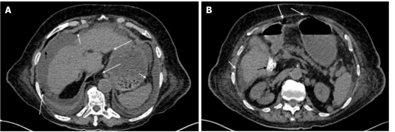
A rectal probe was inserted and a phial of trimebutin was administered without any benefit. The patient was therefore submitted to an abdominal X-ray and computed tomography (CT) (Figure 1), which revealed an abundant abdominopelvic peritoneal effusion and free perigastric air (along the gastrocolic ligament and under the anterior abdominal wall). Moreover, microlithiasis of the gallbladder as well as pericardial and bilateral pleural effusions were also present. Based on the CT findings, the origin of the perforation was suspected to be gastric.
On the basis of this clinical picture, the patient was transferred to our unit and we submitted her to an urgent exploratory laparotomy. The general clinical conditions were very bad, showing hypotension and oliguria. The abdomen exploration revealed a cirrhotic liver, an abundant amount of purulent intraperitoneal liquid, and a fecaloid collection in the pelvic pouch, which was buffered by the uterus. In the anterior distal part of the intraperitoneal rectum, a 2-cm long, full-thickness lesion was evident. The lesion was
surrounded by a necrotic wall, from which a part of a BPP with the pill inside could be seen (Figure 2). In addition, at sigmoid level, some diverticula were filled with coprolites and the wall was rather thin. Therefore, we performed a Hartmann’s procedure, positioning three intraperitoneal drainage routes (Douglas’ pouch, right and left paracolic gutters).
Due to the patient’s worsened condition, she was transferred to the Intensive Care Unit, where she died after 10 d without any surgical complication.
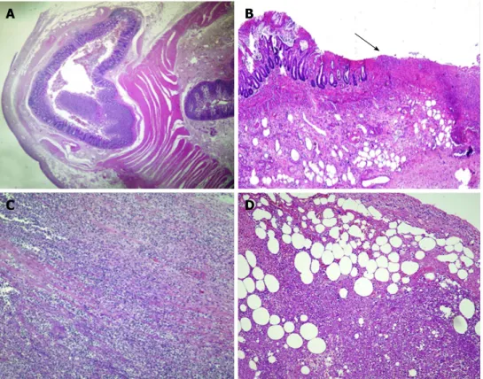
The histological exam (Figure 3) showed a diver-ticular structure consisting of mucosa and submucosa, with a small rim of longitudinal muscle; superficial ulcerations were evident in the pathological area, with full-thickness mucosal necrosis associated with a massive inflammatory cell infiltrate having transmural pattern and involving serosal surface with partial necrosis.
The exploratory laparotomy allowed us to identify the BPP that had been initially missed on the CT scan images; it had appeared as a radiopaque intraluminal body, located in the high rectum and without any evidence of collection or air leakage (Figure 4).
DIsCUssION
Taking into consideration the diagnostic and ma-nagement difficulties of this exceptional clinical case, we have performed a systemic review of the literature to evaluate the diagnostic role of imaging procedures and the management of a GI perforation caused by the inadvertent ingestion of a BPP. Indeed, the goal of this analysis was to identify in the literature the most relevant information about cases of perforation caused by the inadvertent ingestion of a BPP.
For this purpose, we carried out a comprehensive search of citations from PubMed between January 1st 1988 to January 31st 2018, starting from the first
article concerning bowel perforation related to a BPP, using the key words “intestinal perforation blister”, “diagnosis”, “CT blister pill pack” and “rectal perforation blister”. Our search yielded 20 articles, in which 23
Figure 1 CT scan findings. A: Evidence of an important abdominopelvic peritoneal effusion (black arrowhead), perigastric free air along the gastrocolic ligament and
under the anterior abdominal wall (white arrows), and pericardial and bilateral pleural effusions; B: In addition to perigastric free air and under the abdominal wall (white arrows), a microlithiasis (black arrow) of the gallbladder could also be observed.
Figure 2 Postoperative findings. A, B: Blister pill pack with rectal perforation.
September 26, 2018|Volume 6|Issue 10|
A
B
B
A
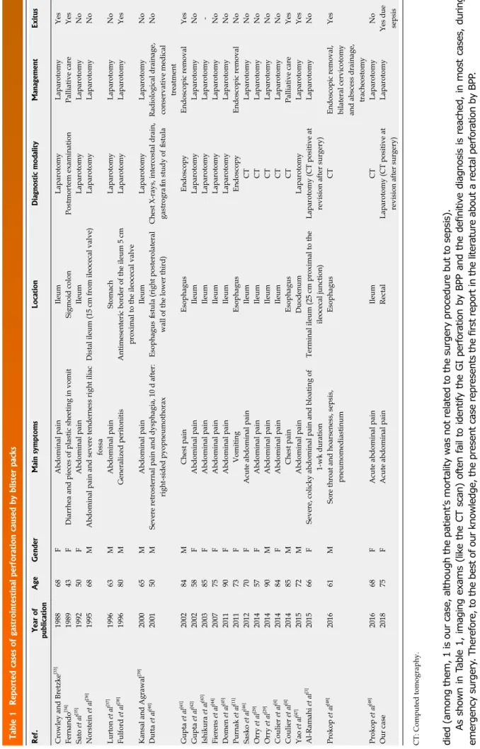
cases of GI perforation were reported, but none of rectal perforation. We excluded articles concerning GI perforation by other types of FBs. In the case of multiple publications on the same group of patients, only the most recent and complete paper was retained, including all types of study design. The following data were analyzed: year of publication; patient’s sex and age (years); location of the perforation; diagnostic modalities; treatment applied; and mortality. We also added our case, in order to compare it with the literature findings.
Based on our search criteria, we identified 23 GI
perforations by BPP in the literature; including our case, we had data on a total of 24 cases (Table 1). The patients were composed of 10 males and 14 females, with a median age of 70 and a standard deviation of 12.9. In particular, the perforations were located as follows: 5 in the esophagus; 1 in the stomach; 1 in the duodenum; 15 in the ileum; 1 in the sigmoid; and 1 in the rectum (represented by our case). In 18 patients, the clinical presentation was characterized by abdominal pain and acute abdomen syndrome, with 1 case presenting only vomiting, 1 presenting diarrhea and vomiting of pieces of plastic, 3 presenting chest pain (1 with right-sided pyopneumothorax), and 1 showing pneumomediastinum.
In this case series, the diagnosis was reached by CT in 7 patients, chest X-rays with gastrografin study of fistula in 1 patient, endoscopy in 2 patients, surgery (laparotomy) in 13 patients (2 with positive findings in CT images upon postsurgery revision 1 of which being our case), and postmortem examination in 1 patient.
With regard to treatment, endoscopic removal was performed in 3 patients (in 1 of them, bilateral cervicotomy, abscess drainage and tracheostomy were also performed), palliative care was undertaken in 2 patients, radiological drainage and conservative medical treatment were adopted in 1 patient, and laparotomy was performed in 18 patients.
The mortality was not reported for 1 patient; for the remaining cases, 15 patients survived and 8 patients
Figure 3 Histological findings. A: The whole histological section shows a diverticular structure consisting of mucosa and submucosa with a small rim of longitudinal
muscle (× 5); B: Evidence of superficial ulcerations (arrow) with full-thickness mucosal necrosis in the pathological area (× 10); C: Massive inflammatory cell infiltration having a transmural pattern and involving the serosal surface (× 20); D: Massive inflammatory cell infiltration having transmural pattern and involving the serosal surface with partial necrosis (× 20). Hematoxylin and eosin staining.
Figure 4 Upon re-reading of the computed tomography imaging, a radiopaque intraluminal body (white arrow) without any evidence of collection or air leakage was visible in the high rectum.
A
B
C
D
388 WJCC|www.wjgnet.com di ed ( am on g th em , 1 is o ur c as e, a lth ou gh th e pa tie nt ’s m or ta lit y w as n ot r el at ed to th e su rg er y pr oc ed ur e bu t t o se ps is ). As s ho w n in T ab le 1 , im ag in g ex am s (li ke t he C T sc an ) of te n fa il to id en tif y th e G I pe rf or at io n by B PP a nd t he d efi ni tiv e di ag no si s is r ea ch ed , in m os t ca se s, d ur in g em er ge nc y su rg er y. T he re fo re , t o th e be st o f o ur k no w le dg e, th e pr es en t c as e re pr es en ts th e fir st r ep or t i n th e lit er at ur e ab ou t a r ec ta l p er fo ra tio n by B PP . G en er al ly, th e in ge st io n of F Bs is s ee n in e m er ge nc y de pa rt m en ts a nd it is ty pi ca lly tr ea te d by o bs er va tio n or e nd os co pi c re m ov al . A pp ro xi m at el y 80 % -9 0% o f i ng es te d FB s pa ss th ro ug h th e G I t ra ct w ith n o co m pl ic at io n, w he re as th e re m ai ni ng 1 0% -2 0% fa il to p ro gr es s. L es s th an 1 % o f F Bs c an d et er m in e an in te st in al p er fo ra tio n [3 ] . S m al l
Table 1 Reported cases of gastrointestinal perforation caused by blister packs Ref.
Year of publication Age Gender Main symptoms Location Diagnostic modality Management Exitus
Crowley and Bretzke
[33] 1988 68 F Abdominal pain Ileum Laparotomy Laparotomy Yes Fernando [34] 1989 43 F
Diarrhea and pieces of plastic sheeting in vomit
Sigmoid colon Postmortem examination Palliative care Yes Sato et al [35] 1992 50 F Abdominal pain Ileum Laparotomy Laparotomy No Norstein et al [36] 1995 68 M
Abdominal pain and severe tenderness right iliac
fossa D is ta l i le um (1 5 cm fr om il eo ce ca l v al ve ) Laparotomy Laparotomy No Lurton et al [37] 1996 63 M Abdominal pain Stomach Laparotomy Laparotomy No Fulford et al [38] 1996 80 M Generalized peritonitis
Antimesenteric border of the ileum 5 cm
proximal to the ileocecal valve
Laparotomy
Laparotomy
Yes
Kansal and Agrawal
[39] 2000 65 M Abdominal pain Ileum Laparotomy Laparotomy No Dutta et al [40] 2001 50 M
Severe retrosternal pain and dysphagia, 10 d after:
right-sided pyopneumothorax
Esophagus fistula (right posterolateral
wall of the lower third)
Chest X-rays, intercostal drain, gastrografin study of fistula Radiological drainage, conservative medical
treatment No Gupta et al [41] 2002 84 M Chest pain Esophagus Endoscopy Endoscopic removal Yes Gupta et al [42] 2002 58 F Abdominal pain Ileum Laparotomy Laparotomy No Ishikura et al [43] 2003 85 F Abdominal pain Ileum Laparotomy Laparotomy -Fierens et al [44] 2007 75 F Abdominal pain Ileum Laparotomy Laparotomy No Domen et al [45] 2011 90 F Abdominal pain Ileum Laparotomy Laparotomy No Purnak et al [11] 2011 73 F Vomiting Esophagus Endoscopy Endoscopic removal No Sasko et al [46] 2012 70 F
Acute abdominal pain
Ileum CT Laparotomy No Orry et al [29] 2014 57 F Abdominal pain Ileum CT Laparotomy No Orry et al [29] 2014 90 M Abdominal pain Ileum CT Laparotomy No Coulier et al [4] 2014 84 F Abdominal pain Ileum CT Laparotomy No Coulier et al [4] 2014 85 M Chest pain Esophagus CT Palliative care Yes Yao et al [47] 2015 72 M Abdominal pain Duodenum Laparotomy Laparotomy Yes Al-Ramahi et al [3] 2015 66 F
Severe, colicky abdominal pain and bloating of
1-wk duration
Terminal ileum (25 cm proximal to the
ileocecal junction)
Laparotomy (CT positive at revision after surgery)
Laparotomy No Prokop et al [48] 2016 61 M
Sore throat and hoarseness, sepsis,
pneumomediastinum
Esophagus
CT
Endoscopic removal, bilateral cervicotomy and abscess drainage,
tracheostomy Yes Prokop et al [48] 2016 68 F
Acute abdominal pain
Ileum CT Laparotomy No Our case 2018 75 F
Acute abdominal pain
Rectal
Laparotomy (CT positive at revision after surgery)
Laparotomy
Yes due sepsis
CT: Computed tomography.
September 26, 2018|Volume 6|Issue 10|
orthodontic appliances and dentures account for 73% of the FBs accidentally ingested by the elderly. Other common FBs are toys, jewelry, nails, gravel, needles/ pins, staples, thumbtacks, wire bristles and magnetic objects.
Particularly, the elderly population is exposed to multiple risk factors that make accidental ingestion of BPPs more likely to happen; these include poor vision, presence of dentures, and polypharmacy. Dentures, in particular, produce a lack of normal palatal and gingival sensation, playing an important role in the accidental ingestion of BPPs[4,5]. In addition, in elderly
patients, diverticular disease is very common in Western countries and predominately affects the distal colon. This condition produces structural abnormalities such as tortuous lumen, strictures and mural pockets that could aggravate an FB-caused injury and the FB itself can be “marooned-in”. Consequently, diverticular disease and FBs may be associated with pathological processes, including inflammation, perforation, abscess and fistula[6].
In practical terms, a BPP appears as an object with sharp and pointed edges and it can produce a bowel perforation. Therefore, sharp, thin, stiff, pointed or long FBs can cause perforation by direct penetration or by remaining stuck within the lumen, thus provoking necrosis of the bowel wall[3].
However, FB intestinal perforation represents a challenging clinical scenario, mainly in patients with intellectual disability or psychiatric disorders cha-racterized by a noncollaborative attitude during medical questioning. Unfortunately, the serious deteriorating symptoms and signs in these patients are the primary cause of admission to hospital.
Frequently, FB ingestion can be asymptomatic or with unspecific symptoms and can vary depending on the sites where the FB arrives and in the related complications. Moreover, the FB ingestion signs can mimic those of other surgical conditions, such as appendicitis, diverticulosis or colonic perforation[7].
Hence, the preoperative diagnosis is frequently a surgical acute abdomen of unknown origin[8].
The various clinical symptoms include dysphagia, odynophagia, chest pain, respiratory distress, hema-temesis, GI bleeding, acute abdominal pain, intestinal perforation, bowel obstruction, localized abscess formation, peritonitis, inflammatory masses or sepsis[9-11],
perineal and scrotal abscess, and enterobladder fistulas[12,13].
Many studies have suggested that FBs may stop in areas of anatomical narrowing (i.e., cricopharyngeal ring, lower esophageal sphincter, pylorus, duodenal sweep, ileocecal valve, and anus), physiological angling (curvature of the duodenum) or pathological stricture or adhesion presence, mainly in patients with previous bowel pathology (e.g., intestinal stricture, Crohn’s disease)[14-17]. Usually, FBs with size exceeding 2-2.5 cm
pass with difficulty through the pyloric canal and those having a dimension of 6-10 cm or greater cannot move
through the duodenum. Even if the most frequent sites of perforation are the lower esophagus and the terminal ileum, a small percentage of perforations can occur at any level, from mouth to anus[14-16,18]. GI mucosa injury
can produce digestive hemorrhage[19], while other
potential complications may arise as a result of FB migration to the liver and pancreas, with pancreatitis, gastric varices development, splenic artery pseudo-aneurysm, or even aspects mimicking locally advanced pancreatic carcinoma[20-23].
Typically, the time from ingestion to perforation is very long; it has been demonstrated that 10.4 d
represents the median time[8]. Most of these patients
occur in the straits and the angles of the GI tract. Therefore, distal ileum, cecum and left colon are other common sites of perforation, although some authors have reported an increased incidence of perforation in association with Meckel’s diverticulum and diverticular disease[13,24-26].
In obese and bedridden patients, the identification of a low calcium opaque FB can be hindered by a large amount of soft tissue or simply by a high level of liquid in the viscera[27]. Nevertheless, the radiographic
appearances of BPPs are typically identifiable by their aluminum foil backing and plastic blister surrounding the pill or tablet along with a thin rim of air. Their lateral appearance has been compared to that of a “UFO”[28]. It
was also observed that the density of the pill itself could be extremely variable, some pills being completely
radiolucent[4]. The most direct assessment for FB
perforation on CT is the identification of the FB near extraluminal gas. Other findings include localized wall thickening, fat stranding and abscess. Multidetector CT, through the possibilities of high-quality multiplanar three-dimensional reconstructions and maximum in-tensity projection, can reach the definitive diagnosis of perforated intestinal structures caused by ingested FBs, often with demonstration of the responsible FB[27,29].
Treatment depends upon patient age, the anatomi-cal location, type and nature of the material ingested along with the presenting symptoms. Therefore, early diagnosis and prompt removal via either endoscopic or surgical measure are necessary. The management may indeed consist of conservative or interventional methods; in almost 20% of the cases, endoscopic treatment is required or possibly laparoscopy or exploratory
laparotomy[30]. The last of these can prove especially
risky in psychiatric patients with a history of ingesting multiple FBs and who have already undergone multiple surgeries[31]. If the FB arrives in the stomach or in the
duodenum, endoscopic removal should be attempted immediately, in order to avoid the risk of perforation of the ileocecal valve, which is approximately 35%[14,15,17].
If the pointed object moves past the duodenum, the patient should be monitored daily through a series of radiographs under close observation. Surgery becomes necessary if the object is no longer progressing radiographically after a 72-h observation. Emergency laparotomy is required if the patient develops clinical
signs of acute peritonitis[14,15].
The case reported herein represents the first case in literature describing a rectal perforation caused by a BBP. The lack of similar situations underlines how difficult it is to reach a correct early diagnosis because of nonspecific and vague symptoms. Moreover, the patient was initially treated for a septic state suspected of having originated from the abdomen. Most certainly, an immediate CT scan would have helped to reach the diagnosis, but it was deemed unnecessary when the patient was taken to the emergency department for observation. Based on an abdomen and thorax X-ray, the hypothesis of acute abdominal perforation had actually been excluded. It is quite hard to establish whether the general outcome would have been diffe-rent had a CT scan been performed, for the patient’s conditions appeared highly critical since her first day of admission and only rarely does a severe abdominal sepsis in a critically ill patient, as in this case, give survival chance.
Thus, as can easily be understood, the diagnosis of our patient was delayed due to her mental impairment, while the CT scan could neither reveal the presence of the BPP nor any air leakage or collection in the proximity of the rectum. Based on the CT findings, our first hypothesis was of a pneumoperitoneum caused by a gastric-duodenal perforation. Probably, as described in the literature, our difficulty in identifying the BPP had to do with the radiolucent characteristics of the BPP itself. In light of the exploratory laparotomy findings, we decided to review the CT scan images. The BPP initially missed on CT scan was identified as a radiopaque intraluminal body in the high rectum, without any evidence of collection or air leakage (Figure 4).
Moreover, the damage caused by the BPP derived both from the BPP itself and from the presence of sigmoid-rectum diverticula. The definitive diagnosis was only reached after exploratory laparotomy and prompt treatment was applied, which included the removal of the causative agent through a Hartmann’s procedure, peritoneal lavage and drainage. Our patient presented the diagnostic criteria of severe sepsis and septic shock. The mortality from severe sepsis and septic
shock is now closer to 20%-30% in many series[32],
and indeed our patient died after 10 d without surgical complications.
The diagnosis of intestinal perforation following the unknown ingestion of an FB is often a difficult challenge in terms of treatment, due to its late diagnosis and a deteriorated clinical condition. Caregivers should be cautious, they should avoid cutting drug blisters in order to not administer the pill with its blister pack to patients. Intraoperative exploration remains critical in most cases.
aRTICLe HIGHLIGHTs
Case characteristics
A 75-year-old woman arrived at our emergency department in poor clinical
390 WJCC|www.wjgnet.com
condition, awake, noncollaborative, oriented only in space, dehydrated, with fever (39 °C) and vomiting, oliguria and hypotension (80/50 mmHg). After 48 h, she presented an acute abdominal pain.
Clinical diagnosis
The diagnostic hypothesis was firstly sepsis of unknown origin.
Differential diagnosis
Based on the computed tomography (CT) findings, the origin of the perforation was suspected to be gastric.
Laboratory diagnosis
The laboratory data revealed white blood cell count of 25920 mmc, with 87% neutrophils, platelet count of 90000 mmc, hemoglobin of 10.9 gr%, C-reactive protein of 10.5 mg/dl and procalcitonin of 34.9 mg/dl.
Imaging diagnosis
The abdominal X-ray and CT revealed an abundant abdominopelvic peritoneal effusion, free perigastric air (along the gastrocolic ligament and under the anterior abdominal wall).
Pathological diagnosis
The abdomen exploration revealed a cirrhotic liver, an abundant amount of purulent intraperitoneal liquid, and a fecaloid collection in the pelvic pouch, which was buffered by the uterus. In the anterior distal part of the intraperitoneal rectum, a 2-cm long, full-thickness lesion was evident. The lesion was surrounded by a necrotic wall from which appeared a part of the blister pill pack (BPP) with the pill inside. In addition, at sigmoid level, some diverticula were filled with coprolites and the wall was rather thin. Exploratory laparotomy findings allowed us to identify the BPP initially missed on CT scan images. Review of the images revealed that it had appeared as a radiopaque intraluminal body in the high rectum, without any evidence of collection or air leakage.
Treatment
The patient was submitted to an urgent exploratory laparotomy. The general clinical conditions were very bad and included hypotension and oliguria. Hence, we performed a Hartmann’s procedure and positioned three intraperitoneal drainage routes. Due to her worsening condition, the patient was transferred to the Intensive Care Unit, where she died after 10 d without any surgical complication.
Related reports
As can easily be understood, the diagnosis was delayed due to the patient’ s mental impairment, while the CT scan could neither reveal the presence of the BPP nor any air leakage or collection in the proximity of the rectum. Based on the CT findings, our first hypothesis was of a pneumoperitoneum caused by a gastric-duodenal perforation. Probably, as described in the literature, our difficulty in identifying the BPP had to do with the radiolucent characteristics of the BPP itself.
Term explanation
In some countries, a BPP is known as a “push-through pack”. Push-through packs consist of two main features: (1) the cover foil being resistant but breaking easily, so that the drug can be pressed out by easily breaking the cover foil; and (2) the semirigid formed cavity can be folded to dispense the drug by pressing it out with a thumb; in both cases, breaking the cover foil with a fingernail will make the pressing-out easier.
Experiences and lessons
To the best of our knowledge, this is the first case report describing a rectal perforation caused by a BBP. In light of the exploratory laparotomy findings, we decided to review the CT scan images. The BPP initially missed on CT scan was identified as a radiopaque intraluminal body in the high rectum, without any evidence of collection or air leakage. The diagnosis of intestinal perforation following the unknown ingestion of an FB is often a difficult challenge in terms of treatment, due to its late diagnosis and a deteriorated clinical condition
aCKNOWLeDGeMeNTs
The authors thank Lucia Lo Conti for proofreading the
September 26, 2018|Volume 6|Issue 10|
aRTICLe HIGHLIGHTs
English.
ReFeReNCes
1 Laeeq SM, Rai AA, Tasneem AA, Luck NH, Majid Z. Pill in the blister pack: a rare cause of dysphagia in an elderly adult. Pan Afr Med J 2015; 22: 176 [PMID: 26918072 DOI: 10.11604/ pamj.2015.22.176.8031]
2 Goh BK, Chow PK, Quah HM, Ong HS, Eu KW, Ooi LL, Wong WK. Perforation of the gastrointestinal tract secondary to ingestion of foreign bodies. World J Surg 2006; 30: 372-377 [PMID: 16479337 DOI: 10.1007/s00268-005-0490-2]
3 Al-Ramahi G, Mohamed M, Kennedy K, McCann M. Obstruction
and perforation of the small bowel caused by inadvertent ingestion of a blister pill pack in an elderly patient. BMJ Case Rep 2015;
2015: [PMID: 26475885 DOI: 10.1136/bcr-2015-212822]
4 Coulier B, Rubay R, Van den Broeck S, Azar AR, Maldague P, Mailleux P, Lismonde Y, Bueres I. Perforation of the gastrointestinal tract caused by inadvertent ingestion of blister pill packs: report of two cases diagnosed by MDCT with emphasis on maximal intensity and volume rendering reformations. Abdom Imaging 2014; 39: 685-693 [PMID: 24643854 DOI: 10.1007/ s00261-014-0120-2]
5 Gunn A. Intestinal perforation due to swallowed fish or meat
bone. Lancet 1966; 1: 125-128 [PMID: 4158955 DOI: 10.1016/ S0140-6736(66)91262-1]
6 Ross E, McKenna P, Anderson JH. Foreign bodies in sigmoid colon diverticulosis. Clin J Gastroenterol 2017; 10: 491-497 [PMID: 29030789 DOI: 10.1007/s12328-017-0786-4]
7 Yao CC, Yang CC, Liew SC, Lin CS. Small bowel perforation caused by a sharp bone: laparoscopic diagnosis and treatment. Surg Laparosc Endosc Percutan Tech 1999; 9: 226-227 [PMID: 10804008 DOI: 10.1097/00129689-199906000-00017]
8 Rodríguez-Hermosa JI, Codina-Cazador A, Sirvent JM, Martín A, Gironès J, Garsot E. Surgically treated perforations of the gastrointestinal tract caused by ingested foreign bodies. Colorectal Dis 2008; 10: 701-707 [PMID: 18005196 DOI: 10.1111/ j.1463-1318.2007.01401.x]
9 Schwartz JT, Graham DY. Toothpick perforation of the intestines. Ann Surg 1977; 185: 64-66 [PMID: 318821 DOI: 10.1097/000006 58-197701000-00010]
10 Maleki M, Evans WE. Foreign-body perforation of the intestinal tract. Report of 12 cases and review of the literature. Arch Surg 1970; 101: 475-477 [PMID: 5457244 DOI: 10.1001/ archsurg.1970.01340280027008]
11 Purnak T, Ozaslan E, Efe C. Concomitant oesophageal perforation and bleeding due to a tiny pill with its blister pack. Age Ageing 2011; 40: 645-646 [PMID: 21771743 DOI: 10.1093/ageing/afr081] 12 Moreira CA, Wongpakdee S, Gennaro AR. A foreign body (chicken
bone) in the rectum causing extensive perirectal and scrotal abscess: report of a case. Dis Colon Rectum 1975; 18: 407-409 [PMID: 1097218 DOI: 10.1007/BF02587433]
13 Akhtar S, McElvanna N, Gardiner KR, Irwin ST. Bowel
perforation caused by swallowed chicken bones--a case series. Ulster Med J 2007; 76: 37-38 [PMID: 17288304]
14 Pavlidis TE, Marakis GN, Triantafyllou A, Psarras K, Kontoulis TM, Sakantamis AK. Management of ingested foreign bodies. How justifiable is a waiting policy? Surg Laparosc Endosc Percutan Tech 2008; 18: 286-287 [PMID: 18574418 DOI: 10.1097/ SLE.0b013e31816b78f5]
15 Eisen GM, Baron TH, Dominitz JA, Faigel DO, Goldstein JL,
Johanson JF, Mallery JS, Raddawi HM, Vargo JJ 2nd, Waring JP, Fanelli RD, Wheeler-Harbough J; American Society for Gastrointestinal Endoscopy. Guideline for the management of ingested foreign bodies. Gastrointest Endosc 2002; 55: 802-806 [PMID: 12024131 DOI: 10.1016/S0016-5107(02)70407-0] 16 Abraham B, Alao AO. An unusual foreign body ingestion in a
schizophrenic patient: case report. Int J Psychiatry Med 2005; 35: 313-318 [PMID: 16480246 DOI:
10.2190/7AE8-3AV0-W3UA-TKV4]
17 Murshid KR, Khairy GE. Laparoscopic removal of a foreign body from the intestine. J R Coll Surg Edinb 1998; 43: 109-111 [PMID: 9621536]
18 Puia IC, Puia VR, Andreescu A, Cristea PG. [Ascending colon perforation by ingested fruit stones]. Chirurgia (Bucur) 2011; 106: 825-827 [PMID: 22308923]
19 Blaho KE, Merigian KS, Winbery SL, Park LJ, Cockrell M. Foreign body ingestions in the Emergency Department: case reports and review of treatment. J Emerg Med 1998; 16: 21-26 [PMID: 9472755 DOI: 10.1016/S0736-4679(97)00229-1] 20 Goh BK, Jeyaraj PR, Chan HS, Ong HS, Agasthian T, Chang
KT, Wong WK. A case of fish bone perforation of the stomach mimicking a locally advanced pancreatic carcinoma. Dig Dis Sci 2004; 49: 1935-1937 [PMID: 15628728 DOI: 10.1007/s10620-004-9595-y]
21 Goh BK, Yong WS, Yeo AW. Pancreatic and hepatic abscess secondary to fish bone perforation of the duodenum. Dig Dis Sci 2005; 50: 1103-1106 [PMID: 15986862 DOI: 10.1007/ s10620-005-2712-8]
22 Kim KH, Woo EY, Rosato EF, Kochman ML. Pancreatic foreign body: Ingested toothpick as a cause of pancreatitis and hemorrhage. Gastrointest Endosc 2004; 59: 147-150 [PMID: 14722574 DOI: 10.1016/S0016-5107(03)02364-2]
23 Rahalkar MD, Pai B, Kukade G, Al Busaidi SS. Sewing needles as foreign bodies in the liver and pancreas. Clin Radiol 2003; 58: 84-86 [PMID: 12565211 DOI: 10.1053/crad.2002.1118]
24 Pinero Madrona A, Fernández Hernández JA, Carrasco Prats M, Riquelme Riquelme J, Parrila Paricio P. Intestinal perforation by foreign bodies. Eur J Surg 2000; 166: 307-309 [PMID: 10817327 DOI: 10.1080/110241500750009140]
25 Mcpherson RC, Karlan M, Williams RD. Foreign body perforation of the intestinal tract. Am J Surg 1957; 94: 564-566 [PMID: 13458636 DOI: 10.1016/0002-9610(57)90580-9] 26 Gómez N, Roldós F, Andrade R. [Intestinal perforation caused by
chicken bone mimicking perforated colonic diverticulitis]. Acta Gastroenterol Latinoam 1997; 27: 329-330 [PMID: 9460513] 27 Coulier B, Tancredi MH, Ramboux A. Spiral CT and
multidetector-row CT diagnosis of perforation of the small intestine caused by ingested foreign bodies. Eur Radiol 2004; 14: 1918-1925 [PMID: 15378256 DOI: 10.1007/s00330-004-2430-1]
28 Tai AW, Sodickson A. Foreign body ingestion of blister pill pack causing small bowel obstruction. Emerg Radiol 2007; 14: 105-108 [PMID: 17342467 DOI: 10.1007/s10140-007-0582-4]
29 Orry X, Balaj C, Lecocq S, Blum A, Delvaux M, Régent D, Claudon M, Laurent V. CT diagnosis of small bowel perforation by ingestion of a blister pack: two case reports. Diagn Interv Imaging 2014; 95: 101-103 [PMID: 23726172 DOI: 10.1016/ j.diii.2013.04.001]
30 Athanassiadi K, Gerazounis M, Metaxas E, Kalantzi N. Management of esophageal foreign bodies: a retrospective review of 400 cases. Eur J Cardiothorac Surg 2002; 21: 653-656 [PMID: 11932163 DOI: 10.1016/S1010-7940(02)00032-5]
31 Wu C, Hungness ES. Laparoscopic removal of a pancreatic foreign body. JSLS 2006; 10: 541-543 [PMID: 17575779]
32 Angus DC, van der Poll T. Severe sepsis and septic shock. N Engl J Med 2013; 369: 840-851 [PMID: 23984731 DOI: 10.1056/ NEJMra1208623]
33 Crowley LV, Bretzke ML. Bowel perforation from ingested unit dose blister-pak. Am J Gastroenterol 1988; 83: 1011-1012 [PMID: 3414640]
34 Fernando GC. Colonic perforation following ingestion of plastic sheeting. Med Sci Law 1989; 29: 263-264 [PMID: 2770479 DOI: 10.1177/002580248902900313]
35 Sato H, Endo T, Tajima K, Sanada Y. A rare case of perineal pain: intestinal perforation caused by a press-through package. Anesth Analg 1992; 75: 456-457 [PMID: 1510270 DOI: 10.1213/0000053 9-199209000-00025]
36 Norstein J, Krajci P, Bergan A, Geiran O. Intestinal perforation after ingestion of a blister-wrapped tablet. Lancet 1995; 346: 1308
392 WJCC|www.wjgnet.com
[PMID: 7475762 DOI: 10.1016/S0140-6736(95)91918-X] 37 Lurton A, Ntiruhungwa J, Saillant H, Surugue J. Stomach
perforation by a blister-wrapped capsule. N Engl J Med 1996; 335: 754 [PMID: 8786776 DOI: 10.1056/NEJM199609053351020] 38 Fulford S, Tooley AH. Intestinal perforation after ingestion of
a blister-wrapped tablet. Lancet 1996; 347: 128-129 [PMID: 8538332 DOI: 10.1016/S0140-6736(96)90259-7]
39 Kansal G, Agrawal V. Intestinal perforation--a unique cause. J Indian Med Assoc 2000; 98: 184, 186 [PMID: 11016184] 40 Dutta U, Gupta NM, Nagi B, Singh K. Blister pack ingestion
resulting in esophago-pleural fistula. Indian J Gastroenterol 2001;
20: 79-80 [PMID: 11305505]
41 Gupta NM, Gupta V, Gupta R, Sudhakar V. Esophageal perforation caused by a blister-wrapped tablet. Asian Cardiovasc Thorac Ann 2002; 10: 87-88 [PMID: 12079986 DOI: 10.1177/021 849230201000127]
42 Gupta V, Manikyam SR, Gupta R, Gupta NM. Pelvic abscess after ingestion of blister-wrapped tablet. Am J Gastroenterol 2002; 97: 2142-2143 [PMID: 12190196 DOI: 10.1111/ j.1572-0241.2002.05940.x]
43 Ishikura H, Sakata A, Sakaki Y, Kimura S, Sumi T, Ichimori T, Uyama K. Intestinal perforation due to ingestion of blister-wrapped tablet in a press-through package. Am J Gastroenterol
2003; 98: 1665-1666 [PMID: 12873609 DOI: 10.1111/ j.1572-0241.2003.07560.x]
44 Fierens K, Van Outryve L, Kint M. Bowel perforation from ingested blister-pack. Acta Chir Belg 2007; 107: 564-565 [PMID: 18074922 DOI: 10.1080/00015458.2007.11680125]
45 Domen H, Ohara M, Noguchi M, Nakanishi Y, Komuro K, Iwashiro N, Ishizaka M. Inadvertent Ingestion of a Press-Through Package Causing Perforation of the Small Intestine within an Incisional Hernia and Panperitonitis. Case Rep Gastroenterol 2011;
5: 391-395 [PMID: 21792348 DOI: 10.1159/000330290]
46 Sasko B, Butz T, Winnekendonk G, Plehn G, Prull M, Liermann D, Trappe HJ. [Bowel perforation because of ingestion of a blister-wrapped tablet after post-interventional coronary perforation]. Dtsch Med Wochenschr 2012; 137: 2637-2640 [PMID: 23225187 DOI: 10.1055/s-0032-1327336]
47 Yao SY, Matsui Y, Shiotsu S. An unusual case of duodenal perforation caused by a blister pack: A case report and literature review. Int J Surg Case Rep 2015; 14: 129-132 [PMID: 26263453 DOI: 10.1016/j.ijscr.2015.07.013]
48 Prokop A, Stepper H, Koll S, Chmielnicki M. [No Blister-Wrapped Pills: Perforation after Ingestion a Blister-Pack]. Z Orthop Unfall 2016; 154: 299-302 [PMID: 27351163 DOI: 10.1055/s-0042-101962]
P- Reviewer: Desai DJ, Kai K, Sergi CM, Vento S, Virk JS S- Editor: Ma YJ L- Editor: A E- Editor: Wu YXJ
September 26, 2018|Volume 6|Issue 10|